🧬 SinoBioData Academic Portal
🏛️ Indexed Academic JournalOriginal: 干细胞研究与转化

Stem Cell Research & Therapy

Premier Chinese Biomedical Journal indexed in SinoBioData: Stem Cell Research & Therapy (干细胞研究与转化).

Total Research Papers: 200
Access: 100% Free Open Access
Browse by Publication Year & Volume

Published Research Papers

Showing 200 of 200 peer-reviewed papers with full Graphical Abstracts.

Original ResearchVol. 17, None • pp. 191DOI: 10.1186/s13287-026-04989-4

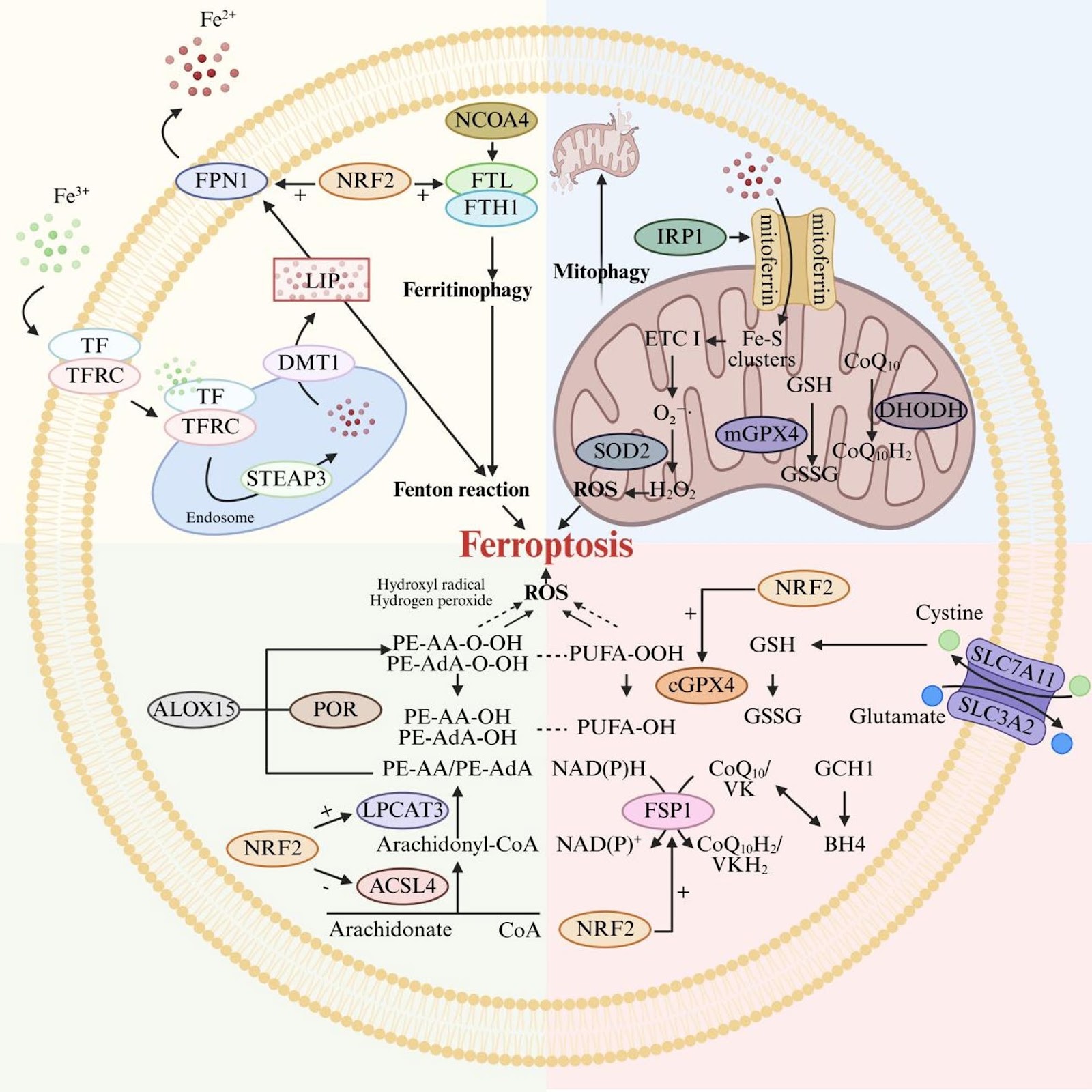
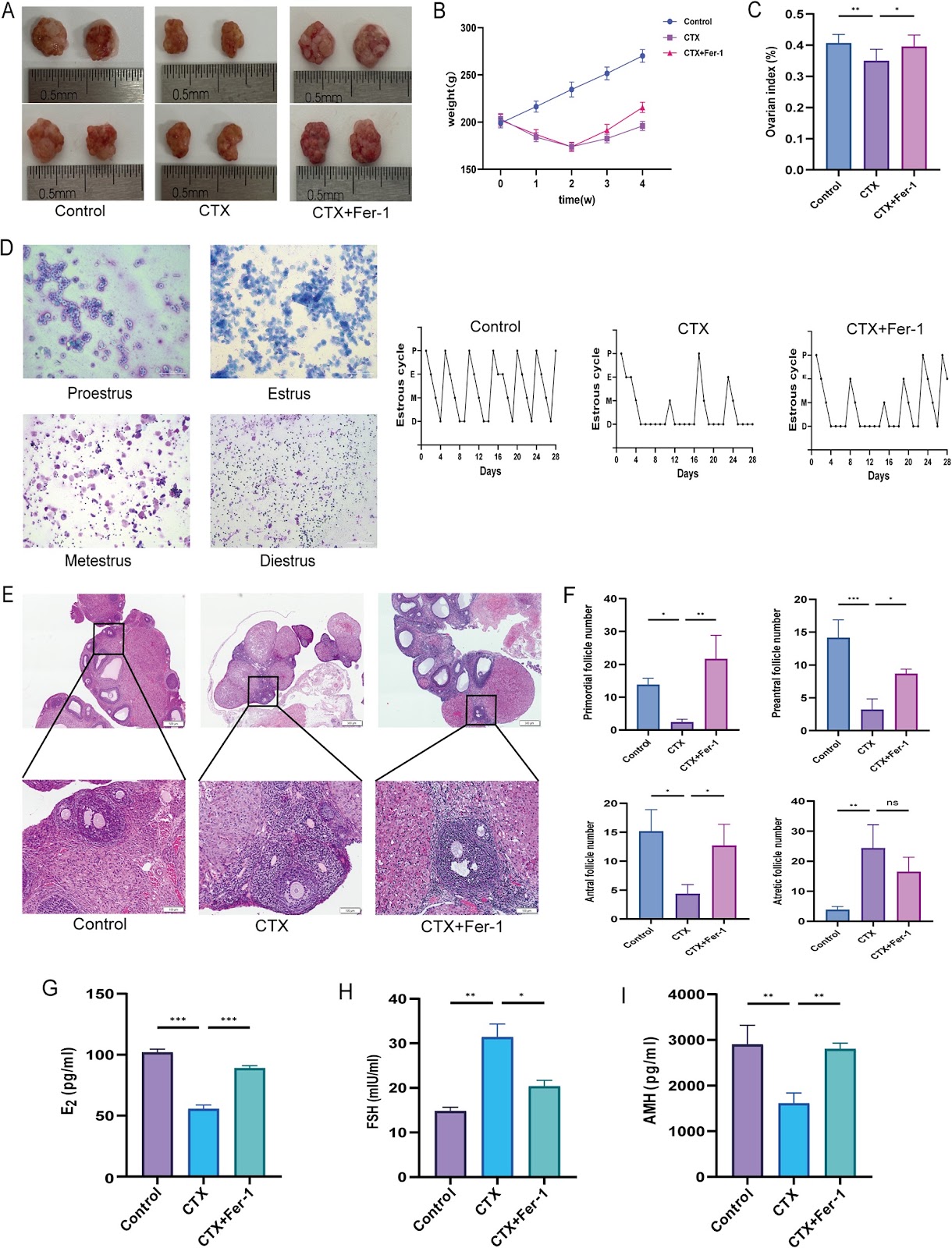
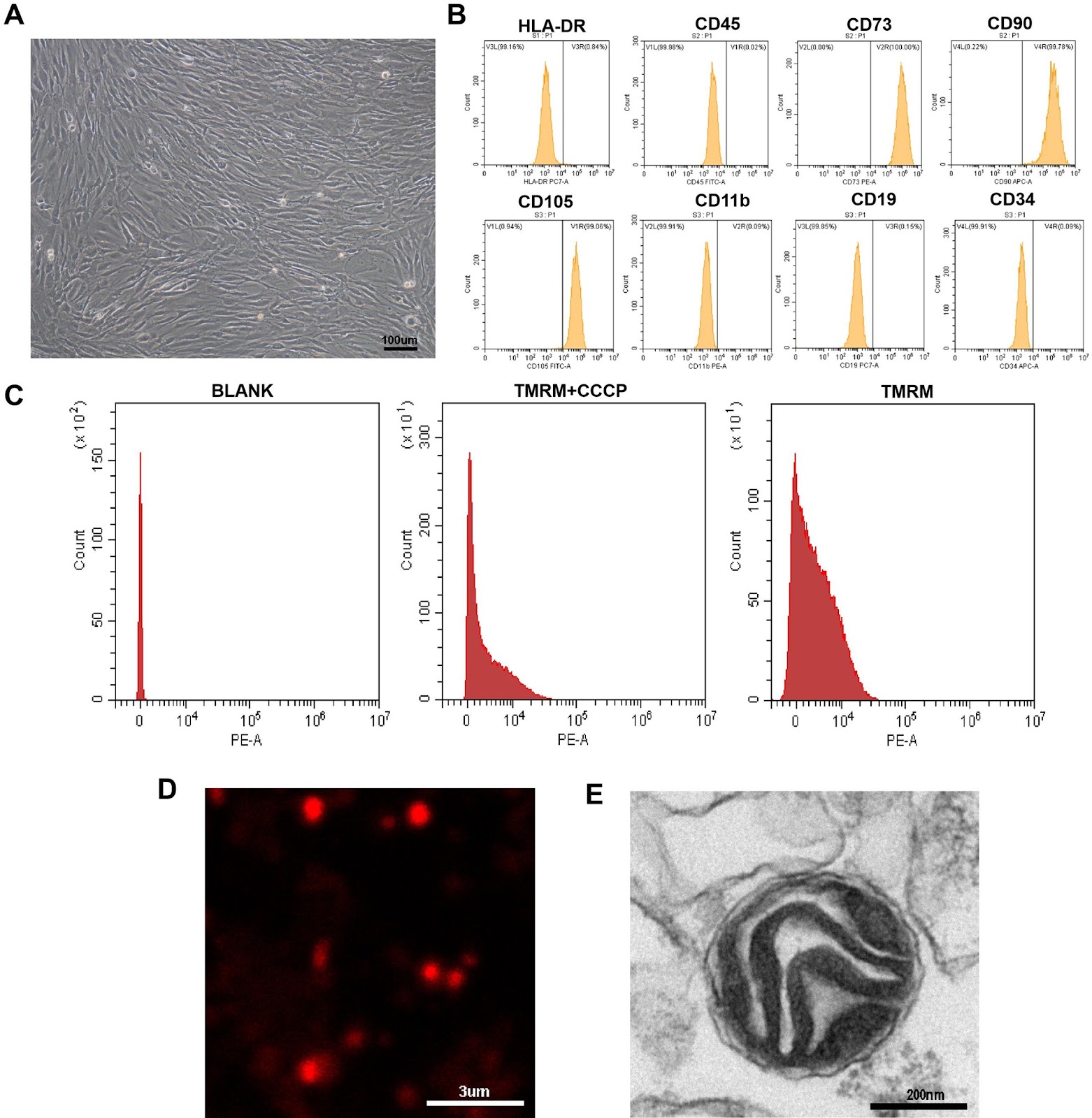
Isorhamnetin-preconditioned MSC-derived exosomes restore ovarian function by inhibiting ferroptosis in chemotherapy-induced POF

Authors: Qiang Zhang, Jinyu Yu, Yan Zheng, Jinlan Jiang, Lianwen Zheng

Background Chemotherapy-induced premature ovarian failure (POF) is a major cause of infertility, with limited treatment options. Mesenchymal stem cell-derived exosomes (MSC-Exos) have therapeutic potential. This study investigated whether preconditioning MSCs with the antioxidant flavonoid isorhamnetin (ISO) enhances the efficacy of their exosomes (ISO-MSC-Exos) against POF. Methods A cyclophosphamide-induced POF rat model was established, and the role of the ferroptosis inhibitor ferrostatin-1 was evaluated. MSC-Exos and ISO-MSC-Exos were isolated by ultracentrifugation and administered via tail vein injection. Ovarian recovery was assessed by monitoring the oestrous cycle, serum hormone levels, and histological findings. Lipid peroxidation and iron metabolism were evaluated by quantifying malondialdehyde, glutathione, iron deposition, and mitochondrial ultrastructure. Immunohistochemistry was used to assess the expression levels of GPX4, ACSL4, and FTH1. Proteomic analyses were performed to explore the underlying mechanisms. Results Ferroptosis plays a pivotal role in the cyclophosphamide-induced POF rat model. Both exosome therapies improved ovarian function and suppressed ferroptosis, with ISO-MSC-Exos showing superior efficacy. ISO-MSC-Exos significantly restored hormone levels, ameliorated oestrous cycle disorders, reduced follicular atresia, and enhanced fertility. Furthermore, ISO-MSC-Exos more effectively elevated glutathione levels, reduced malondialdehyde and Fe2⁺ levels, and reversed the abnormal expression of ferroptosis-related proteins GPX4, ACSL4, and FTH1. Proteomic analysis suggested that ISO-MSC-Exos effectively inhibit ferroptosis by downregulating Alox15 and Tf, thereby reducing lipid peroxidation substrates and cellular iron uptake. This finding represents a potential molecular mechanism underlying their superior efficacy compared with that of MSC-Exos. Conclusions ISO-MSC-Exos showed superior efficacy compared with MSC-Exos in restoring ovarian function and inhibiting ferroptosis, suggesting that ISO pretreatment enhances the therapeutic effect of MSC-Exos in the POF
Isorhamnetin-preconditioned MSC-derived exosomes restore ovarian function by inhibiting ferroptosis in chemotherapy-induced POF
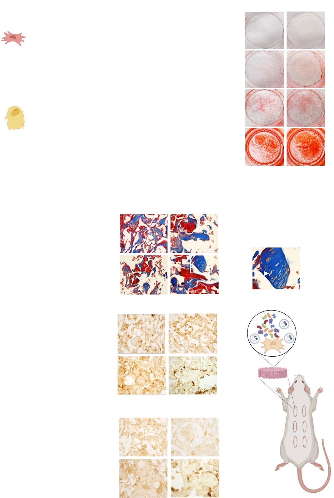
Graphical Abstract
Original ResearchVol. 17, None • pp. 177DOI: 10.1186/s13287-026-04995-6

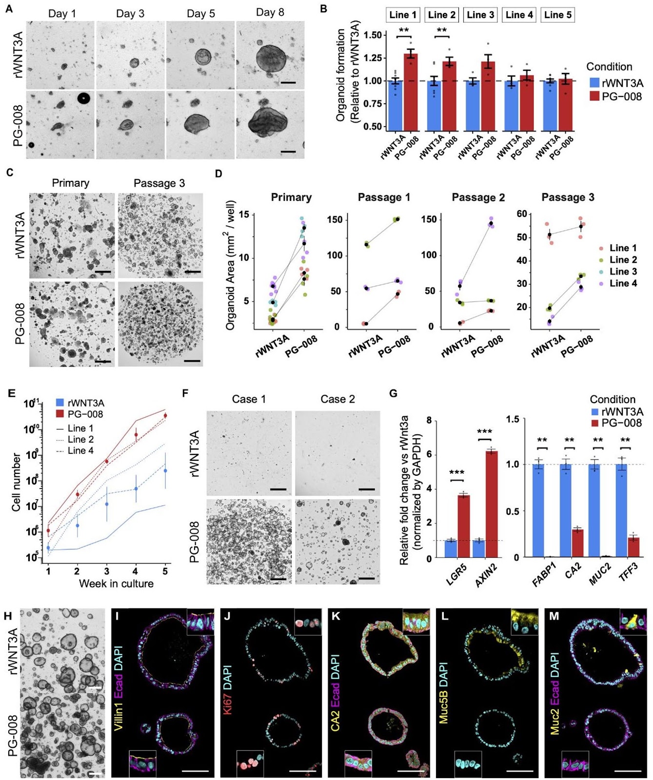
Peptide-based Wnt signal activation enables scalable production of clinical-grade patient-derived intestinal organoids for regenerative cell therapy

Authors: Hady Yuki Sugihara, Sayaka Nagata, Sho Kawasaki, Junichi Takahashi, Yui Hiraguri, Masayoshi Fukuda, Kohei Suzuki, Tatsuro Murano, Satoru Fujii, Toshimitsu Fujii, Hiromichi Shimizu, Kazuo Ohtsuka, Mamoru Watanabe, Ryuichi Okamoto, Tomohiro Mizutani

Background Gastrointestinal diseases often involve cellular damage, degeneration or dysfunction in the tract, frequently requiring surgical interventions risking complications and lowered quality of life. Regenerative medicine holds great promise in improving patient care and providing novel treatment options for previously irreparable and untreatable tissues. Despite the clinical potential of intestinal organoids as a resource for regenerative cell therapy and bioengineering, the lack of clinical-grade cultures has hampered further development. Moreover, strategies to efficiently and reliably expand clinical-grade cultures at the scale required for application is limited. Methods A GMP-compliant protocol was developed to generate patient-derived colonic organoids from endoscopic biopsies. Clinical-grade colonic organoids cultured and expanded in Type-I collagen were compared to conventional Matrigel cultured organoids. To improve the culture-, cost-, and time-efficiency of culture expansion, several strategies were developed including organoid area-based passaging, one well plate culture, and the incorporation of Wnt activating peptide, PG-008. Conventional recombinant WNT3A culture was compared to the peptide PG-008 culture using single cell RNA sequencing. Results Clinical-grade collagen cultured organoids exhibited similar culture efficiency to Matrigel. Organoid establishment rate from 60 patients using the GMP-compliant protocol was 82%. The incorporation of PG-008 significantly enhanced organoid growth and stabilized patient-patient variability through intestinal stem cell (ISC) enrichment. Single cell RNA sequencing revealed that PG-008 resulted in remarkably pure culture consisting of ISCs

Peptide-based Wnt signal activation enables scalable production of clinical-grade patient-derived intestinal organoids for regenerative cell therapy
Graphical Abstract
Original ResearchVol. 17, NoneDOI: 10.1186/s13287-026-04965-y

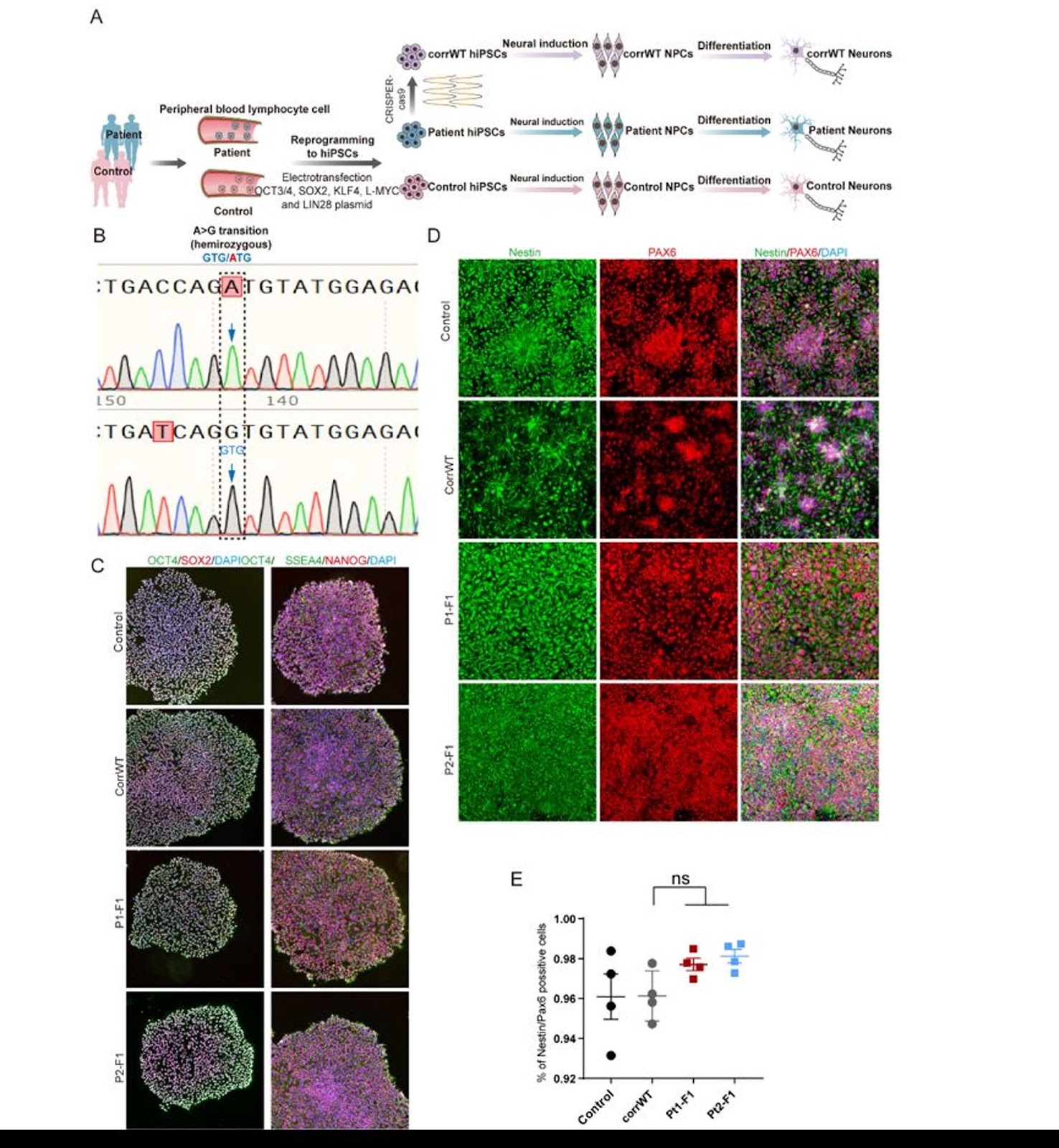
Targeting p75NTR activity alleviates the neurotoxic effect of high glucose on iPSC-derived dopaminergic neurons

Authors: Konstantina Chanoumidou, Ioanna Zota, Maria Anna Papadopoulou, Chrystalla Konstantinou, Alexandros Tsimpolis, Electra Tsagliotis, Maria Tziortziou, Katerina Ntarntani, Anne Grünewald, Matthieu David Lavigne, Achille Gravanis, Ioannis Charalampopoulos

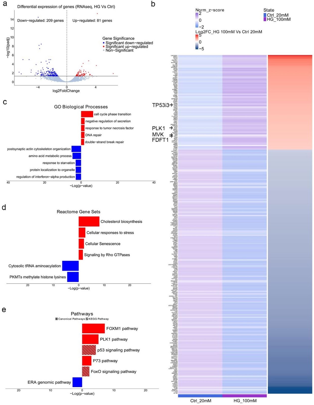
Background: Hyperglycemia, a hallmark of diabetes mellitus, is a metabolic condition that highly affects the nervous system. While evidence from epidemiological and animal studies links diabetes to dopaminergic dysfunction and an increased risk of Parkinson’s disease, the underlying mechanisms remain unclear. Here, we examined the effects of high glucose on human iPSC-derived dopaminergic neurons and glial cells to better understand the pathogenic alterations that lead to neurotoxicity. Previous implication of neurotrophins in the neurological manifestations of diabetes prompted us to focus on the role of p75NTR neurotrophin receptor (p75NTR) in dopaminergic neurodegeneration under hyperglycemic conditions. Methods: iPSC-derived dopaminergic neurons, astrocytes and microglia were treated with high glucose (50mM, 100mM) for 48 h to simulate hyperglycemia. Cytotoxicity assays, RNA sequencing and DNA damage assessments were employed to investigate the pathological alterations induced by high glucose exposure in neurons. Pharmacological targeting of p75NTR activity allowed investigation of its involvement in glucose neurotoxicity. Glial-mediated neurotoxicity was evaluated using conditioned media and inflammatory marker analysis. Results: High glucose treatment led to DNA damage, activation of JNK signaling and cell death in neurons. Importantly, we observed upregulation of p75NTR and its pro-apoptotic ligand pro-NGF, suggesting activation of the pro-NGF/p75NTR axis in high glucose-treated neurons. Inhibition of p75NTR activity rescued neuronal cell death, identifying p75NTR as a central mediator of glucose neurotoxicity. Furthermore, glucose overload sensitized neurons to 6-hydroxydopamine (6-OHDA), increasing their vulnerability to neurotoxic insults—an effect reversed by p75NTR blockade. Treatment with BNN27, a synthetic NGF mimetic, prevented neuronal loss through p75NTR and TrkA receptors, suggesting neurotrophin signaling as a potential therapeutic target for combating high glucose-induced neuronal damage. Finally, we demonstrated the contribution of glial cells to neurodegeneration since high glucose
Targeting p75NTR activity alleviates the neurotoxic effect of high glucose on iPSC-derived dopaminergic neurons
Graphical Abstract
Original ResearchVol. 17, Issue 243 • pp. 1-11DOI: 10.1186/s13287-026-05048-8

Serial brain FDG-PET and IMZ-SPECT following intracerebral MSC transplantation in patients with subacute ischemic stroke

Authors: Masahito Kawabori, Kenji Hirata, Hideo Shichinohe, Shiro Watanabe, Arisa Miura, Yoichi M. Ito, Kohsuke Kudo, Satoshi Kuroda, Kiyohiro Houkin, Miki Fujimura

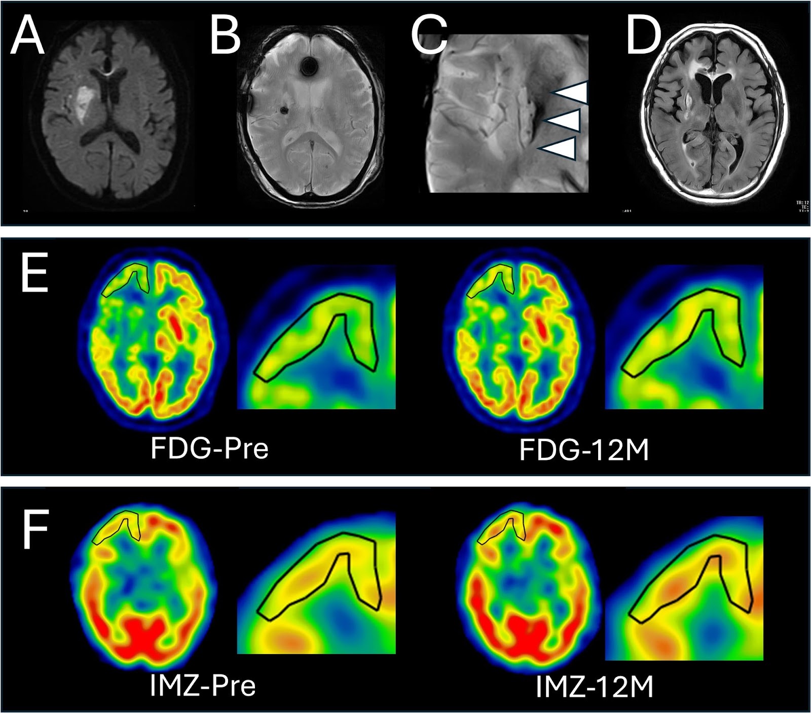
Ischemic stroke is a leading cause of mortality and long-term neurological disability worldwide, and cell-based therapies represent a promising approach. Although clinical studies have reported favorable outcomes following cell transplantation, the effects on host neuronal integrity remain incompletely understood. This study investigated temporal and spatial changes in fluorodeoxyglucose positron emission tomography (FDG-PET) and 123I-iomazenil single-photon emission computed tomography (IMZ-SPECT) after intracerebral cell transplantation in patients with subacute ischemic stroke and examined their relationship with functional recovery. Seven adults with severe post-stroke disability underwent autologous mesenchymal stromal cell (HUNS001-01) transplantation 47–64 days after stroke onset. Brain FDG-PET and IMZ-SPECT were performed preoperatively and at 1, 3, and 12 months post-transplantation. Regions of interest were first manually set in the ipsilateral cortex where the 12-month postoperative-to-preoperative standard uptake value ratio seems increased, and followed by quantitative measurement. Five of seven patients demonstrated 5% or more increase of FDG-PET and/or IMZ-SPECT uptake in peri-infarct cortical regions, predominantly within the frontal or temporal cortex. Transplanted cells localized either within metabolically enhanced regions or in anatomically remote areas. FDG-PET and IMZ-SPECT changes were strongly interacted in each other and were associated with functional improvement. Overall, improvement of glucose metabolism and synaptic density/viability were observed in patient with subacute ischemic stroke, which may have been attributable to cell transplantation. Trial registration: UMIN000026130.

Serial brain FDG-PET and IMZ-SPECT following intracerebral MSC transplantation in patients with subacute ischemic stroke
Graphical Abstract
Original ResearchVol. 17, Issue 260DOI: 10.1186/s13287-026-05147-6

Beyond conventional PRP: a rationale for bioengineered, growth-factor-defined platelet mimetics in alopecia—the precision re-engineered efficacy optimization framework

Authors: Rinky Kapoor, Raji Patil, Debraj Shome, Prashant Anilkumar Singh, Debalina Bose, Alivia Basu, M. S. Sukesh, Imran Ali, Michael Gold

Autologous platelet-rich plasma (PRP) is widely used for alopecia, but outcomes are often inconsistent due to procedural differences and patient-to-patient biological variability, including platelet yield, leukocyte content, and the mixed presence of stimulatory and inhibitory mediators. This commentary outlines a rationale for moving from variable autologous PRP toward defined PRP-inspired, growth-factor-based platelet mimetic formulations with batch-specified concentrations and relative proportions to enable more reproducible dosing and clearer clinical evaluation, aligned with Precision Re-Engineered Efficacy Optimization as a framework for standardizing potency, composition, and performance. Such formulations may improve consistency and scalability, but should be viewed as controlled reconstructions of selected PRP-associated signals rather than complete replicas of platelet releasate. Their translational value will depend on careful formulation characterization, staged proof-of-concept testing, and controlled clinical studies to establish safety, dosing, and comparative effectiveness.

Beyond conventional PRP: a rationale for bioengineered, growth-factor-defined platelet mimetics in alopecia—the precision re-engineered efficacy optimization framework
Graphical Abstract
Original ResearchVol. 17, Issue 101 • pp. 1-16DOI: 10.1186/s13287-025-04892-4

PTPN2 deficiency amplifies inflammatory signalling and impairs functional maturation of human stem cell-derived islets

Authors: Valerie Vandenbempt, Javier Negueruela, Francisco Ribeiro-Costa, Mariana Nunes, Leonardo Traini, Sema Elif Eski, Wadsen St-Pierre-Wijckmans, Stéphane Demine, Nicolas Baeyens, Hazem Ibrahim, Luciano G. Martelotto, Sumeet Pal Singh, Alessandra K. Cardozo, Mayank Bansal, Esteban N. Gurzov

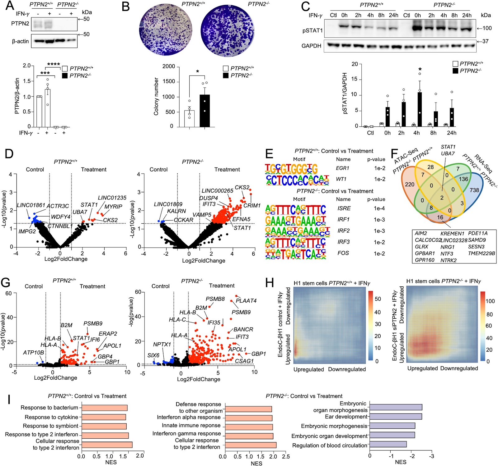
Background Protein tyrosine phosphatases (PTPs) play key roles in β-cell function and diabetes development. PTPN2 is a candidate gene for type 1 diabetes (T1D) that negatively regulates JAK/STAT signalling. However, the impact of PTPN2 deficiency on the differentiation and functionality of human stem cell-derived somatic metabolic cells remains unclear. Methods PTPN2 expression in β cells from T1D organ donors and during the differentiation of human stem cell-derived islets (SC-islets) was evaluated using single-cell RNA-Sequencing (scRNA-Seq) datasets. We differentiated CRISPR-Cas12a genome-edited PTPN2-deficient H1 human embryonic stem cells (H1-hESCs) into SC-islets, and scRNA-Seq was performed. The maturation and functionality of PTPN2-deficient SC-islets were assessed by implantation under the kidney capsule of NOD-SCID mice. Results scRNA-Seq analysis showed that PTPN2 expression was increased in β cells from recently diagnosed T1D and decreased in long-standing T1D organ donors compared with controls. Conversely, we found that PTPN2 expression was decreased at the early stages of SC-islet differentiation and reconstituted at the later stages, suggesting a developmental dynamic. PTPN2 deficiency exacerbated interferon-induced inflammatory signalling in stem cells and differentiated somatic metabolic cells. Interestingly, PTPN2 deficiency increased hedgehog signalling and reduced SC-islet differentiation efficiency in vitro. In addition, PTPN2-knockout SC-islets exhibited reduced glycaemic control after implantation in vivo, mediated by reduced endocrine cell identity and enhanced interferon signalling. Conclusions Our study postulates a key role of PTPN2 in preserving β-cell function during inflammatory and metabolic stress in SC-islets.

PTPN2 deficiency amplifies inflammatory signalling and impairs functional maturation of human stem cell-derived islets
Graphical Abstract
Original ResearchVol. 17, None • pp. 237DOI: 10.1186/s13287-026-05051-z

Awakening endogenous repair: salidroside boosts mitophagy in NPMSCs via SIRT1/FOXO3 to combat intervertebral disc degeneration

Authors: Zhengguang Li, Yiming Wu, Benkui Hua, Hua Sun, Huofeng Wu, Shuangjia Zai, Chen Liu, Yongbo Zhang, Zhaoyu Li, Xuan You, Yufeng Huang, Xuhua Lu, Guoyong Yin, Liang Zhang

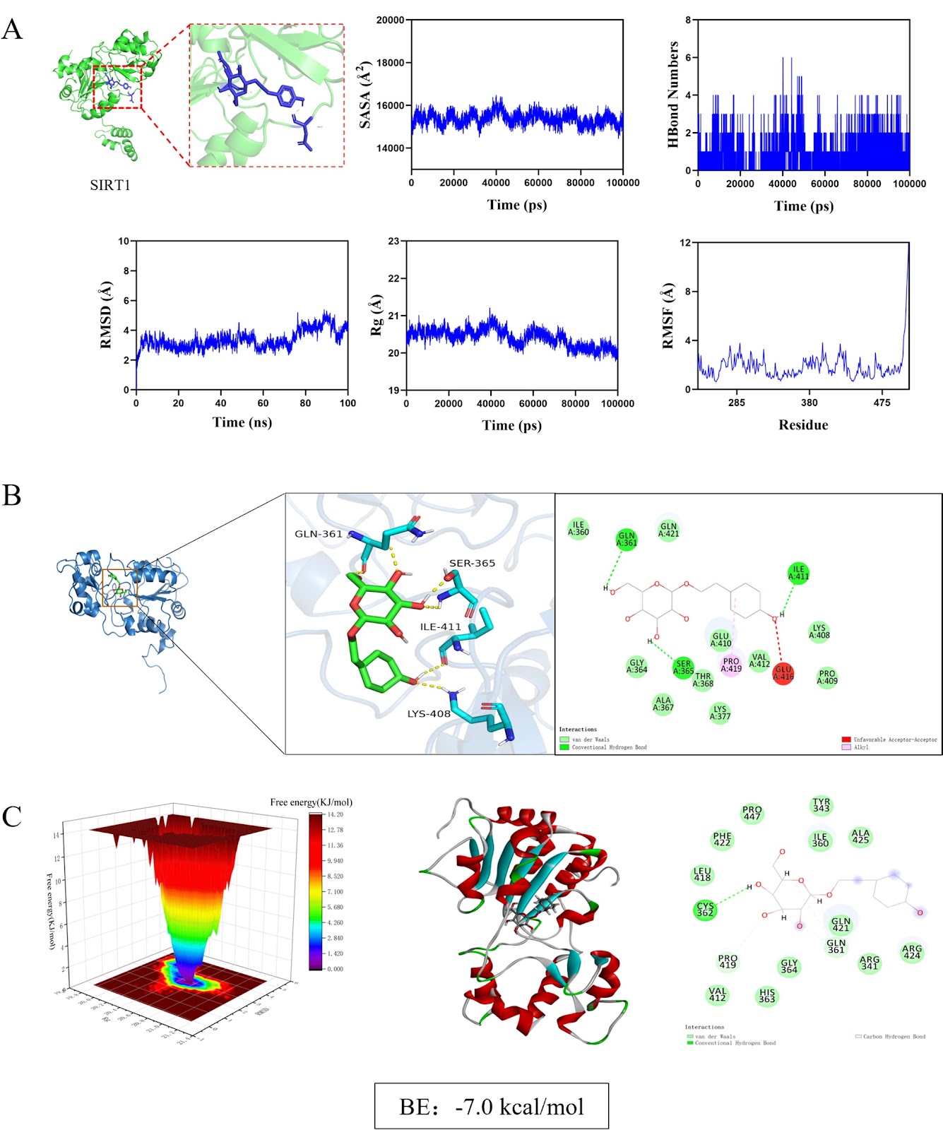
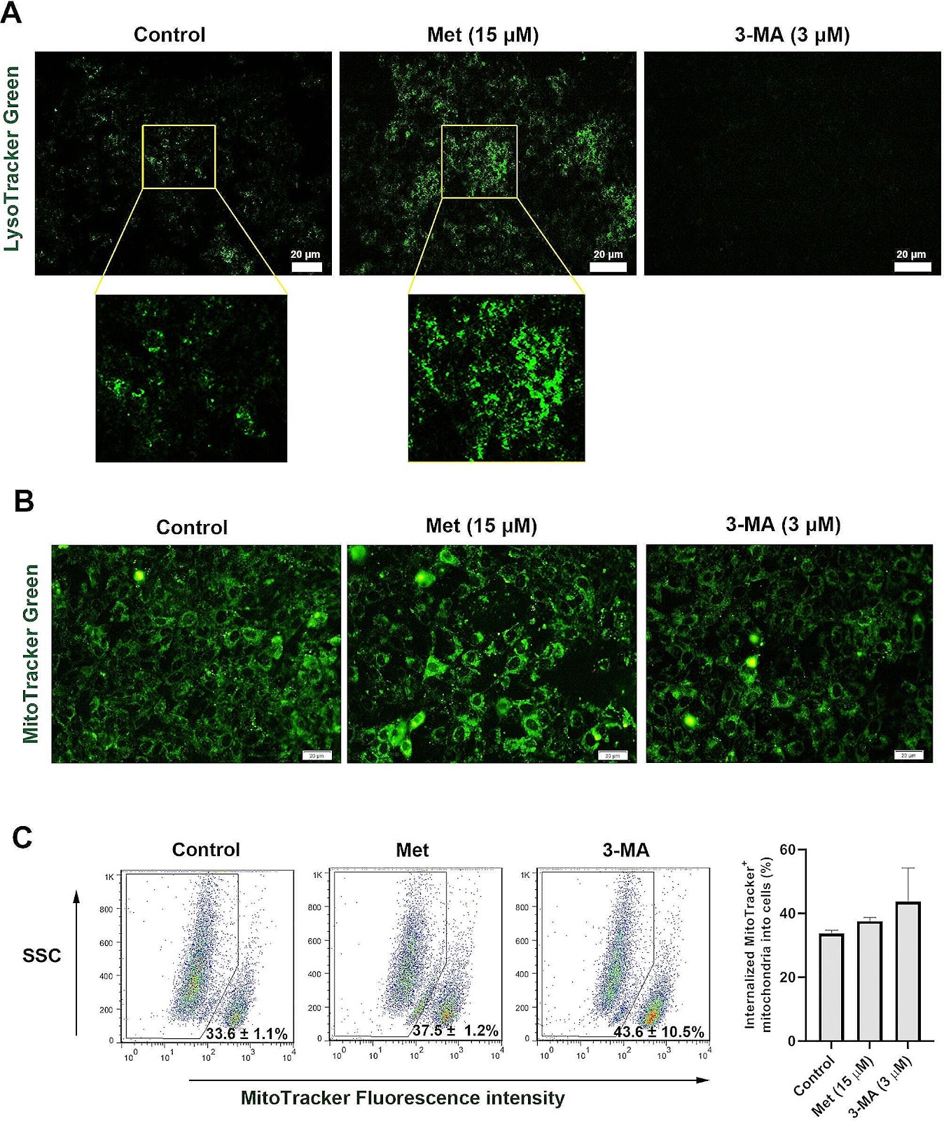
Background Intervertebral disc degeneration (IVDD) is a major cause of low back pain, characterized by mitochondrial dysfunction and impaired mitophagy in nucleus pulposus-derived mesenchymal stem cells (NPMSCs). Sirtuin 1 (SIRT1), a key regulator of mitochondrial quality control, is downregulated in degenerated discs. Salidroside (Sal), a natural compound from Rhodiola rosea, has shown potential in enhancing mitophagy, but its mechanism in IVDD remains unclear. Methods Using network pharmacology, molecular docking, and dynamics simulations, we identified SIRT1 as a key target of Sal. Human and rat NPMSCs were isolated and treated with tert-butyl hydroperoxide (TBHP) to induce degeneration. In vitro assays included CCK-8, EdU, SA-β-Gal, JC-1, Western blot, immunofluorescence, and transmission electron microscope (TEM). An in vivo rat IVDD model was established via needle puncture and treated with Sal and/or the autophagy inhibitor 3-MA. Histological, immunohistochemical, and imaging analyses were performed to evaluate IVDD. Results Sal bound stably to SIRT1 and activated SIRT1/FOXO3 pathway, promoting mitophagic flux, reducing reactive oxygen species accumulation, and suppressing apoptosis in NPMSCs. SIRT1 knockdown or 3-MA treatment abolished Sal’s protective effects. In vivo, Sal treatment preserved disc height, reduced apoptosis, and enhanced mitophagy, while 3-MA exacerbated degeneration.

Awakening endogenous repair: salidroside boosts mitophagy in NPMSCs via SIRT1/FOXO3 to combat intervertebral disc degeneration
Graphical Abstract
Original ResearchVol. 17, Issue 118DOI: 10.1186/s13287-026-04936-3

Ningxue Shengban decoction containing serum alleviates immune thrombocytopenia by modulating CD4+T cells balance via BMSCs-Exo-miR-199a-5p

Authors: Wuxia Yang, Yang Liu, Huiying Kang, Zhen Wang, Yanqi Song, Baoshan Liu, Aidi Wang

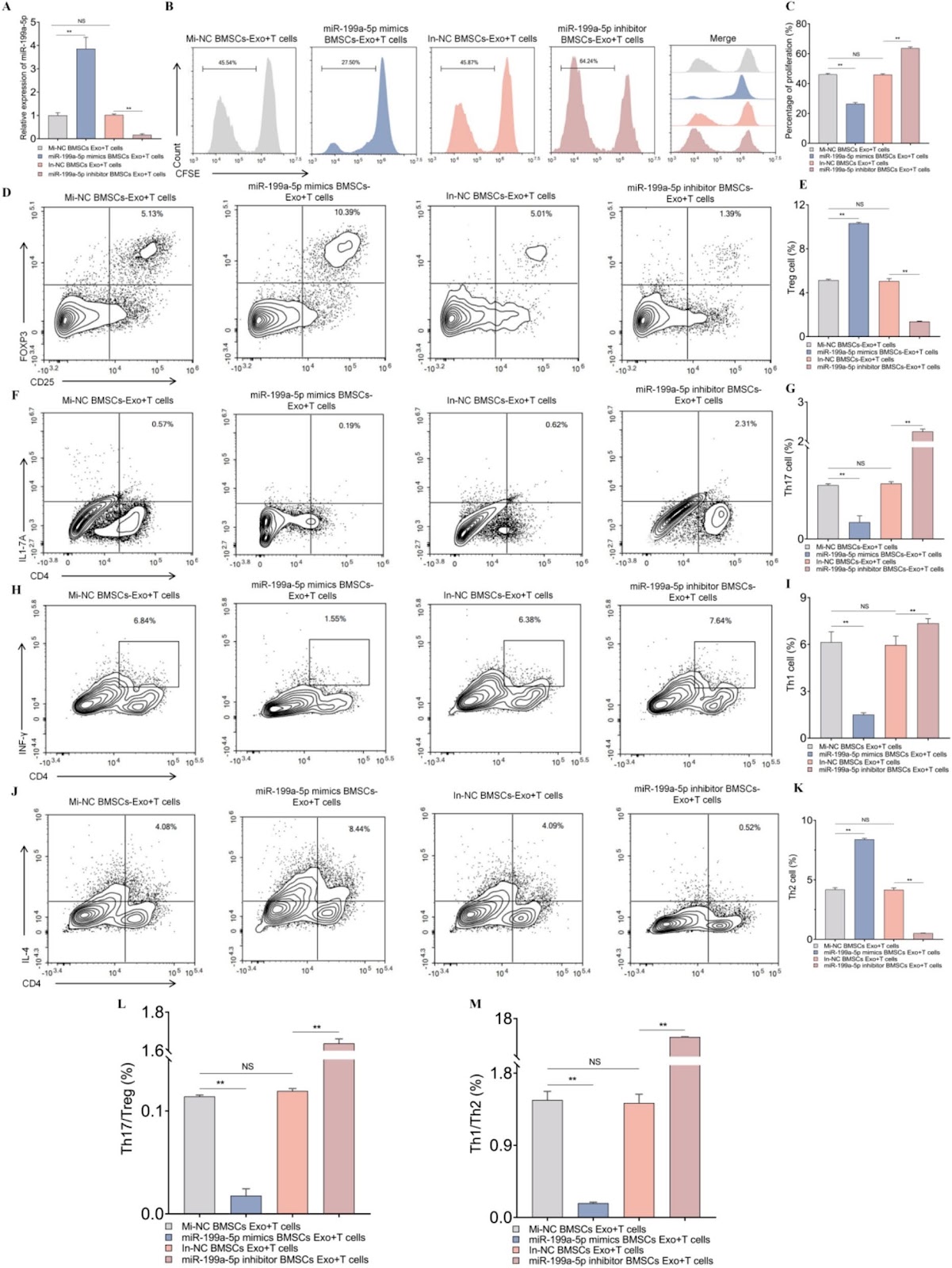
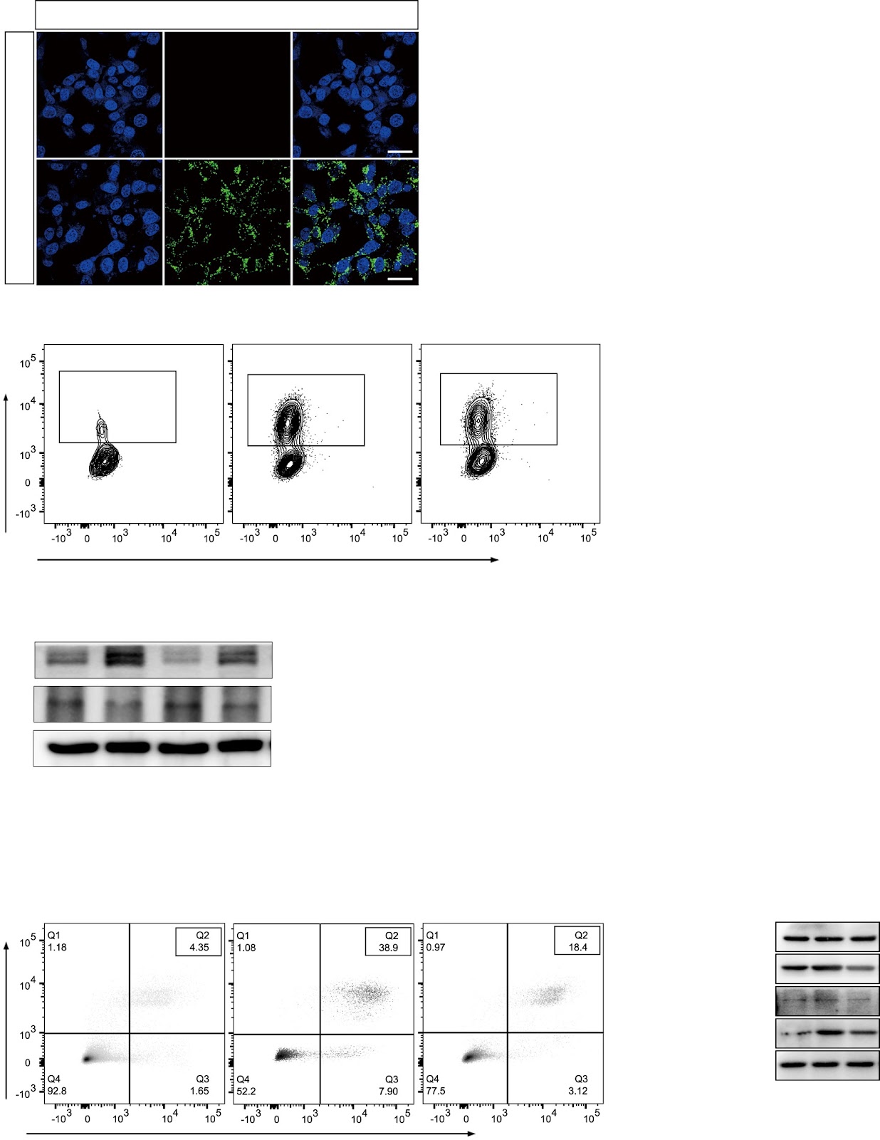
Background The abnormal immune response mediated by CD4+T cells is a key factor in Immune thrombocytopenia(ITP) progression. While Ningxue Shengban Decoction (NXSBD) is an effective therapeutic, its underlying mechanism and targets remain obscure. Aim This study aims to clarify the role of exosomal miR-199a-5p derived from bone marrow mesenchymal stem cells (BMSCs) in immune homeostasis, and to explore the therapeutic effects of exosomes from BMSCs(BMSCs-Exo) pretreated with NXSBD containing serum on ITP. Method We co-cultured CD4+T cells with BMSCs or pre-treated BMSCs-Exo. The proliferation and differentiation of CD4+T cells were then assessed using CFSE staining and flow cytometry (FCM). Additionally, an active ITP murine model was employed to assess the therapeutic efficacy of pre-treated BMSCs-Exo. Platelet counts were measured and organ indices were calculated. Serum autoantibody levels were measured by FCM and ELISA, changes in CD4⁺T cells subsets in the spleen were analyzed by FCM, megakaryocyte number and morphology in bone marrow tissues were examined by H&E staining, and key cytokine levels in mouse serum were quantified by ELISA. Results Our results indicate that the immunomodulatory effect of BMSCs-Exo on CD4+T cells is mediated by miR-199a-5p, and that NXSBD containing serum enhances this effect by increasing miR-199a-5p levels. In an active ITP murine model, BMSCs-Exo treatment significantly ameliorated the pathological features of ITP, as evidenced by increased peripheral platelet counts, reduced spleen and thymus indices, and decreased levels of autoantibodies. Immunophenotypic analysis revealed that an increased percentage of splenic Treg and Th2 cells, and a decreased percentage of Th17 and Th1 cells, were observed after BMSCs-Exo treatment. Additionally, BMSCs-Exo enhanced the production of mature megakaryocytes. Following BMSCs-Exo treatment, the levels of pro-inflammatory cytokines were sharply lowered, whereas anti-inflammatory cytokine levels were markedly elevated. BMSCs-Exo pretreated with NXSBD containing serum exert superior therapeutic efficacy compared with those derived from untreated BMSCs.

Ningxue Shengban decoction containing serum alleviates immune thrombocytopenia by modulating CD4+T cells balance via BMSCs-Exo-miR-199a-5p
Graphical Abstract
Original ResearchVol. 17, None • pp. 153DOI: 10.1186/s13287-026-04954-1

Tanshinone IIA-pretreated mesenchymal stem cells alleviate neuroinflammation in 3×Tg-AD mice via the TREM2/PI3K/Akt pathway

Authors: Jingjing Wu, Ying Ge, Li Zhang, Juan Huang, Nanqu Huang, Yong Luo

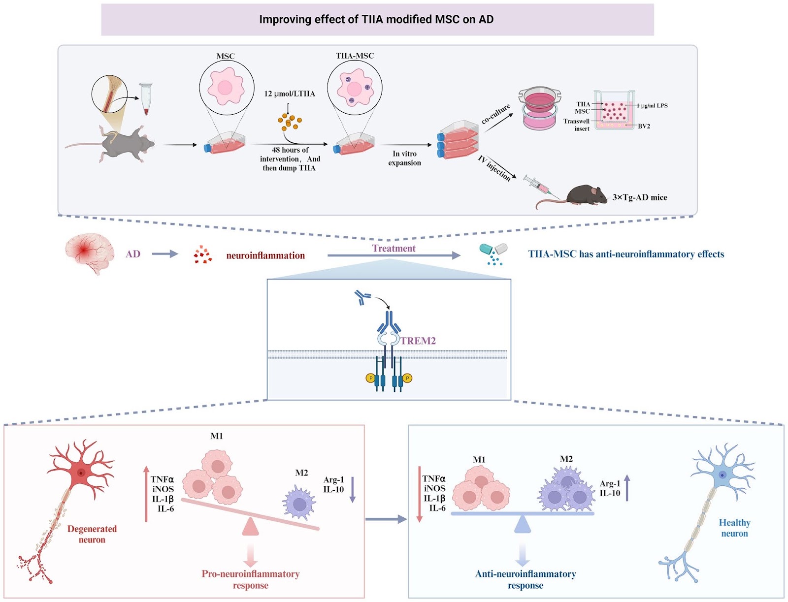
Neuroinflammation is a key pathogenic factor for neurodegenerative diseases. Mesenchymal stem cell (MSC) transplantation, as a potential strategy for regulating neuroinflammation, has received extensive attention. Our previous research revealed that compared with ordinary MSC, MSC pretreated with tanshinone IIA (TIIA), referred to as TIIA-MSC, exhibited superior anti-neuroinflammatory activity, but the mechanism of action remains unclear. To clarify the underlying mechanism, this study integrated in vitro and in vivo experiments and evaluated the therapeutic effect of TIIA-MSC in a triple-transgenic Alzheimer’s disease mouse model (3×Tg-AD mice) and explored its mechanism of action in a lipopolysaccharide (LPS)-induced BV2 microglial cell inflammation model. The results showed that TIIA-MSC could significantly improve the cognitive function of 3×Tg-AD mice, increase brain glucose metabolism levels, promote the recovery of synaptic and mitochondrial structures, and effectively alleviate neuroinflammatory responses. In vitro experiments further verified the superior inhibitory effect of TIIA-MSC on microglial cell activation and proinflammatory factor release. Mechanistic studies have indicated that the triggering receptor expressed on myeloid cells 2 (TREM2) is the key molecule that mediates this process. The knockdown of TREM2 expression significantly weakened the anti-inflammatory effect of TIIA-MSC, suggesting that TREM2 plays a central role in this process. Further analysis revealed that by activating the phosphoinositide 3-kinase (PI3K)/protein kinase B (Akt) signaling pathway downstream of TREM2, TIIA-MSC may promote the transformation of the functional state of microglia from mainly proinflammatory to having neuroprotective and repair properties. This study systematically revealed the molecular mechanism by which TIIA-MSC regulate microglial cell phenotypic transformation through the TREM2/PI3K/Akt pathway and exert anti-neuroinflammatory effects, providing new ideas and an experimental basis for expanding the application of MSC in the treatment of neurodegenerative diseases.

Tanshinone IIA-pretreated mesenchymal stem cells alleviate neuroinflammation in 3×Tg-AD mice via the TREM2/PI3K/Akt pathway
Graphical Abstract
Original ResearchVol. 17, None • pp. 75DOI: 10.1186/s13287-026-04896-8

Deficiency of extracellular vesicles miR-32 from bone marrow mesenchymal stem cells alleviates vascular calcification in type 2 diabetes by inhibiting endothelial ferroptosis

Authors: Zhengjie Lin, Anqi Li, Jie Zheng, Kun Luo, Fei Liang, Shiyan Liu, Zhengfeng Liang, Wei Liu, Jian Tang, Xiaolin Zhong, Jianghua Liu

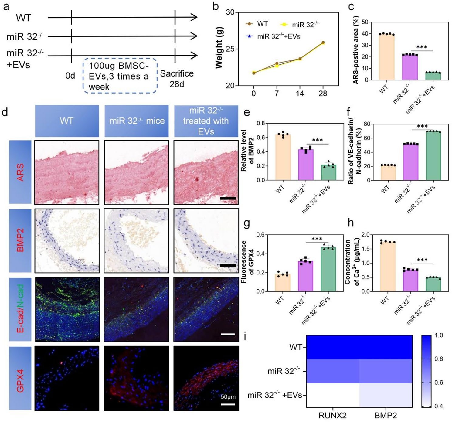
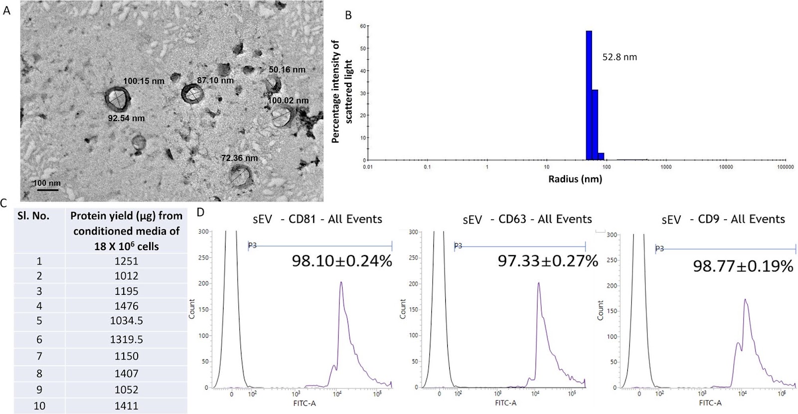
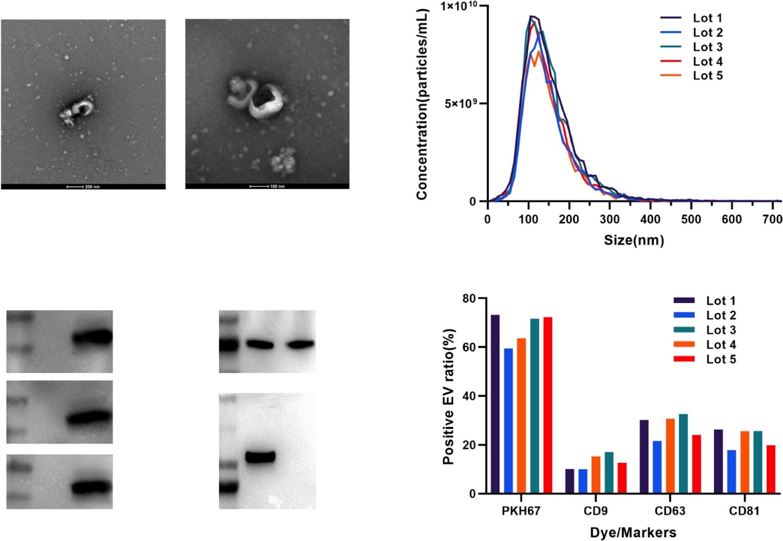
Background The development of vascular calcification (VC) in diabetes is closely related to the endothelial-to-mesenchymal transition (EndMT). We found that microRNA-32-5p (miR-32) was elevated in the plasma of calcification patients. However, it is unclear whether miR-32 mediates the function of bone marrow mesenchymal stem cell-derived extracellular vesicles (BMSC-EVs) in type 2 diabetes (T2D) VC. Methods BMSC-EVs were characterized by TEM, NTA, Western blotting, and confocal microscopy. Alizarin Red and ALP staining assessed the severity of VC. qRT-PCR and Western blotting evaluated the expression of BMP2, RUNX2, GPX4, SLC7A11, VE-cadherin, and N-cadherin, while immunofluorescence was used for detecting VE-cadherin and N-cadherin. In vivo validation was performed using miR-32–/– and ApoE–/– mice. RNA sequencing (RNA-seq) and bioinformatics analysis was conducted to explore underlying mechanisms. Results We demonstrated that BMSC-EVs attenuate VC in endothelial cells (ECs) and inhibit EndMT. In vivo, histological analysis showed that treatment with BMSC-EVs significantly reduced the severity of VC associated with T2D. Notably, knockout of miR-32 further enhanced the inhibitory effect of BMSC-EVs on VC. Mechanistically, transcriptomic and functional analyses suggest that the protective effect of BMSC-EVs on VC is associated with regulation of the MAPK/FoxO signaling pathway, potentially mediated by modulation of ferroptosis. Conclusion These findings demonstrate that BMSC-EVs attenuate T2D-associated VC, partially through miR-32-mediated suppression of EC ferroptosis.

Deficiency of extracellular vesicles miR-32 from bone marrow mesenchymal stem cells alleviates vascular calcification in type 2 diabetes by inhibiting endothelial ferroptosis
Graphical Abstract
Original ResearchVol. 17, None • pp. 78DOI: 10.1186/s13287-026-04895-9

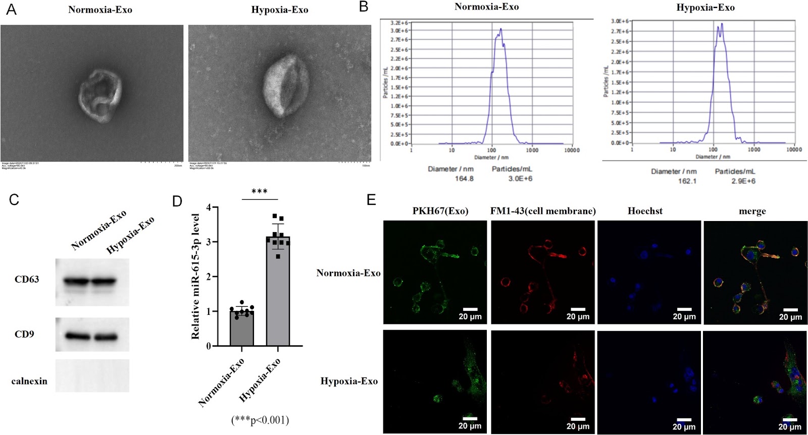
Hypoxia-conditioned BMSC exosomes improve short-term spinal cord injury outcomes via the miR-615-3p/PDE4C-mediated cAMP/PKA pathway

Authors: Wei Bian, Xiangyu Zeng, Ziwen Liu, Mingyan Guan, Tegeleqi Bu, Haoze Li, Zewei Gao, Jianyu Liu

Spinal cord injury (SCI) remains a significant global health challenge with limited effective therapeutic options. Exosomes derived from mesenchymal stem cells (MSCs) have emerged as promising neuroprotective agents due to their biocompatibility and immunomodulatory properties. This study investigated the therapeutic potential of hypoxia-conditioned bone marrow MSC (BMSC)-derived exosomes in both in vitro and in vivo SCI models. Hypoxic preconditioning significantly enriched miR-615-3p in bone marrow mesenchymal stem cell (BMSC)-derived exosomes. In spinal neuron injury models, hypoxic exosomes enhanced cell viability, reduced apoptosis, and ameliorated dysfunction of the mitochondria-associated endoplasmic reticulum membranes (MAMs). Mechanistically, miR-615-3p directly targeted and suppressed phosphodiesterase 4 C (PDE4C), activating the cyclic adenosine monophosphate (cAMP)/protein kinase A (PKA) pathway. This in turn modulated calcium signaling, attenuated mitochondrial calcium overload, and reduced endoplasmic reticulum stress (ERS). In a mouse model of SCI, short-term treatment with hypoxic exosomes promoted functional recovery within a 14-day post-injury period, as evidenced by improved locomotor performance, reduced lesion volume, attenuated tissue edema, and decreased inflammatory cell infiltration. Furthermore, in vivo administration of hypoxic exosomes upregulated miR-615-3p and downregulated PDE4C expression in injured spinal cord tissues. These results demonstrate that hypoxia-conditioned BMSC-derived exosomes exert neuroprotective effects via the miR-615-3p/PDE4C axis, highlighting their potential as a novel therapeutic strategy for SCI by targeting calcium homeostasis and mitochondrial-ER dysfunction. These findings demonstrate the short-term therapeutic potential of hypoxia-conditioned exosomes in SCI. However, further preclinical studies, including long-term follow-up to assess the durability of recovery and potential late-onset effects, alongside clinical validation, are warranted before clinical translation.

Hypoxia-conditioned BMSC exosomes improve short-term spinal cord injury outcomes via the miR-615-3p/PDE4C-mediated cAMP/PKA pathway
Graphical Abstract
Original ResearchVol. 17, None • pp. 209DOI: 10.1186/s13287-026-05020-6

Therapeutic potential of mesenchymal stromal cells in COVID-19: a meta-analysis of clinical trials conducted since the pandemic onset

Authors: Meng-Qi Yuan, Yue-Fei Pan, Zi-Ying Zhang, Ya-Xin Wu, Kai-Di Zhu, Ze-Rui Wang, Ze-Yi Zhang, Jia-Qi Xiong, Zhe Xu, Lei Huang, Fu-Sheng Wang, Lei Shi

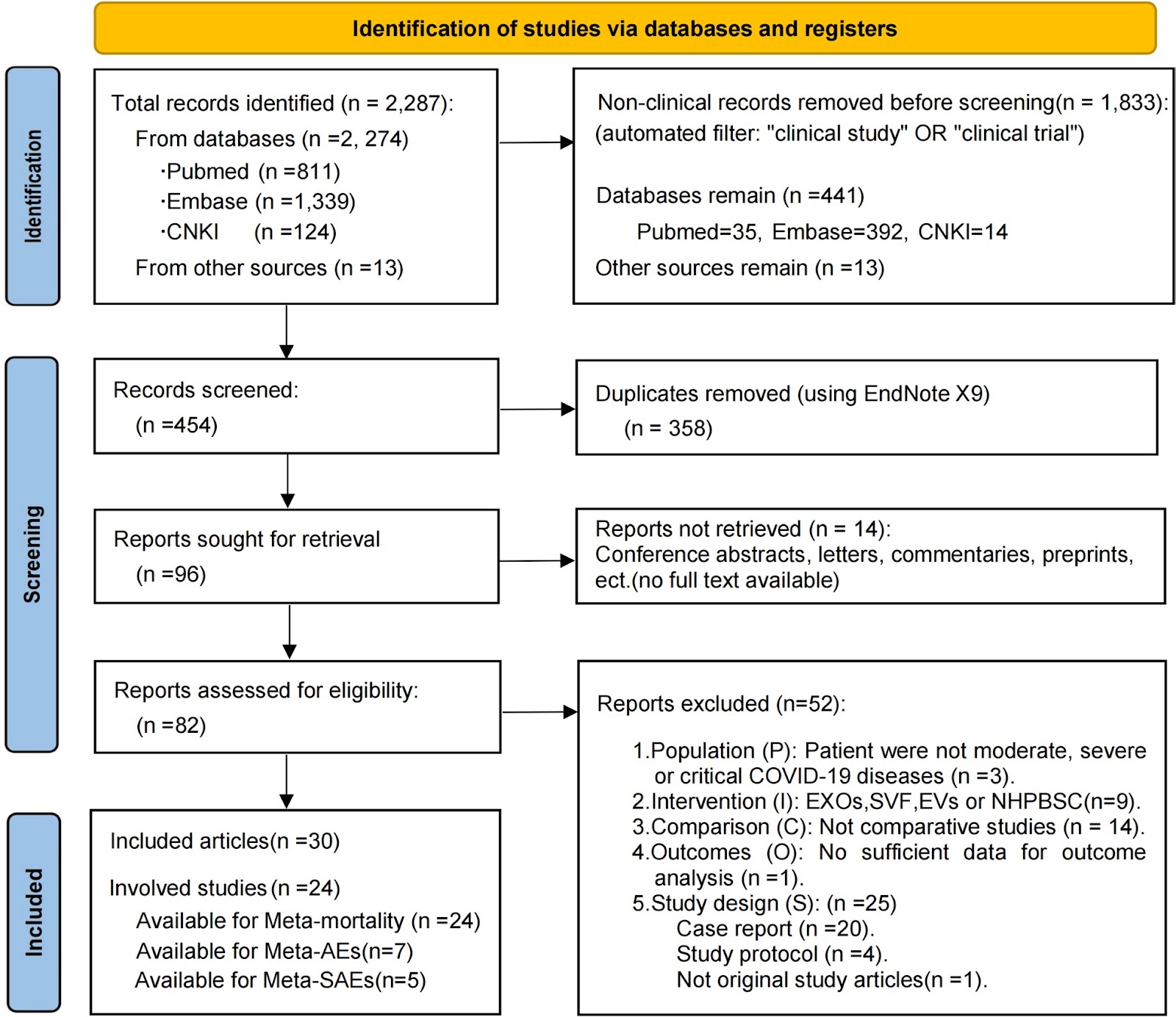
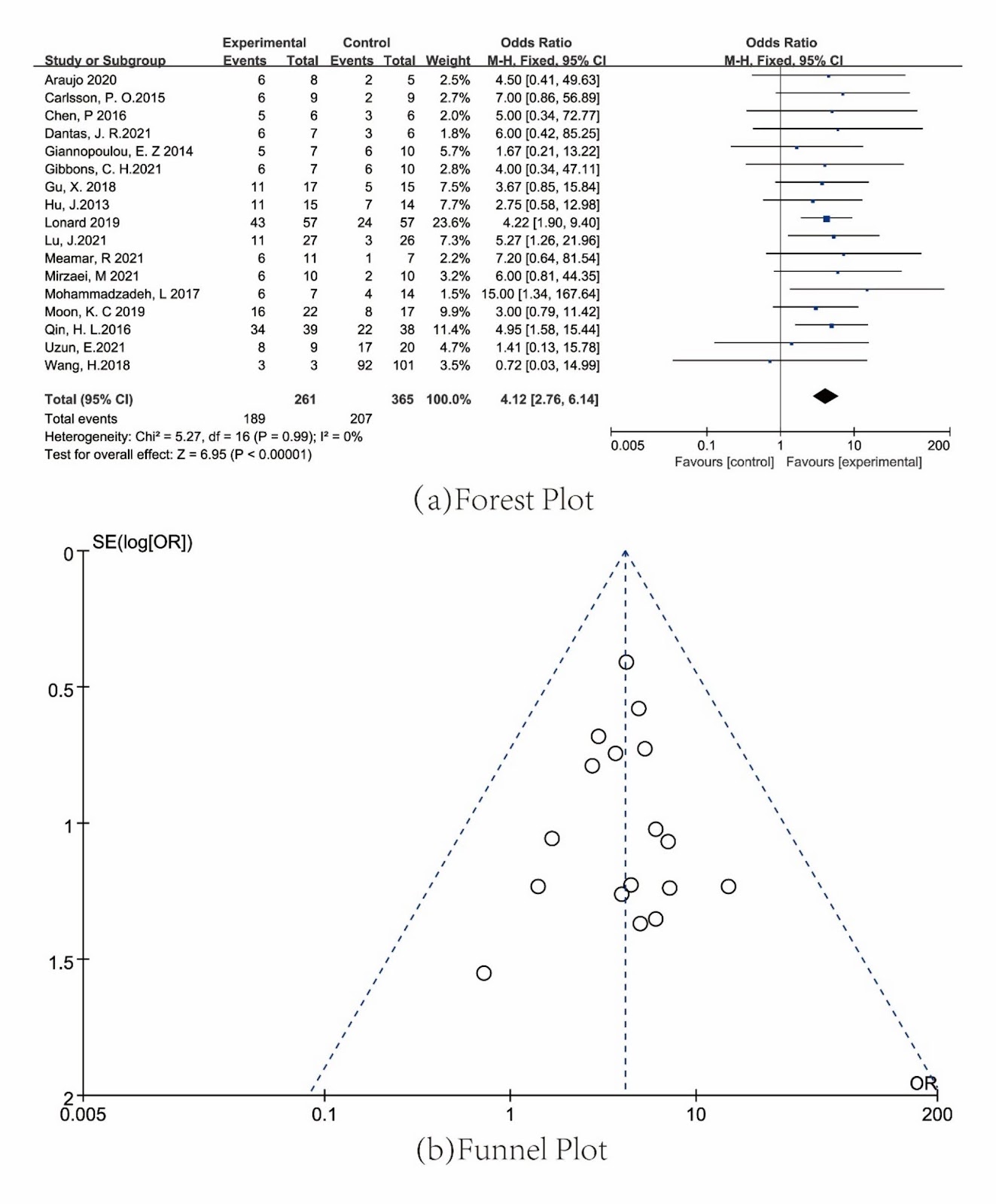
Background Severe acute respiratory syndrome coronavirus 2 (SARS-CoV-2) infection can induce immune dysregulation and multi-organ injury; mesenchymal stromal cell (MSC) therapy has shown promise in clinical trials for COVID-19 and may have broader applicability to pneumonia induced by respiratory viruses (e.g., the influenza virus). This meta-analysis synthesized the available comparative clinical evidence on the safety and efficacy of MSCs in patients with moderate to critical COVID-19 and examined the reported outcomes relevant to Long-COVID. Methods We searched the PubMed, Embase, and CNKI databases for original, comparative studies in moderate, severe, or critical COVID-19 published up to September 2, 2024. Twenty-four eligible studies (13 RCTs and 11 non-randomized controlled trials; n=1080) were included in the mortality meta-analysis. Patients were assigned to either the intervention group (MSC therapy plus standard care) or the control group (standard care with or without placebo). The primary efficacy outcome was all-cause mortality, while the primary safety outcomes were adverse events (AEs) and serious adverse events (SAEs). Secondary outcomes included clinical recovery, hospitalization metrics, chest imaging, and inflammatory biomarkers. We performed a pooled meta-analysis on mortality with subgroup analyses (by disease severity, administration route, dosing frequency, and study design), assessment of publication bias (using funnel plots and Egger’s test), and evaluation of the quality of evidence via the GRADE approach. AEs/SAEs were analyzed using meta-analysis and descriptive statistics, while other secondary outcomes were summarized descriptively. Results MSC therapy significantly reduced all-cause mortality (MSC: 26.4% vs control: 31.9%; fixed-effect OR=0.74, 95% CI 0.55–0.99), with low heterogeneity (I2=2.8%, P=0.422[Q-test]) and no publication bias. The quality of evidence

Therapeutic potential of mesenchymal stromal cells in COVID-19: a meta-analysis of clinical trials conducted since the pandemic onset
Graphical Abstract
Original ResearchVol. 17, None • pp. 107DOI: 10.1186/s13287-026-04921-w

Inhalation of mesenchymal stromal cell-derived extracellular vesicles activates macrophage polarization through the miR-22-3p/NLRP3/IL-1β pathway, ameliorating lung ischemia-reperfusion injury

Authors: Tao Wang, Guodong Wu, Peigen Gao, Fenghui Zhuang, Zeyu Wang, Ziheng Zhou, Chongwu Li, Junqi Wu, Deping Zhao

Background Lung ischemia–reperfusion injury (IRI) is a major contributor to primary graft dysfunction (PGD) after lung transplantation. Mesenchymal stem cell-derived extracellular vesicles (MSC-EVs) have emerged as promising therapeutic agents in inflammatory diseases by ameliorating tissue damage and promoting repair. However, the anti-inflammatory efficacy of these approaches and the underlying mechanisms in lung ischemia–reperfusion injury remain incompletely understood. Methods The protective effects of mesenchymal stromal cell-derived extracellular vesicles (MSC-EVs) against lung ischemia–reperfusion injury were evaluated using two delivery approaches, inhalation and intravenous injection. Both in vivo and in vitro models were employed to assess the biological activity of MSC-EVs and to elucidate the underlying molecular mechanisms. In addition, a rat orthotopic lung transplantation (OLT) model was established to further examine the translational relevance of MSC-EVs. Results MSC–EVs treatment significantly ameliorated lung IRI, with inhalation showing superior efficacy over intravenous delivery. Mechanistically, miR-22-3p within MSC-EVs targeted macrophage NLRP3, suppressing activation of the NLRP3/Caspase-1/IL-1β pathway and promoting M2 polarization. The protective efficacy was confirmed in a clinically relevant rat OLT model, underscoring their translational potential

Inhalation of mesenchymal stromal cell-derived extracellular vesicles activates macrophage polarization through the miR-22-3p/NLRP3/IL-1β pathway, ameliorating lung ischemia-reperfusion injury
Graphical Abstract
Original ResearchVol. 17, Issue 1 • pp. 1-18DOI: 10.1186/s13287-025-04774-9

Bone marrow–derived mesenchymal stem cells alleviate hepatic lipid metabolism disorders after scald injury: integrating liver transcriptome and metabolome

Authors: Zhian Chen, Ya'e Yang, Xiangwen Shi, Rensheng Yang, Wei Fang, Guangjin Liang, Yun Li, Jing Gao, Lihua Ma, Junchun Yang, Rongqing Pang

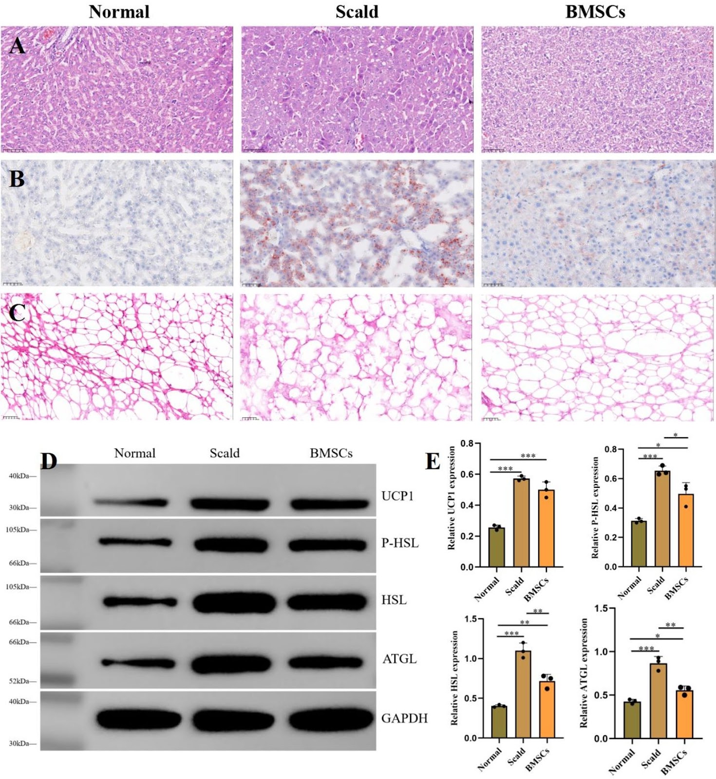
Previous studies have confirmed that scald injuries can lead to disturbances in hepatic lipid metabolism, and bone marrow-derived mesenchymal stem cells (BMSCs) have emerged as a promising therapeutic strategy for alleviating such disorders. However, research focusing on the regulation and restoration of liver lipid metabolic processes remains limited. In this study, we investigated the effects of BMSCs on hepatic lipid metabolism disorders induced by scald injury in rats through integrated transcriptomic and metabolomic analyses. The results demonstrated that portal vein infusion of BMSCs markedly improved body weight recovery, reduced hepatic lipid accumulation, normalized serum lipid profiles, and attenuated liver injury following scalding. Combined transcriptomic and metabolomic data further suggested that the therapeutic mechanism may involve inhibition of NF-κB/Gadd45a signaling in hepatocytes, restoration of sphingolipid metabolism, enhancement of hepatic lipid conversion, and suppression of adipocyte lipolysis. Overall, this study provides a theoretical basis for the potential clinical application of BMSCs in treating hepatic lipid metabolism disorders secondary to severe burn injury.

Bone marrow–derived mesenchymal stem cells alleviate hepatic lipid metabolism disorders after scald injury: integrating liver transcriptome and metabolome
Graphical Abstract
Original ResearchVol. 17, None • pp. 100DOI: 10.1186/s13287-026-04903-y

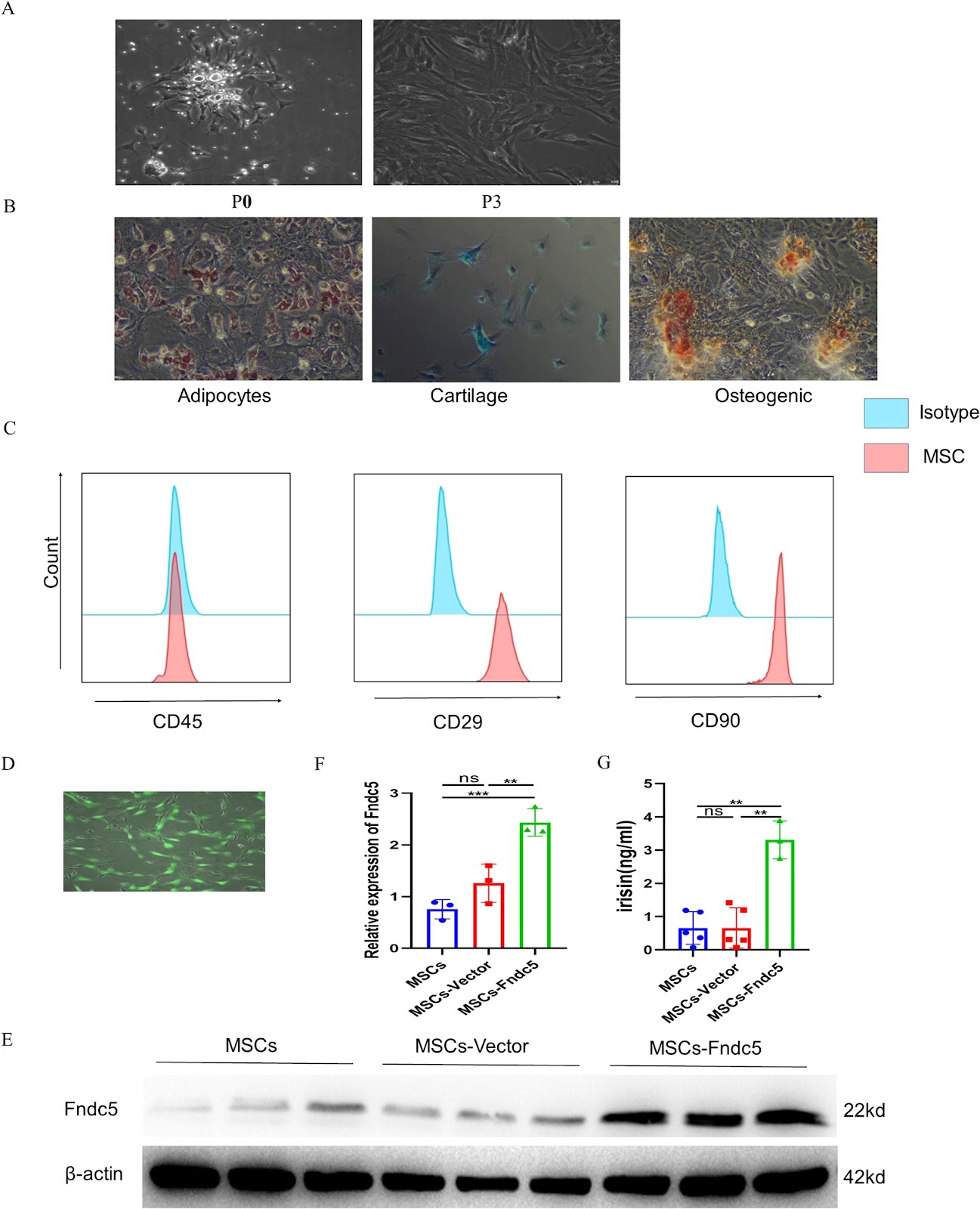
Fndc5 modification optimizes the therapeutic effect of rat MSCs on sepsis-induced ALI/ARDS via activating the PI3K/AKT signaling pathway

Authors: Yuling Luo, Minhua Li, Shan Lin, Zheng Gong, Sumei Wang, Ziqing Zhou, Shiyue Li

Background Acute lung injury/Acute respiratory distress syndrome (ALI/ARDS) is a life-threatening inflammatory lung disorder characterized by high mortality rates and a lack of effective treatment options. Although mesenchymal stem cell (MSC)-based therapies have emerged as a promising approach for ARDS management, optimizing their therapeutic efficacy remains a significant challenge. Recent advances in gene modification techniques have opened new avenues for enhancing MSC functionality. Among these, Fibronectin type III domain-containing protein 5 (Fndc5)/irisin has attracted considerable attention due to its ability to improve endothelial function. This study aims to evaluate the therapeutic potential of Fndc5-modified MSCs in sepsis-induced ALI/ARDS and to elucidate the underlying molecular mechanisms driving their protective effects. Methods To comprehensively evaluate the therapeutic potential of Fndc5-modified MSCs (MSCs-Fndc5) in ARDS, we employed both in vivo and in vitro experimental models. In vivo, a mouse model of sepsis-induced ALI was established through intraperitoneal injection of lipopolysaccharide (LPS), and the protective effects of MSCs-Fndc5 were systematically assessed by analyzing lung histopathology, inflammatory cytokine levels, vascular endothelial integrity, lung wet-to-dry weight ratio, and MSC retention in lung tissue. In parallel, in vitro studies were conducted to investigate the role of MSCs-Fndc5 in mitigating LPS-induced endothelial cell (EC) injury, with a focus on EC proliferation, angiogenesis, barrier permeability, apoptosis, and the regulation of key signaling pathways. Results Fndc5 modification significantly increased the retention rate of MSCs in sepsis-induced ALI murine model while augmenting their in vitro proliferation and migration potential. In vivo, treatment with Fndc5-modified MSCs markedly attenuated lung inflammation, as evidenced by reduced levels of pro-inflammatory cytokines, decreased neutrophil infiltration, and improved lung histopathology. Additionally, MSCs-Fndc5 alleviated pulmonary edema, reduced fibrosis, lowered the lung wet-to-dry weight ratio, and preserved vascular endothelial integrity. In vitro, Fndc5 modification optimizes the therapeutic effect of rat MSCs on sepsis-induced ALI/ARDS via activating the PI3K/AKT signaling pathway.

Fndc5 modification optimizes the therapeutic effect of rat MSCs on sepsis-induced ALI/ARDS via activating the PI3K/AKT signaling pathway
Graphical Abstract
Original ResearchVol. 17, Issue 276 • pp. 1-17DOI: 10.1186/s13287-026-05223-x

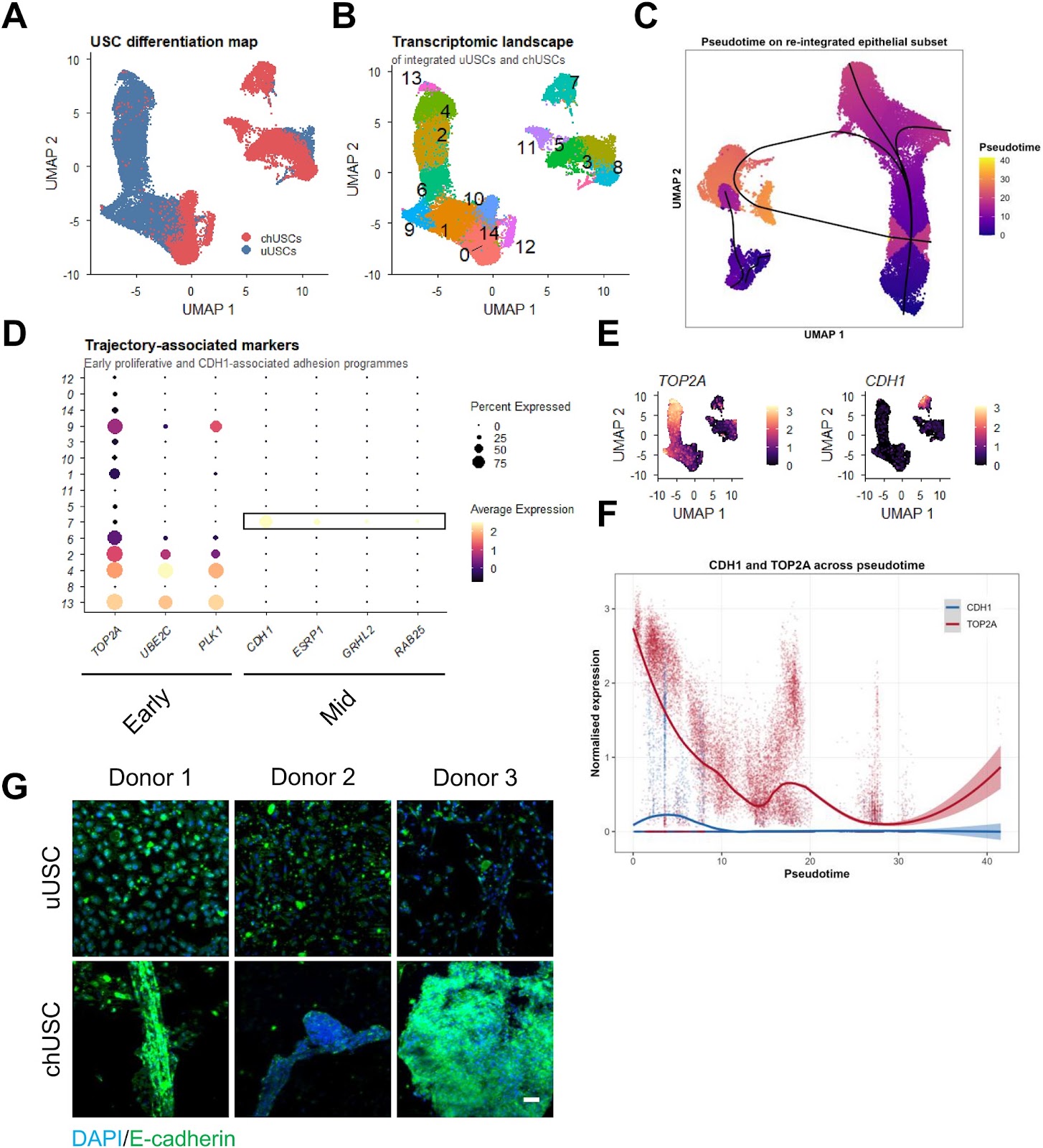
A single-donor proof-of-concept single-cell analysis maps heterogeneous differentiation trajectories toward cartilage-like states in human urine-derived stem cells

Authors: Alexander Schulz, Emily M. Brockmann, Miriam Zentgraf, Andreas S. Baur, Steffen Uebe, Arif B. Ekici, Mark Dedden, Sebastian Zundler, Christian T. Thiel

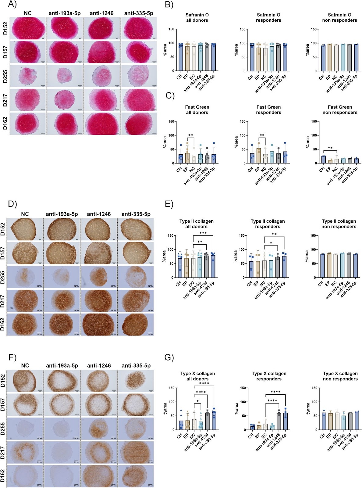
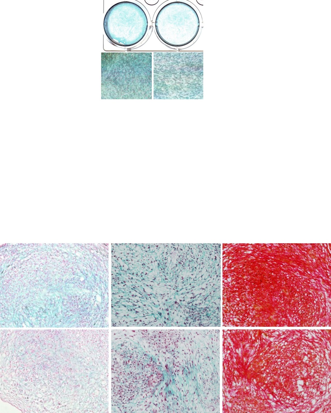
Background Urine-derived stem cells (USCs) represent an accessible and non-invasive cell source with reported chondrogenic differentiation potential. However, the cellular heterogeneity and transcriptional dynamics underlying USC differentiation remain incompletely understood, limiting their translational interpretation. Methods We combined functional differentiation assays with single-cell RNA sequencing to characterize USC differentiation at both phenotypic and transcriptional levels. Chondrogenic and osteogenic differentiation were assessed using histological staining, quantitative PCR, and three-dimensional spheroid cultures. Single-cell transcriptomic analysis was performed on integrated datasets of undifferentiated and differentiated USCs, followed by pseudotime trajectory inference and mapping to a human cartilage reference atlas. Results Chondrogenic induction resulted in reproducible acquisition of cartilage-associated features, including glycosaminoglycan-rich extracellular matrix deposition, increased expression of SOX9, and formation of aggrecan-positive spheroids. In this donor, single-cell analysis mapped an inferred differentiation trajectory from proliferative states towards differentiated populations, although the fine-grained pseudotemporal ordering was sensitive to analytical choices and is therefore interpreted qualitatively. Along this inferred trajectory, we identified a candidate transient transcriptional state associated with elevated CDH1 expression and epithelial-like aggregation features. Probabilistic mapping to a human cartilage reference atlas indicated that overall mapping confidence was low (median prediction score 0.34) and that only a minority of cells showed confident transcriptional similarity (prediction score ≥ 0.5) to mature/articular cartilage-associated reference states (7.8% of all cells and 17.6% of chondrogenically induced cells). This confident similarity was concentrated in a few clusters at the differentiated end of the trajectory rather than representing the bulk of the culture, and label-transfer confidence was not equated with chondrocyte identity. Despite this enrichment, differentiated populations exhibited transcriptional heterogeneity, including subsets

A single-donor proof-of-concept single-cell analysis maps heterogeneous differentiation trajectories toward cartilage-like states in human urine-derived stem cells
Graphical Abstract
Original ResearchVol. 17, NoneDOI: 10.1186/s13287-026-04991-w

Mesenchymal stem cell-derived extracellular vesicles in the treatment of type 2 diabetes and its complications: current progress and future directions

Authors: Sha Zhang, Zong-Yu Zhang, Ruo-Nan Tang, Kai Zhang, Yu Fu, Hua Tian, Jing Ma, Yan Jin, Chen-Xi Zheng, Bing-Dong Sui

Type 2 diabetes (T2D) and its complications represent a complex disorder involving multiple pathophysiological processes. Although conventional therapeutic approaches partially regulate blood glucose, they fail to fundamentally reverse disease progression or effectively prevent complications. This review summarizes the current research advance and challenges of using different forms of mesenchymal stem cell-derived extracellular vesicles (MSC-EVs) in treating T2D and complications. It begins with an introduction to the characteristics of MSC-EVs. Subsequently, the mechanisms and therapeutic prospects of natural MSC-EVs are analyzed, with a focus on their roles in inflammatory modulation, tissue regeneration, and improving insulin resistance. Engineering MSC-EVs, covering strategies including optimizing MSC culture conditions, modifying EV contents, and establishing MSC-EV delivery systems based on bioactive materials are then discussed, which boost EV yield and quality while enhancing therapeutic efficacy. Current challenges, including the limited yield and high heterogeneity of natural MSC-EVs, as well as issues related to long-term safety, immunocompatibility, and large-scale production of engineered MSC-EVs are finally overviewed, with emphasizing artificial intelligence in guiding future research directions. These summaries are crucial for clinical translation of MSC-EVs and will ultimately provide T2D patients with an effective and safe treatment option.

Mesenchymal stem cell-derived extracellular vesicles in the treatment of type 2 diabetes and its complications: current progress and future directions
Graphical Abstract
Original ResearchVol. 17, None • pp. 204DOI: 10.1186/s13287-026-05026-0

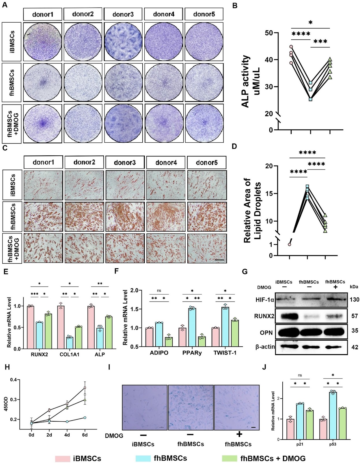
DMOG pretreatment restores osteogenic–adipogenic balance and mitochondrial function in ONFH BMSCs through the HIF-1α/Homer3 pathway

Authors: Qiheng Chen, Lingxian Yi, Penghui Nie, Jie Wang, Jing Zhu, Jiang Peng, Tujun Weng

Background  Osteonecrosis of the femoral head (ONFH) is a progressive orthopedic disorder that often culminates in femoral head collapse and joint failure. Dysfunction of bone marrow mesenchymal stem cells (BMSCs), including impaired osteogenesis, enhanced adipogenesis, and mitochondrial dysfunction, has been increasingly recognized as a central driver of ONFH pathogenesis. However, the molecular mechanisms linking metabolic stress to lineage imbalance remain poorly defined. Methods  Paired BMSCs were isolated from necrotic femoral head regions (fhBMSCs) and the iliac crest (iBMSCs) of ONFH patients. Functional assays, RNA sequencing, and molecular analyses were performed to evaluate the effects of the hypoxia mimetic dimethyloxalylglycine (DMOG) on osteogenic–adipogenic balance, mitochondrial function, and senescence. Loss-of-function experiments targeting hypoxia-inducible factor-1α (HIF-1α) and Homer3 were conducted to elucidate mechanistic pathways. Results  Compared with iBMSCs, fhBMSCs exhibited impaired osteogenesis, enhanced adipogenesis, mitochondrial dysfunction, and increased senescence. DMOG pretreatment restored osteogenic differentiation, suppressed adipogenesis, improved mitochondrial dynamics, reduced oxidative stress, and enhanced bioenergetic metabolism. These protective effects were dependent on HIF-1α stabilization. Transcriptomic profiling identified Homer3 as a downstream negative regulator of HIF-1α. Homer3 was aberrantly upregulated in fhBMSCs but suppressed by DMOG, and its knockdown mimicked the effects of DMOG by promoting osteogenesis, inhibiting adipogenesis, enhancing mitophagy, and restoring mitochondrial function. Conversely, silencing HIF-1α abolished DMOG-mediated benefits and reinstated Homer3 expression.

DMOG pretreatment restores osteogenic–adipogenic balance and mitochondrial function in ONFH BMSCs through the HIF-1α/Homer3 pathway
Graphical Abstract
Original ResearchVol. 17, None • pp. 91DOI: 10.1186/s13287-026-04913-w

Extracorporeal cardiac shock wave stimulation enhances the therapeutic efficacy of intravenously delivered endothelial colony-forming cells via PI3K/AKT signaling in a rat myocardial infarction model

Authors: Mingqiang Wang, Dan Yang, Yiming Ma, Yunke Shi, Jinping Lun, Chaoyue Zhang, Xinbin Li, Yuchen Shi, Hongyan Cai

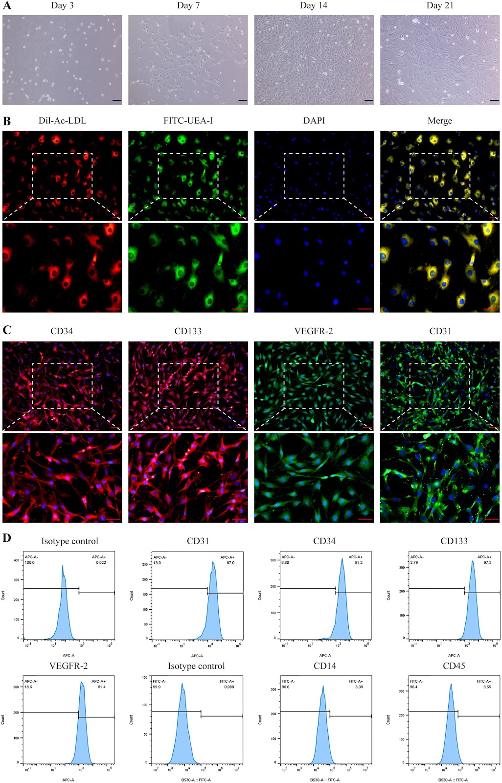
Background Extracorporeal cardiac shock wave (ECSW) therapy enhances the function of endothelial colony-forming cells (ECFCs), but whether it can serve as a preconditioning strategy to enhance myocardial infarction (MI) therapy remains unclear. This study investigated the efficacy and mechanism of intravenously delivered ECSW-preconditioned ECFCs (SW-ECFCs) in a rat MI model. Methods ECFCs were isolated from the bone marrow of ApoE-/- rats and fully characterized. RNA sequencing of control ECFCs versus SW-ECFCs revealed significant enrichment of the PI3K/AKT pathway. We therefore performed a series of in vitro functional assays on these cells, including Transwell migration, Matrigel tube formation, CCK-8 proliferation, flow cytometric apoptosis analysis, and VEGF-A ELISA. The role of the PI3K/AKT pathway was interrogated using the inhibitor LY294002. Subsequently, an acute MI model was established in ApoE-/- rats via left anterior descending coronary artery ligation. Rats were randomized into four groups: MI+PBS, MI+ECFCs, MI+SW-ECFCs, and MI+LY294002-pretreated SW-ECFCs (LY-SW-ECFCs), with sham-operated rats as controls. Comprehensive evaluations included echocardiography, serum injury biomarkers, TTC, and histopathological (H&E, Masson) staining, immunohistochemical detection of cardiomyocyte apoptosis and p-eNOS, immunofluorescence assessment of ECFC homing and vascular markers (CD31, α-SMA, VEGF-A), tissue/plasma nitric oxide measurement, and Western blot analysis of PI3K/AKT signaling proteins. Results Transcriptomic analysis revealed significant enrichment of the PI3K/AKT pathway in SW-ECFCs. Functionally, ECSW enhanced ECFCs migration, tube formation, proliferation, and VEGF-A secretion, while reducing apoptosis;

Extracorporeal cardiac shock wave stimulation enhances the therapeutic efficacy of intravenously delivered endothelial colony-forming cells via PI3K/AKT signaling in a rat myocardial infarction model
Graphical Abstract
Original ResearchVol. 17, None • pp. 146DOI: 10.1186/s13287-026-05022-4

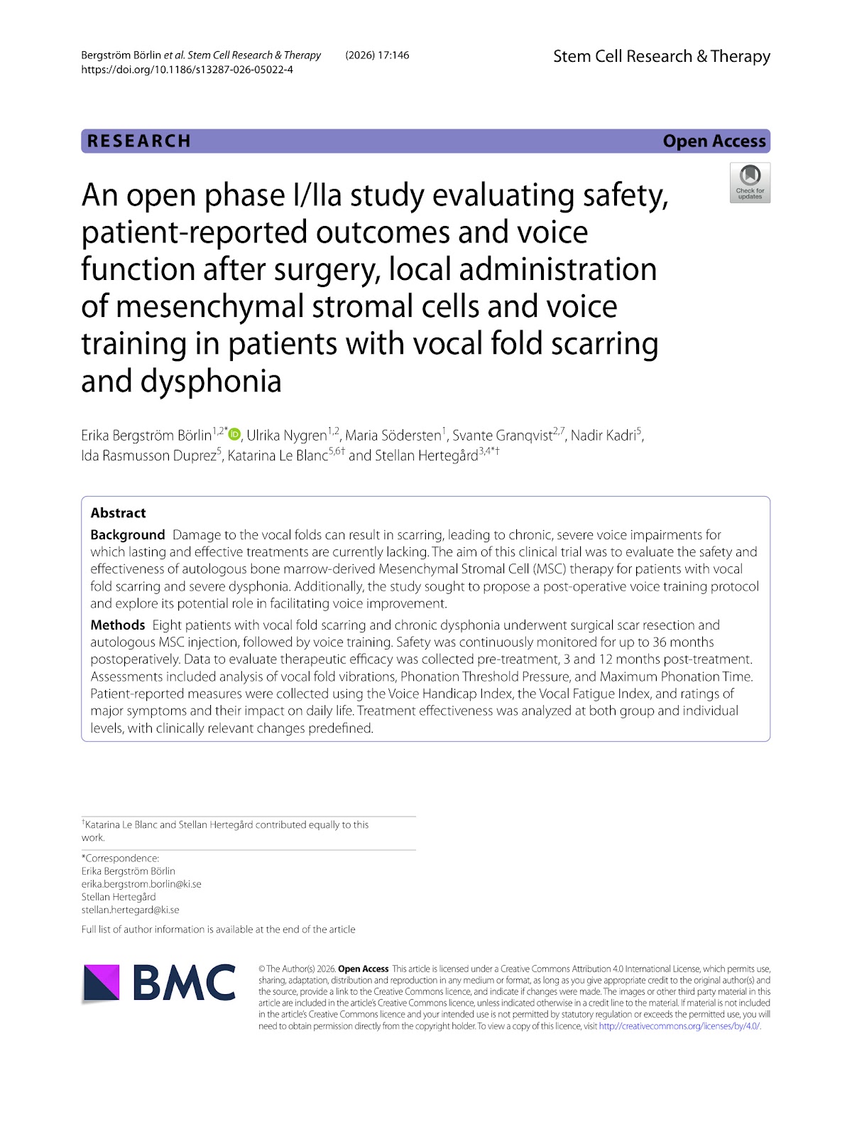
An open phase I/IIa study evaluating safety, patient-reported outcomes and voice function after surgery, local administration of mesenchymal stromal cells and voice training in patients with vocal fold scarring and dysphonia

Authors: Erika Bergström Börlin, Ulrika Nygren, Maria Södersten, Svante Granqvist, Nadir Kadri, Ida Rasmusson Duprez, Katarina Le Blanc, Stellan Hertegård

Background Damage to the vocal folds can result in scarring, leading to chronic, severe voice impairments for which lasting and effective treatments are currently lacking. The aim of this clinical trial was to evaluate the safety and effectiveness of autologous bone marrow-derived Mesenchymal Stromal Cell (MSC) therapy for patients with vocal fold scarring and severe dysphonia. Additionally, the study sought to propose a post-operative voice training protocol and explore its potential role in facilitating voice improvement. Methods Eight patients with vocal fold scarring and chronic dysphonia underwent surgical scar resection and autologous MSC injection, followed by voice training. Safety was continuously monitored for up to 36 months postoperatively. Data to evaluate therapeutic efficacy was collected pre-treatment, 3 and 12 months post-treatment. Assessments included analysis of vocal fold vibrations, Phonation Threshold Pressure, and Maximum Phonation Time. Patient-reported measures were collected using the Voice Handicap Index, the Vocal Fatigue Index, and ratings of major symptoms and their impact on daily life. Treatment effectiveness was analyzed at both group and individual levels, with clinically relevant changes predefined.

An open phase I/IIa study evaluating safety, patient-reported outcomes and voice function after surgery, local administration of mesenchymal stromal cells and voice training in patients with vocal fold scarring and dysphonia
Graphical Abstract
Original ResearchVol. 17, None • pp. 189DOI: 10.1186/s13287-026-05000-w

Innovative strategies for immune thrombocytopenia treatment: immunomodulatory mechanisms and clinical potential of mesenchymal stem cells

Authors: Xin Zhou, Ningning Shan

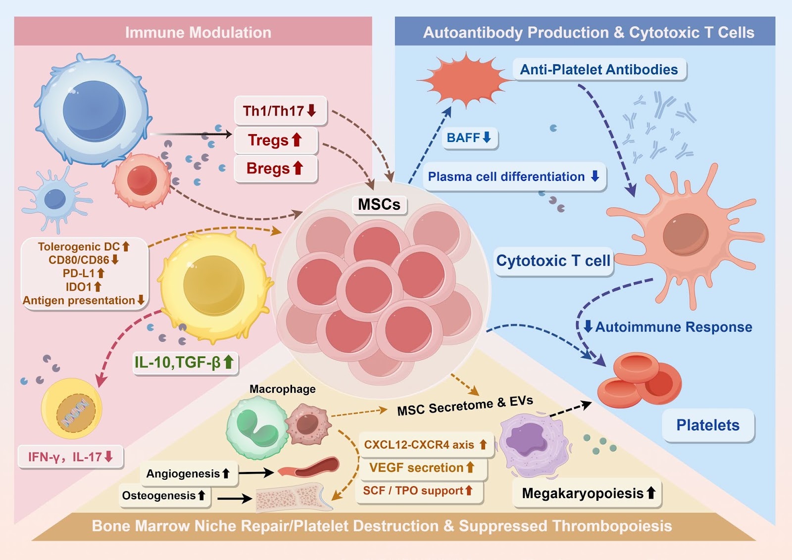
Immune thrombocytopenia (ITP) is a heterogeneous autoimmune disorder characterized by increased platelet destruction and impaired megakaryopoiesis within a dysregulated bone marrow niche. Conventional therapies often achieve only transient platelet recovery, failing to restore immune tolerance, thereby underscoring the need for mechanism-based therapeutic strategies. Mesenchymal stem cells (MSCs) have emerged as promising candidates due to their ability to modulate immune responses and repair the hematopoietic microenvironment. This review synthesizes current evidence regarding the biological properties, immunomodulatory mechanisms, and therapeutic applications of MSCs in ITP, emphasizing intrinsic abnormalities of patient-derived MSCs and the corrective potential of exogenous MSCs from distinct tissue sources. It further integrates emerging insights into MSC functional heterogeneity, optimization of culture conditions, priming strategies, and cellular engineering approaches that may enhance therapeutic efficacy and safety. By highlighting the interplay between immune tolerance restoration and bone marrow niche remodeling, this review provides a translational framework that links mechanistic understanding to the future clinical development of MSC-based therapies for ITP.

Innovative strategies for immune thrombocytopenia treatment: immunomodulatory mechanisms and clinical potential of mesenchymal stem cells
Graphical Abstract
Original ResearchVol. 17, Issue 1 • pp. 182DOI: 10.1186/s13287-026-05006-4

Generation of biologically responsive colon-like intestinal tissue patches from human induced pluripotent stem cells using a rapid co-differentiation platform

Authors: William Dalleywater, Alexander V. Predeus, Batuhan Cakir, Pavel Mazin, Jayakumar Vadakekolathu, Sergio Rutella, Marian L. Meakin, Alison A. Ritchie, Shamir Montazid, Sara Cuevas Ocaña, Nadine Holmes, Victoria Wright, Fei Sang, Silvia Santoni, Adam Bills, Declan Sculthorpe, Rasa Elmentaite, Sarah A. Teichmann, Shazia Irshad, Ian Tomlinson, Andrew Silver, Ricky D. Wildman, Nicholas R. F. Hannan, Felicity R. A. J. Rose, Mohammad Ilyas

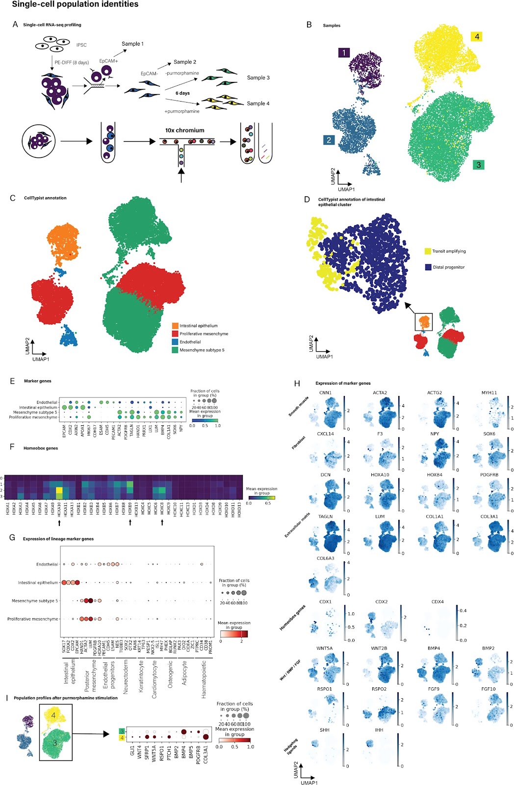
The intestinal mucosa is a complex functional layer which is formed from a diverse range of cell types that include epithelial cells (within crypts and villi) and an array of mesenchymal cells. Many intestinal diseases involve loss of the surface mucosa which can be difficult to restore, and which delays healing and return to normal function. We reason that development of a transplantable intestinal mucosal tissue graft may be a potential therapeutic strategy to aid healing. To be clinically useful, such a tissue graft would need to be capable of rapid production, avoid the risk of host rejection and be demonstrably safe. To create a potential intestinal graft, we developed a novel early-stage human induced pluripotent stem cell (hiPSC) co-differentiation platform capable of generating multiple intestinal cell lineages (epithelial, mesenchymal and endothelial) in 8 days. This protocol is simple to implement, serum-free and greatly reduces the use of animal products. We confirmed the identity of cells by demonstrating that these cells had RNA and protein expression profiles typical of intestinal cell lineages. In particular, we used bulk and single-cell RNA sequencing to characterise global cellular transcriptional profiles robustly and showed that the cells have intestinal identity with early polarisation towards colonic differentiation. The results were replicated across multiple hiPSC lines and in an independent centre. We further cultured the derived cells on collagen hydrogels to form colon-like intestinal patches (CL-IPs). When transplanted into mouse subcutis, CL-IPs formed into colon-like tissue structures, including crypts, stromal and muscle layers. They also developed human-origin vasculature which underwent anastomosis with the murine vasculature to transport murine blood into the graft. Teratoma assays and molecular analyses showed no evidence of residual pluripotency. While at an early stage, this platform shows great potential for further development as a potential source for novel intestinal mucosal regeneration therapy. In addition, the platform is physiologically relevant and thus shows promise as the basis for a new generation of in vitro models of intestinal pathobiology.

Generation of biologically responsive colon-like intestinal tissue patches from human induced pluripotent stem cells using a rapid co-differentiation platform
Graphical Abstract
Original ResearchVol. 17, Issue 233 • pp. 1-14DOI: 10.1186/s13287-026-05125-y

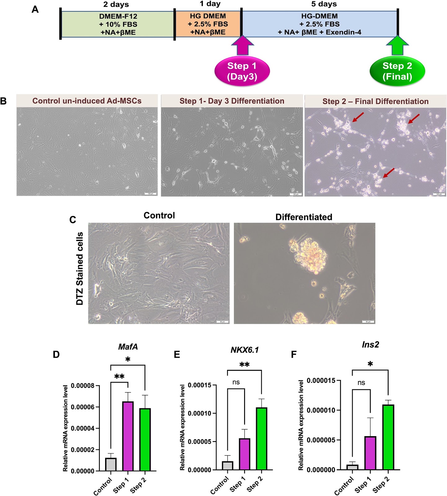
Emerging roles of the long non-coding RNAs MALAT1 and TUG1 during differentiation of adipose tissue-derived mesenchymal stem cells towards insulin-producing cells

Authors: Eman F. Sanad, Alaa Ahmed Saad, Joy Rafeek, Rana Mokbel, Mayar Abdallah, Nadeen Emad, Hagar Adel Mohamed, Yasmin Alaa, Yumna Medhat Mahmoud, Dina H. Kassem

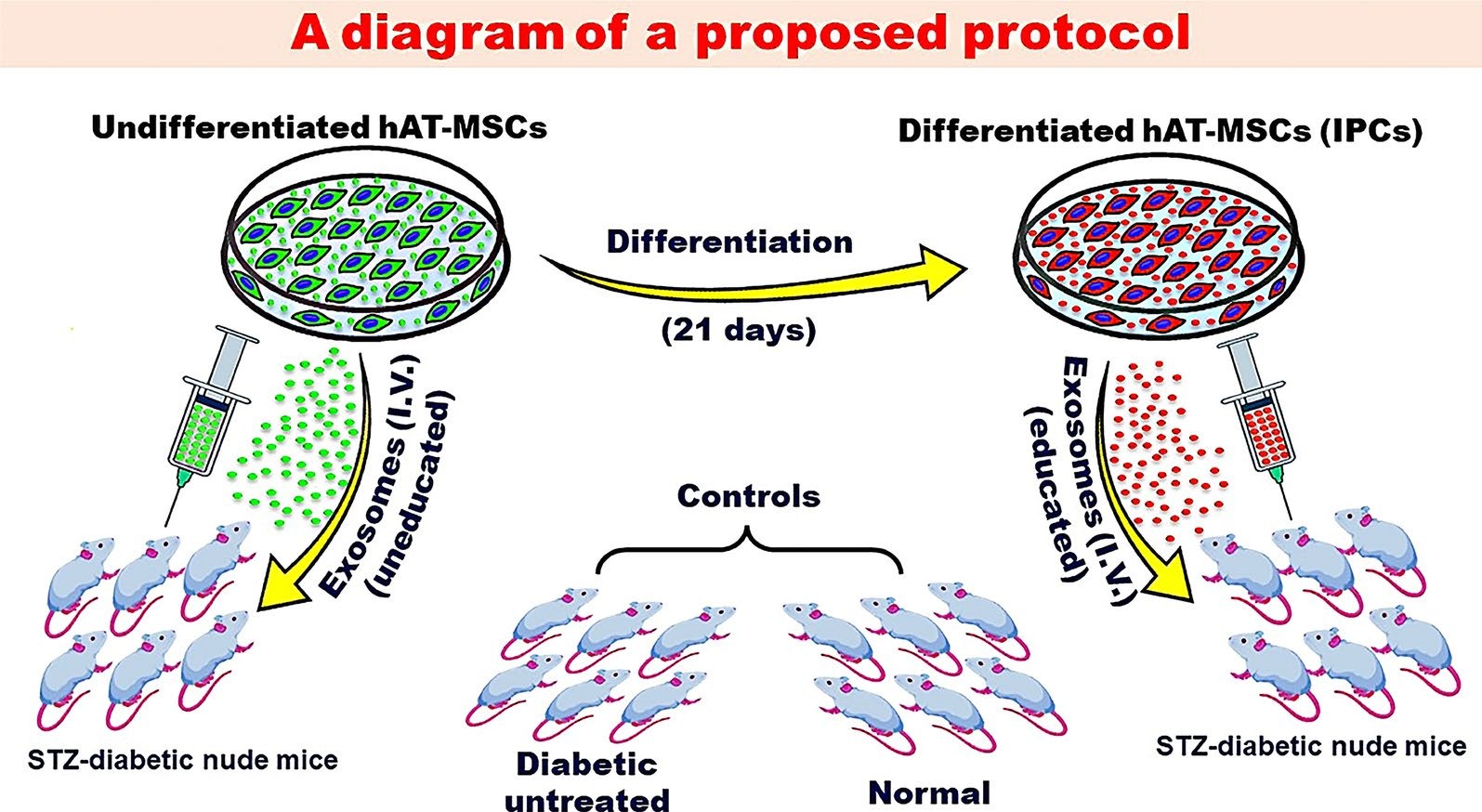
Background: Generation of insulin-producing cells (IPCs) from stem cells provides great hope for patients with diabetes mellitus (DM). Long non-coding RNAs (lncRNAs) ignited much interest regarding their role in determining the fate of stem cells. The lncRNAs MALAT1 and TUG1 have been reported to be interrelated with β-cell dysfunction and/or DM. However, their role during generation of IPCs from stem cells has not been adequately studied. Thus, the current study aimed to investigate the role of MALAT1 and TUG1 during differentiation of adipose tissue-derived mesenchymal stem cells (Ad-MSCs) towards IPCs. Methods: Ad-MSCs were isolated from rat epididymal fat pads, characterized and induced to differentiate towards IPCs. Assessment of differentiation was done by measuring expression levels of various β-cell-related markers using RT-qPCR, as well as morphological changes, and dithizone staining. Expression levels of MALAT1 and TUG1 were also measured by RT-qPCR. Several in-silico analyses were done using RNA–protein Association and Interaction Networks (RAIN) database. Results: MALAT1 and TUG1 expression levels were significantly increased during differentiation of Ad-MSCs into IPCs as compared to control uninduced cells. Furthermore, generated networks from RAIN database revealed an interplay between MALAT1 and TUG1, and between each of them with several common targets like GAS5, HOTAIR and TP53COR1. Conclusions: The current study portrays MALAT1 and TUG1 as novel interrelated molecular mediators and important regulatory nodes enhancing differentiation of Ad-MSCs towards IPCs. Their upregulation during differentiation can be interrelated with competitive endogenous RNA (ceRNA) networks, mediating various epigenetic modifications, orchestrating signaling pathways and overcoming cellular stress during reprogramming/differentiation.

Emerging roles of the long non-coding RNAs MALAT1 and TUG1 during differentiation of adipose tissue-derived mesenchymal stem cells towards insulin-producing cells
Graphical Abstract
Original ResearchVol. 17, Issue 247 • pp. 1-16DOI: 10.1186/s13287-026-05061-x

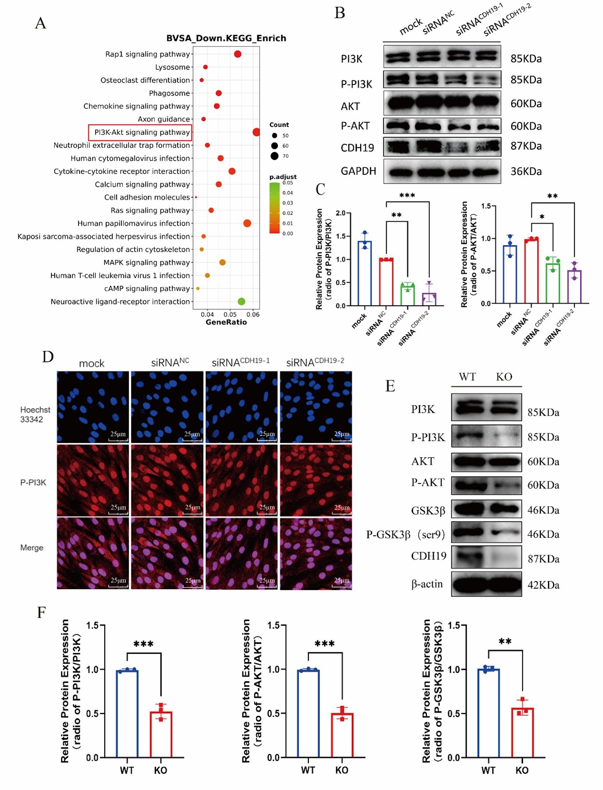
Cadherin 19 deficiency inhibits osteogenic differentiation and bone formation by regulating PI3K/AKT signaling pathway

Authors: Huan Zhou, Yizhao Cheng, Liangxing Chen, Yaqin Zhang, Huimin Xiao, Chenhui Zhou, Hui Fu, Xiaoyan Zhang, Liangliang Xu, Daohua Xu

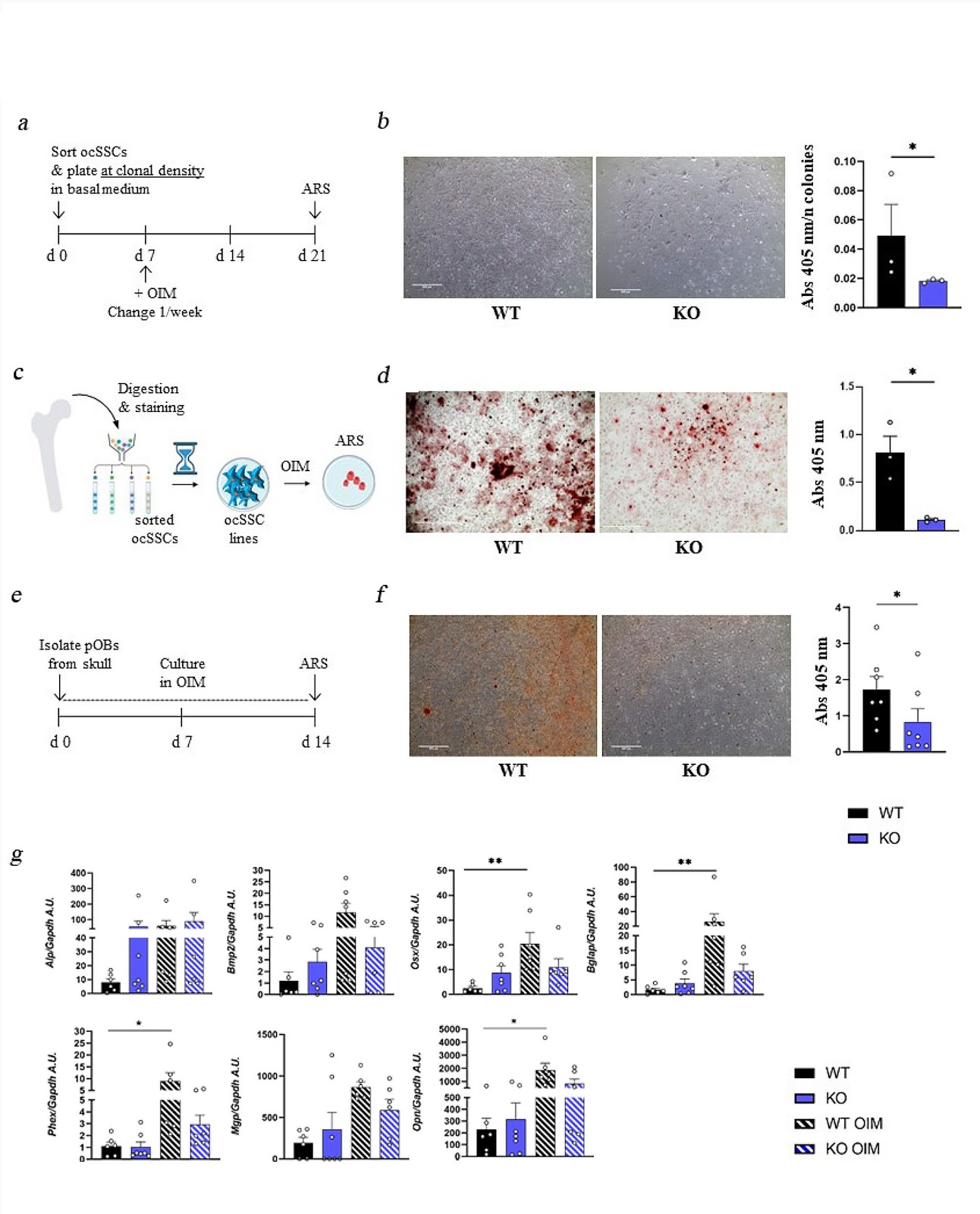
Background Osteoporosis (OP) is a systemic bone disease characterized by damage to bone strength, leading to increased bone fragility and fracture risk. Cadherin 19 (CDH19) is located on chromosomes 18q22-q23, and 18q deletion is associated with terminal deletion diseases, including foot/hand deformities. However, the role of CDH19 in bone remains undefined. Methods A conditional knockout mouse model of the CDH19 gene was constructed using the Cre-loxP system, and the bone mass and bone morphology in mice were investigated using microCT and histological staining. Osteoblasts were isolated and cultured from wild-type and CDH19 knockout mice. Cell proliferation and differentiation were explored through EdU labeling, qPCR, alkaline phosphatase (ALP)/ alizarin red S (ARS) staining, and Western blot assays. The expression of genes altered in CDH19 gene knockout osteoblast was checked by RNA sequencing (RNA-seq), and subsequently confirmed by immunofluorescence and Western blot. Results We found that CDH19 could maintain the normal proliferation and differentiation in osteoblasts. After knocking out the CDH19 gene, the abilities of proliferation and osteogenesis were significantly inhibited in osteoblasts. Moreover, the bone mass of CDH19 knockout mice was significantly reduced, characterized by decreases in bone density, trabecular number, and bone volume fraction. The RNAseq analysis and western blot showed the PI3K/AKT signaling pathway was significantly inhibited in osteoblasts with CDH19 deletion. Furthermore, we demonstrated that administration of PI3K/AKT signaling pathway agonist 740Y-P partially alleviated the inhibition of osteogenic differentiation caused by CDH19 deletion in vitro and in vivo. Conclusion This study demonstrated that CDH19 regulated osteogenic differentiation by modulating the PI3K/AKT signaling pathway in osteoblasts. CDH19 may become a novel target for the treatment of bone diseases.

Cadherin 19 deficiency inhibits osteogenic differentiation and bone formation by regulating PI3K/AKT signaling pathway
Graphical Abstract
Original ResearchVol. 17, Issue 274 • pp. 1-19DOI: 10.1186/s13287-026-05093-3

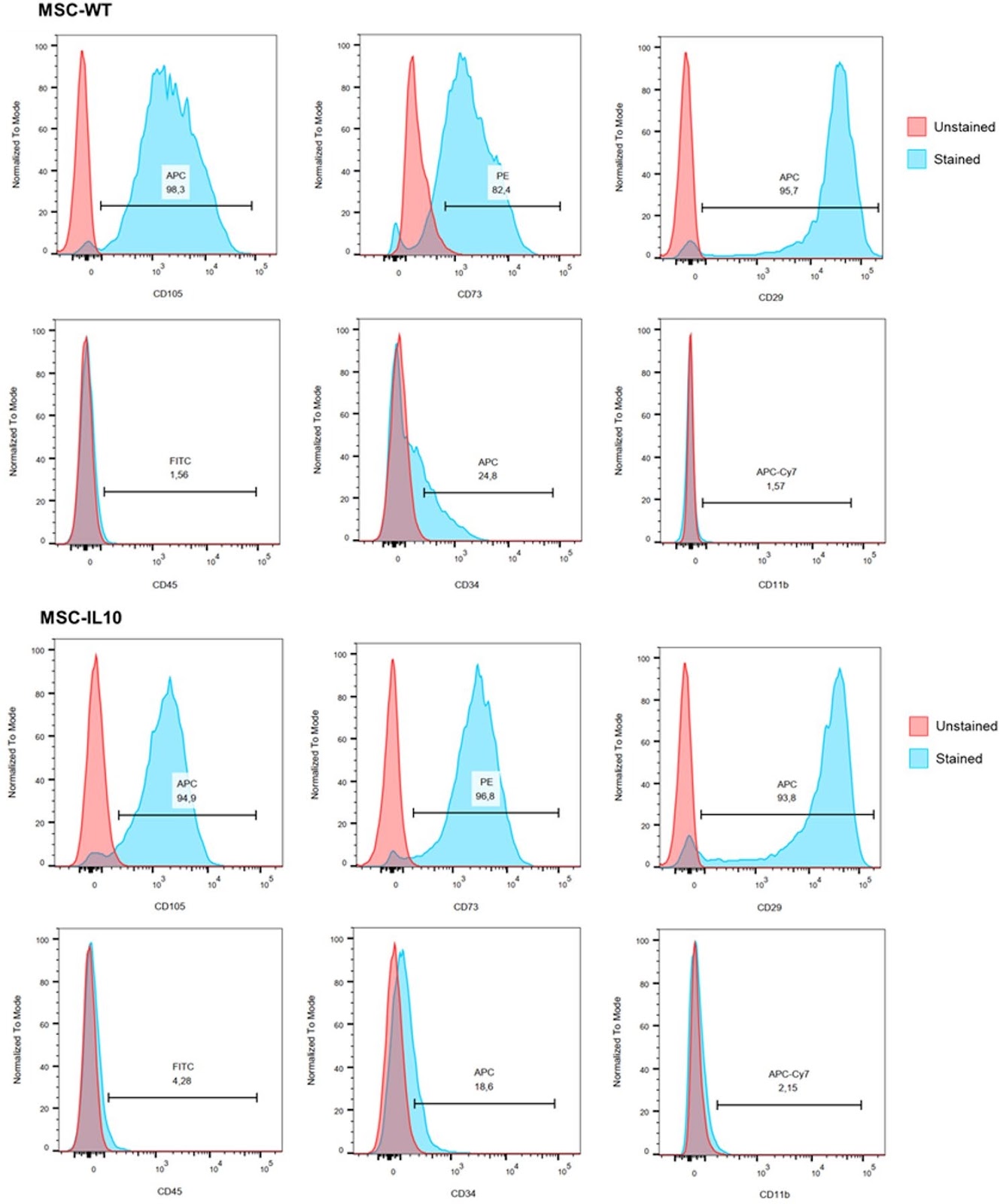
Interleukin-10-engineered mesenchymal stem/stromal cells exhibit robust immunomodulatory effects in vitro and in vivo

Authors: Diego de Carvalho Carneiro, Cássio Santana Meira, Rosane Borges Dias, Vinícius Pinto Costa Rocha, Patrícia Kauanna Fonseca Damasceno, Josiane Dantas Viana Barbosa, Milena Botelho Pereira Soares

Introduction  A dysregulated inflammatory response to infection can lead to sepsis, a leading cause of mortality worldwide, and effective anti-inflammatory therapies remain limited. Mesenchymal stem/stromal cells (MSCs) are attractive candidates as immunomodulatory agents. This study evaluated whether genetic modification of MSCs to express interleukin-10 (IL-10), a key anti-inflammatory cytokine, enhances their immunomodulatory effects. Methods  Bone marrow-derived MSCs from C57Bl/6 mice were genetically engineered by lentiviral transduction to express mouse IL-10 (MSC-IL-10). The immunomodulatory activity in vitro was assessed by co-cultures with macrophages stimulated with LPS and IFN-γ, as well as in Con A–stimulated splenocytes. BALB/c mice subjected to lipopolysaccharide (LPS)-induced endotoxemia were treated with vehicle, dexamethasone, wild-type MSCs (MSC-WT), or MSC-IL-10. Survival, plasma cytokines, leukocyte profiles, CD11b⁺ inflammatory cells, and organ histopathology and biodistribution were evaluated in vivo. Results  MSC-IL-10 maintained the mesenchymal phenotype and multipotent characteristics while exhibiting robust IL-10 expression. In in vitro assays, MSC-IL-10 significantly decreased the production of the cytokines TNF-α, IL-1β, IL-6, IL-12 or Nos2 expression by stimulated macrophages or splenocytes, demonstrating superior immunomodulatory effects compared to MSC-WT. In in vivo mice models, MSC-IL-10 significantly reduced systemic pro-inflammatory cytokines, restored circulating leukocyte counts, and attenuated CD11b⁺ (Mac-1 integrin) inflammatory cell recruitment, surpassing MSC-WT-treated groups. Importantly, MSC-IL-10 mitigated tissue damage mainly to lungs and exhibited biodistribution to liver, lungs and spleen in LPS-challenged mice. Conclusions  These results support an enhanced immunomodulatory effect of IL-10-expressing MSCs as a promising cell-based therapeutic approach for sepsis and other inflammatory and immune mediated disorders.

Interleukin-10-engineered mesenchymal stem/stromal cells exhibit robust immunomodulatory effects in vitro and in vivo
Graphical Abstract
Original ResearchVol. 17, Issue 1 • pp. 79DOI: 10.1186/s13287-026-04900-1

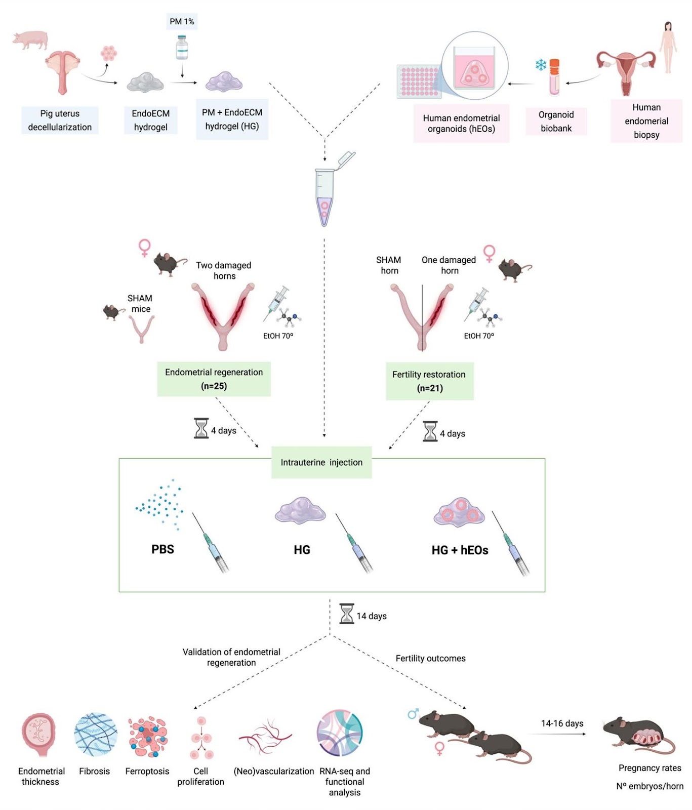
Hybrid endometrial-derived hydrogel and human endometrial organoids synergize for uterine regeneration in an immunocompetent murine model

Authors: María Gómez-Álvarez, Clara Bueno-Fernandez, Emilio Francés-Herrero, Marcos Agustina-Hernández, Paula Alonso-Frías, Nadaya Corpas, Amparo Faus, Ana Díaz, Antonio Pellicer, Hortensia Ferrero, Irene Cervelló

Background The human endometrium is a regenerative tissue essential for fertility, but pathological conditions like Asherman syndrome, endometrial atrophy, and thin endometrium can impair its function. Current therapies lack efficacy, driving demand for innovative regenerative therapies. In this context, endometrial-derived hydrogels and organoids have shown promise individually for tissue regeneration, but their combined therapeutic potential has not been previously evaluated in vivo. This study explores a dual regenerative strategy combining a hybrid hydrogel — composed of synthetic PuraMatrix® and endometrial extracellular matrix hydrogel — with human endometrial organoids in an immunocompetent murine model with uterine damage. Methods Endometrial damage model was established in female C57BL/6 mice (n = 46) via uterine injury using 70° ethanol. After 4 days of endometrial damage, human endometrial organoids were co-injected with the hybrid hydrogel into the uterine horns. Two weeks post-injection, a subset of mice (n = 25) was sacrificed for biocompatibility, histological, and transcriptomic analyses. Functional recovery of the endometrium was assessed in the remaining animals (n = 21) through fertility outcome evaluation. For endometrial regeneration analyses, normally distributed data were analyzed by one-way ANOVA and Tukey’s multiple comparisons, while non-normally distributed data were analyzed by the Kruskal–Wallis test with Dunn’s multiple comparisons. For fertility outcomes, t-test or Mann–Whitney U tests for 2-by-2 comparisons were performed.

Hybrid endometrial-derived hydrogel and human endometrial organoids synergize for uterine regeneration in an immunocompetent murine model
Graphical Abstract
Original ResearchVol. 17, Issue 104DOI: 10.1186/s13287-026-04962-1

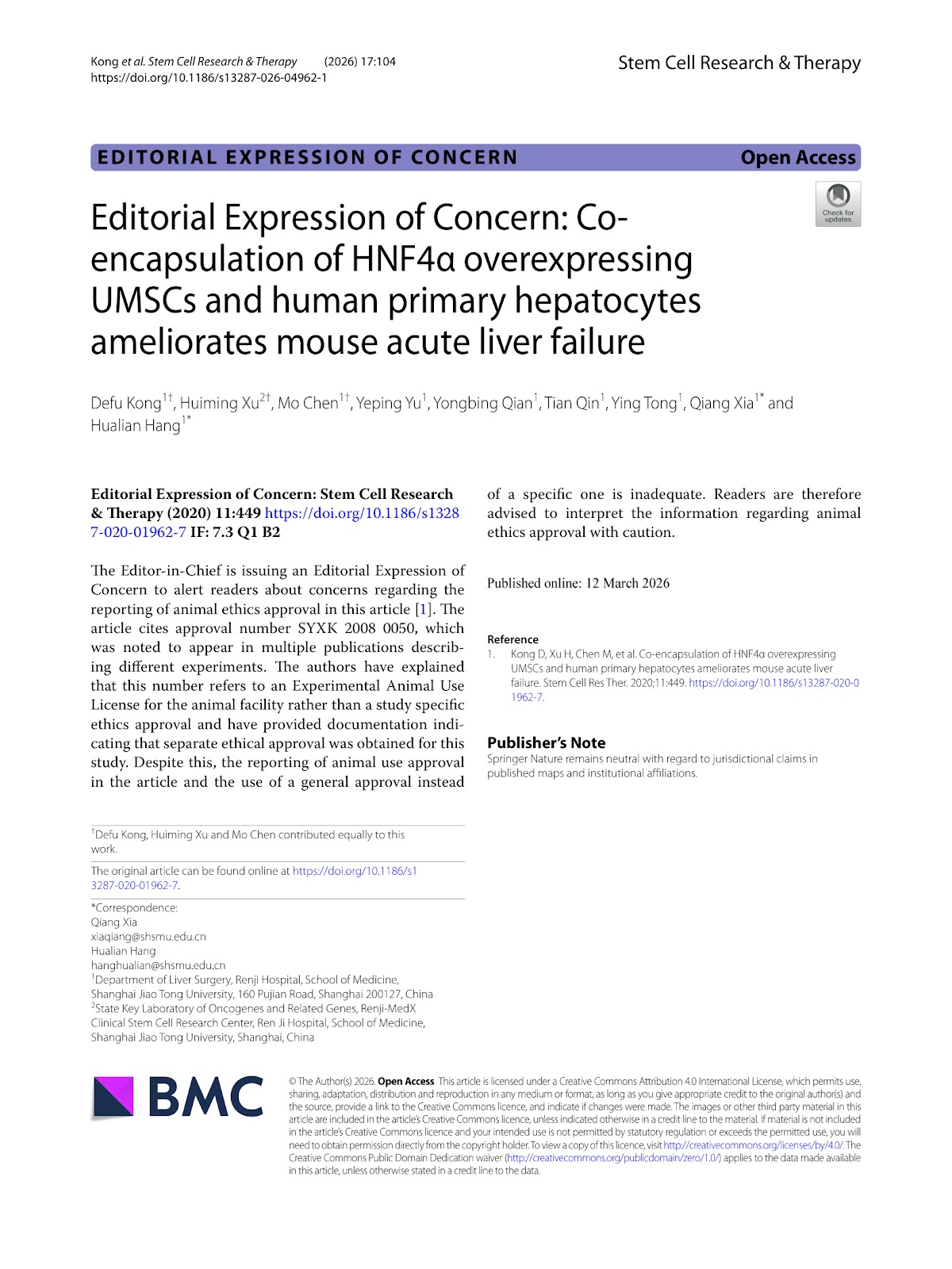
Editorial Expression of Concern: Co-encapsulation of HNF4α overexpressing UMSCs and human primary hepatocytes ameliorates mouse acute liver failure

Authors: Defu Kong, Huiming Xu, Mo Chen, Yeping Yu, Yongbing Qian, Tian Qin, Ying Tong, Qiang Xia, Hualian Hang

The Editor-in-Chief is issuing an Editorial Expression of Concern to alert readers about concerns regarding the reporting of animal ethics approval in this article [1]. The article cites approval number SYXK 2008 0050, which was noted to appear in multiple publications describing different experiments. The authors have explained that this number refers to an Experimental Animal Use License for the animal facility rather than a study specific ethics approval and have provided documentation indicating that separate ethical approval was obtained for this study. Despite this, the reporting of animal use approval in the article and the use of a general approval instead of a specific one is inadequate. Readers are therefore advised to interpret the information regarding animal ethics approval with caution.

Editorial Expression of Concern: Co-encapsulation of HNF4α overexpressing UMSCs and human primary hepatocytes ameliorates mouse acute liver failure
Graphical Abstract
Original ResearchVol. 17, Issue 265 • pp. 1-22DOI: 10.1186/s13287-026-05073-7

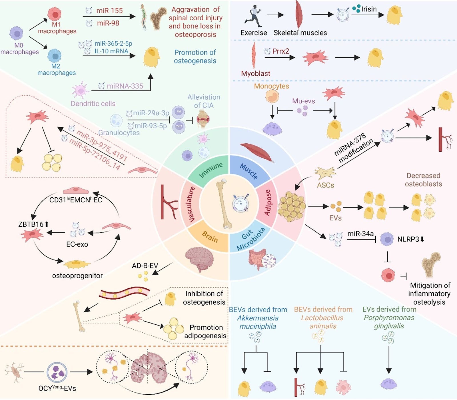
Exosomes in bone health and disease: cellular crosstalk, systemic signaling, and AI-driven advances in regenerative therapy

Authors: Xueying Liu, Wenqing Yu, Chengyu Huang, Zhenmin Wang, Ying Qian, Xin Chen, Gaoyang Chen

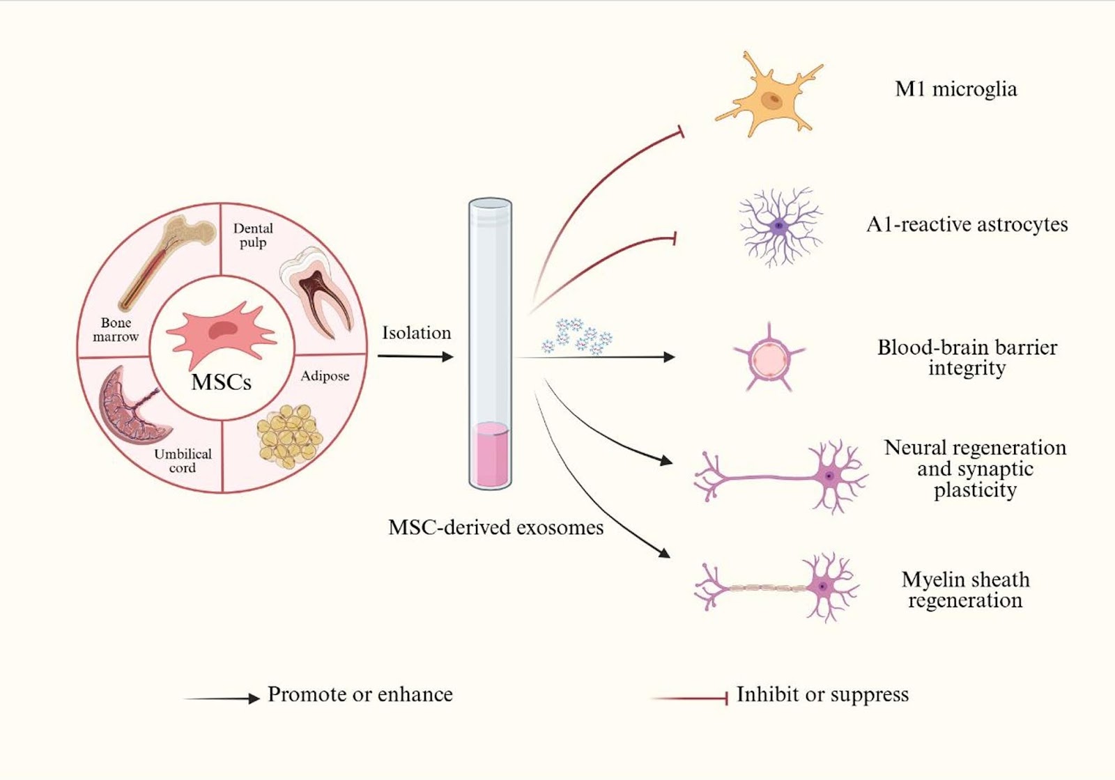
Exosomes have emerged as critical mediators of intercellular and inter-organ communication in bone biology. Secreted by bone-resident cells such as osteoblasts, osteoclasts, osteocytes, and mesenchymal stem cells (MSCs), these nanosized vesicles carry diverse molecular cargos that regulate bone remodeling, regeneration, and skeletal homeostasis. In addition to mediating local communication within the bone microenvironment, exosomes also participate in systemic crosstalk communication between bone and other tissues, including skeletal muscle, adipose tissue, gut microbiota, the immune system, the nervous system, and vasculature. Disruption of these exosome-mediated pathways contributes to the development and progression of bone diseases, including osteoporosis, osteoarthritis, osteonecrosis of the femoral head, and bone metastases. This review summarizes current advances in exosome-mediated signaling in both physiological and pathological contexts, with particular emphasis on their roles as biomarkers, therapeutic agents, and drug delivery vehicles. We also discuss the emerging contribution of artificial intelligence (AI) to exosome research, especially in biomarker discovery, disease classification, and target identification, as well as the major challenges that currently limit clinical translation. Together, these insights highlight the potential of exosome-based strategies for precision medicine in bone diseases.

Exosomes in bone health and disease: cellular crosstalk, systemic signaling, and AI-driven advances in regenerative therapy
Graphical Abstract
Original ResearchVol. 17, Issue 250 • pp. 1-16DOI: 10.1186/s13287-026-04941-6

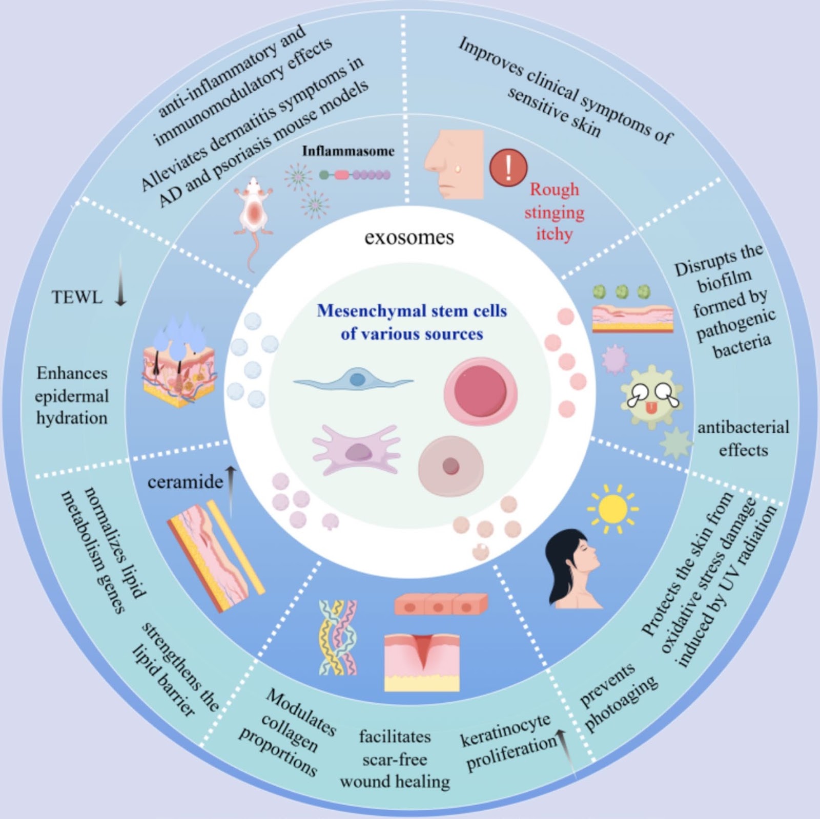
Targeting skin barrier repair: mechanisms of action, therapeutic evidence, and clinical translation challenges of mesenchymal stem cell-derived exosomes

Authors: Yujin Li, Jian Huang, Zhibing Fu, Lihua Gao, Xiaoliang Tong, Lu Zhou, Jinrong Zeng, Lina Tan

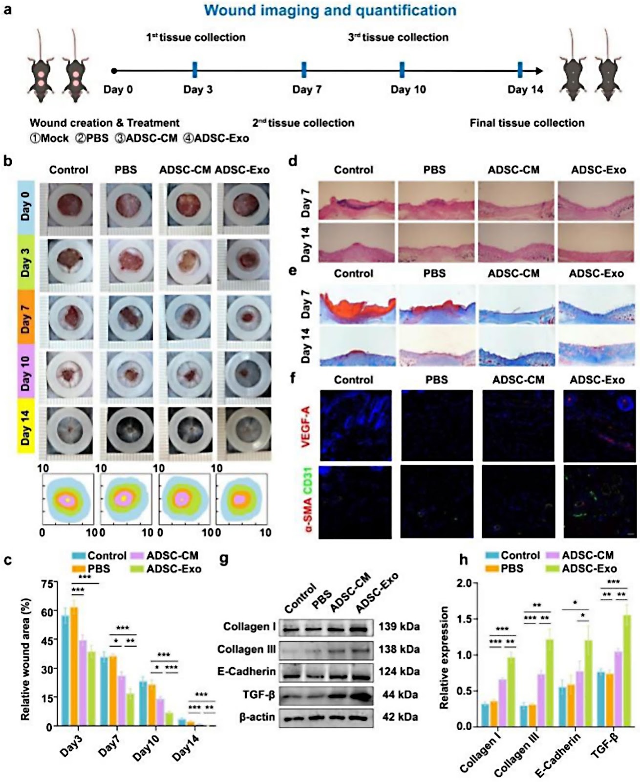
Dysfunction of the skin barrier is a central pathological feature in dermatology, driving the need for innovative repair strategies. Mesenchymal stem cell-derived exosomes (MSC-exos) represent a promising cell-free therapeutic paradigm, leveraging their innate cargo to modulate regeneration and immune responses. This review systematically examines the multifaceted role of MSC-exos in restoring skin barrier integrity. We delineate their molecular mechanisms in repairing physical, immunological, and microbial barrier components, supported by evidence from preclinical disease models. The influence of MSC source and preconditioning on exosome efficacy is analyzed, alongside emerging bioengineering approaches. Crucially, we identify and discuss the key translational challenges—including standardization, scalable manufacturing, and regulatory pathways—that must be addressed to advance these nanotherapeutics toward clinical application. This synthesis provides a critical framework for future research aimed at harnessing MSC-exos for targeted barrier repair.

Targeting skin barrier repair: mechanisms of action, therapeutic evidence, and clinical translation challenges of mesenchymal stem cell-derived exosomes
Graphical Abstract
Original ResearchVol. 17, None • pp. 82DOI: 10.1186/s13287-026-04902-z

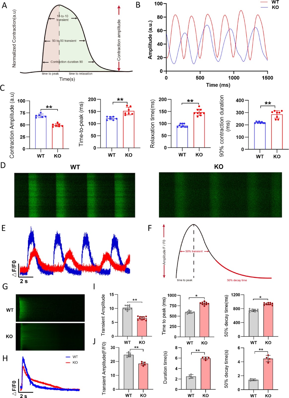
FHOD3 deficiency disrupts sarcomere organization and activates CaMKII signaling in human stem cell-derived cardiomyocytes

Authors: Mingyu Wei, Xiaojie Hou, Siyao Zhang, Xianjing Hu, Xi Chen, Zhen Gao, Shuwan Xu, Zhan Shi, Min Zhu, Feng Lan, Ming Cui

Background Inherited cardiomyopathy (ICM) is a genetic disorder characterized by abnormal myocardial structure and function, often progressing to heart failure. FHOD3, a member of the Formin gene family, plays a crucial role in cardiomyocyte cytoskeletal organization. Mutations in FHOD3 have been associated with various cardiomyopathies, including hypertrophic cardiomyopathy (HCM), dilated cardiomyopathy (DCM) and left ventricular noncompaction (LVNC). However, the molecular mechanisms underlying FHOD3 deficiency-induced cardiomyopathy remain elusive. Methods A FHOD3 knockout (FHOD3-/-) human embryonic stem cell (hESC) line was generated using the CRISPR/Cas9 system and subsequently differentiated into cardiomyocytes (hESC-CMs). Sarcomere structure, calcium handling, mitochondrial function, and contractility were evaluated via immunofluorescence, electron microscopy, Seahorse metabolic analysis, and high-definition video analysis, respectively. Transcriptomic sequencing was performed to identify differentially expressed genes and enriched pathways. Results FHOD3-deficient hESC-CMs exhibited marked sarcomere disorganization and degradation, impaired calcium handling and compromised mitochondrial function, ultimately leading to reduced contractility. Transcriptomic analysis revealed significant downregulation of sarcomere-related genes and calcium-handling genes, with enrichment in pathways associated with cardiomyopathy and calcium signaling. Furthermore, FHOD3 deficiency triggered the phosphorylation of CaMKII (Thr286), a key regulator of cardiac hypertrophy and remodeling, contributing to the progression of heart failure. Treatment with the myosin activator Omecamtiv mecarbil (OM) partially restored contractility without affecting calcium handling, highlighting its potential as a therapeutic strategy.

FHOD3 deficiency disrupts sarcomere organization and activates CaMKII signaling in human stem cell-derived cardiomyocytes
Graphical Abstract
Original ResearchVol. 17, NoneDOI: 10.1186/s13287-026-04979-6

A review of the circadian regulation of stem cells: harnessing the internal body clock for enhanced regenerative therapies

Authors: Sulaiman Mohammed Alnasser

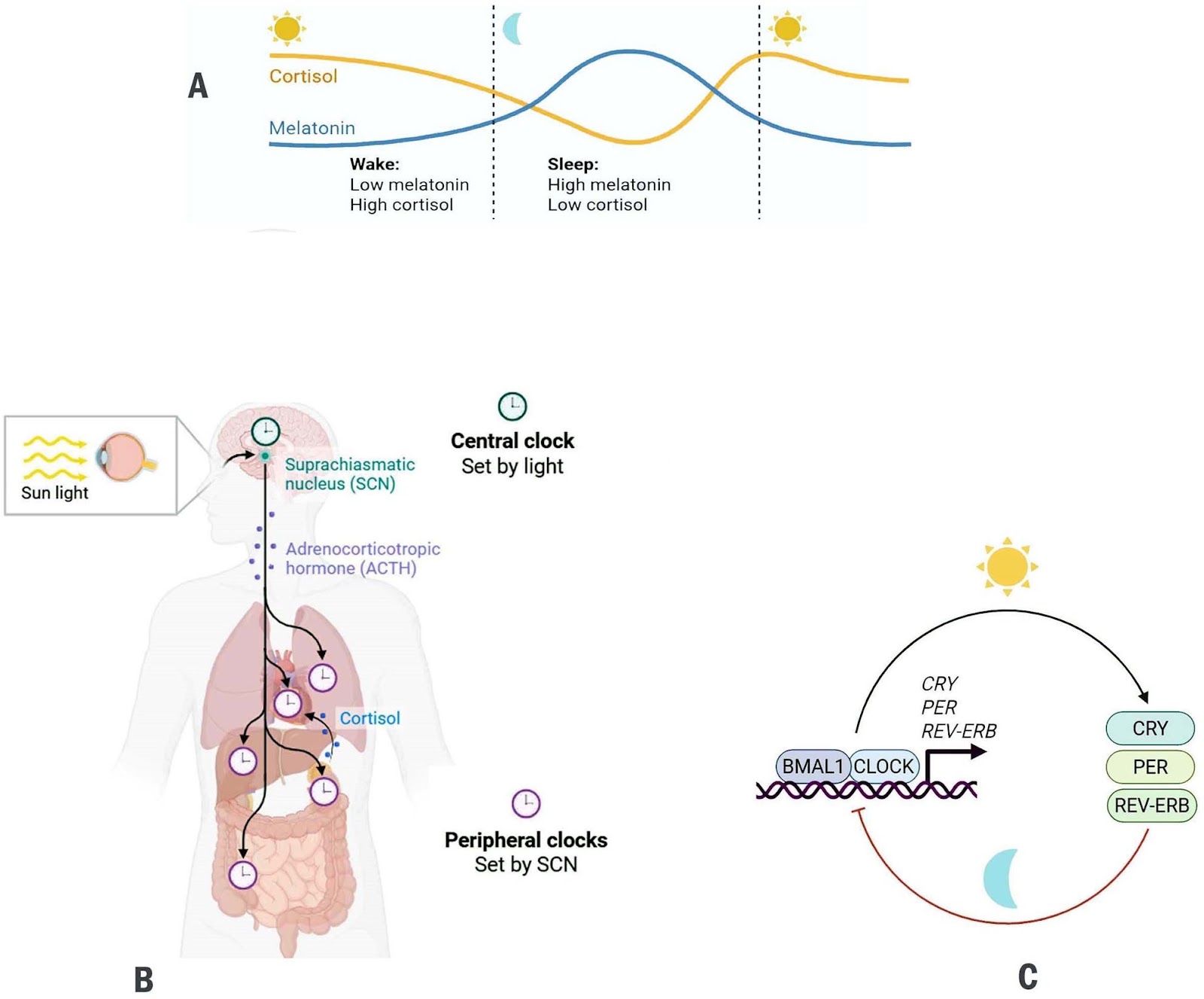
Background Circadian rhythms are endogenous, transcription-translation feedback loops that align cellular activities with the 24-h light–dark cycle. Stem-cell populations across tissues exhibit circadian oscillations that influence their self-renewal, proliferation, and differentiation. Key developmental pathways (Wnt/β-catenin, Notch, and Hedgehog) are increasingly recognized as both regulators and targets of circadian machinery. Objectives This review synthesizes current knowledge on the bidirectional crosstalk between circadian clock components and major stem-cell regulatory pathways, and evaluates how this interplay shapes tissue homeostasis, regenerative capacity, and therapeutic potential. Methods Literature examining molecular interfaces between circadian clock genes and Wnt, Notch, and Hedgehog signaling was surveyed, with emphasis on transcriptional regulation, chromatin dynamics, post-translational control, and functional outcomes for stem-cell behavior and regeneration. Results Evidence indicates that core clock components modulate stem-cell pathways through direct transcriptional control, shared enhancer architecture, altered chromatin accessibility, and rhythmic protein modification. In turn, Wnt, Notch, and Hedgehog signals feed back onto clock genes, influencing circadian amplitude and phase within stem-cell niches. Perturbation of this reciprocal regulation disrupts tissue maintenance, diminishes regenerative responses, alters metabolic equilibrium, and may promote tumorigenesis. Conclusions Circadian oscillators act as temporal gatekeepers of stem-cell function. Mapping the molecular interfaces between clock genes and developmental signaling pathways reveals new opportunities to refine regenerative therapies. Chronotherapeutic strategies, i.e. timing interventions to intrinsic circadian phases may enhance the efficacy, precision, and safety of stem-cell–based treatments.

A review of the circadian regulation of stem cells: harnessing the internal body clock for enhanced regenerative therapies
Graphical Abstract
Original ResearchVol. 17, Issue 263 • pp. 1-12DOI: 10.1186/s13287-026-05207-x

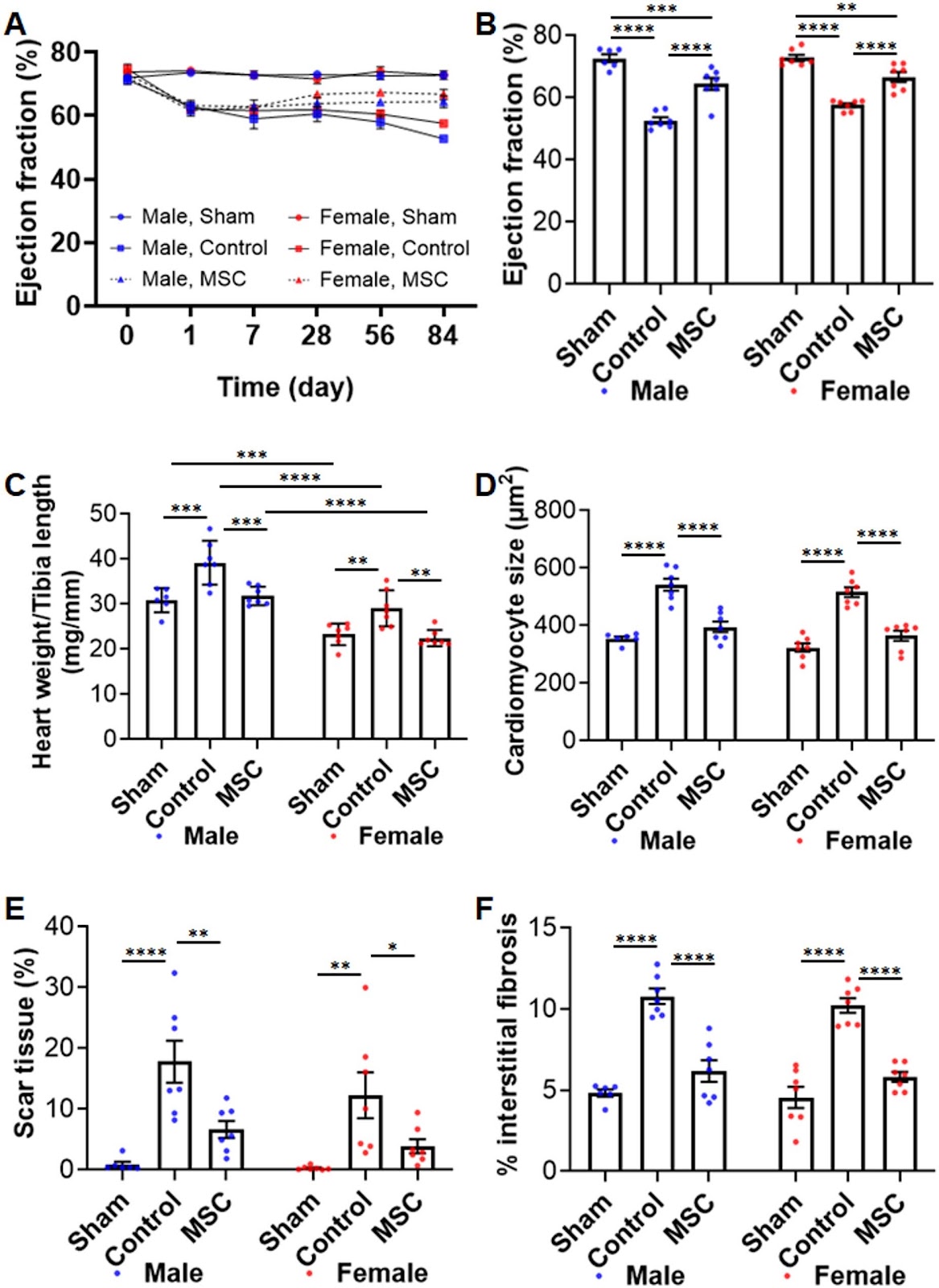
Efficacy of multi-layered human iPS cell-derived cardiovascular cell sheets in a pacing-induced canine dilated cardiomyopathy model

Authors: Yu Shimoyama, Kenji Kakuta, Kiho Araki, Hyoe Komae, Minoru Ono, Jun K. Yamashita

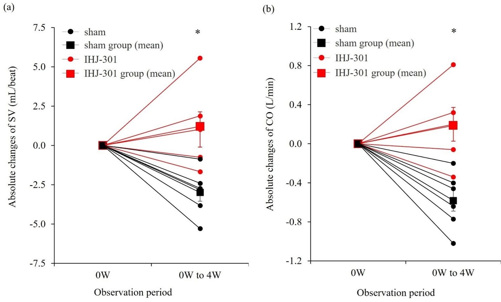
Background Dilated cardiomyopathy (DCM) is a progressive, intractable disease that leads to heart failure. Heart transplantation is the only curative treatment; however, access is limited by donor scarcity. Induced pluripotent stem cell (iPSC)-based therapies are attracting attention for DCM, but suitable large-animal models and robust preclinical data have been limited. Methods We generated multi-layered cardiovascular cell sheets from human iPSCs by combining cardiomyocytes with endothelial and stromal cells and overcoming stacking limits using interleaved gelatin hydrogel microspheres, yielding a thicker cardiac tissue-like construct (product code: IHJ-301). To enable rigorous testing in non-ischemic heart failure, we established a modified canine rapid-pacing heart failure model that maintains depressed function without mortality by continuing pacing at a slightly reduced rate after induction (Step-Down Pacing Heart Failure model). IHJ-301 was implanted epicardially onto the left ventricular surface via thoracotomy, and cardiac function was assessed by echocardiography and right-heart catheterization. Results After 4 weeks of rapid pacing (230±10 bpm), left ventricular ejection fraction (LVEF) was reduced from 77.8±1.1% (pre-pacing) to 44.9±1.9% (n=11) (0 W). Continued pacing at 210±10 bpm for additional 4 weeks resulted in no mortality and maintained depressed function (4 W LVEF 47.3±2.6%). IHJ-301 was implanted at 0 W. At 4 weeks post-implantation (4 W), all animals in the IHJ-301 group (n=5) showed greater functional improvement than sham (n=6). Absolute changes from 0 W to 4 W were: ΔLVEF (%) 9.38±1.47 vs. 1.90±0.34; Δfractional shortening (%) 4.84±0.75 vs. 0.97±0.18; stroke volume (mL/beat) 1.21±1.26 vs. −2.99±0.60; cardiac output (L/min) 0.19±0.19 vs. −0.58±0.12 (all p<0.05). Conclusions We established a non-ischemic large-animal heart failure model that sustains depressed function for one month, enabling clear therapeutic readouts. IHJ-301 significantly improved multiple parameters of cardiac function, providing preclinical evidence that IHJ-301 could offer a promising therapeutic option for DCM.
Efficacy of multi-layered human iPS cell-derived cardiovascular cell sheets in a pacing-induced canine dilated cardiomyopathy model
Graphical Abstract
Original ResearchVol. 17, None • pp. 176DOI: 10.1186/s13287-026-04964-z

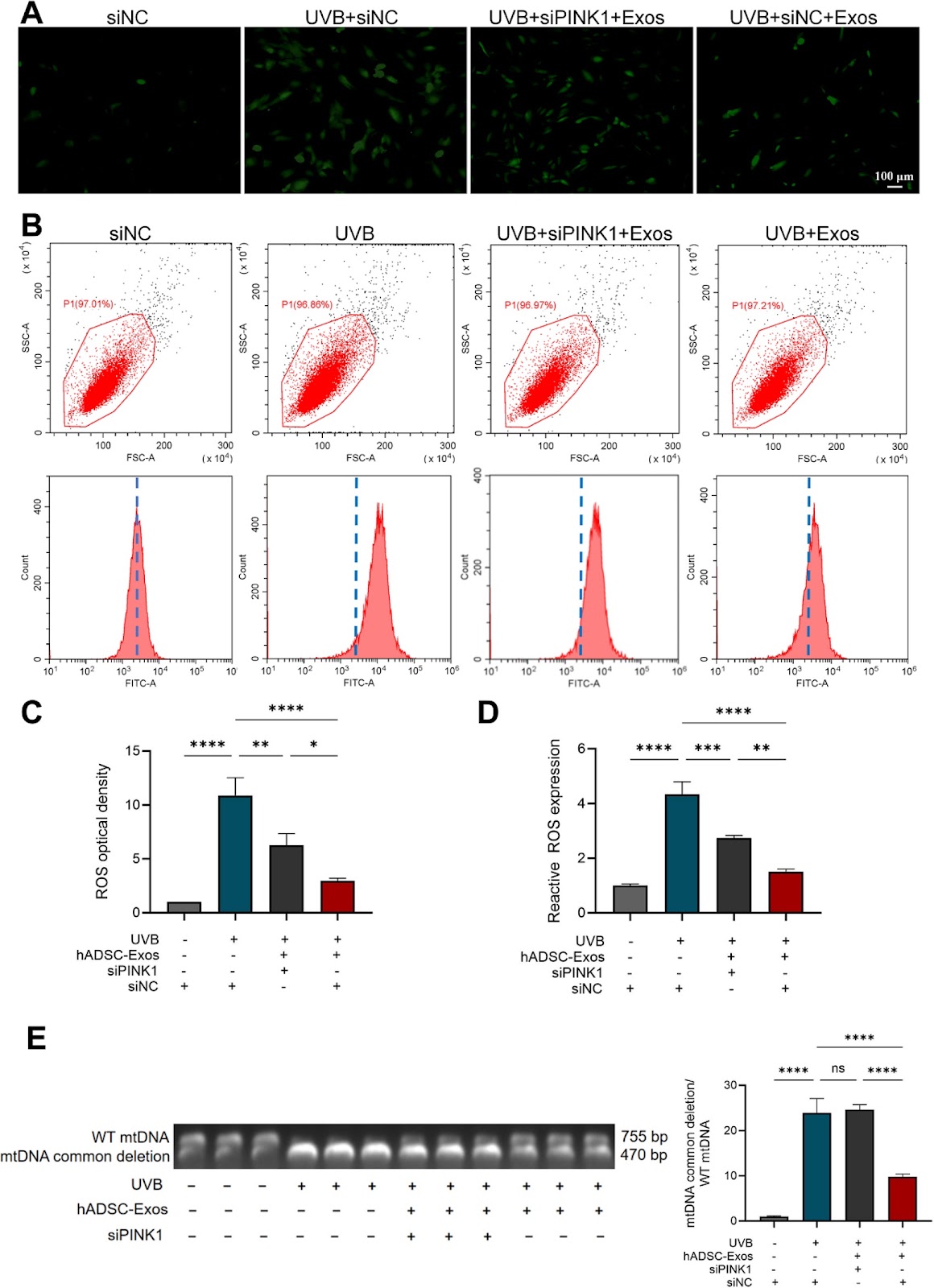
Research based on serine metabolism indicates mesenchymal stem cells alleviate psoriasis by regulating the PSPH-PINK1-Parkin-NLRP3 pathway in HaCaT

Authors: Qing Lin, Yunfei Ji, Bin Yang, Rongjia Zhu, Ping Song, Robert chunhua Zhao

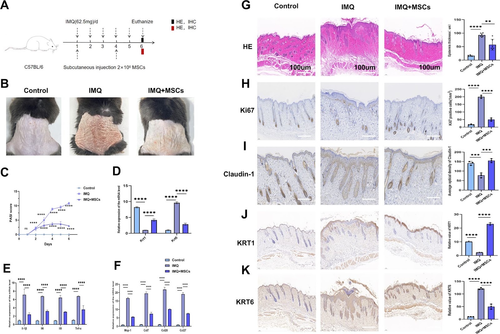
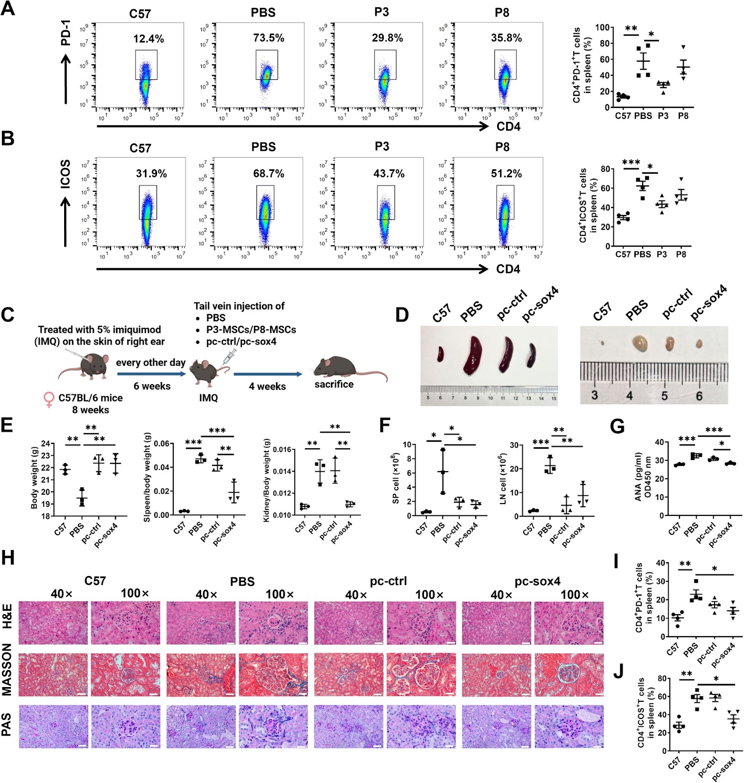
Background Psoriasis is a refractory immune-related disease. In recent years, it has been discovered that mesenchymal stem cells (MSCs) can be used as a new therapeutic approach for psoriasis, but their potential therapeutic mechanism remains unclear. This study aims to explore the role of MSCs in the treatment of psoriasis. Methods We employed a mouse psoriasis model induced by imiquimod (IMQ) in vivo and a co-culture system of MSCs and HaCaT keratinocytes (KCs) cell line in vitro. These approaches allowed us to investigate the effect of MSCs on the levels of inflammatory factors and the activation of inflammasomes in both contexts. Mouse-targeted amino acid sequencing, transmission electron microscopy for in vitro observation, immunofluorescence for both in vivo and in vitro analyses, and siRNA transfection in vitro were employed in this study. Results Our results showed that MSCs significantly improved the skin lesion of mice with psoriasis, and reduced the levels of inflammatory factors and chemokines including IL-1β, IL-6, IL-8, TNF-α, MCP-1, CCL7, CCL20 and CCL27 in the mouse skin lesion areas and M5- induced psoriatic KCs models in vitro. Likewise, MSCs repaired the skin barrier by enhancing claudin-1 expression in vivo. In addition, MSCs increased KRT1 and decreased KRT6 levels in vivo and in vitro. Amino acid metabolism analysis showed that MSCs could improve the serine metabolism level in the mouse skins and upregulated the key enzyme phosphoserine phosphatase (PSPH) in serine metabolism. In vitro experiments demonstrated that knockdown of PSPH could reverse the therapeutic effects of MSCs on psoriasis. Furthermore, studies in vitro and in vivo revealed that MSCs can activate the PINK1-Parkin pathway. It was specifically manifested by elevated levels of PINK1, Parkin, p-Parkin, Beclin-1, and LC3B-II/I, coupled with a reduction in P62 protein. Subsequently, the activation of PINK1-Parkin led to decreased expressions of IL-1β, IL-6, IL-8, TNF-α, CCL7, CCL20, CCL27, and MCP-1. In vitro and in vivo experiments indicated that MSCs can reduce the levels of IL-1β, IL-6, IL-8, TNF-α, CCL7, CCL20, CCL27, and MCP-1 by inhibiting the activation of NLRP3 inflammasomes. Meanwhile, PSPH knockdown in vitro can reverse the activating effects of MSCs on the PINK1-Parkin, as shown by decreased levels of PINK, Parkin, p-Parkin, Beclin-1, and LC3B-II/I, concurrently with an elevation in P62. Conclusions The results of this study indicated that MSCs can alleviate IMQ-induced psoriasiform dermatitis in mice by upregulating serine metabolism. The key serine metabolism enzyme PSPH may enhance PINK1/Parkin-mediated mitochondrial autophagy in psoriatic HaCaT and inhibit NLRP3 inflammasome activation in HaCaT cells, thereby alleviating skin inflammatory responses and suppressing skin proliferation in psoriatic mice.

Research based on serine metabolism indicates mesenchymal stem cells alleviate psoriasis by regulating the PSPH-PINK1-Parkin-NLRP3 pathway in HaCaT
Graphical Abstract
Original ResearchVol. 17, Issue 1 • pp. 86DOI: 10.1186/s13287-026-04942-5

Correction: Development of a robust induced pluripotent stem cell atrial cardiomyocyte differentiation protocol to model atrial arrhythmia

Authors: Jordan Thorpe, Matthew D. Perry, Osvaldo Contreras, Emily Hurley, George Parker, Richard P. Harvey, Adam P. Hill, Jamie I. Vandenberg

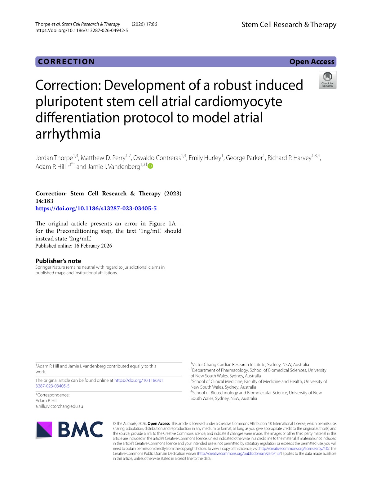
The original article presents an error in Figure 1A—for the Preconditioning step, the text ‘1ng/mL’ should instead state ‘2ng/mL’.

Correction: Development of a robust induced pluripotent stem cell atrial cardiomyocyte differentiation protocol to model atrial arrhythmia
Graphical Abstract
Original ResearchVol. 17, Issue 1 • pp. 67DOI: 10.1186/s13287-025-04883-5

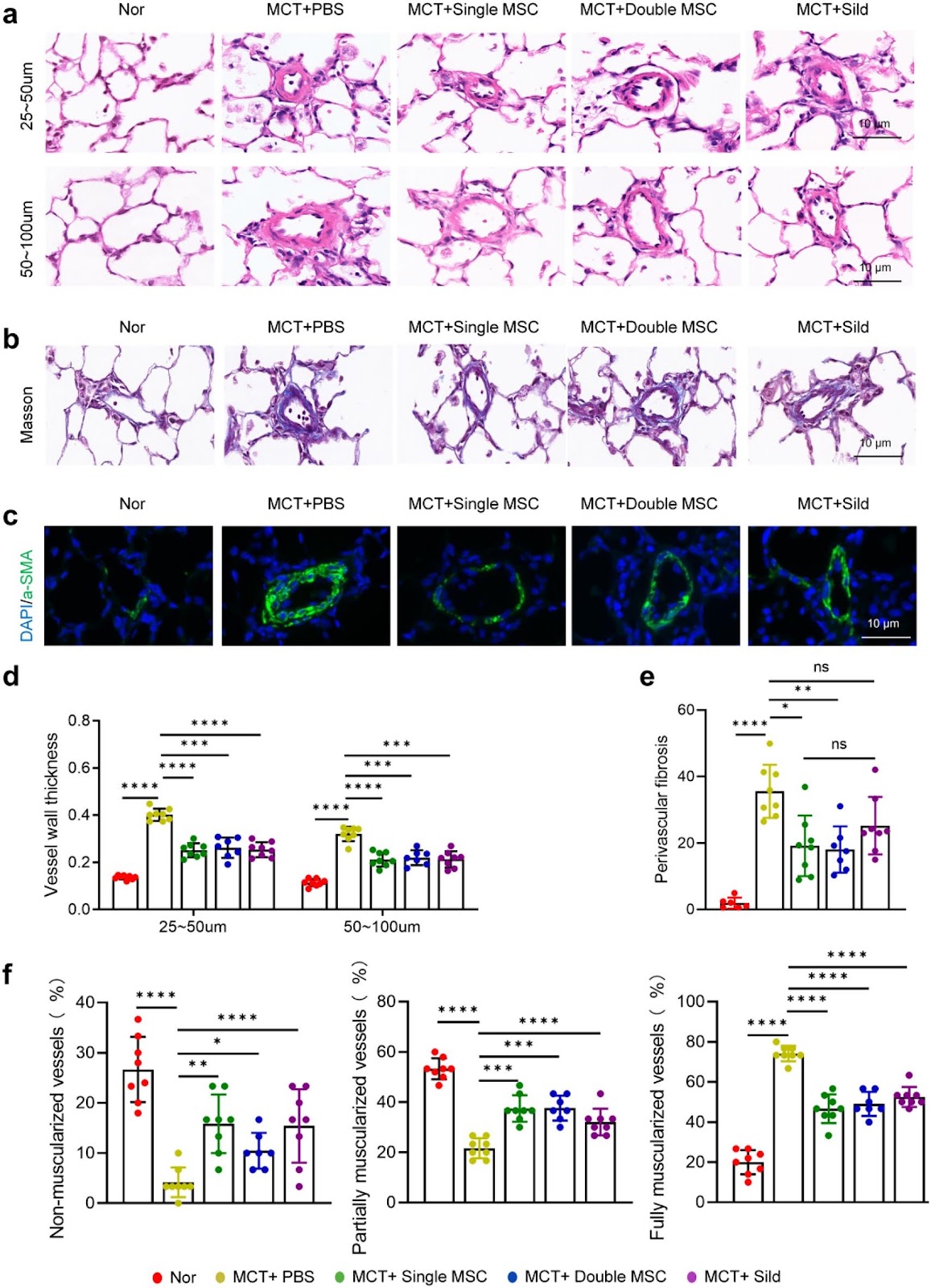
Mesenchymal stromal cells alleviate pulmonary arterial hypertension by suppressing pulmonary arterial adventitial fibroblast activation and extracellular matrix remodeling via the SOCS3/STAT3 pathway

Authors: Jiaojiao Wang, Jing Jin, Mengni Zhang, Xinyuan Chen, Sheng Du, Xiaoxiao Mao, Changlei Bao, Jinsheng Zhu, Xinyu Song, Shiyue Li

Background Pulmonary arterial hypertension (PAH) is a fatal condition characterized by progressive vascular remodeling in the pulmonary arteries, eventually leading to right heart failure and death. Dysregulated extracellular matrix (ECM) remodeling is central to PAH pathogenesis and represents a potential therapeutic target. Mesenchymal stromal cells (MSCs) have shown promise in preclinical studies; however, the optimal therapeutic window, dosing frequency, and mechanistic basis for their regulation of vascular ECM remain unclear. Methods We employed a monocrotaline (MCT)-induced rat model of PAH to evaluate different MSC treatment regimens, including early administration (day 1 post-MCT), delayed administration (days 7 and 14), and repeated dosing (days 1 and 11). Additionally, we combined in vivo and in vitro approaches to investigate how MSCs modulate the activation of pulmonary arterial adventitial fibroblasts (PAAFs) and influence ECM remodeling. Results Biodistribution studies indicated that MSC retention in lung tissue peaked within 24 h and gradually declined by day 21. A single early dose of MSCs (on day 1) significantly ameliorated PAH progression, increasing the 28-day survival rate, reducing right ventricular systolic pressure (RVSP), improving right ventricular function, and attenuating small pulmonary vascular remodeling, including reductions in medial thickening, excessive muscularization, and collagen deposition. Repeated MSC administration did not provide additional therapeutic benefit. Both in animal models and cell cultures, MSCs effectively suppressed PAAF activation and reduced ECM protein production. This anti-fibrotic effect was mediated via the SOCS3/STAT3 signaling pathway. Conclusions Early single-dose MSC therapy effectively alleviates PAH by suppressing PAAF activation and ECM remodeling through the SOCS3/STAT3 pathway, offering a potential therapeutic strategy for PAH.

Mesenchymal stromal cells alleviate pulmonary arterial hypertension by suppressing pulmonary arterial adventitial fibroblast activation and extracellular matrix remodeling via the SOCS3/STAT3 pathway
Graphical Abstract
Original ResearchVol. 17, NoneDOI: 10.1186/s13287-026-05059-5

Autologous bone marrow mesenchymal stem cell mitochondrial transplantation in recurrent assisted reproductive technology failure: a randomized controlled trial

Authors: Xiaoping Liu, Dandan Wang, Lei Jia, Weixi Chen, Rui Huang, Cong Fang, Cijie Du, Liang Yang, Xingguo Liu, Xiaoyan Liang

Background Mitochondrial dysfunction contributes to poor embryo quality and recurrent assisted reproductive technology (ART) failure. Mitochondrial transplantation (MIT), which involves supplementing oocytes with exogenous mitochondria, has been proposed as a novel strategy to improve ART outcomes. However, both its clinical efficacy and safety remain unclear. Methods In this single-center trial, 151 patients with a history of ≥ 2 failed ART cycles provided 1178 metaphase II (MII) oocytes. Sibling oocytes were randomized 1:1 to receive autologous bone marrow mesenchymal stem cells (BMSCs) mitochondria co-injection during intracytoplasmic sperm injection (ICSI) or standard ICSI. The primary outcome was the rate of day-3 good-quality embryos. Results MIT significantly accelerated early embryonic cleavage at the 3-cell stage and 5-cell stage, but this morphokinetic alteration did not translate into improvements in good-quality embryo rate, clinical pregnancy rate, or live birth rate. Long-term follow-up of 23 live births revealed no adverse effects, with all offspring exhibiting normal growth and development. Exploratory analysis revealed that oocytes yielding ≥ 70% transferable embryos after MIT harbored an elevated higher burden of medium frequency (0.05–0.5) mtDNA point mutations. Conclusions While autologous BMSCs-MIT transiently alters early cleavage kinetics, it does not demonstrate a clinical advantage in unselected patients with recurrent ART failure. Nevertheless, its observed safety profile and the identification of mtDNA mutation burden as a potential predictive biomarker provide a foundation for shifting future MIT research from a universal approach toward precision application in molecularly stratified populations.

Autologous bone marrow mesenchymal stem cell mitochondrial transplantation in recurrent assisted reproductive technology failure: a randomized controlled trial
Graphical Abstract
Original ResearchVol. 17, None • pp. 192DOI: 10.1186/s13287-026-05008-2

ATG5 overexpression enhances the therapeutic efficacy of mesenchymal stem cells in a mouse colitis model by augmenting anti-inflammatory and antioxidative mechanisms

Authors: Yang Sun, Huiyan Sun, Tengfei Zhao, Yanyun Zhao, Fengjun Xiao, Lisheng Wang

Background The therapeutic efficacy of mesenchymal stem cells (MSCs) can be improved by enhancing their adaptation to the inflammatory microenvironment. Autophagy maintains MSCs functionality, and autophagy-related gene 5 (ATG5) mediates autophagy and regulates the biological functions and therapeutic efficacy of these cells. The aim of this study was to investigate the role of ATG5 in the antioxidant capacity and evaluate the therapeutic effect of ATG5-engineered MSCs for colitis treatment. Methods Cell viability was assessed using a Cell Counting Kit-8. The mRNA expression of autophagy-, antioxidant-, and polarization-related genes was determined through real-time quantitative polymerase chain reaction, and protein expression was analyzed via western blotting. Macrophage polarization markers were analyzed using flow cytometry. Multiomics approaches, including RNA transcriptome sequencing, untargeted metabolomics, and 16S ribosomal RNA microbiota analysis, were also used. Mice with dextran sulfate sodium-induced colitis were used to evaluate the therapeutic efficacy of MSCs. Results Preconditioning MSCs with hypoxia (1% O₂) and serum deprivation significantly enhanced autophagy and upregulated ATG5 expression. Adenovirus-mediated ATG5 overexpression in MSCs (MSCs-ATG5) enhanced their autophagic activity and antioxidant capacity, upregulated HMOX-1, SOD2, and CAT expression, and increased glutathione peroxidase and catalase enzymatic activity, while enhancing cell proliferation, without altering surface marker expression. Further, MSCs-ATG5 significantly promoted M2 macrophage polarization and regulated oxidative stress-related signaling pathways. Additionally, MSCs-ATG5-based therapy markedly ameliorated colitis disease signs in mice. Transcriptome analysis revealed that MSCs-ATG5 suppressed the IL-17/NF-κB inflammatory signaling pathway. This treatment also regulated levels of the anti-inflammatory metabolite prostaglandin D2 (PGD2) in colon tissues.

ATG5 overexpression enhances the therapeutic efficacy of mesenchymal stem cells in a mouse colitis model by augmenting anti-inflammatory and antioxidative mechanisms
Graphical Abstract
Original ResearchVol. 17, None • pp. 116DOI: 10.1186/s13287-026-04938-1

Research progress on the effects of M1/M2 macrophages on the differentiation and maturation of stem cell-derived cardiomyocytes: a review

Authors: Xi Wu, Fan Zhou, Junsheng Mu

Stem cell-derived cardiomyocytes (SC-CMs) represent a promising cell source for cardiac regenerative medicine, disease modeling, and drug screening. However, their clinical translation faces significant challenges, including functional immaturity, poor long-term survival, and inadequate integration with host tissue following transplantation. The immune microenvironment, particularly the dynamic polarization of macrophages into pro-inflammatory (M1) or reparative (M2) phenotypes, is increasingly recognized as a critical regulator of cardiac repair, yet a systematic understanding of its specific effects on SC-CM fate remains incomplete. This review aims to comprehensively evaluate the dual regulatory roles of M1 and M2 macrophages on the differentiation efficiency, structural and functional maturation, and in vivo transplantation efficacy of SC-CMs. A systematic literature search was conducted in PubMed, Web of Science, Nature, and CNKI for relevant studies published from database inception to July 2025. After screening, 92 articles were included for analysis. The synthesized evidence demonstrates that M1 macrophages and their secreted factors (e.g., TNF-α, IL-1β) impede cardiac differentiation by inhibiting the Wnt/β-catenin pathway, disrupt sarcomeric organization and calcium handling, and maintain SC-CMs in a glycolytic, immature state. In contrast, M2 macrophages enhance SC-CM maturation by providing trophic support (e.g., IGF-1, HGF), promoting electrophysiological maturation and metabolic reprogramming towards oxidative phosphorylation, and facilitating angiogenesis via VEGF. The novelty of this review lies in its integrated perspective on macrophage-driven immunomodulation as a central axis for SC-CM maturation. Furthermore, it discusses emerging therapeutic strategies—such as optimized transplantation timing, co-transplantation with immunomodulatory cells, engineered exosomes, and smart biomaterials—that leverage macrophage polarization to create a favorable microenvironment for SC-CMs. Ultimately, harnessing macrophage-SC-CM crosstalk is a crucial step toward advancing clinically effective and immunologically informed cardiac regeneration therapies.

Research progress on the effects of M1/M2 macrophages on the differentiation and maturation of stem cell-derived cardiomyocytes: a review
Graphical Abstract
Original ResearchVol. 17, None • pp. 155DOI: 10.1186/s13287-026-04974-x

A novel OTUD5 variant disrupts neural progenitor cell homeostasis: mechanistic insights from HEK293T cell-based analyses

Authors: Na Xu, Shihao Wang, Tingting Yang, Meiping Yu, Yu Sun, Yongkun Zhan, Yongguo Yu

Background Variants in OTUD5 are associated with neurodevelopmental disorders (NDDs), yet the underlying molecular mechanisms remain unclear. This study aimed to investigate the pathogenicity of a novel OTUD5 variant (c.697G > A, p.Val233Met) and elucidate its regulatory role in neural progenitor cell (NPC) proliferation and differentiation, thereby uncovering the function of OTUD5 in neurodevelopment. Methods The OTUD5 variant was identified in two NDD patients via exome sequencing. Patient-derived induced pluripotent stem cells (iPSCs) and CRISPR/Cas9-corrected isogenic controls were generated. NPC proliferative activity was assessed by Ki67 immunofluorescence staining, cell-cycle distribution was analyzed by flow cytometry, and neuronal differentiation was evaluated by Tuj1/MAP2 immunofluorescence staining. Substrate screening was conducted in HEK293T cells using co-immunoprecipitation (Co-IP) and mass spectrometry. Deubiquitination capacity and protein stability were validated through ubiquitination assays and cycloheximide (CHX) chase experiments. Results The p.Val233Met variant, located within the catalytic OTU domain, induced a marked conformational alteration in the OTUD5 protein. Functionally, the variant caused aberrant NPC proliferation (1.8-fold increase in Ki67+ cells, accompanied by release of G1 arrest) and impaired neuronal differentiation (60% reduction in Tuj+ cells). Mechanistically, wild-type OTUD5 stabilized GSK3β by removing K48-linked ubiquitin chains, whereas the mutant isoform exhibited diminished deubiquitinase activity, accelerating GSK3β degradation and shortening its half-life by 40%. Conclusion This study establishes a novel disease mechanism whereby OTUD5 mutations disrupt NPC homeostasis through GSK3β destabilization, highlighting the critical role of ubiquitination regulation in neurodevelopment. Our iPSC model provides a platform for testing GSK3β-targeted therapies in OTUD5-related NDDs.

A novel OTUD5 variant disrupts neural progenitor cell homeostasis: mechanistic insights from HEK293T cell-based analyses
Graphical Abstract
Original ResearchVol. 17, NoneDOI: 10.1186/s13287-026-05080-8

Construction of liver organoid models by hepatobiliary differentiation from human induced pluripotent stem cells: state of the art, challenges and improving strategies

Authors: Yi-Hang Wu, Yang Wang, Wen-Jie Zhang, Li-Li Yuan, Yan Chen

Physiologically relevant liver models are essential for advancing hepatic disorder research, especially for disease modeling and drug development, yet current in vitro systems fail to adequately recapitulate the architecture and function of the liver. Owing to the accessibility, robust proliferation and multilineage differentiation potential of human induced pluripotent stem cells (iPSCs), liver organoids derived from iPSCs have emerged as a promising resource in hepatology. Despite this promise, the field still faces persistent bottlenecks including incomplete hepatic maturation, insufficient incorporation of non-parenchymal cells (notably immune and stromal populations), phenotypic instability, and a lack of consensus on standardized differentiation protocols. Therefore, this review systematically analyzes the challenges and strategies of iPSC differentiation into liver organoids and the related influencing factors by focusing on multidimensional regulation of hepatobiliary development as well as the effects of cellular origin, culture system and liver microenvironment on hepatic differentiation of iPSCs. Moving forward, priority should be given to the following directions: (1) Elucidating the self-assembly mechanism of liver organoids to enable precise control of hepatobiliary differentiation, thereby better governing organoid morphology and improving reproducibility; (2) Replacing exogenous cytokines with small-molecule compounds at different stages of iPSC differentiation to simplify and standardize differentiation protocols; (3) Advancing liver organoid transplantation as a means to validate physiological functionality and shift cell therapy from passive replacement toward active tissue reconstruction; (4) Integrating artificial intelligence to achieve intelligent and precise regulation of hepatic differentiation.

Construction of liver organoid models by hepatobiliary differentiation from human induced pluripotent stem cells: state of the art, challenges and improving strategies
Graphical Abstract
Original ResearchVol. 17, Issue 284 • pp. 1-24DOI: 10.1186/s13287-026-05044-y

Stem cell-driven biomedical technologies for tooth regeneration: engineering scaffolds, organoid models, and molecular targeted strategies

Authors: Zhaorui Jin, Bo Yang, Siyuan Zhang, Zhi Liu, Yuhao Wang, An Lin, Kexin Yang, Mei Yu, Weidong Tian, Fangjun Huo

Tooth loss remains a major unmet clinical challenge, and current prosthetic approaches cannot restore the biological complexity, sensory function, or regenerative capacity of natural teeth. Recent progress in stem cell biology, developmental engineering, and regenerative biomaterials has opened new possibilities for biological tooth regeneration. This review integrates advances across three major research domains that together define the current landscape of translational regenerative dentistry. First, we discuss stem cell-based, scaffold-guided strategies for tooth regeneration. These approaches combine dental and nondental stem cells, including DPSCs, SCAPs, PDLSCs, SHED, and iPSC-derived lineages, with bioactive materials such as HA/TCP ceramics, dentin-derived extracellular matrix scaffolds, and natural or synthetic polymers to promote odontogenic differentiation, vascularization, and periodontal attachment. Second, we summarize emerging tooth organoid and bioengineered tooth germ technologies that recapitulate epithelial–mesenchymal interactions and enable controlled reconstruction of dentin–pulp and periodontal compartments for modeling human odontogenesis. Third, we highlight molecular regulation-driven therapeutic strategies, focusing on the modulation of Wnt, BMP, FGF, TGF-β, and USAG-1 pathways to stimulate endogenous tooth regeneration and correct developmental defects. Despite marked progress, challenges remain, including stable neurovascular integration, optimization of stem cell–material crosstalk, precise control of spatiotemporal signaling, and long-term functional stability in vivo. Finally, we outline future directions involving smart biomaterials, gene- and protein-based molecular targeting, organoid-guided regeneration, and iPSC-enabled personalized therapies, which may further accelerate the clinical translation of stem cell-based tooth regeneration.

Stem cell-driven biomedical technologies for tooth regeneration: engineering scaffolds, organoid models, and molecular targeted strategies
Graphical Abstract
Original ResearchVol. 17, Issue 226 • pp. 1-23DOI: 10.1186/s13287-026-05087-1

Endothelial progenitor cell susceptibility to DNA damaging and DDR-modulating compounds determines endothelial differentiation accuracy

Authors: Sina Federmann, Michelle Westerhoff, Andreas S. Reichert, Gerhard Fritz

The clinical use of the anticancer drug doxorubicin (Dox) is limited by irreversible cardiotoxicity. The pathophysiological relevance of different cardiac cell types, including endothelial progenitor cells (EPC), in this process is unclear. Since progenitor cells are particularly relevant for tissue regeneration, we hypothesize that residual damage resulting from Dox-based therapeutic regimen may influence their endothelial differentiation accuracy. Therefore, we comparatively investigated the response of murine embryonic stem cells (mESC), endothelial progenitor cells (EC d4) and terminally differentiated endothelial-like cells (EC d6) following exposure to Dox and selected pharmacological inhibitors of DNA repair/DNA damage response (DDR) (RAD51i B02; HDACi entinostat (EST)). We show that EC d4 exhibit enhanced Dox sensitivity as compared to mESC and EC d6. EdU incorporation and replication fork progression analyses revealed pronounced agent-specific differences between mESC, EC d4 and EC d6. Furthermore, DNA damage formation varied in a drug-dependent manner, with mESC showing enhanced residual levels of DNA single-strand breaks (SSB) as compared to EC d4 and EC d6 while EC d6 revealed highest levels of DNA double-strand breaks (DSB). Dox treatment of EC d4 did not prevent their further differentiation into EC d6. However, it caused several functional impairments in the surviving EC d6 progeny, including defects in mitochondrial homeostasis, endothelial barrier function related to cell-cell adhesion factors (ZO1, VE-cadherin), cytokine response and low-density lipoprotein (LDL) uptake. This is accompanied by increased senescence. Summarizing, we demonstrate both overlapping and agent-specific responses of mESC, EC d4 and EC d6 to Dox and DNA repair/DDR inhibitors. Notably, drug treatment of EPC (EC d4) causes multiple dysfunctions in differentiated EC d6. Hence, pharmacological measures aiming to specifically protect EPC from Dox-induced damage are suggested to foster the maintenance of healthy endothelial functionality during regeneration, thereby lowering the risk of detrimental late cardiotoxicity resulting from Dox-based anticancer regimen.

Endothelial progenitor cell susceptibility to DNA damaging and DDR-modulating compounds determines endothelial differentiation accuracy
Graphical Abstract
Original ResearchVol. 17, Issue 235 • pp. 1-24DOI: 10.1186/s13287-026-04980-z

Mesenchymal stem cell derived exosomes mitigate COVID-19 cytokine storm via Annexin A1 and TGF-β mediated MAPK pathway inhibition

Authors: Nesrine Ebrahim, Hajir A. Al Saihati, Arigue A. Dessouky, Yasmeen Mohammed Ismail, Ashraf A. Shamaa, Shereen A. Mohamed, Mohamed E. Mohamed, Nermine Nosseir, Mohamed Ahmed Eladl, Gianpiero Di Leva, Omnia A. Badr

Background Severe COVID-19 is marked by a dysregulated inflammatory response, known as a cytokine storm, resulting in acute respiratory distress syndrome (ARDS) and multiple organ failure. Mesenchymal stem cell-derived exosomes (MSC-Exos) have demonstrated potential as immunomodulatory agents. This work investigates the possibility of MSC-Exos to mitigate excessive inflammation in COVID-19 by targeting the mitogen-activated protein kinase (MAPK) signalling pathway. Methodology We integrated molecular docking analysis between TGF-β and Annexin A1 as exosomal proteins and key component proteins of the MAPK pathway (p38, ERK1/2, JNK1). The in-silico results were then validated in vivo using a Syrian hamster model of SARS-CoV-2 infection. Quantitative PCR (qPCR), western blotting, and histological examination were employed to evaluate the effects of MSC-Exos therapy on MAPK pathway activation, cytokine production, and lung tissue pathology. Results The in-silico study revealed extensive hydrogen bonding and hydrophobic interactions at the protein–protein interfaces between exosomal proteins and MAPK components. These interactions suggest that exosomal proteins may modulate MAPK signaling pathways. In vivo, MSC-Exos administration led to marked downregulation of pivotal genes in the MAPK signaling pathway (MEKK1, MEKK2, MEKK3), diminished phosphorylation of JNK1, p38, and ERK1/2, and lowered production of pro-inflammatory cytokines (IL-1β, IL-6, TNF-α). Histopathological examination demonstrated ameliorated lung tissue structure, characterized by diminished alveolar wall thickness and decreased immune cell infiltration.

Mesenchymal stem cell derived exosomes mitigate COVID-19 cytokine storm via Annexin A1 and TGF-β mediated MAPK pathway inhibition
Graphical Abstract
Original ResearchVol. 17, None • pp. 252DOI: 10.1186/s13287-026-05066-6

Prevascularization of electrospun PCL/PLA scaffolds using human adipose-derived stem and endothelial cells enhances vascular integration and host angiogenesis in vivo

Authors: Lavinia Grob, Dagmar Brislinger, Marc Mueller, Anja Högler, Kathrin Galistl, Monika Sundl, Daniel Kummer, Nassim Ghaffari-Tabrizi-Wizsy, Hannah Müller, Melanie Pichlsberger, Lars-Peter Kamolz, Ingrid Lang-Olip

Background Inadequate vascularization remains a major limitation in tissue engineering, often leading to graft failure due to limited oxygen and nutrient supply. Prevascularization, the formation of microvascular networks within scaffolds before implantation, aims to accelerate perfusion and improve graft integration. We developed bilayer electrospun poly(ε-caprolactone)/poly(l-lactide) (PCL/PLA) scaffolds prevascularized by co-culture of human adipose-derived mesenchymal stem cells (AD-MSCs) and human placental arterial endothelial cells (HPAECs). Methods AD-MSCs were isolated from lipoaspirates and characterized by flow cytometry and functional assays. Bilayered PCL/PLA scaffolds were engineered with a wide-meshed layer for cell infiltration and a fine-meshed layer for mechanical stability. Scaffolds were seeded with AD-MSCs, HPAECs, or both (co-culture). Cell viability, adhesion, and apoptosis were analyzed histologically. Angiogenic and vasculogenic potential was evaluated in vitro and in vivo using the chick chorioallantoic membrane (CAM) assay. Results AD-MSCs expressed characteristic markers, demonstrated adipogenic and osteogenic differentiation, and promoted angiogenesis in 2D co-culture. ELISA analyses indicated dynamic secretion of VEGF, HGF, and bFGF, reflecting both paracrine and contact-dependent AD-MSC–HPAEC interactions. On scaffolds, cells primarily adhered to the wide-meshed layer. Co-culture induced vessel-like structures within a multicellular stromal environment; monocultures did not support prevascularization. Five days post-implantation, prevascularized scaffolds exhibited human microvessels at the scaffold–CAM interface and in adjacent tissue, closely associated with AD-MSCs and containing chicken erythrocytes—indicating successful anastomosis and functional perfusion. Quantitative analysis showed a significant increase in vessel branching points in the host CAM tissue in response to AD-MSC-only (2.8-fold)

Prevascularization of electrospun PCL/PLA scaffolds using human adipose-derived stem and endothelial cells enhances vascular integration and host angiogenesis in vivo
Graphical Abstract
Original ResearchVol. 16, None • pp. 663DOI: 10.1186/s13287-025-04780-x

Extracellular vesicles from Yiguanjian-primed bone-marrow mesenchymal stem cells ameliorate chronic liver fibrosis via miR-7045-5p

Authors: Zhejun Liu, Xiaodan Jiang, Hongjie You, Zuoqing Tang, Yun Ma, Niancong Che, Chongyang Ma, Wenlan Liu

Background Liver fibrosis is a crucial pathological stage in the progression of chronic liver diseases. Yiguanjian (YGJ), a Chinese herbal formula, exhibits anti-inflammatory, anti-fibrotic, and hepatoprotective effects. Extracellular vesicles from bone-marrow mesenchymal stem cells (BMSC-EVs) have shown potential in treating various disorders, including liver fibrosis. This study investigated the regulatory effects of EVs from YGJ-preconditioned BMSCs (YGJ-EVs) on TGF-β1-stimulated hepatic stellate cells (HSCs) and their therapeutic potential in a mouse model of liver fibrosis, with a focus on identifying the causative microRNA cargo. Methods YGJ-EVs and control EVs were isolated from BMSC culture supernatants and characterized via western blotting, transmission electron microscopy, and nanoparticle tracking analysis. Their cellular uptake in vitro and in vivo was evaluated using DIR labeling. To identify candidate miRNAs mediating YGJ-EV bioactivity, miRNA microarray analysis was conducted. To assess the effect of YGJ-EVs on liver fibrosis, TGF-β1-activated HSC cells were treated with YGJ-EVs or control-EVs for 24 h, and then the expression of proteins related to fibrotic activation (COL1-A1 and α-SMA), lysosomal biogenesis (LAMP1, TPP1, CTSD, and CTSB) mitophagy (p62, LC3, PINK1, and Parkin), and the Akt/AMPK/TFEB pathway was assessed. To determine whether miR-7045-5p is the causative factor, HSC cells transfected with miR-7045-5p were similarly analyzed. Results miRNA microarray analysis revealed miR-7045-5p upregulation in YGJ-EVs versus control EVs. In CCl4-treated mice, YGJ-EV-derived miR-7045-5p ameliorated the liver fibrosis, improved the hepatic function, and suppressed the HSC activation by inhibiting the Akt/AMPK/TFEB pathway. In vitro, miR-7045-5p overexpression attenuated TGF-β1-induced HSC activation. Conclusion YGJ increases miR-7045-5p abundance in BMSC-EVs. YGJ-EVs alleviate liver fibrosis by delivering the anti-fibrotic miRNA miR-7045-5p, which inhibits the Akt/AMPK/TFEB pathway, thereby promoting lysosomal biogenesis and mitophagy in HSCs.

Extracellular vesicles from Yiguanjian-primed bone-marrow mesenchymal stem cells ameliorate chronic liver fibrosis via miR-7045-5p
Graphical Abstract
Original ResearchVol. 16, Issue 669 • pp. 1-18DOI: 10.1186/s13287-025-04767-8

Targeting cellular senescence in progenitor cells as a strategy to enhance bone regeneration by cell therapies: a systematic review of pre-clinical investigations

Authors: Mayu Morita, Eshan B. Damle, Issei Shinohara, Masatoshi Murayama, Yosuke Susuki, Qi Gao, Chao Ma, Simon Kwoon-Ho Chow, Stuart Barry Goodman

Background With the global population aging, optimizing bone regeneration is becoming increasingly important for enhancing the quality of life among elderly individuals. Progenitor cell-based therapies, such as mesenchymal stromal cells and induced pluripotent stem cells for bone regeneration have shown challenges due to cellular senescence and the control of the differentiation processes remain significant hurdles. In particular, elevated expression of senescence markers may play a pivotal role in limiting bone regeneration. This systematic review examines how these senescence markers influence the efficacy of progenitor cell therapies and whether targeting them could improve outcomes. Methods We conducted a systematic literature review following the PRISMA guidelines, using the PubMed, Web of Science, Embase and Scopus with the algorithm of “bone regeneration AND senescence AND marker”. Data synthesis focused on human cell sources and specifically examined senescence markers related to bone regeneration. Results Studies using human cells were discussed in 101 papers. Based on our inclusion and exclusion criteria, 13 papers remained for our review on senescence markers in human cells within the context of bone regeneration and senescence, with and without interventional strategies. More than half of the cell sources in current aging-related studies are derived from bone marrow. Markers of aging relevant to bone regeneration include changes in cell size and morphology, increased levels of β-galactosidase (β-Gal) and Reactive Oxygen Species (ROS), and the presence of a senescence-associated secretory phenotype (SASP). Additionally, distinct senescence markers such as p16Ink4a, p21, and p53, and mitochondrial dysfunction were associated with reduced osteogenic potential and impaired regenerative capacity. Conclusion Bone marrow is the most common source of cells for studies of senescence. Cellular senescence characterized by elevated expression of specific markers was consistently shown to be negatively associated with osteogenic capacity and regenerative outcomes. The most common strategies to rejuvenate senescent cells include

Targeting cellular senescence in progenitor cells as a strategy to enhance bone regeneration by cell therapies: a systematic review of pre-clinical investigations
Graphical Abstract
Original ResearchVol. 16, None • pp. 610DOI: 10.1186/s13287-025-04745-0

Engineering hypoimmune stem cell-derived beta cells

Authors: Benedikt J. M. Licht, Garry P. Duffy, Ruth E. Levey

In type 1 diabetes (T1D), autoimmune targeting destroys insulin-producing β cells in the pancreas, creating a chronic state of insulin deficiency. Islet transplantation presents a regenerative cell therapy approach that can re-establish insulin production and intrinsic glycemic control. However, islet transplantation is currently limited by a lack of cadaveric human islet donors and a requirement for life-long immune suppression following transplant. Developments in stem cell maturation and differentiation protocols have enabled production of insulin-producing cells ‘on demand’, thereby addressing the pancreatic donor tissue shortage. Continued reliance on immune suppression to avoid graft rejection, however, can result in opportunistic infection and malignancy, thus remaining a major obstacle for wide-spread application of insulin-producing β cell transplantation. As such, there has been significant interest in identifying alternative strategies for avoiding graft rejection without immune suppression including encapsulation and co-transplantation of accessory immunomodulating cells. However, these approaches are limited by incomplete immune isolation as well as concerns over maintenance of effector function and graft survival in vivo, respectively. Genetically engineering hypoimmune stem cell-derived β cells has thus emerged as a promising strategy for improving islet transplantation outcomes. These approaches leverage our understanding of pathways involved in immune regulation to selectively protect the transplanted insulin-producing cells without affecting systemic immune function. This review will summarize recent bioengineering approaches for generating hypoimmune stem cell-derived β cells. It will also discuss relevant safety concerns and potential genetic targets for future investigation that take inspiration from the development of immune evasive primary islets and chimeric antigen receptor (CAR) T cells.

Engineering hypoimmune stem cell-derived beta cells
Graphical Abstract
Original ResearchVol. 16, None • pp. 625DOI: 10.1186/s13287-025-04732-5

Therapeatic evaluation and single cell analysis of adipose stromal vascular fraction isolation from a commercial cell separation system

Authors: Shunxin Han, Qian Zhang, Feng Lu, Junrong Cai

Background In the field of regenerative therapy, the stromal vascular fraction (SVF) extracted from adipose tissue has been widely recognized for its significant benefits. However, the cellular composition and therapeutic effect of SVF products prepared via different methods are unclear. Methods SVF cells were obtained via three approaches: (1) generation of the SVF via mechanical emulsification (M-SVF), (2) generation of the SVF via laboratory enzymatic digestion (L-SVF), and (3) generation of the SVF via commercial cell separation systems (C-SVF). We evaluated their healing effects on mouse wounds. Additionally, we utilized single-nucleus RNA sequencing (snRNA-seq) technology to explore the cellular composition of the C-SVF. Results The cell yield of C-SVF was comparable to that of L-SVF. During in vitro culture, C-SVF exhibited enhanced proliferation and a reduced proportion of apoptotic cells. In a mouse wound model, the application of C-SVF facilitated the closure of mouse wounds and improved collagen remodeling and angiogenesis in the wound area. Additional snRNA-seq analysis revealed that APOE+ adipose-derived stem cells and immune cells, especially M2 anti-inflammatory macrophages, are enriched in C-SVF, which together promote wound repair, and that APOE+ adipose-derived stem cells (ADSCs) and immune cells, especially M2 anti-inflammatory macrophages, are enriched in C-SVF, which jointly regulate and promote wound repair. Conclusion A commercial extraction system is an effective method for isolating viable SVF cells enriched with APOE+ ADSCs and M2 macrophages.

Therapeatic evaluation and single cell analysis of adipose stromal vascular fraction isolation from a commercial cell separation system
Graphical Abstract
Original ResearchVol. 16, None • pp. 94DOI: 10.1186/s13287-025-04148-1

Long-term outcomes of mesenchymal stem cell therapy in severe COVID-19 patients: 3-year follow-up of a randomized, double-blind, placebo-controlled trial

Authors: Meng-Qi Yuan, Le Song, Ze-Rui Wang, Zi-Ying Zhang, Ming Shi, Junli He, Qiong Mo, Ning Zheng, Wei-Qi Yao, Yu Zhang, Tengyun Dong, Yuanyuan Li, Chao Zhang, Jinwen Song, Lei Huang, Zhe Xu, Xin Yuan, Jun-Liang Fu, Cheng Zhen, Jianming Cai, Jinghui Dong, Jianzeng Zhang, Wei-Fen Xie, Yonggang Li, Bo Zhang, Lei Shi, Fu-Sheng Wang

Background The long-term effects and outcomes of human mesenchymal stem cell (MSC) therapy in patients with severe coronavirus disease 2019 (COVID-19) remain poorly understood. This study aimed to evaluate the extended safety and efficacy of MSC treatment in severe patients with COVID-19 who participated in our earlier randomized, double-blind, placebo-controlled clinical trial, with follow-up conducted over 3 years. Methods One hundred patients with severe COVID-19 were randomized to receive either an MSC infusion (n=65, 4×10^7 cells/dose, on days 0, 3, and 6) or a placebo, with both groups receiving the standard of care. At 36 months post-MSC therapy, patients were followed up to long-term safety and efficacy, particularly the effects of MSC therapy on persistent COVID-19 symptoms. Evaluated outcomes included lung imaging results, 6-min walking distance (6-MWD), pulmonary function test results, quality of life scores based on the Short Form-36 (SF-36) health survey, Long COVID symptoms, new-onset comorbidities, tumor marker levels, and rates of COVID-19 reinfection. Results Three years post-treatment, 46.94% (23/49) of patients in the MSC group and 34.48% (10/29) in the placebo group showed normal findings on computed tomography (CT) images (odds ratio [OR]=1.68, 95% confidence interval [CI]: 0.65–4.34). The general health (GH) score from the SF-36 was higher in the MSC group (67.0) compared to the placebo group (50.0), with a difference of 12.86 (95% CI: 1.44–24.28). Both groups showed similar results for total lung severity scores (TSS), 6-MWD, pulmonary function tests, and Long COVID symptoms. No significant differences between groups were observed in new-onset complications (including tumorigenesis) or tumor marker levels. After adjusting for China’s dynamic zero-COVID-19 strategy, severe acute respiratory syndrome coronavirus 2 (SARS-CoV-2) reinfection rates were 53.06% (26/49) in the MSC group and 67.86% (19/28) in the placebo group (OR=0.54, 95% CI: 0.20–1.41). Conclusions These findings support the long-term safety of MSC therapy in patients with severe COVID-19 over 3 years. MSC treatment may offer potential benefits for lung recovery and improved quality of life in patients experiencing Long COVID symptoms. Trial registration: ClinicalTrials.gov, NCT04288102. Registered 28 February 2020, https://clinicaltrials.gov/study/NCT04288102.

Long-term outcomes of mesenchymal stem cell therapy in severe COVID-19 patients: 3-year follow-up of a randomized, double-blind, placebo-controlled trial
Graphical Abstract
Original ResearchVol. 16, None • pp. 606DOI: 10.1186/s13287-025-04621-x

Effects of miR-210-3p/SDF2 and miR-31-5p/FGF7 from hypoxic endometrial exosomes on UCB-MSC proliferation, migration, and differentiation

Authors: Simiao Liu, Wanyu Zhang, Chengyan Deng, Hanbi Wang

Background Mesenchymal stem cells (MSCs) isolated from umbilical cord blood (UCB) exhibit significant therapeutic efficacy in endometriosis; however, the molecular mechanisms governing their regulation remain incompletely elucidated. This study delves into the regulatory functions of miR-210-3p and miR-31-5p, which are secreted via exosomes from hypoxia-damaged endometrial epithelial cells, in modulating the behavior of UCB-MSCs. Methods UCB-MSCs were transfected with specific inhibitors targeting miR-210-3p and miR-31-5p. Proliferation and migratory capacities were quantified using CCK8, EdU incorporation, Transwell, and scratch wound healing assays. Western blotting was employed to assess the expression of endometrial epithelial markers (CD9 and CK19) and stromal markers (Vimentin and CD13), alongside the phosphorylation status of JAK2 and STAT3. Dual-luciferase reporter assays were conducted to validate SDF2 and FGF7 as direct targets of miR-210-3p and miR-31-5p, respectively. Results Suppression of miR-210-3p and miR-31-5p significantly augmented the proliferative and migratory abilities of UCB-MSCs, while simultaneously enhancing their differentiation into endometrial epithelial cells and attenuating their transition into stromal cells. Concurrently, the phosphorylation levels of JAK2 and STAT3 were markedly elevated. Overexpression of SDF2 and FGF7 further amplified the proliferative, migratory, and epithelial differentiation capacities of UCB-MSCs, accompanied by heightened activation of the JAK2/STAT3 signaling pathway. Notably, SDF2 overexpression and FGF7 overexpression effectively counteracted the inhibitory effects exerted by miR-210-3p and miR-31-5p mimics on UCB-MSC proliferation, migration, and epithelial differentiation, mediated through the modulation of JAK2/STAT3 signaling. Conclusion miR-210-3p and miR-31-5p orchestrate the functional dynamics of UCB-MSCs by targeting SDF2 and FGF7, respectively, through the JAK2/STAT3 pathway. These findings unveil novel mechanistic insights into the regenerative potential of UCB-MSCs, offering promising avenues for therapeutic advancements in endometriosis.

Effects of miR-210-3p/SDF2 and miR-31-5p/FGF7 from hypoxic endometrial exosomes on UCB-MSC proliferation, migration, and differentiation
Graphical Abstract
Original ResearchVol. 16, None • pp. 214DOI: 10.1186/s13287-025-04350-1

Phase separation participates in the genetic regulation mechanism of hematopoietic stem cells: potential therapeutic methods

Authors: XinYu Tang, Yan Wang, RuiRong Xu

Hematopoietic stem cells (HSCs) are the primitive cells that give rise to common precursors for all blood cell lineages. Abnormalities in their number and/or function are important factors leading to the decline of immune function and the occurrence of various systemic diseases. Phase separation refers to a physicochemical mechanism in which intracellular liquid-liquid phase separation (LLPS) forms membrane-less organelles. It participates in various physiological activities and is related to the occurrence of diseases. Studies have shown that the functional activity of HSCs is regulated by complex mechanisms, and phase separation is closely related to these complex mechanisms such as genetic regulation, epigenetic regulation, microenvironment regulation, gene expression, autophagy degradation, and cell proliferation. With the deepening of research, the importance of phase separation in the pathogenesis and treatment of diseases such as leukemia and tumors has gradually emerged, but the deep mechanism of its regulation of HSCs genetic regulation still lacks exploration, and the direction of clinical targeted therapy is not yet clear. Here, we will summarize and elaborate the genetic regulation mechanism of HSCs, discuss the relationship between phase separation and the functional regulation of HSCs, and analyze the possibility of phase separation participating in the genetic regulation of HSCs to treat diseases, in order to provide help for the clinical implementation of targeted therapy for HSCs regulation.

Phase separation participates in the genetic regulation mechanism of hematopoietic stem cells: potential therapeutic methods
Graphical Abstract
Original ResearchVol. 16, None • pp. 174DOI: 10.1186/s13287-025-04282-w

Erythropoietin delivery through kidney organoids engineered with an episomal DNA vector

Authors: Z. Du, A. Bas-Cristóbal Menéndez, M. Urban, A. Hartley, D. Ratsma, M. Koedam, T. P.P. van den Bosch, M. Clahsen-van Groningen, J. Gribnau, J. Mulder, M. E.J. Reinders, C. C. Baan, B. van der Eerden, R. P. Harbottle, Martin J. Hoogduijn

Background The kidney’s endocrine function is essential for maintaining body homeostasis. Erythropoietin (EPO) is one of the key endocrine factors produced by the kidney, and kidney disease patients frequently experience anemia due to impaired EPO production. In the present study we explored the potential of human induced pluripotent stem cell (iPSC)-derived kidney organoids to restore EPO production. Methods EPO secretion by kidney organoids was examined under 1% and 20% oxygen levels. To increase the EPO secreting capacity of kidney organoids, iPSC were genetically engineered with a non-integrating scaffold/matrix attachment region (S/MAR) DNA vector containing the EPO gene and generated EPO-overexpressing (EPO+) kidney organoids. To assess the physiological effects of EPO+ organoids, 2–8 organoids were implanted subcutaneously in immunodeficient mice. Results Kidney organoids produced low amounts of EPO under 1% oxygen. EPO S/MAR DNA vectors persisted and continued to robustly express EPO during iPSC expansion and kidney organoid differentiation without interfering with cellular proliferation. EPO+iPSC demonstrated efficient differentiation into kidney organoids. One-month post-implantation, EPO+ organoids displayed continuously elevated EPO mRNA levels and significantly increased endothelial cell numbers compared to control organoids. Hematocrit levels were notably elevated in mice implanted with EPO+ organoids in an organoid number-dependent manner. EPO+ organoids furthermore influenced bone homeostasis in their hosts, evidenced by a change in trabecular bone composition. Conclusion Kidney organoids modified by EPO S/MAR DNA vector allow stable long-term delivery of EPO. The observed physiological effects following the implantation of EPO+ organoids underscore the potential of gene-edited kidney organoids for endocrine restoration therapy.

Erythropoietin delivery through kidney organoids engineered with an episomal DNA vector
Graphical Abstract
Original ResearchVol. 16, None • pp. 460DOI: 10.1186/s13287-025-04613-x

β-Sitosterol preconditioning enhances the resistance of BMSCs and chondrocyte to oxidative stress and promotes cartilage repair in osteoarthritis

Authors: Chengyin Liu, Xiaoman Wang, Yanyan Zhang, Hongfan Ge, Qi Chang, Zhenlei Zhou

Background Osteoarthritis (OA) is a joint disorder that severely affects patients’ mobility, overall health, and ability to perform daily activities. Despite advancements in therapeutic strategies, stem cell-based therapies for OA still face challenges, particularly in enhancing the antioxidative capacity of stem cells to improve therapeutic outcomes. Therefore, this study aimed to explore the potential of β-sitosterol in this context. Methods This study evaluated the protective effects of β-sitosterol on bone marrow-derived mesenchymal stem cells (BMSCs) and chondrocytes under oxidative stress conditions and assessed its potential in promoting cartilage repair in a rabbit OA model. Cell viability, gene expression, oxidative stress markers, and mitochondrial function were examined. In vivo therapeutic effects were evaluated through histological and immunohistochemical analyses. Results The results revealed that β-sitosterol significantly enhanced BMSC viability, upregulated the expression of Col2a1 and aggrecan, while inhibiting MMP13 expression. Furthermore, β-sitosterol effectively alleviated oxidative stress and preserved mitochondrial function in BMSCs. Notably, BMSCs pretreated with β-Sitosterol exhibited a higher potential for facilitating cartilage regeneration in the OA model, as evidence by histopathological analysis. Conclusions These findings suggest that β-sitosterol possesses significant antioxidative and chondroprotective properties, which enhance the therapeutic efficacy of BMSCs in addressing OA-related cartilage damage.

β-Sitosterol preconditioning enhances the resistance of BMSCs and chondrocyte to oxidative stress and promotes cartilage repair in osteoarthritis
Graphical Abstract
Original ResearchVol. 16, None • pp. 119DOI: 10.1186/s13287-025-04258-w

The local pulsatile parathyroid hormone delivery system induces the osteogenic differentiation of dental pulp mesenchymal stem cells to reconstruct mandibular defects

Authors: Yuanyuan Jia, Mianmian Duan, Yan Yang, Duchenhui Li, Dongxiang Wang, Zhenglong Tang

Background Tumors and injuries often lead to large mandibular defects. Accelerating the osteogenesis of large bone defect areas is a major concern in current research. In this study, dental pulp mesenchymal stem cells (DPSCs) were used as seed cells, and the local pulsatile parathyroid hormone (PTH) delivery system was used as an osteogenic-inducing active ingredient to act on DPSCs and osteoblasts, which were applied to the jaw defect area to evaluate its therapeutic effect on bone regeneration. Methods Pulsatile delivery systems, both with and without PTH, were developed following the protocols outlined in our previous study. In vitro, the biocompatibility of the pulsatile delivery system with DPSCs was assessed using the Cell Counting Kit-8 (CCK8) assay and live/dead cell staining. Osteogenic differentiation was evaluated through alkaline phosphatase staining and alizarin red staining. In vivo, critical bone defects with a diameter of 10 mm were created in the mandibles of white rabbits. The osteogenic effect was further assessed through gross observation, X-ray imaging, and histological examination. Results In vitro experiments using CCK8 assays and live/dead cell staining demonstrated that DPSCs successfully adhered to the surface of the PTH pulsatile delivery system, showing no significant difference compared to the control group. Furthermore, alkaline phosphatase staining and Alizarin Red staining confirmed that the localized pulsatile parathyroid hormone delivery system effectively induced the differentiation of DPSCs into osteoblasts, leading to the secretion of abundant calcium nodules. Animal studies further revealed that the PTH pulsatile delivery system promoted the osteogenic differentiation of DPSCs, facilitating the repair of critical mandibular bone defects.

The local pulsatile parathyroid hormone delivery system induces the osteogenic differentiation of dental pulp mesenchymal stem cells to reconstruct mandibular defects
Graphical Abstract
Original ResearchVol. 16, None • pp. 580DOI: 10.1186/s13287-025-04681-z

Spatial self-organization of cancer stem cell niches revealed by live single-cell imaging

Authors: Mathilde Brulé, Anais Horochowska, Emeline Fontaine, Raoul Torero-Ibad, Flavie Woesteland, Marie Denoulet, Jean Pesez, Eric Adriaenssens, Robert-Alain Toillon, Xuefen Le Bourhis, Benjamin Pfeuty, Chann Lagadec, François Anquez

Background: Phenotypic plasticity is a major factor in tumor heterogeneity and treatment resistance. In particular, cancer stem cells (CSCs) represent a small subpopulation within tumors that possesses self-renewal and tumor-forming capabilities. Understanding reprogramming, maintenance, and lineage properties of CSCs requires dedicated tools to disentangle the respective influences of phenotypic inheritance and cell-cell interactions. Methods: Here, we set up ultra-wide field microscopy to image breast cancer cell lines expressing a stemness fluorescent reporter over several days. The fluorescent reporter distinguishes three phenotypes: CSCs, cancer differentiated cells (CDCs), and intermediate/transiting cancer cells (iCCs). Results: Spatial statistics indicate significant zonation in which CSCs cluster together and are spatially separated from CDCs, forming patterns resembling niches. Surprisingly, single-cell time series reveal spontaneous reprogramming events from CDC to CSC even in unperturbed populations. We identify that such transitions are prone to arise during the cell cycle. Moreover, lineage analysis shows that the phenotype is partially inherited from ancestor cells. However, such heredity is not sufficient to explain the spatial properties of the cell population, which also depend on cell-cell interactions. Indeed, we find that phenotypic transitions of cancer cells are influenced by the phenotypic state of neighboring cells. Reprogramming into CSCs is respectively promoted and inhibited by the presence of CSCs and CDCs in the neighborhood. Conclusions: Altogether, our results disentangle how phenotypic inheritance and intercellular interactions orchestrate the spatio-temporal self-organization of cancer cell heterogeneity, maintaining a subpopulation of CSCs within niches.

Spatial self-organization of cancer stem cell niches revealed by live single-cell imaging
Graphical Abstract
Original ResearchVol. 16, None • pp. 249DOI: 10.1186/s13287-025-04377-4

A meta-analysis on application and prospect of cell therapy in the treatment of diabetes mellitus

Authors: Hanluo Li, Cheng Chen, Yuansheng Wang, Wei Yi, Peipei Guo, Chenguang Yao, Jinbiao Liu, Yanhong Wei, Kanghong Hu, Xiaoke Shang, Sini Kang

Objective Diabetes mellitus (DM) is a grave autoimmune disorder because of no insulin self-generation. Currently, mainly clinical methods exist, serious adverse effects leading to stem cell therapy are considered. The mesenchymal stem cells (MSCs), require high differentiation capacity and are judged as crucial in DM treatment. The meta-analysis aimed to systemically analyze the particular types of MSCs which play a more important role in DM and which DM is treated more effectively. Method A systematic review was conducted on the published literature, clinical trials and observational studies, utilizing databases such as PubMed, Embase, Cochrane and clinicaltrial.gov. RevMan software was adopted to draw Forest Plot and Funnel Plot, and subgroup analysis were employed to evaluate heterogeneity between different groups. Results We identified the meta-analyses of 34 unique random controlled trials and divided our own systematic reviews into 8 groups. The MSCs were associated with placebo (OR=2.79, 95% CI [1.63, 4.75]), Standard Clinical Treatment (SCT) (OR=4.12, 95% CI [2.76, 6.14]), and monocyte (OR=6.52, 95% CI [3.56, 9.48]). The comparison between Autologous MSCs and Allogenic MSCs (OR=4.64, 95% CI [3.42, 6.31]), Autologous BMMSCs and other MSCs (OR=5.28, 95% CI [3.64, 7.66]), Allogenic ASCs and UCMSCs (OR=3.54, 95% CI [1.83, 6.86]), Type I DM and Type II DM (OR=3.10, 95% CI [1.79, 5.38]), intravenous injection and other injections (OR=4.81, 95% CI [3.34, 6.94]), diabetic foot ulcers and diabetic neurological disease (OR=3.88,,95% CI [2.53,5.95]). Conclusion Current evidence suggests that MSCs hold significant potential for treating DM, demonstrating considerably high safety and efficacy. MSCs exhibit higher therapeutic benefits compared to monocytes, with autologous MSCs offering better clinical outcomes than allogenic sources. MSCs (BMMSCs) proved more effective than other types of MSCs. However, no significant differences were observed between adipose-derived MSCs (ASCs)

A meta-analysis on application and prospect of cell therapy in the treatment of diabetes mellitus
Graphical Abstract
Original ResearchVol. 16, Issue 1 • pp. 115DOI: 10.1186/s13287-025-04213-9

Purine metabolism in bone marrow microenvironment inhibits hematopoietic stem cell differentiation under microgravity

Authors: Xiru Liu, Hao Zhang, Jinxiao Yan, Penghui Ye, Yanran Wang, Nu Zhang, Zhenhao Tian, Bin Liu, Hui Yang

Background Spaceflight and microgravity environments have been shown to cause significant health impairments, including bone loss, immune dysfunction, and hematopoietic disorders. Hematopoietic stem cells (HSCs), as progenitors of the hematopoietic system, are critical for the continuous renewal and regulation of immune cells. Therefore, elucidating the regulatory mechanisms governing HSC fate and differentiation in microgravity environments is of paramount importance. Methods In this study, hindlimb unloading (HU) was employed in mice to simulate microgravity conditions. After 28 days of HU, cells were isolated for analysis. Flow cytometry and colony-forming assays were utilized to assess changes in HSC proliferation and differentiation. Additionally, transcriptomic and untargeted metabolomic sequencing were performed to elucidate alterations in the metabolic pathways of the bone marrow microenvironment and their molecular regulatory effects on HSCs fate. Results Our findings revealed that 28 days of HU impaired hematopoietic function, leading to multi-organ damage and hematological disorders. The simulated microgravity environment significantly increased the HSCs population in the bone marrow, particularly within the long-term and short-term subtypes, while severely compromising the differentiation capacity of hematopoietic stem/progenitor cells. Transcriptomic analysis of HSCs, combined with metabolomic profiling of bone marrow supernatants, identified 1,631 differentially expressed genes and 58 metabolites with altered abundance. Gene set enrichment analysis indicated that HU suppressed key pathways, including hematopoietic cell lineage and MAPK signaling. Furthermore, integrated analyses revealed that metabolites affected by HU, particularly hypoxanthine enriched in the purine metabolism pathway, were closely associated with hematopoietic cell lineage and MAPK signaling pathways. Molecular docking simulations and in vitro experiments confirmed that hypoxanthine interacts directly with core molecules within these pathways, influencing their expression. Conclusions These findings demonstrate that hypoxanthine in the bone marrow supernatant acts as a signaling mediator under microgravity, influencing HSCs fate by modulating hematopoietic cell lineage and MAPK signaling pathways. This study offers novel insights into the impact of microgravity on HSC fate and gene expression, underscoring the pivotal role of bone marrow microenvironmental metabolic changes in regulating key signaling pathways that determine hematopoietic destiny.

Purine metabolism in bone marrow microenvironment inhibits hematopoietic stem cell differentiation under microgravity
Graphical Abstract
Original ResearchVol. 16, None • pp. 444DOI: 10.1186/s13287-025-04549-2

Multi-function of adipose-derived stem cells on gut disorder: from bench to bedside

Authors: Jiatong Zhao, Shuzi Xin, Xiaohui Liu, Weikai Shi, Boya Wang, Hongli Wang, Yi Wang, Rongxuan Hua, Yize Li, Lei Gao, Xin Lu, Jingdong Xu

Adipose-derived stem cells (ADSCs) are a specific type of mesenchymal stem cells (MSCs) obtained easily from adipose tissue (AT). Compared with MSCs, ADSCs are easier to obtain, have fewer ethical issues, and have a higher proliferative capacity, which makes them a promising type of stem cell in regenerative medicine. ADSCs possess impressive capabilities in cell regeneration as well as differentiation, making them promising candidates for injury repair, tissue regeneration and alleviation of inflamed tissues. At present, most clinical studies on ADSCs focus on the treatment of wounds, multiple sclerosis, soft tissue trauma, aging, diabetes, Parkinson’s disease, bone and cartilage regeneration, stroke, and spinal cord injury, while its clinical applications in the gastrointestinal tract are relatively few. Therefore, this review summarizes the findings of preclinical experiments, clinical trials, and areas that may require further development of ADSCs in the treatment of digestive disorders, including inflammatory bowel disease (IBD), colorectal cancer (CRC), colorectal fibrosis, hepatocellular carcinoma, hepatic fibrosis, gastric cancer (GC), gastrostomy closure and radiation-induced proctitis. The review is concluded by discussing the goals for improvement and future directions for ADSCs before large-scale clinical application.

Multi-function of adipose-derived stem cells on gut disorder: from bench to bedside
Graphical Abstract
Original ResearchVol. 16, Issue 1 • pp. 63DOI: 10.1186/s13287-025-04179-8

Induction of the p21/CDK6 pathway and alteration of the immune microenvironment by the stem cell marker CBX3 in melanoma

Authors: Wanxian Chen, Linsa Zhou, Jingjing Jiang, Jiasheng Chen, Deyi Geng, Yaokun Chen, Xiaosha Han, Qihu Xie, Genghong Guo, Xuefen Chen, Shijie Tang, Xiaoping Zhong

Background As one of the stem cell markers, chromobox protein homolog 3 (CBX3) participates in multiple signaling pathways that affect the progression of various tumors. However, the role of CBX3 in melanoma remains unclear, and the mechanisms by which CBX3 may regulate immunotherapy outcome remain largely unknown. Methods We used the Cancer Genome Atlas, Genotype-Tissue Expression portal, and Gene Expression Omnibus database to estimate CBX3 expression and its prognostic effect in melanoma. The role of CBX3 in proliferation and migration of melanoma cells were examined using the CCK8, cloning, wound healing, and transwell assays. The effect of CBX3 on melanoma tumorigenesis was assessed using an in vivo animal model. The role of CBX3 in cell cycle was examined using flow cytometry, and expression levels of cell cycle-related genes and proteins in cells with altered CBX3 levels were analyzed using qPCR and western blotting. The function of CBX3 in the immune microenvironment of melanoma was studied using single-cell RNA sequencing and public databases. Results We found that CBX3 was highly expressed in melanoma with poor prognosis. CBX3 promoted the proliferation and migration of melanoma cells in vivo and in vitro. Functional analysis revealed that CBX3 regulates cell cycle, as it accelerated the G1 to S transition, decreased p21 expression, and increased CDK6 expression. Finally, single-cell sequencing and immune-related assays showed that CBX3 is immunogenic and can change the immune microenvironment of melanoma. Conclusions We conclude that the stem cell marker, CBX3 activates the p21/CDK6 pathway and alters the immune microenvironment in melanoma.

Induction of the p21/CDK6 pathway and alteration of the immune microenvironment by the stem cell marker CBX3 in melanoma
Graphical Abstract
Original ResearchVol. 16, None • pp. 389DOI: 10.1186/s13287-025-04501-4

Highly efficient XIST reactivation in female hPSC by transient dual inhibition of TP53 and DNA methylation during Cas9 mediated genome editing

Authors: Nami Motosugi, Keita Hasegawa, Natsumi Kurosaki, Erika Kawaguchi, Kenji Izumi, Yumi iida, Misaki Higashiseto, Keiko Yokoyama, Ayumi Sasaki, Kazuhiko Nakabayashi, Atsushi Fukuda

The irreversible erosion of X-chromosome inactivation (XCI) due to repression of the long non-coding RNA XIST presents a major challenge for disease modeling and raises safety concerns for the clinical application of female human pluripotent stem cells (hPSCs) due to the aberrant overexpression of X-linked genes. While Cas9-mediated non-homologous end joining (NHEJ) targeting the XIST promoter can induce DNA demethylation and restore XCI by reactivating XIST, its efficiency remains low. Here, we introduce a highly efficient strategy for XIST reactivation by combining TP53 inhibition with suppression of DNA methylation maintenance during Cas9-mediated NHEJ. This dual-inhibition approach increased the proportion of XIST-positive hPSCs from ~5 to ~43.7%, providing a robust method for stabilizing XCI in female hPSCs for diverse applications.

Highly efficient XIST reactivation in female hPSC by transient dual inhibition of TP53 and DNA methylation during Cas9 mediated genome editing
Graphical Abstract
Original ResearchVol. 16, None • pp. 321DOI: 10.1186/s13287-025-04426-y

Blastocyst complementation: current progress and future directions in xenogeneic organogenesis

Authors: Paula Barlabé, Xabier L. Aranguren, Giulia Coppiello

The generation of organs derived from pluripotent stem cells can be achieved in vivo through the blastocyst complementation technique. This method is based on the introduction of pluripotent stem cells into organogenesis-disabled pre-implantation embryos, where environmental signals instruct donor cells to colonize the vacant niche and to develop into the missing organ. When applied interspecies, this approach has the potential to produce human organs in genetically engineered livestock, offering a promising solution to the global transplants’ shortage crisis. In this review, we summarize the current progress in blastocyst complementation research and highlight the key challenges that must be addressed to advance this field.

Blastocyst complementation: current progress and future directions in xenogeneic organogenesis
Graphical Abstract
Original ResearchVol. 16, Issue 277 • pp. 1-15DOI: 10.1186/s13287-025-04398-z

CD73-expressing endometrial regenerative cell-derived exosomes mitigate acute cardiac allograft rejection through regulating adenosine metabolism in mice

Authors: Chenglu Sun, Dejun Kong, Hong Qin, Shilong Li, Conglin Wang, Shaohua Ren, Yini Xu, Hongda Wang, Hao Wang

Background Organ transplantation is a life-saving option for end-stage organ dysfunction, but long-term graft survival is limited by unavoidable allograft rejection. While endometrial regenerative cells (ERCs) have been shown to alleviate acute rejection, the underlying mechanisms are not fully understood. This study explored whether ERC-derived exosomes contribute to this effect through CD73-mediated immunoregulation. Methods ERCs were pretreated with GW4869, an exosome inhibitor, to block exosome secretion, and CRISPR-Cas9-based CD73 knockout was performed to validate the role of CD73 in the ERC and ERC-exos. CD73 enzyme activity was measured using an AMP assay in vitro, whereas ATP, AMP, and adenosine levels were quantified using mass spectrometry in vivo. A murine allogeneic heart transplantation model (BALB/c to C57BL/6) was established to evaluate the immunoregulatory effects of ERC-exos in vivo. Graft tissues were analyzed by H&E staining, and immunohistochemistry and flow cytometry analysis of the spleens were performed to assess graft rejection. In vitro, flow cytometry was used to examine CD4+ T-cell activation, proliferation, differentiation, and subsets. Adenosine receptor inhibitors were used to identify receptor-mediated CD73-exosome signaling, and the potential of combining CD73-expressing exosomes with rapamycin to promote long-term graft survival was explored. Results GW4869 reduces the ability of ERCs to inhibit CD4+ T-cell activation and proliferation in vitro and attenuates the ERC-mediated suppression of acute allograft rejection in vivo. ATP, AMP and ADO increase adenosine 2a receptor (A2aR) but not A2bR expression on CD4+ T cells. CD73-expressing ERC-derived exosomes (ERC-exos) metabolize AMP into adenosine, leading to the inhibition of CD4+ T-cell activation, proliferation, and Th1 differentiation in vitro. This regulatory effect is reversed by the A2a receptor inhibitor CPI444. Furthermore, CD73 depletion blocks ERC-derived exosome-mediated adenosine production and impairs the ability of these cells to inhibit CD4+ T-cell activation and proliferation in vitro, as well as attenuate acute cardiac allograft rejection in vivo. Finally, the combination of ERC-exos with rapamycin significantly prolonged allograft survival from 15 days with rapamycin monotherapy to 38 days. Conclusion CD73 expression is crucial for the ability of ERC-exos to generate adenosine to mitigate acute cardiac allograft rejection in mice. ERC-exos combined with rapamycin can prolong allograft survival.

CD73-expressing endometrial regenerative cell-derived exosomes mitigate acute cardiac allograft rejection through regulating adenosine metabolism in mice
Graphical Abstract
Original ResearchVol. 16, Issue 611 • pp. 1-11DOI: 10.1186/s13287-025-04703-w

A phase I, open-label study of intravenous human dental pulp stem cells (NestaCell®) at two dose levels in patients with Huntington’s disease

Authors: Joyce Macedo Sanches Fernandes, Eduardo Pagani, Cristiane Valverde Wenceslau, Leandro Hideki Ynoue, Luciana Ferrara, Irina Kerkis

Background Huntington’s disease (HD) is a progressive neurodegenerative disorder with no approved disease-modifying therapies. Human dental pulp stem cells (hDPSCs) offer potential therapeutic benefits due to their neurogenic, neurotrophic, and immunomodulatory properties. This prospective, open-label, single-centre, first-in-human clinical trial evaluated the safety, tolerability, and preliminary efficacy of intravenous hDPSC in patients with HD. Methods Six male patients with HD received intravenous infusions of hDPSCs in two dosage cohorts: three patients received 1 million cells/kg, and three received 2 million cells/kg. The treatment protocol consisted of cycles of three infusions at monthly intervals followed by subsequent administration cycles every six months, as per a protocol amendment based on the initial favourable safety outcomes. The total number of infusions ranged from 4 to 26 over the five years. During the first year, all patients underwent intensive multiparametric monitoring in an intensive care unit (ICU) for 48 h after each infusion. Results No adverse events occurred during the 48-h ICU monitoring or within 15 days post-infusion. Of 41 treatment-emergent adverse events (TEAEs) reported during follow-up, 35 were judged unrelated to the hDPSCs, mainly reflecting disease progression or incidental findings. Six treatment-emergent adverse events (TEAEs) were considered treatment-related, involving transient changes in hair pigmentation or regrowth. One patient discontinued due to a serious adverse event—lung cancer arising from a pre-existing pulmonary nodule identified at enrolment. Genetic analysis of the excised tumour showed no evidence of investigational product engraftment, supporting its non-tumorigenic nature. The same patient experienced a severe depressive episode

A phase I, open-label study of intravenous human dental pulp stem cells (NestaCell®) at two dose levels in patients with Huntington’s disease
Graphical Abstract
Original ResearchVol. 16, None • pp. 584DOI: 10.1186/s13287-025-04706-7

Mesenchymal stem cells inhibit mitochondrial fission by upregulating armadillo repeat containing 1, ameliorating oxidative stress in renal fibrosis

Authors: Tao Li, Chuan Guo, Qin Liu, Fengting Jing, Min Zhao, Hemin Xiong, Chang Li, Wei Zhang, Bo Chen

Background Oxidative stress damage is the important mechanism that promotes the process of fibrosis. Whether mesenchymal stem cells (MSCs) regulate mitochondrial dynamics and oxidative stress via armadillo repeat containing 1 (ARMC1) in renal fibrosis? Methods Using proteomics analysis, compare the significant differences in renal tissue proteins before and after MSCs intervention in adenine-induced nephropathy. Using a lentiviral vector to overexpress the ARMC1 gene in HK-2 cells, with the empty vector as a control. MSCs conditioned media (MSCs-CM) was applied to TGF-β1 treated cells, and MSCs were used in a cisplatin-induced nephropathy mouse model to assess mitochondrial dynamics, ROS generation, antioxidant stress, and fibrosis indicators, with Mdivi-1 (a Drp1 inhibitor) and Apocynin (a selective NADPH oxidase inhibitor) as positive controls. Results Renal proteomics showed that MSCs increased ARMC1 protein in the renal tissue of adenine nephropathy (3.521 times). In vitro, MSCs-CM increased ARMC1, reduced DRP1, and enhanced OPA1 and MFN2, lowering ROS, boosting mitochondrial bioactivity, and increasing antioxidant proteins NRF2, SOD1, SOD2, and CAT while decreasing fibrosis markers α-SMA, FN, COL-I, and KIM-1, and raising E-cadherin. The indicator variations in ARMC1-OE cells and OE-Con cells were similar between subgroups; Notably, under identical treatment conditions, the shifts in indicators within ARMC1-OE cells were more significant than those observed in OE-Con cells. In cisplatin-induced nephropathy mice, MSCs, Apocynin, and Mdivi-1 improved renal function and reduced interstitial collagen deposition, inhibited mitochondrial fission, enhanced antioxidant capacity, and reduced fibrosis. However, individual interventions were found to be less effective than their combined counterparts, with the synergistic impact of MSCs and Mdivi-1 achieving the most remarkable outcomes. Conclusion MSCs have the potential to improve renal fibrosis by influencing mitochondrial dynamics and oxidative stress through the upregulation of ARMC1 expression. ARMC1 may be an effective target for anti-fibrosis.

Mesenchymal stem cells inhibit mitochondrial fission by upregulating armadillo repeat containing 1, ameliorating oxidative stress in renal fibrosis
Graphical Abstract
Original ResearchVol. 16, None • pp. 335DOI: 10.1186/s13287-025-04474-4

Engineering biomimetic bone marrow niche with gene modified mesenchymal stromal cells for ex vivo culture of human hematopoietic stem and progenitor cells

Authors: Sevanthy Suresh, Vigneshwaran Venkatesan, Manoj Kumar K. Azhagiri, Gokulnath Mahalingam, Prathibha Babu Chandraprabha, Mohankumar K. Murugesan, Sanjay Kumar, Saravanabhavan Thangavel, Srujan Marepally

Background: Hematopoietic Stem and Progenitor Cells (HSPCs) gene therapy has shown significant progress, with commercial approval for at least four distinct haematological disorders, and poised for a rapid expansion in the upcoming years. Despite these advancements, the ex vivo culture of HSPCs continues to present significant challenges. The stress induced by ex vivo culture can negatively impact transplantation outcomes, while the need for exogenous cytokine supplementation contributes to the high costs associated with gene therapy products. Methods: We developed genetically modified human bone marrow MSCs (GM-MSCs) secreting cytokines such as Stem cell factor (SCF), Thrombopoietin (TPO), FMS-like tyrosine kinase-3-ligand (FLT3L), and Interleukin-3 (IL3), closely resembling bone marrow cellular niche to augment HSPCs culture. Results: HSPCs proliferate on GM-MSCs akin to standard conditions, devoid of external cytokine supplementation and these HSPCs retain their stem cell characteristics, colony-forming potential, stemness gene signatures, and capacity for long-term multilineage reconstitution in NBSGW mice. We demonstrate that our biomimetic feeder layer supports and alleviates stress associated with Homology Directed Repair (HDR) mediated gene-editing of HSPCs for fetal haemoglobin reactivation for a potential application in β-hemoglobinopathies gene therapy. Conclusion: Our GM-MSCs offer a compelling alternative to traditional cytokine supplementation by establishing a biomimetic bone marrow niche that fosters HSPC expansion while maintaining their stemness. These findings underscore the potential of engineered MSCs to revolutionize ex vivo HSPCs culture, ultimately enhancing their therapeutic value for gene therapy applications.

Engineering biomimetic bone marrow niche with gene modified mesenchymal stromal cells for ex vivo culture of human hematopoietic stem and progenitor cells
Graphical Abstract
Original ResearchVol. 16, None • pp. 96DOI: 10.1186/s13287-025-04173-0

Melatonin-pretreated mesenchymal stem cell-derived exosomes alleviate cavernous fibrosis in a rat model of nerve injury-induced erectile dysfunction via miR-145-5p/TGF-β/Smad axis

Authors: Xiaolin Zhang, Mengbo Yang, Xinda Chen, Ming Zhang, Yiliang Peng, Mujun Lu

Background Cavernous nerve injury-induced erectile dysfunction (CNI-ED) is a common complication after radical prostatectomy. Conventional treatment approaches have had little success in treating the severe cavernous fibrosis which is a consequence of CNI-ED. Methods Pre-treatment of adipose-derived stem cells with melatonin allows for the extraction of active exosomes (MT-hASC-EVs) from the conditioned medium. The therapeutic effects of MT-hASC-EVs were assessed in a rat model of CNI-ED, and the anti-fibrotic properties were evaluated. MicroRNA sequencing was used to identify specific microRNAs highly expressed in MT-hASC-EVs, and differential microRNAs were screened for regulatory pathways through target gene enrichment analysis. Finally, the conclusions from bioinformatics analysis were validated through in vitro experiments. Results Intracavernous injection of MT-hASC-EVs significantly restored erectile function and reduced the extent of corpus cavernosum fibrosis in the CNI-ED rat model. MT-hASC-EVs promoted the proliferation and anti-apoptotic effects of corpus cavernosum smooth muscle cells (CCSMCs) in vitro. Mechanistically, MT-hASC-EVs inhibit fibrosis by delivering miR-145-5p, which targets TGF-β2/Smad3 axis. Conclusions MT-hASCs-EVs can inhibit cavernous fibrosis and improve erectile function in a rat model of CNI-ED by targeting the miR-145-5p/TGF-β/Smad axis.

Melatonin-pretreated mesenchymal stem cell-derived exosomes alleviate cavernous fibrosis in a rat model of nerve injury-induced erectile dysfunction via miR-145-5p/TGF-β/Smad axis
Graphical Abstract
Original ResearchVol. 16, None • pp. 316DOI: 10.1186/s13287-025-04463-7

The influence of femtosecond laser intrastromal lenticules on the characteristics and maturity in tissue-engineered stem cell-derived retinal pigment epithelium sheets

Authors: Gu Jianing, Su Zhanyu, Wang Yini, Chen Yuexi, Cui Zekai, Li Shengguo, Ding Chengcheng, Sheng Wang, Li Kangjun, Tang Shibo, Chen Jiansu

Background: Recent advances in clinical trials have involved the transplantation of induced retinal pigment epithelium (iRPE) cells from stem cells in creating a functional monolayer that mimics the characteristics of natural adult RPE cells. One method of achieving this goal is through the use of tissue engineering. In this research, decellularised femtosecond laser intrastromal lenticules (dfLEN) were employed as a scaffold for cultivating a bioengineered iRPE monolayer sheet. Methods: iRPE cells were obtained by differentiating induced pluripotent stem cells (iPSC). These cells were then seeded on decellularized FLI-lenticules (dfLEN). The functionality, characterization, and oxidative stress of iRPE cultured on dfLEN were compared with those cultured on plates (TCP) using various assays such as immunofluorescence (IF), Edu, CCK8, ELISA, DFCH-DA, and JC-1. Additionally, RNA-seq assays and electron microscope (SEM and TEM) were used to test the iRPE characteristic on engineered dfLEN. Finally, we evaluated the biocompatibility of iRPE-dfLEN sheets by transplanting them into the subretinal space of New Zealand white rabbits. Results: The iRPE cells cultured on dfLEN exhibited morphology and physiology similar to that of native RPE tissue. The dfLEN not only increased the resistance capacity of iRPE cells but also improved their functional properties compared to TCP. In addition, our results indicate that dfLEN enhances the expression of genes associated with cilium assembly, resulting in notable improvements in ciliogenesis in iRPE cells. Finally, the dfLEN-iRPE sheets demonstrated favorable biocompatibility and some viability when transplanted into the subretinal space of rabbits for a period of 14 days.

The influence of femtosecond laser intrastromal lenticules on the characteristics and maturity in tissue-engineered stem cell-derived retinal pigment epithelium sheets
Graphical Abstract
Original ResearchVol. 16, None • pp. 527DOI: 10.1186/s13287-025-04627-5

Transcriptomic insights and feeder-free culturing of porcine expanded potential stem cells from cloned embryos

Authors: Lian Cai, Mirae Kim, Hyerin Choi, Haneul Kim, Sang-Hwan Hyun, Eunhye Kim

Background Generating expanded potential stem cells from cloned porcine embryos (pEPSCsNT) represents a notable advancement in regenerative medicine and agricultural biotechnology. However, challenges, including low derivation efficiency, limited understanding of transcriptomic features, and unknown feasibility of culturing under feeder-free conditions, remain. This study aimed to generate pEPSCs using blastocysts derived from parthenogenetic activation, in vitro fertilization, and somatic cell nuclear transfer (SCNT) using a modified culture system. Methods We derived pEPSCNT lines using an optimized culture system. We characterized the pEPSC lines from all three origins by analyzing pluripotent marker expression, performing karyotyping, and assessing their differentiation potential into the three germ layers. Furthermore, we performed a comparative transcriptomic analysis using in vivo and cloned embryo data, with a major focus on cell lines derived from SCNT (pEPSCsNT). We optimized feeder-free culture conditions for the pEPSCNT line and derived the pEPSCNT lines using an optimized culture system with an efficiency of ~14%. Results The cells were closely correlated with 8-cell to morula-stage embryos and exhibited significant enrichment of EPSC signature genes, suggesting a unique pluripotent state relatively close to the naïve state, specifically within a formative state. The pEPSCsNT possessed broad differentiation capacity, indicative of Hippo signaling pathway enrichment, blastocyst-like structure formation ability, and potential differentiation into trophoblast lineage cells. Conclusions Our modified culture medium combined with the 2× Matrigel coating system facilitated the transition to feeder-independent culture conditions. These findings facilitate the establishment of a feeder-free culture system while preserving pluripotency and differentiation potential.

Transcriptomic insights and feeder-free culturing of porcine expanded potential stem cells from cloned embryos
Graphical Abstract
Original ResearchVol. 16, Issue 1 • pp. 609DOI: 10.1186/s13287-025-04755-y

Apremilast improves cardiomyocyte cohesion and arrhythmia in different models for arrhythmogenic cardiomyopathy

Authors: Konstanze Stangner, Orsela Dervishi, Janina Kuhnert, Carl Wendt, Soumyata Pathak, Maria Shoykhet, Silvana Olivares-Florez, Sina Moztarzadeh, Jens Opsteen, Ni Luh Cathrin Suniasih Wohlfarth, Ruth Biller, Elisabeth Graf, Dominik S. Westphal, Tatjana Williams, Brenda Gerull, Tomo Šarić, Sunil Yeruva, Jens Waschke

Background Arrhythmogenic cardiomyopathy (ACM) is a genetically inherited desmosome heart disease leading to life-threatening arrhythmias and sudden cardiac death. Currently, ACM treatment paradigms are merely symptom targeting. Recently, apremilast was shown to stabilize keratinocyte adhesion in the desmosomal disease pemphigus vulgaris. Therefore, this study investigated whether apremilast can be a therapeutic option for ACM. Methods Human induced pluripotent stem cells from a healthy control (hiPSC) and an ACM index patient (ACM-hiPSC) carrying a heterozygous desmoplakin (DSP) gene mutation (c.2854G > T, p.Glu952Ter), confirmed by whole exome sequencing (WES), were established. Cyclic-AMP ELISA, dissociation assay, immunostaining, and Western blotting analyses were performed in human iPSC-derived cardiomyocytes (hiPSC-CMs), murine HL-1 cardiomyocytes, and cardiac slices derived from wild-type (WT) mice, plakoglobin (PG, Jup) knockout (Jup−/−) (murine ACM model) or PG Serine 665 phosphodeficient (JUP-S665A) mice. Microelectrode array (MEA) analyses in ventricular cardiac slices and Langendorff heart perfusion were performed to analyze heart rate variability and arrhythmia. Results ACM-hiPSC derived cardiomyocytes (ACM-hiPSC-CMs) revealed a significant loss of cohesion, which was rescued by apremilast. Further, treatment with apremilast strengthened basal cardiomyocyte cohesion in HL-1 cells and WT murine cardiac slices, paralleled by phosphorylation of PG at Serine 665 in human and murine models. In HL-1 cells, apremilast in addition activated ERK1/2, inhibition of which abolished apremilast-enhanced cardiomyocyte cohesion. Further, dissociation assays in slice cultures from JUP-S665A and Jup−/− mice revealed that PG is crucial for apremilast's effects. Additionally, apremilast reduced arrhythmic events in ventricular cardiac slices and Langendorff-perfused hearts. Conclusion Apremilast improves cardiomyocyte cohesion and reduces arrhythmia in different models of ACM, suggesting a novel therapeutic strategy for this disease.

Apremilast improves cardiomyocyte cohesion and arrhythmia in different models for arrhythmogenic cardiomyopathy
Graphical Abstract
Original ResearchVol. 16, None • pp. 136DOI: 10.1186/s13287-025-04168-x

Reviving hope: unlocking pancreatic islet immortality by optimizing a trehalose-based cryopreservation media and cell-penetrating peptide

Authors: Zakieh Samsonchi, Roshanak Amirian, Lobat Tayebi, Hossein Derakhshankhah, Zhila Izadi, Ensiyeh Hajizadeh-Saffar

Background Diabetes mellitus remains a pervasive global health concern, urging a deeper exploration of islet transplantation as a potential enduring solution. The efficacy of this therapeutic approach pivots on the precision of cryopreservation techniques, ensuring both the viability and accessibility of pancreatic islets. This study delves into the merits of cryopreserving these islets using the disaccharide trehalose, accompanied by an inventive strategy involving poly L proline (PLP) as a cell-penetrating peptide to overcome the cryoprotectant limitations inherent to trehalose. Methods In our experiments with rat islets, we conducted meticulous viability assessments for fresh and frozen samples. We employed a spectrum of methods, including live/dead staining, insulin/glucagon staining, and measurement of reactive oxygen species (ROS) levels. To gauge functional integrity, we executed glucose-stimulated insulin secretion tests. Subsequently, we transplanted thawed islets into diabetic mice to scrutinize their performance in clinically relevant conditions. Results Our study yielded compelling results, affirming the successful cryopreservation of pancreatic islets using trehalose and PLP. Viability, as corroborated through live/dead and insulin/glucagon staining, underscored the sustained preservation of frozen islets. Moreover, these preserved islets exhibited functional integrity by releasing insulin responsively to glucose stimulation. Significantly, upon transplantation into diabetic mice, the thawed islets proficiently restored euglycemia, evidenced by a substantial reduction in fasting blood glucose and an enhanced glucose tolerance. Conclusion Our findings accentuate the potential of trehalose and PLP as sophisticated cryoprotectants for preserving pancreatic islets. Beyond highlighting viability and functionality, the preserved islets demonstrated a remarkable capacity to restore euglycemia post-transplantation. This research holds promise in addressing the inherent limitations of islet transplantation, particularly in the realm of Type 1 diabetes treatment.

Reviving hope: unlocking pancreatic islet immortality by optimizing a trehalose-based cryopreservation media and cell-penetrating peptide
Graphical Abstract
Original ResearchVol. 16, None • pp. 524DOI: 10.1186/s13287-025-04655-1

A GelMA/polydopamine hydrogel with PTH and osteogenically stimulated alveolar mucosa-derived stem cells promotes bone regeneration in MRONJ-affected wounds

Authors: Che-Chang Tu, Ming-Hsu Chen, Guan-Yu Lan, Yu-Ting Lin, Yu-Tse Lin, Wei-Chiu Tai, Jiashing Yu, Po-Chun Chang

Background Medication-related osteonecrosis of the jaw (MRONJ) is a serious complication in patients taking bisphosphonates. This study aimed at developing a mesenchymal stem cell-based strategy to reduce the incidence of MRONJ and recover regeneration capability of MRONJ-affected wounds by using a gelatin methacryloyl/polydopamine hydrogel (GelMA/PD) to adhere alveolar mucosa-derived stem cells (AMCs) on the bone surface, with the osteogenically stimulated AMCs modulated by microRNA (miR) transfection, and the osteoanabolic environment activated by parathyroid hormone (PTH). Methods GelMA/PD was synthesized by photo-crosslinking, and the incorporation of PD onto GelMA as well as mechanical properties were assessed. Rat AMCs were isolated, and the stemness was characterized. AMCs were osteogenically stimulated by miR transfection. Maxillary osteotomy was created in rats administrated with zoledronic acid and dexamethasone to simulate MRONJ-affected wounds, and osteotomy in rats without ZA served as healthy controls. Wounds were unfilled or filled with GelMA/PD alone, GelMA/PD with AMCs (GA), GelMA/PD with OAMCs (GO), or GelMA/PD with OAMCs and PTH (PO), and were assessed by gross observation, micro-CT imaging, histology, and immunohistochemistry for osteoblast-osteoclast coupling. Results GelMA/PD exhibited modestly decreased compressive strength and superior adhesion strength compared with GelMA. AMCs were double positive for CD73 and CD90, showed trilineage differentiation capability, and were osteogenically stimulated by miR-218 transfection. Among MRONJ-affected wounds, soft tissue coverage was accelerated, with reduced sequestra and significantly greater bone volume in PO group (38.46 ± 10.02%) relative to unfilled group (21.81 ± 6.18%), and osteoblast-osteoclast coupling was evident in GO and PO groups. Soft tissue recovery, inflammation reduction, and matrix deposition on defect surfaces were more prominent in PO group.

A GelMA/polydopamine hydrogel with PTH and osteogenically stimulated alveolar mucosa-derived stem cells promotes bone regeneration in MRONJ-affected wounds
Graphical Abstract
Original ResearchVol. 16, None • pp. 571DOI: 10.1186/s13287-025-04665-z

Extracellular vesicle bioactivity and potential for clinical development are determined by mesenchymal stromal cell clonal subtype

Authors: Savvas Ioannou, Alasdair G. Kay, Andrew P. Stone, Emma Rand, Samuel Elberfeld, William Bolton, Tony Larson, Rachel E. Crossland, Oksana Kehoe, David A. Mentlak, Xiao-Nong Wang, Chris MacDonald, Paul G. Genever

Background Mesenchymal stromal cells (MSCs) have been used in numerous clinical trials but very few reach phase 3 or market authorisation. Progress is often hampered by the use of non-clonal, heterogeneous and uncharacterised MSC cultures and lack of mechanistic understanding. There is limited evidence of MSC engraftment in vivo and disease resolution may be the result of the paracrine effects of the MSC secretome, rather than the cells per se. Extracellular vesicles (EVs) are key components of the MSC secretome and there is growing interest in the use of EVs as cell-free therapies. However, like MSCs, heterogeneity can exist within any therapeutic EV pool. Here we used immortalised clonal MSC lines, termed Y201 and Y202, to examine how MSC phenotype influences EV character and function. Methods EVs were isolated by ultracentrifugation and characterised by nano-sizing, ultrastructural morphometric analysis, western blotting, mass spectrometry and miRNA screening. Bioactivity was determined by phosphorylation of ERK1/2, proliferation and T cell polarisation assays and using two in vivo models of inflammatory disease. Results EVs from Y201 and Y202 MSCs were morphologically similar, however, Y201 EVs were more abundant in EV biomarkers versus Y202 EVs, with an enhanced miRNA and proteomic content. Computational analysis of the Y201 EV proteome identified significant enrichment in matrix-associated proteins, predicted to contribute to an elaborate EV corona particularly abundant in RGD-containing proteins fibronectin and MFG-E8, which was confirmed by western blotting. Y201 EVs, but not Y202 EVs, significantly increased the proliferation of articular chondrocytes in a dose-dependent manner, and the proliferative effect of Y201 EVs was mediated at least in part via an RGD (integrin)-FAK-ERK1/2 axis. Both Y201 and Y202 EV subsets significantly reduced proliferative index scores of activated T cells. However, only Y201 EVs, not Y202 EVs, suppressed disease activity compared to controls in different in vivo models of inflammatory peritonitis and arthritis.

Extracellular vesicle bioactivity and potential for clinical development are determined by mesenchymal stromal cell clonal subtype
Graphical Abstract
Original ResearchVol. 16, None • pp. 570DOI: 10.1186/s13287-025-04573-2

Intranasal delivery of DPSC-derived small extracellular vesicles-encased phloroglucinol attenuates non-motor and motor deficits and promotes neurogenesis in an in vivo rat model of Parkinson’s disease

Authors: Kallolika Mondal, Rituparna Ghanty, Anita Mahadevan, Girish Waghmare, Rashmi Santhoshkumar, Nandeesh BN, Indrani Datta

Background  Parkinson’s disease (PD) is characterized by dopaminergic (DA) neuron degeneration in the substantia nigra pars compacta (SNpc) driven by oxidative stress, inflammation, and impaired neurogenesis. Phloroglucinol, a polyphenolic antioxidant, has demonstrated neuroprotective effects in PD models but suffers from limited clinical applicability due to poor blood-brain barrier (BBB) permeability. Small extracellular vesicles (sEV) derived from dental pulp stem cells (DPSCs) exhibit neuroprotective and immunomodulatory properties and serve as promising vehicles for targeted drug delivery across the BBB. This study aimed to evaluate the therapeutic efficacy of intranasally administered sEV-encased phloroglucinol (sEV-Phl) in a chronic MPTP rat model of PD. Methods  DPSC-derived sEV were isolated via density gradient ultracentrifugation and characterized using Transmission Electron Microscopy (TEM), Dynamic-Light-Scattering (DLS), and CD marker expression. Phloroglucinol was encased in sEV (sEV-Phl) using sonication. Antioxidant properties were tested in vitro using an H2DCF.DA assay in SH-SY5Y cells exposed to 6-OHDA. Chronic MPTP-treated male Wistar rats received intranasal sEV-Phl, with motor and non-motor behaviours evaluated up to 4-weeks post-MPTP treatment. TH-positive neurons, neurogenesis (Ki67, BrdU and FOXA2), lipid-peroxidation, and neurotransmitter-levels were analyzed. sEV biodistribution was tracked via near-infrared imaging and localization in neuronal and glial cells was confirmed with PKH-26 labelling, with confocal-imaging further verifying localization in neuronal and glial cells. TNF-α expression was assessed as a marker of neuroinflammation. Results  sEV displayed high purity and homogeneity. sEV-Phl significantly reduced oxidative stress both in vitro and in vivo, as indicated by decreased ROS and lipid peroxidation levels. sEV-Phl treated MPTP rats demonstrated marked improvement in motor and non-motor behaviours compared to MPTP rats. Immunohistochemical analysis revealed

Intranasal delivery of DPSC-derived small extracellular vesicles-encased phloroglucinol attenuates non-motor and motor deficits and promotes neurogenesis in an in vivo rat model of Parkinson’s disease
Graphical Abstract
Original ResearchVol. 16, None • pp. 206DOI: 10.1186/s13287-025-04322-5

Nano-zinc oxide (nZnO) targets the AMPK-ULK1 pathway to promote bone regeneration

Authors: Xiu Chen, Zhenkun Weng, Hongchao Zhang, Jian Jiao, Jingjia Liang, Jin Xu, Dongmei Wang, Qian Liu, Qing Yan, Aihua Gu

Background Nano-zinc oxide (nZnO) has attracted significant attention in bone tissue engineering due to its antibacterial properties, anti-inflammatory effects, biocompatibility, and chemical stability. Although numerous studies have demonstrated the enhancement of osteogenic differentiation by nZnO-modified tissue engineering materials, the underlying mechanisms remain poorly characterized. Methods This study aimed to identify the molecular mechanisms how nZnO promoted osteogenic differentiation and bone regeneration using transcriptome analysis, drug intervention, and shRNA knockdown techniques, etc. First, the study evaluated the in vivo effects of gelatin methacryloyl (GelMA) containing nZnO on bone regeneration using a mouse calvarial defect model. The impact of nZnO exposure on the osteogenic differentiation of mesenchymal stem cells (MSCs) was then assessed. The combined treatment of nZnO and MSCs in GelMA for bone regeneration was assessed in the mouse calvarial defect model thereafter. Results nZnO induced osteoblastic differentiation to promote bone regeneration. nZnO activated the AMP-dependent protein kinase (AMPK)-ULK1 signals to stimulate autophagosomes formation and facilitate autophagy flow, which was the essential pathway to induce osteogenic differentiation. The combined treatment of MSCs and nZnO significantly enhanced bone regeneration in calvarial defect mice. Conversely, AMPK inhibitor Compound C (C.C) reversed the effects on autophagy flow and osteogenic potentiality induced by nZnO. Conclusions These results highlight that nZnO can regulate bone regeneration by activating autophagy through the AMPK/ULK1 signaling pathway, which may provide a novel therapeutic strategy for addressing bone defects using nZnO.

Nano-zinc oxide (nZnO) targets the AMPK-ULK1 pathway to promote bone regeneration
Graphical Abstract
Original ResearchVol. 16, None • pp. 483DOI: 10.1186/s13287-025-04578-x

BACH1 recruits STAT3 to enhance leukemia inhibitory factor receptor activity and augments the self-renewal capacity of mouse embryonic stem cells

Authors: Jinghua Ma, Siyu Ma, Cong Niu, Siqing Wang, Xiangxiang Wei, Dan Meng, Xiuling Zhi, Jieyu Guo

Background Genomic studies have linked single nucleotide variants in the enhancer region of the leukemia inhibitory factor receptor (Lifr) gene to chromatin accessibility and the regulation of self-renewal in mouse embryonic stem cells (mESCs). However, the underlying mechanisms remain unclear. This study investigates the role of the transcription factor BTB and CNC homology 1 (BACH1) in regulating the Lifr enhancer and its impact on mESC pluripotency. Methods We performed RNA-sequencing (RNA-seq) to assess the impact of Bach1 knockout on gene expression in mESCs. Additionally, chromatin immunoprecipitation (ChIP), co-immunoprecipitation (co-IP), and luciferase reporter gene analysis were employed to investigate the mechanism by which BACH1 regulates Lifr expression. Results Genomic analyses identified BACH1 binding at the Lifr enhancer proximal to rs50454566 in mESCs. Integrated single-cell RNA sequencing (scRNA-seq) data revealed co-upregulation of Bach1 and Lifr in inner cell mass (ICM) cells. RNA-seq analyses demonstrated that Bach1 depletion attenuated Lifr expression and impeded LIFR-signal transducer and activator of transcription 3 (STAT3) signaling. Mechanistically, BACH1 recruited STAT3 to the Lifr enhancer, driving Lifr transcription and facilitating the LIFR-STAT3 signaling pathway, thereby enhancing mESC self-renewal. Conclusion Our findings demonstrate that BACH1 enhances Lifr enhancer activity by recruiting STAT3 and activates the LIFR-STAT3 signaling pathway by promoting the LIFR expression, thereby maintaining mESC self-renewal.

BACH1 recruits STAT3 to enhance leukemia inhibitory factor receptor activity and augments the self-renewal capacity of mouse embryonic stem cells
Graphical Abstract
Original ResearchVol. 16, None • pp. 399DOI: 10.1186/s13287-025-04480-6

From inflammation to healing: the crucial role of GPR91 activation and SDH inhibition in chronic diabetic wound recovery

Authors: Hengdeng Liu, Shixin Zhao, Hanwen Wang, Xuefeng He, Suyue Gao, Minmin Su, Miao Zhen, Shuying Chen, Lei Chen, Julin Xie

Background Diabetic foot ulcers (DFU) typically exhibit impaired healing due to dysregulated re-epithelialization and excessive inflammation. Succinate, a key metabolic intermediate, is now understood to regulate inflammation through G Protein-Coupled Receptor 91 (GPR91) and succinate dehydrogenase (SDH), although its role in DFU remains unclear. Methods Co-cultures of M2 macrophages and epithelial cells, along with clinical samples, were used to analyze the expression of GPR91 and SDH. Functional assays were performed using high glucose (HG)-treated M2 macrophages (HG-M2) and an in vivo model. Cytokine and growth factor levels in cell supernatant were measured, and molecular mechanisms were explored via qRT-PCR, flow cytometry, and western blot analysis. Results Elevated glucose concentrations increased succinate levels and disrupted M2 macrophage–epidermal stem cells (EpSCs) interactions. GPR91 knockdown worsened HG-M2 dysfunction, while GPR91 overexpression (OE-GPR91) enhanced anti-inflammatory responses and reduced succinate. OE-GPR91-conditioned medium preserved EpSCs stemness and promoted migration mediated by hepatocyte growth factor (HGF). SDH inhibition (via Dimethyl malonate, DMM) boosted M2 macrophage activity by reducing reactive oxygen species (ROS) and upregulating Gpr91 expression. Mechanistically, GPR91 activated the pAkt/pGSK3β/β-catenin pathway, while DMM enhanced M2 macrophage function via the PI3K-Akt/pERK1/2 pathway. Conclusions GPR91 upregulation and SDH inhibition improve HG-M2 macrophage function, reduce inflammation, and enhance HGF-mediated EpSCs repair. Targeting both pathways may represent a promising approach to promote DFU healing.

From inflammation to healing: the crucial role of GPR91 activation and SDH inhibition in chronic diabetic wound recovery
Graphical Abstract
Original ResearchVol. 16, None • pp. 53DOI: 10.1186/s13287-025-04154-3

MSCs with upregulated lipid metabolism block hematopoietic stem cell differentiation via exosomal CTP-1A in MDS

Authors: Chunlai Yin, Xue Yan, Jinyi Ren, Cheng Zhang, Jiaqing Liu, Zilong Wang, Jing Liu, Weiping Li, Xia Li

Background Myelodysplastic syndrome (MDS) is a clonal disorder of hematopoietic stem cells (HSCs), characterized by ineffective hematopoiesis and a high risk of progression to acute myeloid leukemia. Elucidating the mechanism underlying the dysfunction of MDS-HSCs is crucial for exploring the pathogenesis of the syndrome. While previous studies have implicated mesenchymal stem cells (MSCs), a principal component of the bone marrow (BM) microenvironment, in the inhibition of normal hematopoiesis, the precise molecular mechanisms have not been fully elucidated. In this study, we investigated the effects of MSCs from MDS patients on hematopoietic functions of HSCs from a metabolic perspective. Methods MSCs were isolated from BM of MDS patients. The proliferation, apoptosis, differentiation and support for hematopoiesis of these cells were analyzed using CCK-8 assay, FC and induction medium and CFU (colony forming units) assay, respectively. Expression levels of metabolic molecules were used as indicators to screen MSCs with different metabolic pathways and were detected by RT-PCR and Western blotting. Exosome derived from MSCs were isolated from the culture supernatant and confirmed by Transmission Electron Microscope, Dynamic Light Scattering and Western blotting. The effects of these exosomes on HSCs were analyzed using the same methods as those used to assess MSCs function. Results Our findings demonstrated that MDS-MSCs exhibited significant functional impairments, including reduced proliferation, impaired differentiation, diminished support for hematopoiesis, and increased apoptosis. Notably, we observed an upregulation of lipid metabolism in these MSCs, which appears to contribute to their dysfunction. Intriguingly, the aberrant lipid metabolic profile can be effectively reversed by the administration of etomoxir (ETO), an inhibitor of carnitine palmitoyltransferase 1A (CPT-1A). Furthermore, MSCs with enhanced lipid metabolism could transmit this dysfunction to HSCs through the secretion of exosomes that are enriched in CPT-1A. Conclusions We suggest that the MDS BM microenvironment disrupts MSCs metabolism by increasing the expression of CPT-1A, which impairs the ability to support normal HSCs. Interestingly, the suppressive effect is mediated by exosomes rich in CPT-1A, which derived from MSCs. These findings provide novel insights into MDS MSCs-metabolism-Exosome axis in ineffective hematopoiesis and offer new strategies for the treatment of MDS.

MSCs with upregulated lipid metabolism block hematopoietic stem cell differentiation via exosomal CTP-1A in MDS
Graphical Abstract
Original ResearchVol. 16, None • pp. 376DOI: 10.1186/s13287-025-04504-1

Small extracellular vesicles secreted from TGF-β1-licensed mesenchymal stromal cells reduce inflammation-associated injury following corneal alkali burn

Authors: Ellen Donohoe, Aoife Canning, Eanna Johnston, Seyedmohammad Moosavizadeh, Jiemin Wang, Martin Leahy, Oliver Treacy, Aideen E. Ryan, Thomas Ritter

Background It is well established that the mesenchymal stromal cell (MSC) therapeutic potency can be enhanced by cytokine pre-activation or licensing. However, its effects on therapeutic efficacy of small extracellular vesicles (MSC-sEV) have not yet been well established. Here we report on two different cytokine licensing strategies, using either a pro-inflammatory or anti-inflammatory cytokine and evaluate their therapeutic potency in vitro and in a preclinical model of corneal chemical burn. Methods BALB/c MSCs were cultured with no supplement, recombinant IFNγ, or recombinant TGFβ1 for 72 h. sEV, sEVIFNγ, and sEVTGFβ were then isolated from conditioned medium of parental cells by a combination of ultrafiltration and size exclusion chromatography. Following isolation MSC-sEV were thoroughly characterized for size, marker expression and therapeutic efficacy. To evaluate their immunomodulatory capacity, both naïve and licensed MSC-sEV were tested in in vitro macrophage and T cell assays and in a preclinical corneal injury model. Results Relative to unlicensed sEV, sEVIFNγ exhibited increased expression of MHC I and PD-L1 on their surface, whereas sEVTGFβ expressed higher levels of CD44, CD29, and CD73. For immunomodulatory capacity, only sEVTGFβ was found to reduce macrophage expression of MHC II and CD80 and induced the secretion of anti-inflammatory macrophage cytokines. sEVTGFβ were also found to increase Treg expansion and FOXP3 expression. Given the superior efficacy observed of sEVTGFβ in vitro, this product was evaluated in a preclinical mouse model of corneal chemical burn. sEVTGFβ were applied either topically (day 0, 1, and 3) or subconjunctivally (day 0, and 3), and mice were monitored for 14 days. sEVTGFβ ameliorated burn-induced structural damage and accelerated restoration of normal corneal thickness, compared to PBS-treated controls. sEVTGFβ also resulted in reduced inflammatory mediators (IL-1β, iNOS) and minimised levels of fibrosis-associated collagen in the cornea. Mice that received subconjunctival, but not topical, administration of sEVTGFβ exhibited regulatory immune cell profiles with reduced pro-inflammatory-

Small extracellular vesicles secreted from TGF-β1-licensed mesenchymal stromal cells reduce inflammation-associated injury following corneal alkali burn
Graphical Abstract
Original ResearchVol. 16, None • pp. 105DOI: 10.1186/s13287-025-04245-1

Apoptotic vesicles derived from bone marrow mesenchymal stem cells increase angiogenesis in a hind limb ischemia model via the NAMPT/SIRT1/FOXO1 axis

Authors: Jinxing Chen, Zekun Shen, Bingyi Chen, Shuang Liu, Yifan Mei, Kai Li, Ziyang Peng, Chaoshuai Feng, Weiyi Wang, Shaoying Lu

Background Chronic limb-threatening ischemia (CLTI) is the most severe form of peripheral arterial disease (PAD). Mesenchymal stem cell (MSC) transplantation holds promise as a treatment for CLTI; however, the harsh local environment poses challenges to its effectiveness. Apoptotic vesicles (ApoVs) are extracellular vesicles produced by cells undergoing apoptosis, and they can carry various biomolecules from their parent cells, including proteins, RNA, DNA, lipids, ions, and gas neurotransmitters. ApoVs play significant roles in anti-inflammatory responses, anti-tumor activities, and tissue regeneration through intercellular communication, and they have demonstrated potential as drug carriers. In this study, we investigated the potential of bone marrow stem cell (BMSC)-derived ApoVs for treating CLTI. Methods In vivo, we explored the therapeutic effect of ApoVs on a hindlimb ischemia model through Laser Doppler, matrigel plug assay, and histological analysis. In vitro, we analyzed the effects of ApoVs on the proliferation, migration, and angiogenesis of HUVECs and explored the uptake process of ApoVs. In addition, Proteomic analysis, western blotting, quantitative real-time PCR, shRNA, and siRNA were used to analyze ApoVs-induced HUVECs activation and downstream signaling pathways. Results BMSCs transplantation showed improvement in a hind limb ischemia model, and this effect still exists after apoptosis of BMSCs. Subsequently, ApoVs of BMSCs were isolated and found to improve mouse hind limb ischemia in vivo. In vitro, ApoVs can be ingested by HUVECs through dynamin-, clathrin-, and caveolin-mediated endocytosis and promote its proliferation, migration, and angiogenesis. Mechanistically, ApoVs transferred NAMPT to HUVECs, therefore activating the NAMPT/SIRT1/FOXO1 axis, influencing the transcriptional activity of FOXO1, and promoting angiogenesis. Conclusions Our results demonstrate that the transplanted BMSCs can ameliorate hindlimb ischemia by releasing ApoVs during apoptosis. The main mechanism of this effect is promoting the proliferation, migration, and angiogenesis of endothelial cells via the NAMPT/SIRT1/FOXO1 axis.

Apoptotic vesicles derived from bone marrow mesenchymal stem cells increase angiogenesis in a hind limb ischemia model via the NAMPT/SIRT1/FOXO1 axis
Graphical Abstract
Original ResearchVol. 16, Issue 472 • pp. 1-8DOI: 10.1186/s13287-025-04608-8

Integrin signaling pathways in mesenchymal stem cells

Authors: Dafina Najwa Mohd Arizam, Fazlina Nordin, Azlina Ahmad, Khairul Bariah Ahmad Amin Noordin

This review provides an overview of the integrin signaling pathways and their roles in mesenchymal stem cell differentiation into adipocytes, chondrocytes, and osteoblasts. In these three differentiated cells, the cell extracellular matrix plays an important role in regulating the integrin signaling pathway, as the presence of growth factors and other molecules in the extracellular matrix will affect the cell differentiation. The focus of this review is to elucidate the role of the integrin signaling pathway in adipogenesis, chondrogenesis, and osteogenesis, highlighting its diverse contributions to tissue homeostasis and repair. By synthesizing current knowledge, this paper aims to inspire further research into the therapeutic potential of targeting integrin pathways in stem cell-based tissue engineering.

Integrin signaling pathways in mesenchymal stem cells
Graphical Abstract
Original ResearchVol. 16, None • pp. 403DOI: 10.1186/s13287-025-04532-x

Human embryonic stem cell-derived Sertoli cells as an immune modulator of cell transplantation therapy in a diabetic mice model

Authors: Jeoung Eun Lee, Chang Woo Lee, A-Reum Han, Jina Kim, Dong Ryul Lee

Objective Sertoli cells (SCs) are somatic cells that are a part of the seminiferous tubules in the testes and support germ cell development and maturation. Additionally, SCs play another role in protecting male germ cells from immune destruction via the formation of the blood-testis barrier and the secretion of several immunoregulatory factors. Based on these characteristics, SCs have been suggested to create a tolerogenic environment to protect co-transplanted cells as immune modulators. Because mature SCs are quiescent somatic cells and show lower proliferation activity in vitro, it is difficult to obtain the number of human cells needed for clinical applications. Materials and methods We established a protocol for mass production of SCs from human ESCs (hESC-SCs) and their functional properties were analyzed in vitro and in diabetic-induced mice after their co-transplantation with human insulin-secreting cells. Results hESC-SCs were successfully produced via a stepwise differentiation protocol. In addition, a mass culture method was established to secure the number of hESC-SCs available for cell therapy. hESC-SCs obtained from in vitro derivation highly express marker genes of SCs, such as GATA4, SOX9, CLDN11, and AR, and have shown immune-modulation activity similar to that of human bone marrow-mesenchymal stem cells. In diabetic-induced mice subcutaneously co-transplanted with EndoC-βH1 cells (insulin-secreting cells) and hESC-SCs, lower blood glucose levels were maintained for 6 months than in those transplanted with EndoC-βH1 cells alone. Conclusions We believe that hESC-SCs could be useful tool for securing cell therapy to treat human diseases in the future.

Human embryonic stem cell-derived Sertoli cells as an immune modulator of cell transplantation therapy in a diabetic mice model
Graphical Abstract
Original ResearchVol. 16, None • pp. 300DOI: 10.1186/s13287-025-04401-7

Synergistic potential of bone marrow mesenchymal stem cells and miR181-a combinational therapy against multiple sclerosis

Authors: Xin Xiu, Sijia Chen, Yumei Liu, Bo Sun, Hulun Li, Sifan Zhang, Xixi Yang, Yu Wei, Xichen Peng, Yan Wang, Yanping Wang, Junfeng Wu, Yao Zhang, Lili Mu, Qingfei Kong, Xijun Liu

Background: Multiple sclerosis (MS) is a progressive autoimmune disease characterized by massive inflammatory infiltration, demyelination, and subsequent axonal injury and neuronal damage in the central nervous system (CNS). The etiology of MS remains unclear and there is not yet a definitive therapeutic schedule for the disease. Bone marrow mesenchymal stem cells (BMSCs), exhibiting neuroimmune-modulatory functions to alleviate various autoimmune diseases, show great potential in the treatment of MS. However, the instability of BMSCs-mediated immunosuppression in vivo has limited their application. MiR181-a, a positive regulator of immune balance, which has a preference for T cells and B cells differentiation, but degrade rapidly upon entering systemic circulation due to their unstable molecular structure. Methods: We propose a synergistic therapy approach that combines the penetrative targeting capability of BMSCs with the immuno-modulatory effects of miR181-a by overexpressing miR181-a to BMSCs through lentivirus packaging system. With this strategy, on the basis of the establishment of the experimental autoimmune encephalomyelitis (EAE) model, miR181-a overexpressing BMSCs (miR181a-BMSCs) would have a stronger immuno-modulatory treatment benefit, in terms of attenuating MS development. Results: Indicate that this method prolongs the modulatory effects of BMSCs and resulted in significantly enhancements of the proliferation of regulatory B cells (Bregs), regulatory T cells (Tregs) and the inhibition of Th17 cells compared to the traditional BMSCs group. Moreover, 10-fold miRNA’s concentration in the exosome of miR181a-BMSCs, leading to an increased duration of miRNAs to exert their biological effects. By immunotherapy and synergistic treatment, the effectiveness of the treatment is significantly enhanced, showing consistent results in different groups of the animal model.

Synergistic potential of bone marrow mesenchymal stem cells and miR181-a combinational therapy against multiple sclerosis
Graphical Abstract
Original ResearchVol. 16, Issue 1 • pp. 84DOI: 10.1186/s13287-025-04229-1

OCT4 translationally promotes AKT signaling as an RNA-binding protein in stressed pluripotent stem cells

Authors: Wenjie Chen, Xinyu Chen, Cheng Chen, Shiqi She, Xia Li, Lina Shan, Xiaobing Zhang, Songsong Dan, Yisha Wang, Yan-Wen Zhou, Qingyi Cao, Wenxin Wang, Jianwen Hu, Yaxun Wei, Yaqiang Xue, Yi Zhang, Songying Zhang, Ying-Jie Wang, Bo Kang

Background Despite numerous studies addressing the molecular mechanisms by which pluripotent stem cells (PSCs) maintain self-renewal and pluripotency under normal culture conditions, the fundamental question of how PSCs manage to survive stressful conditions remains largely unresolved. Post-transcriptional/translational regulation emerges to be vital for PSCs, but how PSCs coordinate and balance their survival and differentiation at translational level under extrinsic and intrinsic stress conditions is unclear. Methods The high-throughput sequencing of cross-linking immunoprecipitation cDNA library (HITS-CLIP) was employed to decipher the genome-wide OCT4-RNA interactome in human PSCs, a combined RNC-seq/RNA-seq analysis to assess the role of OCT4 in translational regulation of hypoxic PSCs, and an OCT4-protein interactome to search for OCT4 binding partners that regulate cap-independent translation initiation. By taking the Heterozygous Knocking In N-terminal Tags (HKINT) approach that specifically disrupts the 5'-UTR secondary structure and tagging its protein product of the mRNA from one allele while leaving that from the other allele intact, we examined the effect of disrupting the OCT4/5'-UTR interaction on translation of AKT1 mRNA. Results We revealed OCT4 as a bona fide RNA-binding protein (RBP) in human PSCs that bound to the 5'-UTR, 3'-UTR and CDS regions of mRNAs. Multiple known proteins participating in IRES-mediated translation initiation were detected in the OCT4-protein interactome, and a combined RNC-seq/RNA-seq analysis further confirmed a crucial role of OCT4 in translational regulation of PSCs in response to hypoxic stress. Remarkably, OCT4 bound to the GC-rich elements in the 5'-UTR of AKT1 and multiple PI3K/AKT-pathway-gene mRNAs, and promoted their translation initiation via IRES-mediated pathways under stress conditions. Specifically disrupting the AKT1 mRNA 5'-UTR structure and the OCT4/5'-UTR interaction by the HKINT approach significantly reduced the translation level of AKT1 that led to a higher susceptibility of PSCs to oxidative stress-induced apoptotic death and prioritized differentiation toward ectoderm and endoderm. Conclusions Our results reveal OCT4 as an anti-stress RBP for translational regulation that critically coordinates the survival and differentiation of PSCs in response to various stressors.

OCT4 translationally promotes AKT signaling as an RNA-binding protein in stressed pluripotent stem cells
Graphical Abstract
Original ResearchVol. 16, None • pp. 521DOI: 10.1186/s13287-025-04642-6

A traditional herbal decoction regulates skeletal muscle satellite cell osteogenesis and myogenesis for repairing osteosarcopenic fractures via β-catenin

Authors: Zhenxiong Jin, Weiwei Da, Yi Shen, Yongjian Zhao, Hao Xu, Hongbo Wan, Xuequn Wu, Xiang Gao, Yan Li, Qi Shi, Dezhi Tang

Introduction: Osteosarcopenic fractures, an emerging geriatric syndrome characterized by sarcopenia-osteoporotic fractures coexistence, delayed fracture healing, and elevated risk of re-fracture. Limited research has investigated the mechanisms by which skeletal muscle satellite cells (SMSCs) promote muscle regeneration and osteoporotic fracture healing. The aim of this study was to investigate the impact of a traditional herbal decoction (HD), the Invigorate the Spleen and Tonify the Kidney Formula, on SMSC regulation, muscle regeneration, and fracture healing. Method: Using conditional knockout mice, the role of SMSCs in promoting fracture healing and mitigating sarcopenia was evaluated by visualizing the fracture area and surrounding muscle tissue. The signaling pathways involved were comprehensively analyzed using a combination of Western blotting, real-time PCR analysis, immunohistochemical staining, and immunofluorescent staining. And the key elements and compounds facilitating osteogenesis and myogenesis were identified using HPLC and network pharmacology analysis. Results: This study demonstrated that the herbal decoction mediates the β-catenin signaling pathway, mobilizes SMSCs to migrate to the fracture area, facilitates their osteogenic and myogenic differentiation, and enhances osteoporotic fracture healing. Knockdown of β-catenin in SMSCs in Pax7-CreERT2/+;β-cateninfx/fx conditional knockout mice led to sarcopenia and osteoporosis. Additionally, the herbal decoction significantly increased bone mass, repaired bone microstructure, and promoted muscle fiber remodeling around fractures in mice. Conclusions: These findings provide the first evidence that the HD, as a β-catenin agonist, not only promotes fracture healing by modulating the osteogenic and myogenic effects of SMSCs but also ameliorates sarcopenia.

A traditional herbal decoction regulates skeletal muscle satellite cell osteogenesis and myogenesis for repairing osteosarcopenic fractures via β-catenin
Graphical Abstract
Original ResearchVol. 16, None • pp. 573DOI: 10.1186/s13287-025-04737-0

Human induced pluripotent stem cells for in vitro modeling of impaired mucociliary clearance in cystic fibrosis lung disease

Authors: Mark-Christian Klassen, Anita Balázs, Janina Zöllner, Nicole Cleve, Laurien Czichon, Laura von Schledorn, Jan Hegermann, Janna C. Nawroth, Doris Roth, Mia Mielenz, Silke Hedtfeld, Frauke Stanke, Tihomir Rubil, Fabio Ius, Danny Jonigk, John W. Hanrahan, Arjang Ruhparwar, Ruth Olmer, Marcus A. Mall, Sylvia Merkert, Ulrich Martin

Severely impaired mucociliary airway function is the primary pathomechanism in Cystic Fibrosis (CF) lung disease. Despite significant advances in CF therapy, there is still a critical need for alternative, individualized treatment options, especially for patients with untreatable CFTR mutations. Although intestinal organoids and primary airway cells are widely used as preclinical models of CF, both systems exhibit limitations with regard to the proper modelling of mucociliary clearance or the availability of sufficient cell quantities. Patient-specific human induced pluripotent stem cells (hiPSCs) are a promising alternative due to their unlimited expansion potential and capacity to differentiate into airway epithelia. However, cellular inhomogeneities in iPSC-derived airway cultures complicated conventional assays that determine CFTR function such as Ussing chamber measurements, and a comprehensive demonstration of CF pathophysiology in hiPSC-derived airway models has been largely lacking. This study provides comprehensive data demonstrating very similar gene expression, (ultra)structure and CFTR function in CF iPSC-derived airway (iALI) and primary airway (pALI) cultures. Addressing current limitations, we have implemented a sensitive, straightforward, and automatable ciliary beat frequency (CBF) assay, which is largely unaffected by inhomogeneities and directly reflects disturbed mucus viscosity and mucociliary transport in CF lung disease. Electron microscopy images confirmed the disease phenotype showing a highly dense and dehydrated mucus layer on top of CF iALI cultures. Furthermore, established CFTR modulator drugs partially rescued the disease phenotype in CF iALI cultures, which validated the utility of iALI cultures as a scalable, patient-specific platform for CF research and personalized drug development.

Human induced pluripotent stem cells for in vitro modeling of impaired mucociliary clearance in cystic fibrosis lung disease
Graphical Abstract
Original ResearchVol. 17, None • pp. 42DOI: 10.1186/s13287-025-04865-7

Modeling pathogenesis and progression of metabolic dysfunction-associated steatotic liver disease and therapeutic drug screening using hESC-derived mature polarized hepatocyte organoids

Authors: Changlu Qin, Haibin Wu, Peilin Liao, Wenjiao Yan, Xinyi Shi, Jinghe Xie, Shoupei Liu, Sen Chen, Xiangting Cao, Yongjian Zhou, Yuyou Duan

Background: Metabolic dysfunction-associated steatotic liver disease (MASLD), the most prevalent chronic liver disorder worldwide, exhibits complex pathogenesis and lacks effective targeted therapeutics. Existing animal models are limited by prolonged induction periods and interspecies discrepancies, while conventional monolayer hepatocyte cultures fail to recapitulate disease pathology due to inadequate polarization and functional immaturity. Methods: To overcome these limitations, we established an in vitro MASLD model by treating human embryonic stem cell (hESC)-derived mature polarized hepatocyte organoids (P-hep-orgs) with free fatty acids (FFAs). Pathogenesis and progression of MASLD in this model were characterized using multiple assays, and its utility for drug screening was validated with three known antioxidant or lipid-lowering agents. Results: P-hep-orgs derived from hESCs expressed mature hepatocyte markers (e.g., ALB), exhibited polarized architecture (e.g., MRP2) and demonstrated functionalities of mature hepatocytes (e.g., urea production). Moreover, we developed an in vitro MASLD model by treating P-hep-orgs with FFAs. This model recapitulated key pathological progression hallmarks, including disrupted glucose/lipid metabolism, oxidative stress, apoptosis, loss of polarization, impaired liver function, and ductular reaction. Furthermore, transcriptomic analysis revealed that P-hep-orgs treated with FFAs for 10 days shared similar molecular signatures with human MASH liver tissues (581 overlap DEGs). Finally, this model was used to assess the potential efficacy of established antioxidant or lipid-lowering agents (e.g., Vitamin E) in alleviating pathological phenotypes, including lipid accumulation and oxidative stress.

Modeling pathogenesis and progression of metabolic dysfunction-associated steatotic liver disease and therapeutic drug screening using hESC-derived mature polarized hepatocyte organoids
Graphical Abstract
Original ResearchVol. 16, None • pp. 291DOI: 10.1186/s13287-025-04396-1

hUMSC-derived exosomes alleviate follicular interstitial cell autophagy by let-7a-5p/AMPK/mTOR axis in POI rats

Authors: Yu Tang, Yu He, Xingyu Huo, Juntong Chen, Maojiao Qian, Haoyu Huang, Yixuan Meng, Lianshuang Zhang, Feibo Xu, Yukun Zhang, Hongchu Bao, Yanlian Xiong

Background  One major factor contributing to infertility in women of childbearing age is premature ovarian insufficiency (POI). Exosomes produced from human umbilical cord mesenchymal stem cells (hUMSC-Exos) have drawn a lot of attention lately as a potential treatment for ovarian dysfunction brought on by POI. However, its therapeutic mechanism is still unclear and needs further exploration. Methods  POI model was established by intraperitoneal injection of cyclophosphamide (CTX) in female Wistar rats. These POI rats were treated with hUMSC-Exos for one week. In addition to in vivo experiments, in vitro POI models were also established. In vitro experiments, theca interstitial cells (TICs) treated with CTX were exposed to normal as well as let-7a-5p inhibitory hUMSC-Exos. The ovary structure, morphology, endocrine function, and reproductive ability of POI rats were observed by H&E staining and ELISA. Western blot, immunofluorescence staining (IF), and quantitative real-time polymerase chain reaction (qRT-PCR) were used to evaluate the autophagy-related indexes in ovary and TICs of POI rats in each group. Results  CTX induced abnormalities of ovarian morphology, structure, endocrine, and reproductive function in rats, and accompanied by autophagy of TICs. Notably, hUMSC-Exos diminishes ovarian structural and functional damage in POI rats and TICs autophagy via targeting the AMPK/mTOR pathway. Furthermore, downregulating let-7a-5p in hUMSC-Exos weakened their ability to prevent TICs autophagy. Conclusions  Overall, the findings suggested that hUMSC-Exos improves ovarian function in POI rats by inhibiting TICs autophagy via the let-7a-5p/AMPK/mTOR pathway. Our study provided further evidence that POI patients can benefit from hUMSC-Exos-mediated therapy.

hUMSC-derived exosomes alleviate follicular interstitial cell autophagy by let-7a-5p/AMPK/mTOR axis in POI rats
Graphical Abstract
Original ResearchVol. 16, Issue 1 • pp. 81DOI: 10.1186/s13287-025-04202-y

Human spindle-shaped urine-derived stem cell exosomes alleviate severe fatty liver ischemia–reperfusion injury by inhibiting ferroptosis via GPX4

Authors: Shangheng Shi, Cunle Zhu, Shangxuan Shi, Xinqiang Li, Imran Muhammad, Qingguo Xu, Xinwei Li, Ziyin Zhao, Huan Liu, Guangming Fu, Meiying Song, Xijian Huang, Feng Wang, Jinzhen Cai

Background Severe hepatic steatosis can exacerbate Ischemia–reperfusion injury (IRI), potentially leading to early graft dysfunction and primary non-function. In this study, we investigated the heterogeneity of different subpopulations of Urine-derived stem cells (USCs) to explore the most suitable cell subtype for treating severe steatotic liver IRI. Methods This study utilized scRNA-seq and Bulk RNA-seq to investigate the transcriptional heterogeneity between Spindle-shaped USCs (SS-USCs) and Rice-shaped USCs (RS-USCs). Additionally, rat fatty Liver transplantation (LT) model, mouse fatty liver IRI model, and Steatotic Hepatocyte Hypoxia-Reoxygenation (SHP-HR) model were constructed. Extracellular vesicles derived from SS-USCs and RS-USCs were isolated and subjected to mass spectrometry analysis. The therapeutic effects of Spindle-shaped USCs Exosomes (SS-USCs-Exo) and Rice-shaped USCs Exosomes (RS-USCs-Exo) were explored, elucidating their potential mechanisms in inhibiting ferroptosis and alleviating IRI. Results Multiple omics analyses confirmed that SS-USCs possess strong tissue repair and antioxidant capabilities, while RS-USCs have the potential to differentiate towards specific directions such as the kidney, nervous system, and skeletal system, particularly showing great application potential in renal system reconstruction. Further experiments demonstrated in vivo and in vitro models confirming that SS-USCs and SS-USCs-Exo significantly inhibit ferroptosis and alleviate severe fatty liver IRI, whereas the effects of RS-USCs/RS-USCs-Exo are less pronounced. Analysis comparing the proteomic differences between SS-USCs-Exo and RS-USCs-Exo revealed that SS-USCs-Exo primarily inhibit ferroptosis and improve cellular viability by secreting exosomes containing Glutathione Peroxidase 4 (GPX4) protein. This highlights the most suitable cell subtype for treating severe fatty liver IRI. Conclusions SS-USCs possess strong tissue repair and antioxidant capabilities, primarily alleviating ferroptosis in the donor liver of fatty liver through the presence of GPX4 protein in their exosomes. This highlights SS-USCs as the most appropriate cell subtype for treating severe fatty liver IRI.

Human spindle-shaped urine-derived stem cell exosomes alleviate severe fatty liver ischemia–reperfusion injury by inhibiting ferroptosis via GPX4
Graphical Abstract
Original ResearchVol. 16, None • pp. 296DOI: 10.1186/s13287-025-04417-z

KRT5high TP63-expressing urothelial basal cells act as a driver to bladder urothelium regeneration in rabbit

Authors: Jiasheng Chen, Mingming Yu, Lin Wang, Hua Xie, Yiqing Lv, Yichen Huang, Yue Hong, Fang Chen

Background  Urothelial regeneration is a crucial part of bladder tissue engineering. However, there is a lack of ideal “seed cells” in current practices. Here, we demonstrated that a sub-population of p63 positive basal cells could be activated and differentiate into intermediate and superficial umbrella cells after full-thickness mucosal resection in rabbit. Methods  A focal mucosal resection model was used to characterize the role of different urothelial cells during regeneration. Urothelial basal cells were isolated from rabbit bladder mucosa and cultured in vitro. The basal cells were then transplanted in vivo in a manner of cell sheet for reconstruction. Results  Via single-cell RNA sequencing (scRNA-seq), it has been confirmed that the cluster of KRT5high TP63-expressing cells possesses a ‘stemness’ signature which can give rise to lineage cell types sequentially. With a strong support from the underneath pre-set capsule vascular bed, the transplanted cell sheet could develop into a physio-morphology resembled to the native mucosa in vivo. Importantly, we validated that the bioengineered urothelium implemented perfect barrier function after implanted to bladder. Conclusions  In summary, bioengineering urothelium with KRT5high TP63-expressing basal cells on a capsule vascular bed offers a promising strategy for bladder tissue engineering and provides a model for drug screening and bladder disease research.

KRT5high TP63-expressing urothelial basal cells act as a driver to bladder urothelium regeneration in rabbit
Graphical Abstract
Original ResearchVol. 17, Issue 1 • pp. 37DOI: 10.1186/s13287-025-04851-z

Intratracheal administration of mesenchymal stem cells ameliorates hyperoxia-induced bronchopulmonary dysplasia by inhibiting NLRP3 inflammasome activation: the critical role of Aldh1a2

Authors: Xuejing Xu, Linghong Liu, Na Dong, Tianqing Xin, Qing Shi, Dong Li, Xiuli Ju

Background Bronchopulmonary dysplasia (BPD) is a chronic lung disease driven by inflammation and oxidative stress. Mesenchymal stem cells (MSCs) have shown protective effects against hyperoxic lung injury. However, few studies have thoroughly examined the significantly differentially expressed genes (DEGs) in the lungs before and after MSC treatment. In this study, we analyzed the significant DEGs in lung tissues during both in vivo and vitro umbilical cord-derived mesenchymal stem cells (UCMSCs)-mediated repair of hyperoxic lung injury and investigated their potential mechanisms of action. Methods Neonatal rats were exposed to hyperoxia and subsequently treated with UCMSCs. Inflammatory responses were quantified via ELISA and RT‒qPCR, while Western blotting (WB) and immunohistochemistry (IHC) were used to examine NLRP3 inflammasome and IL-1β expression. Transcriptomic analysis of UCMSC-mediated lung repair revealed 46 DEGs, which were validated by RT‒qPCR, and WB verified the significant differential expression of ALDH1A2. In RLE-6TN cells, Aldh1a2 expression was reduced during MSC-mediated repair of H2O2-induced oxidative stress injury. Functional evaluations were performed. WB further analyzed NLRP3 inflammasome and IL-1β expression in these processes. A recombinant adenoviral overexpression vector was intratracheally administered to hyperoxia-exposed neonatal rats. Arterial blood gas and RT‒qPCR were performed, and ELISA, WB, and IHC were used to evaluate the impact of Aldh1a2 overexpression on lung inflammation and oxidative stress, focusing on the NLRP3 inflammasome. Results UCMSCs ameliorated hyperoxia-induced alveolar simplification and microvessel loss, reduced inflammation and oxidative stress injury, and inhibited the expression of the NLRP3 inflammasome. RT‒qPCR and WB analyses revealed significant differential expression of Aldh1a2 in UCMSC-treated hyperoxia-induced lung injury. UCMSCs also mitigated H2O2-induced oxidative stress injury in RLE-6TN cells. Inhibition of Aldh1a2 expression exacerbated oxidative stress, upregulated NLRP3 inflammasome and IL-1β expression, and impaired the reparative effects of UCMSCs. Conversely, Aldh1a2 overexpression or UCMSC intervention ameliorated hyperoxia-induced alveolar simplification and microvascular abnormalities, suppressed inflammation, and enhanced lung ventilation and angiogenesis. These findings indicated that Aldh1a2 overexpression inhibits NLRP3 inflammasome activation and IL-1β release. Conclusions Aldh1a2 was significantly differentially expressed in UCMSC-mediated repair of hyperoxic lung injury, and its overexpression ameliorates BPD by inhibiting NLRP3 inflammasome activation, suggesting a novel therapeutic target for BPD.

Intratracheal administration of mesenchymal stem cells ameliorates hyperoxia-induced bronchopulmonary dysplasia by inhibiting NLRP3 inflammasome activation: the critical role of Aldh1a2
Graphical Abstract
Original ResearchVol. 16, None • pp. 692DOI: 10.1186/s13287-025-04806-4

The human umbilical cord–mesenchymal stem cell secretome regulates hair growth and cycle transition by promoting methylthioadenosine synthesis via the PI3K/AKT/mTOR pathway

Authors: Yarui Zhang, Xueer Wang, Qimei Chen, Shan Zhao, Lingwei Bu, Shenhua Wu, Jianyuan Huang, Xunhong Xu, Xinyu Yang, Yong Miao, Lin Zhang, Min Zhang

Background The human umbilical cord (hUC)–mesenchymal stem cell (MSC) secretome (SCT) is a cell-free therapy that may emerge as a novel therapeutic strategy for hair loss prevention. Here, we aimed to elucidate the underlying mechanisms through which SCT regulates hair growth and cycle transition. Methods Using C57BL/6 mice, ex vivo follicles, and cell experiments, we studied the effects and mechanisms of SCT on hair growth and cycling using untargeted metabolomics and phosphoproteomics. A three-month double-blind clinical study was conducted to validate the effects of SCT on human hair. Results SCT promotes the telogen-to-anagen transition, hair thickening, and elongation of the vibrissae in mice; regulates dermal papilla cells and hair matrix cells through cysteine and methionine metabolism; and stimulates methylthioadenosine synthesis in hair matrix cells by activating the PI3K/AKT/mTOR signaling pathway. Clinical studies demonstrated that SCT increased human hair density and average hair diameter. Scalp physiological tests and subjective feedback indicated no related adverse reactions on the scalp or hair. Conclusions SCT promoted hair growth, thickening, and the hair follicle cycle via the PI3K/AKT/mTOR signaling pathway. This research provides a basis for the application of cell-free alternatives in hair care and hair loss prevention.

The human umbilical cord–mesenchymal stem cell secretome regulates hair growth and cycle transition by promoting methylthioadenosine synthesis via the PI3K/AKT/mTOR pathway
Graphical Abstract
Original ResearchVol. 16, None • pp. 456DOI: 10.1186/s13287-025-04570-5

A systematic review of preclinical studies on therapeutic potential of mesenchymal stem/stromal cells and their secretome in bacterial infections

Authors: Leen Ali, Fatimah Shaaban, Elie Salem Sokhn, Fatima A. Saleh

Background  Bacterial infections are a globally growing health issue, with an estimated 7.7 million deaths attributed to these infections worldwide. These life-threatening infections, primarily linked to antimicrobial resistance, are difficult to treat, and the growing reliance on last-resort antibiotics is exacerbating the problem. For this reason, numerous preclinical studies have been conducted using mesenchymal stem/stromal cells (MSCs) and their secretome as an alternative new therapeutic strategy for treating bacterial infections. However, these studies exhibit substantial disparities, often due to the lack of a consensus definition for MSCs and the broad variability in their reported characteristics. Thus, the purpose of this systematic review was to summarize studies that have used various sources of human MSCs and their secretome to treat bacterial infection in rodent models, to present an overview of evidence to proceed with clinical studies. Methods  This systematic review was registered with PROSPERO and conducted following the Preferred Reporting Items for Systematic Reviews and Meta-Analyses (PRISMA) guidelines. Using search terms related to “mesenchymal stem cells”, “mesenchymal stromal cells” as recommended by ISCT, “bacterial infections”, and “therapy”, candidate articles were identified through the PubMed database, and data were gathered using a narrative approach. Results  Of the 517 articles retrieved, only thirty-seven studies met the inclusion criteria, and their analysis revealed several main findings. Human MSCs demonstrated positive effects mainly in decreasing bacterial load, reducing injuries, and improving the overall survival rate in rodents, with bone marrow-derived MSCs being the most used and effective type. All studies demonstrated that MSCs and their secretome can modify and enhance the immune response in rodents after bacterial infection. Conclusions  This study showed that employing both stem cell-based and cell-free therapies for the treatment of bacterial infections has significant results in preclinical studies, offering promising potential as alternative treatment options. However, the findings are based solely on rodent models and the absence of donor-related investigations, necessitating further research to translate these findings into clinical applications.

A systematic review of preclinical studies on therapeutic potential of mesenchymal stem/stromal cells and their secretome in bacterial infections
Graphical Abstract
Original ResearchVol. 16, NoneDOI: 10.1186/s13287-025-04260-2

Editorial Expression of Concern: Any closer to successful therapy of multiple myeloma? CAR-T cell is a good reason for optimism

Authors: Stem Cell Research & Therapy Editorial Board

The Editor-in-Chief of Stem Cell Research & Therapy is issuing an editorial expression of concern to alert readers that this article shows indication of irregularities in authorship during the publication process. The substantial authorship change that took place at revision does not appear to match the extent of the revision itself, and no sufficient rationale for the change has been provided by the authors. Navid Shomali, Roza Motavali did not state explicitly whether he agrees to this Expression of Concern. Faroogh Marofi, Safa Tahmasebi, Heshu Sulaiman Rahman, Max Stanley Chartrand, Rebar N. Mohammed, Yashwant Pathak and Roza Motavalli do not agree to this Expression of concern. Denis Kaigorodov, Alexander Markov, Alexei Valerievich Yumashev, Mostafa Jarahian and Farhad Motavalli Khiavi did not reply to correspondence from the Editor about this Expression of Concern.

Editorial Expression of Concern: Any closer to successful therapy of multiple myeloma? CAR-T cell is a good reason for optimism
Graphical Abstract
Original ResearchVol. 16, None • pp. 235DOI: 10.1186/s13287-025-04366-7

Subchondral injection of human umbilical cord mesenchymal stem cells ameliorates knee osteoarthritis by inhibiting osteoblast apoptosis and TGF-beta activity

Authors: Congzi Wu, HuiHui Xu, Zhen Wu, Haipeng Huang, Qinwen Ge, Jianbo Xu, Jiali Chen, Pinger Wang, Wenhua Yuan, Hongting Jin, Peijian Tong

Background Osteoarthritis (OA) is a common degenerative disease caused by multiple pathological mechanisms wherein subchondral bone malfunction plays a substantial role. Recently, subchondral (SC) injection of orthobiologics has been attracting growing interest albeit the mainstream delivery method of mesenchymal stem cells (MSCs) is through intra-articular (IA). This study investigates the effect of SC injection of human umbilical cord mesenchymal stem cells (UCMSCs) on OA and its possible therapeutic mechanism compared to IA injection. Methods Male Sprague-Dawley rats with anterior cruciate ligament transection (ACLT) received saline or UCMSC injections via SC or IA. Consecutive injections once a week for three weeks and withdrawal for another four weeks, followed by Radiographical scanning, histopathological, immunohistochemical, and terminal deoxynucleotidyl transferase (TdT)-mediated dUTP nick-end labelling (TUNEL) staining. Cell counting Kit-8 (CCK-8) assay, alkaline phosphatase (ALP), alizarin red staining (ARS), TUNEL, flow cytometry, quantitative real-time polymerase chain reaction (qRT-PCR) and Western blotting were employed in TNFα-induced MC3T3-E1 cells to illustrate the exact pathogenesis mechanism. Results IA and SC UCMSC injections preserved cartilage, synovium, and subchondral bone parameters like trabecular bone volume fraction (BV/TV). SC injection uniquely improved Trabecular separation (Tb.Sp) and Trabecular number (Tb.N). SC and IA injections of UCMSCs demonstrated equivalent efficacy in promoting osteoblastic bone formation and attenuating aberrant angiogenesis of subchondral bone. In addition, we demonstrated that osteoblast apoptosis and Smad2-dependent TGF-beta (TGF-β) are crucial and interactive subchondral bone pathological features in OA. In vivo and vitro studies further revealed that UCMSCs inhibited excessive TGF-β/pSmad2 signaling to regulate osteoblast apoptosis and bone remodeling, thereby ameliorating OA progression. These findings suggest that SC injection of UCMSCs is a promising therapeutic strategy for OA, potentially offering advantages over IA injection in improving subchondral bone microarchitecture.

Subchondral injection of human umbilical cord mesenchymal stem cells ameliorates knee osteoarthritis by inhibiting osteoblast apoptosis and TGF-beta activity
Graphical Abstract
Original ResearchVol. 16, None • pp. 439DOI: 10.1186/s13287-025-04560-7

Mechanisms and clinical progress of adipose-derived stem cells and their derivatives in the treatment of hair loss

Authors: Jiale Zhang, Feng Chen, Yue Hu, Xianling Cong

The rising prevalence of alopecia poses a significant challenge for both clinicians and researchers. As the global incidence of hair loss continues to increase, research into hair biology and regenerative mechanisms has gained considerable attention. However, current treatment options for alopecia are often constrained by limited efficacy and notable adverse effects. This underscores an urgent need for innovative therapeutic strategies to address these gaps. Adipose-derived stem cells (ADSCs), a subset of mesenchymal stem cells, represent a promising new approach in the treatment of alopecia. This review provides a detailed examination of the fundamental properties of ADSCs and their derivatives, exploring their mechanisms of action in alopecia therapy. Analysis of the efficacy of ADSCs and their derivatives in both preclinical and clinical settings highlight their potential to stimulate hair regeneration. Additionally, the review discusses various pre-treatment methods designed to enhance the regenerative capacity of ADSCs in hair growth, elucidating the mechanisms involved. The review also addresses the challenges and future directions for the use of ADSCs in alopecia treatment, aiming to offer valuable insights for both theoretical research and clinical practice. Ultimately, this work seeks to contribute to the development of more effective treatment regimens for alopecia.

Mechanisms and clinical progress of adipose-derived stem cells and their derivatives in the treatment of hair loss
Graphical Abstract
Original ResearchVol. 16, None • pp. 616DOI: 10.1186/s13287-025-04751-2

CXCR5-engineered mesenchymal stromal cells home to spleen and mitigate post-sepsis syndrome by preventing secondary infection

Authors: Jiahao Shi, Lu Zhu, Xin Sui, Xiaomeng Yi, Xiaoyong Chen, Zhishan Li, Minjie Liu, Yingying Bao, Dongyan Zhu, Yuantao Li, Heshe Li, Ying Wang, Lin Nie, Rouchen Lin, Yunli Tong, Jing Huang, Jianqi Feng, Rui Fang, Tao Wang, Andy Peng Xiang, Xiaoran Zhang

Background Sepsis survivors frequently develop compromised humoral immunity, manifesting as prolonged immunosuppression that increases susceptibility to secondary infections with high associated mortality. While mesenchymal stromal cells (MSCs) have demonstrated efficacy in mitigating initial inflammatory responses and improving early survival rates in murine cecal ligation and puncture (CLP) models, they show limited effectiveness against subsequent secondary infections. Given that lymphopenia represents a primary driver of this infection vulnerability, we sought to investigate whether spleen-targeted MSC therapy could prevent lymphocyte depletion and enhance protection against secondary infections. Methods In this study, we developed CXCR5-overexpressing MSCs (MSCCXCR5) to enhance their targeted migration toward splenic CXCL13. Using a CLP mouse model followed by secondary Staphylococcus aureus (S. aureus) infection, we systematically evaluated the therapeutic potential of MSCCXCR5 in combating post-sepsis immunosuppression. Results MSCCXCR5 demonstrated enhanced splenic migration, specifically homing to CXCL13-rich B-cell zones, and conferred significant protection against both S. aureus secondary infection and sepsis-induced lymphopenia. Mechanistic investigations revealed that MSCCXCR5 treatment preserved follicular and germinal center B-cell populations, maintaining splenic architecture and potentiating antigen-specific B-cell responses to CLP challenge. These effects collectively contributed to markedly improved survival outcomes in CLP mice following secondary infection. Conclusion Our findings demonstrate that MSCCXCR5 confers dual-phase protection in sepsis, improving survival during both the initial hyperinflammatory phase and subsequent immunosuppressive period. This comprehensive

CXCR5-engineered mesenchymal stromal cells home to spleen and mitigate post-sepsis syndrome by preventing secondary infection
Graphical Abstract
Original ResearchVol. 16, None • pp. 160DOI: 10.1186/s13287-025-04293-7

A perfect islet: reviewing recent protocol developments and proposing strategies for stem cell derived functional pancreatic islets

Authors: Sujitha Sali, Leen Azzam, Taraf Jaro, Ahmed Ali Gebril Ali, Ali Mardini, Omar Al-Dajani, Shahryar Khattak, Alexandra E. Butler, Juberiya M. Azeez, Manjula Nandakumar

The search for an effective cell replacement therapy for diabetes has driven the development of “perfect” pancreatic islets from human pluripotent stem cells (hPSCs). These hPSC-derived pancreatic islet-like β cells can overcome the limitations for disease modelling, drug development and transplantation therapies in diabetes. Nevertheless, challenges remain in generating fully functional and mature β cells from hPSCs. This review underscores the significant efforts made by researchers to optimize various differentiation protocols aimed at enhancing the efficiency and quality of hPSC-derived pancreatic islets and proposes methods for their improvement. By emulating the natural developmental processes of pancreatic embryogenesis, specific growth factors, signaling molecules and culture conditions are employed to guide hPSCs towards the formation of mature β cells capable of secreting insulin in response to glucose. However, the efficiency of these protocols varies greatly among different human embryonic stem cell (hESC) and induced pluripotent stem cell (hiPSC) lines. This variability poses a particular challenge for generating patient-specific β cells. Despite recent advancements, the ultimate goal remains to develop a highly efficient directed differentiation protocol that is applicable across all genetic backgrounds of hPSCs. Although progress has been made, further research is required to optimize the protocols and characterization methods that could ensure the safety and efficacy of hPSC-derived pancreatic islets before they can be utilized in clinical settings.

A perfect islet: reviewing recent protocol developments and proposing strategies for stem cell derived functional pancreatic islets
Graphical Abstract
Original ResearchVol. 16, NoneDOI: 10.1186/s13287-025-04214-8

Correction: Adenovirus-mediated transfer of hepatocyte growth factor gene to human dental pulp stem cells under good manufacturing practice improves their potential for periodontal regeneration in swine

Authors: Yu Cao, Zhenhai Liu, Yilin Xie, Jingchao Hu, Hua Wang, Zhipeng Fan, Chunmei Zhang, Jingsong Wang, Chu-Tse Wu, Songlin Wang

The authors wish to note the following correction: The images in Fig. 2D of our paper, which were intended to show Annexin V staining for apoptosis of human dental pulp stem cells (hDPSCs) and HGF-transfected hDPSCs (HGF-hDPSCs) under hypoxic conditions or serum-free media, were incorrect. The original results demonstrated that more apoptotic cells were observed in the hDPSCs group compared to the HGF-hDPSCs group. However, we inadvertently used images of Annexin V staining for apoptosis in human bone marrow mesenchymal stem cells (hBMSCs) and HGF-transfected hBMSCs (HGF-hBMSCs). Upon reviewing the original experimental records, we discovered that the incorrect images were included during the manuscript preparation process due to insufficient verification. We have now provided the correct images for hDPSCs and HGF-hDPSCs in Fig. 2D (see attachment). We sincerely apologize for this oversight. This error occurred because our research group has been extensively engaged in studying the biological characteristics of HGF gene-transfected mesenchymal stem cells. Unfortunately, due to carelessness, we mistakenly selected the wrong images. Nevertheless, our research consistently demonstrates that the anti-apoptotic ability of mesenchymal stem cells (including rBMSCs, hBMSCs, and hDPSCs) is enhanced under hypoxic conditions or serum-free media following HGF gene transfection. The methodology and results remain consistent with our previous studies. After thoroughly reviewing all data and experimental records, we confirm that this correction does not affect the validity of the original study’s results or conclusions.

Correction: Adenovirus-mediated transfer of hepatocyte growth factor gene to human dental pulp stem cells under good manufacturing practice improves their potential for periodontal regeneration in swine
Graphical Abstract
Original ResearchVol. 17, None • pp. 22DOI: 10.1186/s13287-025-04847-9

Device encapsulated MSCs for adaptive secretome therapy to effectively target ischaemic heart injury

Authors: Andrew R. Kompa, David W. Greening, Jarmon G. Lees, Anne M. Kong, Jonathon Cross, Ashley Nowland, Ren J. Phang, Saba Naghipour, Yali Deng, Jack R. T. Darby, Lina Mariana, Cameron Kos, Tanya Hall, Andrew Newcomb, James J. H. Chong, Rebecca H. Ritchie, Janna L. Morrison, Klearchos K. Papas, Kilian Kelly, Derek J. Hausenloy, Thomas Loudovaris, Shiang Y. Lim

Background: Effective long-term strategies to protect the ischaemic heart remain a significant challenge. Mesenchymal stromal cells (MSCs) offer therapeutic potential primarily through their secretome, a bioactive factor-rich milieu with broad beneficial effects. However, existing delivery methods have not demonstrated sustained cardioprotection. The objective of this study was to evaluate a clinically translatable approach for sustained MSC-secretome delivery to achieve long-term cardioprotection. Methods: Cymerus MSCs, derived from human induced pluripotent stem cells (iPSCs), were encapsulated in a Procyon immunoisolation device and implanted subcutaneously in adult Sprague Dawley rats with chronic myocardial ischaemia-reperfusion injury. A human iPSC-derived engineered cardiac microtissue model was used to simulate ischaemia-reperfusion injury and assess cardioprotective effects in a human context. Proteomic analysis was performed to characterize adaptive changes in MSCs and their secretome post-implantation. Results: The MSC-loaded Procyon device significantly improved cardiac function and reduced adverse left ventricular remodelling over 12 weeks in both young and middle-aged, male and female rats. The encapsulated MSCs remained viable and retained the ability to release therapeutic secretome at 12 weeks post-implantation. In vitro, the MSC secretome protected human engineered cardiac microtissues from simulated ischaemia-reperfusion injury by restoring contractile function, improving cell viability, and reducing oxidative stress. Proteomic profiling of encapsulated MSC identified 179 unique cellular proteins post-implantation, associated with adaptive immune and inflammatory responses as well as wound healing. MSC secretome profiling revealed increased protein diversity associated with tissue repair and immune regulation, suggesting MSCs undergo an adaptive response to ischaemic conditions.

Device encapsulated MSCs for adaptive secretome therapy to effectively target ischaemic heart injury
Graphical Abstract
Original ResearchVol. 16, Issue 1 • pp. 493DOI: 10.1186/s13287-025-04577-y

Allogeneic bone marrow-derived mesenchymal stem cells in the aging kidney: secondary results of a Parkinson's disease clinical trial

Authors: Juan D. Martinez-Lemus, Donald A. Molony, Jessika Suescun, Emily Tharp, Tia S. Thomas, Charles Green, Chiamaka Onuigbo, Robert Ritter III, Mya C. Schiess

Background Kidney function declines with age, largely due to chronic low-grade inflammation. Mesenchymal stem cells (MSCs) have demonstrated immunomodulatory effects in certain immune-mediated kidney diseases, but their role in preserving renal function in aging individuals without chronic kidney disease (CKD) remains unclear. This study presents secondary outcome findings from a randomized clinical trial in Parkinson's disease (PD), evaluating the impact of allogeneic human bone marrow-derived MSCs (allo-hMSCs) on kidney function in an aging population with PD with preserved renal function. Methods Subjects with PD aged 50–79 years with baseline estimated glomerular filtration rate (eGFR) > 60 mL/min/1.73 m2 were randomized to receive either three allo-hMSC infusions, one placebo followed by two allo-hMSC infusions, or three placebo infusions at 18-week intervals. Kidney function was assessed using eGFR, serum creatinine (SCr), and blood urea nitrogen (BUN) at baseline, 9 weeks after the first two infusions, and at weeks 40 and 88. eGFR was calculated using the 2021 CKD-EPI equation. A Bayesian modeling approach was used to estimate posterior probabilities (PP) of treatment effects. Results Of 45 randomized patients, 44 were analyzed; 43 completed infusions, and 40 completed the 88-week follow-up. The three-infusion group (N=16) showed an average annual eGFR increase of 3.29 mL/min/1.73 m2, versus declines of –1.46 and –2.92 in the two-infusion (N=14) and placebo (N=15) groups. SCr decreased by –0.12 mg/dL at both weeks 40 (PP: 93.9%) and 88 (PP: 86.2%) in the three-infusion group versus placebo, with no significant SCr differences between the two-infusion and placebo groups. BUN levels did not differ significantly between treatment and placebo groups. Conclusion In older adults with PD and preserved kidney function, repeated allo-hMSC infusions were associated with improved kidney function measures. While promising, these findings are preliminary and may be specific to PD. Further studies are needed to assess potential benefits in the broader aging population. Trial Registration ClinicalTrials.Gov. NCT04506073. November 09, 2020. https://clinicaltrials.gov/study/NCT04506073

Allogeneic bone marrow-derived mesenchymal stem cells in the aging kidney: secondary results of a Parkinson's disease clinical trial
Graphical Abstract
Original ResearchVol. 16, None • pp. 607DOI: 10.1186/s13287-025-04727-2

SPARC-modified mesenchymal stem cells promote recovery of β-cells and insulin secretion by calcium ion homeostasis

Authors: Jiaqi Gao, Balun Li, Hongkai Tian, Chenchen Li, Nikita Merzlikin, Dongyao Han, Zixi Ling, Zengyu Zhang, Wenlong Zhu, Jianqi Dai, Lydmila Gerunova, Changrong Lv, Na Li, Jinlian Hua

Introduction Type 1 diabetes (T1D) results from the destruction of pancreatic β-cells, leading to insulin deficiency. As insulin therapy does not affect disease progression, advancements in immune regulation therapies have emerged, including the reconstitution of the insulin secretory system. Cysteine-rich acidic secretory protein (SPARC) is an extracellular matrix glycoprotein that regulates cell adhesion, facilitating cell migration, and mediating interactions between cells and their extracellular matrix. SPARC is overexpressed during tissue repair and is involved in β-cells survival. However, the potential of SPARC-modified mesenchymal stem cells (MSCs) to improve insulin secretion has not been thoroughly investigated. This study investigated the therapeutic effects of SPARC-MSCs in vivo and in vitro and assessed whether SPARC enhances survival and insulin secretion after β-cells injury. Methods In vivo, we established T1D models in mice and canine using SPARC-MSCs for cell transplantation. In vitro, MIN6 cells were damaged with STZ, and SPARC-MSC supernatant was co-cultured with MIN6 for various assays. Results Our study demonstrated that SPARC enhanced the regenerative capacity and migratory efficiency of MSCs after H2O2 injury and improved their morphology. In STZ-induced canine and mice diabetes models, SPARC-MSCs therapy significantly reduced hyperglycemia, improved oral glucose tolerance test (OGTT), and reversed weight loss in canine. Biochemical analyses showed improved liver function, and histological examination revealed restored islet area was significantly restored. Transcriptome and proteome sequencing indicated significant enrichment in calcium binding and cell migration pathways. Co-culturing SPARC-MSC supernatant with MIN6 cells after STZ injury restored their regenerative ability, enhancing insulin secretion and ATP content under high glucose stimulation. SPARC treatment also significantly increased intracellular Ca2+ levels in MIN6 cells. Conclusion SPARC significantly promotes cell regeneration and stimulates insulin secretion by increasing intracellular ATP and Ca2+ influx. In diabetic canine and mice models, it alleviated hyperglycemia, improved glucose tolerance, and enhanced pancreatic islet area and insulin secretion.

SPARC-modified mesenchymal stem cells promote recovery of β-cells and insulin secretion by calcium ion homeostasis
Graphical Abstract
Original ResearchVol. 16, None • pp. 365DOI: 10.1186/s13287-025-04475-3

Human adipose-derived stem cell exosomes reduce mitochondrial DNA common deletion through PINK1/Parkin-mediated mitophagy to improve skin photoaging

Authors: Yihao Wang, Wanxing Liao, Yiping Wang, Junlin Liao, Nian Chen, Chiyu Jia, Li Zeng

Background: Mitochondrial DNA (mtDNA) deletion and oxidative stress are key contributors to skin photoaging. Mitophagy helps mitigate oxidative stress. Human adipose-derived stem cell exosomes (hADSC-Exos) have been shown to counteract skin photoaging. This study aimed to explore the role and mechanism of hADSC-Exos in addressing skin photoaging. Methods: hADSC-Exos were isolated, and their surface markers were identified. Human dermal fibroblasts (HDFs) and nude mice were exposed to ultraviolet-B (UVB) irradiation, and treated with hADSC-Exos. Oxidative stress and photoaging were assessed through SA-β-gal staining, p21 expression, mtDNA deletion, reactive oxygen species (ROS) levels, and histological analysis. The PINK1, Parkin, LC3b, and p62 protein levels were measured to evaluate mitophagy. The PINK1 small-interfering RNA (siPINK1) was then used in HDFs to investigate the role of hADSC-Exos in mitophagy. Results: In UVB-exposed HDFs and nude mice, the number of SA-β-gal-positive cells, along with levels of p21, ROS, and mtDNA deletion, were significantly increased, but these effects were reduced by hADSC-Exos. Moreover, hADSC-Exos treatment significantly elevated PINK1 and Parkin levels, as well as the LC3bII/I ratio, while reducing p62 expression. In photoaged HDFs treated with hADSC-Exos, PINK1 knockout using siRNA decreased the LC3bII/I ratio and levels of PINK1 and Parkin, while increasing p62, ROS, and mtDNA deletion compared to the negative control (NC) group. Conclusion: hADSC-Exos can mitigate skin photoaging by promoting PINK1/Parkin-mediated mitophagy, thereby reducing mtDNA deletion and oxidative stress.

Human adipose-derived stem cell exosomes reduce mitochondrial DNA common deletion through PINK1/Parkin-mediated mitophagy to improve skin photoaging
Graphical Abstract
Original ResearchVol. 16, None • pp. 518DOI: 10.1186/s13287-025-04635-5

Hyaluronic acid–ornithine crosslinked hydrogel as a superior 3D culture platform for high-quality exosome production in advanced wound healing

Authors: Shuangquan Qu, Rui Zhao, Ke Li, Yutao Tan, Pei Li, Biwu Liu, Jieru Yang, Wenhu Zhou

Background: Extracellular vesicle (EV)-based cell-free therapies have emerged as a powerful alternative to stem cell transplantation in regenerative medicine, owing to their ability to promote tissue repair while avoiding safety concerns associated with live-cell therapies. However, traditional two-dimensional (2D) cell cultures used for EV production are constrained by low exosome (Exo) yields and limited biological activity. Objective: This study introduces a novel and scalable three-dimensional (3D) culture platform based on a hyaluronic acid (HA) and L-ornithine methyl ester (Orn) hydrogel to enhance the production and therapeutic efficacy of stem cell-derived exosomes. Methods: The HA-Orn hydrogel was fabricated via a simple and mild crosslinking strategy, forming a biomimetic matrix that promotes spontaneous spheroid formation. Exosomes derived from 3D cultures (3D-Exo) were compared with those from 2D cultures (2D-Exo) in terms of yield, molecular composition, and biological functions. Results: 3D-Exo exhibited significantly increased yield and superior functional properties, including enhanced stimulation of cell proliferation, migration, angiogenesis, and extracellular matrix remodeling. In vivo, 3D-Exo treatment accelerated wound closure and reduced inflammation in a mouse skin injury model, demonstrating robust therapeutic efficacy and safety. Mechanistic studies revealed distinct miRNA expression profiles and activation of regenerative signaling pathways in 3D-Exo. Conclusion: This work presents a cost-effective, scalable, and bioinspired 3D culture system for high-yield and functionally enhanced Exo production. The HA-Orn hydrogel platform offers significant translational potential for advancing cell-free regenerative therapies, particularly in the context of wound healing.

Hyaluronic acid–ornithine crosslinked hydrogel as a superior 3D culture platform for high-quality exosome production in advanced wound healing
Graphical Abstract
Original ResearchVol. 16, Issue 150 • pp. 1-16DOI: 10.1186/s13287-025-04264-y

From gut to liver: organoids as platforms for next-generation toxicology assessment vehicles for xenobiotics

Authors: Sulaiman Mohammed Alnasser

Traditional toxicological assessment relied heavily on 2D cell cultures and animal models of study, which were inadequate for the precise prediction of human response to chemicals. Researchers have now shifted focus on organoids for toxicological assessment. Organoids are 3D structures produced from stem cells that mimic the shape and functionality of human organs and have a number of advantages compared to traditional models of study. They have the capacity to replicate the intricate cellular microenvironment and in vivo interactions. They offer a physiologically pertinent platform that is useful for the researchers to monitor cellular responses in a more realistic manner and evaluate drug toxicity. Additionally, organoids can be created from cells unique to a patient, allowing for individualized toxicological research and providing understanding of the inter-individual heterogeneity in drug responses. Recent developments in the use of gut and liver organoids for assessment of the xenobiotics (environmental toxins and drugs) is reviewed in this article. Gut organoids can reveal potential damage to the digestive system and how xenobiotics affect nutrient absorption and barrier function. Liver is the primary site of detoxification and metabolism of xenobiotics, usually routed from the gut. Hence, these are linked and crucial for evaluating chemical or pollutant induced organ toxicity, forecasting their metabolism and pharmacokinetics. When incorporated into the drug development process, organoid models have the potential to improve the accuracy and efficiency of drug safety assessments, leading to safer and more effective treatments. We also discuss the limitations of using organoid-based toxicological assays, and future prospects, including the need for standardized protocols for overcoming reproducibility issues.

From gut to liver: organoids as platforms for next-generation toxicology assessment vehicles for xenobiotics
Graphical Abstract
Original ResearchVol. 16, None • pp. 666DOI: 10.1186/s13287-025-04788-3

Mesenchymal stem cell therapy for end-stage liver disease: adversity and opportunity

Authors: Shiqi Li, Yichen Wang, Su-meng Li, Yaxin Zhu, Yan-qin Du, Xin Zheng, Jun Wu

End-stage liver disease (ESLD) is one of the predominant diseases contributing to high morbidity and mortality worldwide, with etiologies including alcoholic liver disease, viral hepatitis, non-alcoholic fatty liver disease, and metabolic-associated liver disease. Currently, liver transplantation remains the only effective treatment, however, its clinical application is significantly limited by donor shortages, immune rejection, and high medical costs. Among the five types of stem cells that have been experimentally applied to liver diseases, mesenchymal stem cells (MSCs) have emerged as the most extensively studied, with the largest number of experimental and clinical research platforms worldwide. This review compiles findings from 25 preclinical and clinical studies on MSCs in the treatment of ESLD, aiming to elucidate the core mechanisms of action and then outline both the challenges in MSC clinical translation and the novel opportunities arising from cutting-edge research.

Mesenchymal stem cell therapy for end-stage liver disease: adversity and opportunity
Graphical Abstract
Original ResearchVol. 16, None • pp. 205DOI: 10.1186/s13287-025-04329-y

Human platelet lysate produced from leukoreduction filter contents enables sufficient MSC growth

Authors: Shinobu Wakamoto, Tomoko Furukawa, Masahito Kawabori, Mitsuaki Akino, Shiho Kato, Hisae Fuse, Sumio Ohtsuki, Yoshihiro Torimoto, Miki Fujimura, Shuichi Kino

Background Stem cell therapy holds significant potential for promoting recovery, with numerous products currently under development. Blood-derived supplements are often essential for successful stem cell expansion, with fetal bovine serum (FBS) being the most commonly used supplement. However, FBS has drawbacks, including the risk of immune responses, ethical concerns about animal welfare, and potential zoonotic infections. Human platelet lysate (hPL), derived from lysed platelets, contains various growth factors and has been proposed as an alternative to FBS. However, obtaining sufficient human platelets for clinical use remains challenging. Leukoreduction filters, used during blood transfusion manufacturing to remove leukocytes, also retain significant amounts of platelets and plasma. This study investigates the feasibility and efficacy of filter-derived hPL (f-hPL) for mesenchymal stem cell (MSC) expansion. Methods Leukoreduction filters were collected after their use in the manufacturing of whole blood transfusion products. Each filter was reverse-perfused with saline to extract residual blood contents. Platelets (f-platelet) and supernatant were separated by multiple centrifugation steps. f-Platelet were lysed with varying concentrations of fresh frozen plasma (FFP) to determine the optimal protein concentration for the lysate solution. Then, plasma left in the leukoreduction filters were used to generate lysate solution (f-plasma) at optimal protein concentration. f-Platelet (1.1 × 10^9/mL) and f-plasma (27 mg/mL protein) were combined in a freezing bag and subjected to three freeze-thaw cycles to produce f-hPL. Both small- and large-scale f-hPL were manufactured, and MSCs expansion and quality assessments were perfomed to evaluate the efficacy of f-hPL. Results A total of 3.5 ± 0.6 × 10^10 f-platelets were obtained from a single leukoreduction filter, yielding a collection efficiency of 37.1 ± 5.3%. The optimal protein concentration of lysate solution for cell expansion was > 27 mg/mL. Subsequently, six leukoreduction filters used to produce enough f-platelet and p-plasma for 100 mL of f-hPL. MSCs cultured in medium supplemented with 10% f-hPL demonstrated superior expansion, with cell proliferation rates 20% higher than those observed with commercial hPL and 300% higher than those cultured with FBS. The expanded MSCs met the International Society for Cell & Gene Therapy criteria for cell surface markers and differentiation potential.

Human platelet lysate produced from leukoreduction filter contents enables sufficient MSC growth
Graphical Abstract
Original ResearchVol. 16, None • pp. 497DOI: 10.1186/s13287-025-04629-3

Fucoxanthin protects placenta-derived human mesenchymal stem cells against oxidative stress-induced apoptosis by modulating genes involved in DNA damage repair, ER stress response, p53-induced apoptosis

Authors: Gunticha Suwanmanee, Pakpoom Kheolamai, Chairat Tantrawatpan, Daniel Grimes, Ioan Valentin Matei, Luminita Paraoan, Sirikul Manochantr

Human mesenchymal stem cells (hMSCs) hold significant promise in regenerative medicine due to their ability to reduce inflammation and promote tissue repair. However, their therapeutic potential is often compromised by their high susceptibility to apoptosis under oxidative stress, prevalent in the microenvironment of the target tissues. Our previous study showed that fucoxanthin, a carotenoid derived from brown algae, can improve the viability of placenta-derived mesenchymal stem cells (PL-MSCs) by reducing intracellular ROS levels through the activation of the PI3K/Akt/Nrf-2 signaling pathway. In this study, we further investigate the mechanisms underlying the protective effect of fucoxanthin against oxidative stress-induced apoptosis in PL-MSCs, using an in vitro model. PL-MSCs were cultured with 750 µM H2O2 to induce oxidative stress and treated with various concentrations of fucoxanthin for 48 h. The effect of fucoxanthin on PL-MSC apoptosis under oxidative stress conditions was determined using CCK-8, Annexin V/DRAQ7™ apoptosis assays, as well as the expression of apoptosis-related genes and proteins. The effect of fucoxanthin on the transcriptome of PL-MSCs under oxidative stress conditions was also assessed by high-throughput Nanostring analysis. The results showed that fucoxanthin significantly decreased the apoptosis of PL-MSCs under oxidative stress in a dose-dependent manner by reducing the expression of pro-apoptotic proteins and inhibiting their activation, while increasing the expression of anti-apoptotic proteins in these cells. Furthermore, fucoxanthin also downregulates the expression of genes associated with the endoplasmic reticulum stress, p53-induced apoptosis, while increasing the expression of genes involved in the regulation of the cell cycle, DNA damage repair, cytokine signaling, nucleotide synthesis, PI3K/mTOR pathway and AMPK pathway in PL-MSCs under oxidative stress conditions. Taken together, the findings provide compelling evidence that fucoxanthin protects PL-MSCs against oxidative stress-induced apoptosis by modulating the expression of various genes involved in DNA damage repair, ER stress response, p53-induced apoptosis in these cells. This suggests that fucoxanthin could be used as a potential supplement to enhance the therapeutic efficacy of MSC-based therapies.

Fucoxanthin protects placenta-derived human mesenchymal stem cells against oxidative stress-induced apoptosis by modulating genes involved in DNA damage repair, ER stress response, p53-induced apoptosis
Graphical Abstract
Original ResearchVol. 16, None • pp. 644DOI: 10.1186/s13287-025-04764-x

Morphometric prediction of mesenchymal stromal cell-like immunosuppressive capacity of human hair follicle dermal sheath cup cells: an implication for regenerative medicine in hair loss diseases

Authors: Hyuma Tsuji, Hsing-Yi Lin, Suguru Kinoshita, Hidemasa Jinguji, Yosuke Nakazawa, Masashi Ogo, Manabu Ohyama

Background Human hair follicle dermal sheath cup cells (DSCCs) hold promise as a cell source of regenerative medicine treatment for hair loss owing to their ability to secrete growth factors and/or signal pathway activators. The therapeutic effect of autologous DSCCs transplantation for male/female pattern hair loss (PHL) was demonstrated in a phase III equivalent clinical study. Intralesional inflammation has been implicated in the pathophysiology of various hair loss diseases, including PHL. As DSCCs possess mesenchymal stem/stromal cell (MSC)-like properties and MSCs are immunosuppressive, we investigated whether they exhibit immunoregulatory capabilities comparable to MSCs and developed an in vitro morphometric assay to predict this capability. Methods DSCCs were isolated via microdissection and propagated in vitro. Their conformity to MSC criteria was assessed based on cell surface antigen expression and differentiation potential. Furthermore, immunoregulatory capabilities were assessed by co-culturing DSCCs with anti-CD3/28 antibody-stimulated peripheral blood mononuclear cells (PBMCs) and examining the suppression mechanisms through pharmacological intervention. Multiple lots of DSCCs derived from various donors and manufacturing conditions were cultured and analyzed by phase-contrast microscopy to obtain their morphometric profiles. Parameters correlating with the expression levels of immunomodulatory factors were used to create a predictive model. Additional DSCC lots were manufactured to validate the predictive model. Results Similar to MSCs, cell differentiation assays revealed that DSCCs exhibited multipotency, and they did not express co-stimulatory molecules in response to immunogenic stimuli, suggesting low immunogenicity. Moreover, co-culture experiments with allogeneic PBMCs revealed that DSCCs reduced T cell proliferation (from 78 to 5%) and

Morphometric prediction of mesenchymal stromal cell-like immunosuppressive capacity of human hair follicle dermal sheath cup cells: an implication for regenerative medicine in hair loss diseases
Graphical Abstract
Original ResearchVol. 16, None • pp. 391DOI: 10.1186/s13287-025-04527-8

CD317 stabilizes TNFR1 and confers the anti-inflammatory functions of MSCs via NF-κB/TSG6 pathway

Authors: Si Chen, Lan Yao, Xin Wen, Xianqi Wang, Lin Chen, Jingting Zhang, Jianyong Xu

Background Although both pre-clinical and clinical studies show promising outcomes, resulting in rapid growth of clinical trials of MSC-based therapies in recent years, the heterogeneity and therapeutic inconsistency of MSCs have severely hampered their clinical applications. Purifying homogenous MSC populations with enhanced specific functions represents one promising approach. We have demonstrated recently that the CD317+ MSCs have enhanced anti-inflammatory functions and improved therapeutic efficacy and consistency. Methods In the current study, we performed both in vitro and in vivo investigations to delineate whether and how CD317 regulates the immune modulation function of MSCs. Results Our data here indicate that the CD317 directly contributes to the immune suppression function of MSCs stimulated by TNF-α through up-regulating TSG6 via CD317/lipid-raft/TNFR1 complex. The CD317 stabilizes the TNFR1 complex, resulting in hyper-activation of the NF-κB pathway and up-regulation of TSG6, which confers the therapeutic effects of MSCs on the mouse model of ALI (acute lung injury) and IBD (inflammatory bowel disease). Conclusions Thus, the CD317 stabilizes TNFR1 and confers the anti-inflammatory functions of MSCs via NF-κB/TSG6 Pathway.

CD317 stabilizes TNFR1 and confers the anti-inflammatory functions of MSCs via NF-κB/TSG6 pathway
Graphical Abstract
Original ResearchVol. 16, None • pp. 265DOI: 10.1186/s13287-025-04360-z

Clinical application of mesenchymal stem cells in immunosenescence: a qualitative review of their potential and challenges

Authors: Xu Wang, Dan Guo, Chengmei He, Xiaoxi Wang, Yi Wei, Fengchun Zhang, Li Wang, Yanlei Yang

Aging leads to a gradual decline in immune function, termed immunosenescence, which significantly elevates the susceptibility to infections, cancers, and other aging-related diseases. Recent advancements have shed light on the molecular underpinnings of immune aging and pioneered novel therapeutic interventions to counteract its effects. Mesenchymal stem cells (MSCs)-a type of multipotent stromal cells with regenerative potential, low immunogenicity, and strong immunomodulatory properties-are increasingly recognized as a promising therapeutic option to reverse or alleviate immunosenescence-related dysfunction. This review systematically summarizes recent discoveries on how MSCs counteract immune aging, particularly their ability to rejuvenate aged immune cells and restore immune homeostasis. It also addresses key challenges, such as variations in MSC sources, donor variability, and the lack of standardized protocols, while proposing future directions to enhance therapeutic precision. Although preclinical and clinical studies highlight the potential of MSC-based strategies for delaying immunosenescence, critical issues remain unresolved, including long-term safety and efficacy, optimizing cell delivery systems, and elucidating context-specific mechanisms. Addressing these challenges will accelerate the development of MSC-based therapies to combat aging-associated immune decline.

Clinical application of mesenchymal stem cells in immunosenescence: a qualitative review of their potential and challenges
Graphical Abstract
Original ResearchVol. 16, None • pp. 502DOI: 10.1186/s13287-025-04660-4

Three-dimensional midbrain organoids: a next-generation tool for Parkinson's disease modelling and drug discovery

Authors: Rosalie Elvira, Eng King Tan, Zhi Dong Zhou

Parkinson's disease (PD), a progressive neurodegenerative disorder marked by dopaminergic (DA) neuron loss and Lewy body formation, lacks therapies to halt neurodegeneration. Current models, including 2D cultures and animal studies, fail to fully recapitulate human midbrain complexity, underscoring the need for advanced human-relevant disease modelling systems. Midbrain organoids (MOs), three-dimensional (3D) stem cell-derived neuronal structures mimicking midbrain architecture, have emerged as transformative tools for modelling PD. These organoids replicate key pathological hallmarks and enable disease mechanistic studies and drug screening for PD. Recent advances of research in MOs include genetic modelling of PD-linked mutations (e.g., LRRK2, GBA1, DNAJC6), optogenetics-assisted α-synuclein (α-syn) protein aggregation systems, and high-throughput drug testing platforms. MOs also show promise for cell replacement therapy, with successful integration and functional recovery in animal PD models. However, challenges such as batch variability, limited vascularization, incomplete neuronal maturation, and high costs hinder reproducibility and scalability. Future directions focus on integrating vascular networks, microglia co-cultures, automated workflows, and assembloid technologies to enhance pathophysiological relevance and translational potential in PD. By addressing these limitations, research in MOs could revolutionize PD research, offering critical insights into disease mechanisms and accelerating therapeutic discovery for PD patients.

Three-dimensional midbrain organoids: a next-generation tool for Parkinson's disease modelling and drug discovery
Graphical Abstract
Original ResearchVol. 16, None • pp. 599DOI: 10.1186/s13287-025-04718-3

L-Glutamate enables the EGFR-MEK-ERK-mTFB2 axis to enhance mitochondrial biogenesis in intestinal stem cells

Authors: Cai-xia Dou, Hao-zhan Qu, Ying-chao Qin, Xiao-fan Wang, Jia-yi Zhou, Xiu-qi Wang, Hui-chao Yan

Background Intestinal stem cells (ISCs) sustain epithelial homeostasis through rapid mitochondrial metabolism, however, how they sense nutrient signals to regulate mitochondrial function remains unclear. Methods We examined the role of L-glutamate (Glu) in regulating cell mitochondrial biosynthesis using in vivo piglets, ex vivo porcine intestinal organoids (IOs), and in vitro IPEC-J2 cells. Results Glu enhanced jejunal development in weaned piglets. Isobaric tags for relative and absolute quantitation (iTRAQ) analysis revealed the significant enrichment of mitochondrial functions and activation of EGFR-MEK-ERK-mTFB2 signaling pathway in the jejunum. In vitro, 5 mM Glu promotes mitochondrial biosynthesis and potentiates the EGFR-MEK-ERK-mTFB2 axis. Whereas inhibition of EGFR with Osimertinib and silencing EGFR abolished these effects in IOs and IPEC-J2 cells. Colocalization and biochemical studies demonstrated interaction between Glu and EGFR in IOs. Conclusions Glu promotes mitochondrial biogenesis and ISC expansion by activating the EGFR–MEK–ERK–mTFB2 axis, highlighting a nutrient-sensing mechanism that couples energy availability to ISC function.

L-Glutamate enables the EGFR-MEK-ERK-mTFB2 axis to enhance mitochondrial biogenesis in intestinal stem cells
Graphical Abstract
Original ResearchVol. 16, None • pp. 380DOI: 10.1186/s13287-025-04525-w

Overexpression of SOX4 in MSCs inhibits cellular senescence and enhances therapeutic efficacy in systemic lupus erythematosus

Authors: Jingjing Qi, Xiangge Zhao, Xiaoyu Gao, Xiaolu Zhu, Junli Wang, Jiaqing Liu, Jing Wei, Xia Li, Bihu Gao

Background Mesenchymal stem cells (MSCs) are widely used in treating autoimmune diseases. However, replicative senescence limits the quantity and quality of MSCs during population doublings in vitro. Transcription factor SOX4 is a crucial regulator of cell fate and stemness. This study aims to explore the role of SOX4 in senescence of MSCs and enhance their therapeutic efficacy in systemic lupus erythematosus (SLE). Methods In early-passage MSCs (P3), late-passage MSCs (P8), SOX4 downregulated P3-MSCs or SOX4 overexpressed P8-MSCs, cell morphology, mitochondrial reactive oxygen species (mtROS), senescence-associated β-galactosidase (SA-β-Gal) activity, cell proliferation rate, senescence-associated secretory phenotype (SASP) factors, cell cycle suppressors, the immunosuppressive effects on T cell activation and proliferation and the expression levels of SOX4 were determined. Imiquimod induced SLE mice were transplanted with P3-MSCs and P8-MSCs or control and SOX4 overexpressed P8-MSCs, and clinical symptoms were assessed. Results Compared to P3-MSCs, P8-MSCs display a senescent phenotype, increased mtROS, SA-β-Gal activity, SASP factors, and cell cycle suppressors p53, p21, and p16. Additionally, P8-MSCs have a reduced immunosuppressive function on T cell activation and proliferation, and express lower levels of SOX4. Downregulation of SOX4 in P3-MSCs promotes cellular senescence and impairs their immunosuppressive function. Conversely, overexpression of SOX4 in P8-MSCs ameliorates cellular senescence and enhances their immunosuppressive function. Furthermore, transplantation of P3-MSCs or SOX4-overexpressing P8-MSCs demonstrates greater therapeutic significantly efficacy in SLE mice compared to P8-MSCs.

Overexpression of SOX4 in MSCs inhibits cellular senescence and enhances therapeutic efficacy in systemic lupus erythematosus
Graphical Abstract
Original ResearchVol. 16, None • pp. 93DOI: 10.1186/s13287-025-04241-5

aFGF gene-modified adipose-derived mesenchymal stem cells promote healing of full-thickness skin defects in diabetic rats

Authors: Yiren Zhu, Pinhua Chen, Zhengchao Zhang, XueYi He, Ruoli Wang, Qi Fang, Zhixian Xu, Wubing He

Background Chronic diabetic wounds pose a significant clinical challenge due to the limited efficacy of current treatments. This study aimed to investigate the role and potential mechanisms of adipose-derived mesenchymal stem cells (ADSCs) overexpressing acidic fibroblast growth factor (aFGF) in diabetic wound healing in a rat model. Methods ADSCs were genetically modified to achieve stable overexpression of aFGF. Varying doses of aFGF-ADSCs (1×10⁶, 2×10⁶, 3×10⁶, 4×10⁶) were injected into the muscular tissue surrounding diabetic rat wounds. We assessed aFGF expression and its impact on various stages of wound healing, including angiogenesis, inflammatory response, epithelialization, and collagen deposition. Transcriptomic sequencing was performed to explore the underlying mechanisms driving enhanced wound healing. Results Lentiviral transduction successfully induced stable aFGF overexpression in ADSCs. In vivo experiments revealed that varying doses of aFGF-ADSCs markedly enhanced wound healing in diabetic rats in a dose-dependent manner. The dose of 3×10⁶ aFGF-ADSCs demonstrated the most significant effect. In the 3×10⁶ aFGF-ADSCs group, expression levels of aFGF, CD31, and CD163 were significantly higher than in other groups (p < 0.05), while CD86 expression was significantly lower (p < 0.05). Conclusion Single doses of aFGF-ADSCs comprehensively improved various aspects of wound repair in diabetic rats, offering a potential new approach for treating chronic diabetic wounds. The mechanism of action involves promoting angiogenesis, modulating inflammatory responses, accelerating epithelialization, and optimizing collagen deposition.

aFGF gene-modified adipose-derived mesenchymal stem cells promote healing of full-thickness skin defects in diabetic rats
Graphical Abstract
Original ResearchVol. 16, Issue 1 • pp. 14DOI: 10.1186/s13287-025-04131-w

Bone marrow mesenchymal stem cells derived cytokines associated with AKT/IAPs signaling ameliorate Alzheimer’s disease development

Authors: Yalan Lu, Yanfeng Xu, Li Zhou, Siyuan Wang, Yunlin Han, Kewei Wang, Chuan Qin

Background Alzheimer’s disease (AD) is a progressive neurodegenerative condition affecting around 50 million people worldwide. Bone marrow-derived mesenchymal stem cells (BMMSCs) have emerged as a promising source for cellular therapy due to their ability to differentiate into multiple cell types and their paracrine effects. However, the direct injection of BMMSCs can lead to potential unpredictable impairments, prompting a renewed interest in their paracrine effects for AD treatment. The specific mechanism and central role of cytokines in this process have not been fully elucidated. Methods Mouse BMMSCs were isolated, validated, and then transplanted intracerebrally into APP/PS1 female mice. The behavioral tests, including open-field test, novel object recognition test, and Morris water maze were performed, followed by β-amyloidosis plaque and neuron apoptosis analyses. Then the tissue RNA sequencing and mBMMSC cytokine analysis were performed. A cytokine antibody array for BMMSCs and the brain slice models were performed with AD model tissues were used to elucidate the molecular mechanisms. Finally, APP/PS1 mice were administrated with cytokine mixture for cognitive recovery. Results Our results demonstrated that BMMSCs significantly improved cognitive function, reduced beta-amyloid plaque deposition, and decreased apoptotic neurons through the activation of the AKT signaling pathway. Using a cytokine antibody array, we identified three highly expressed AKT pathway regulated neuroprotective factors in BMMSCs: IGF1, VEGF, and Periostin2. These cytokines were found to upregulate inhibitors of apoptosis family proteins (IAPs) and suppress Caspase-3 activity in brain slices induced with beta amyloidosis (Aβ), okadaic acid (OA), and lipopolysaccharide (LPS). When injection of this cytokine mixture to APP/PS1 mice also resulted in a mitigation of cognitive impairment. Conclusions These findings suggest that the secretory factors IGF1, VEGF, and Periostin2 derived from BMMSCs play a crucial role in neuroprotection by modulating the AKT/IAPs pathway to restore neuronal function. These cytokine sets could be a potential therapeutic strategy for AD and lay the groundwork for promising clinical applications.

Bone marrow mesenchymal stem cells derived cytokines associated with AKT/IAPs signaling ameliorate Alzheimer’s disease development
Graphical Abstract
Original ResearchVol. 16, None • pp. 346DOI: 10.1186/s13287-025-04428-w

MSCs engineered with secreted Klotho alleviate blood–brain barrier disruption and reduce neuroinflammation more effectively than MSCs in experimental autoimmune encephalomyelitis

Authors: Narges Maleki, Maryam Rezapour Kalkhorann, Mohammad Sajad Sajad Emami Aleagha, Amir Emami, Abdolamir Allameh

Background The anti-aging protein, Klotho, has been shown to exert neuroprotective effects in neurodegenerative disorders. This study was designed to evaluate the effects of MSCs engineered with secreted Klotho (SKL-MSCs) on neuroinflammation in experimental autoimmune encephalomyelitis (EAE) mouse model and to investigate underlying molecular mechanisms. Methods EAE was induced in female C57BL/6 mice, and animals were then randomized to receive PBS, MSCs, or SKL-MSCs at the onset of disease. BBB permeability assay was performed. The mRNA and protein expression of inflammatory factors was detected in the brain of animals by real-time PCR and immunohistochemistry, respectively. The mRNA and protein expression of BBB-associated factors was detected in the brain of animals by real-time PCR and Western blotting, respectively. Results The results showed that SKL-MSCs slowed EAE progression and attenuated the disease severity more effectively than MSCs. SKL-MSCs also decreased the expression of TNF-α, IFN-γ, and IL-17 but increased the expression of IL-10 more potently than MSCs in the brain of EAE animals. Furthermore, SKL-MSCs reduced BBB permeability more significantly than MSCs, which was accompanied by decreased levels of BBB-associated factors, ICAM-1, VCAM-1, MMP-9, and CCL2, in the brain of EAE animals. However, in mice treated with MSCs, the reduction in the expression of BBB-associated factors was limited to ICAM-1 and MMP-9. Conclusions Our study highlighted the significantly greater therapeutic power of SKL-MSCs compared with MSCs in attenuating EAE disease severity and reducing neuroinflammation, which might be mediated through a more marked reduction in the BBB permeability and BBB-associated factors expression levels in the brain of animals.

MSCs engineered with secreted Klotho alleviate blood–brain barrier disruption and reduce neuroinflammation more effectively than MSCs in experimental autoimmune encephalomyelitis
Graphical Abstract
Original ResearchVol. 17, Issue 1 • pp. 39DOI: 10.1186/s13287-025-04862-w

FTO-mediated m6A modification regulates the osteogenic differentiation of ADSCs by targeting FOXO1

Authors: Zhaohua Wang, Si Wen, Huizheng Li, Xiaosu Wang, Shu Guo, Shude Yang

Using adipose-derived stem cells (ADSCs) has recently become a crucial approach for treating bone defects owing to their ease of accessibility and substantial differentiation potential. N6-methyladenosine (m6A) modification greatly influences biological processes and determines the differentiation fate of stem cells. However, the specific mechanisms by which m6A modification influences the osteogenic differentiation of ADSCs remain unclear. We identified FOXO1 as the key m6A-modified gene during the osteogenesis of ADSCs. Furthermore, demethylase FTO enhanced RUNX2 expression while inhibiting PPARG expression by modifying FOXO1, thereby facilitating ADSC osteogenesis. FTO knockdown inhibited ADSC migration and proliferation and impaired osteogenesis by suppressing FOXO1. At the mechanistic level, we first revealed that FTO was exported to the cytoplasm and then directly bound with FOXO1 mRNA at its 1760th bp site. Consistent use of non-steroidal anti-inflammatory drugs (NSAIDs) containing FTO inhibitors impeded ADSC-mediated bone formation both in vivo and in vitro. In summary, our study reveals the role of m6A modification based on the FTO–FOXO1–RUNX2/PPARG axis in regulating the osteogenic differentiation of ADSCs, thereby improving the clinical use of ADSCs and providing strategies for related drug applications in bone regeneration.

FTO-mediated m6A modification regulates the osteogenic differentiation of ADSCs by targeting FOXO1
Graphical Abstract
Original ResearchVol. 16, None • pp. 254DOI: 10.1186/s13287-025-04358-7

Combining sodium-glucose co-transporter-2 inhibitor with mesenchymal stem cells and brown adipose tissue (BAT) and white adipose tissue (WAT) transplantation to mitigate the progression of diabetic kidney disease: a pre-clinical approach

Authors: Stephany Beyerstedt, Marcella L. Franco, Alanah K. G. Carlos, Jaqueline Arjona, Gleice R. Josefi-Rocha, Bruno S. Barbosa, Maria Theresa A. Balby-Rocha, Andrei Furlan da Silva, Tuany Marques Reiter Alves, Melise Oliveira Mariano, Maria Clara Soares Klein, Érika Bevilaqua Rangel

Introduction The increasing prevalence of Diabetes Mellitus (DM) correlates with a rising incidence of Diabetic Kidney Disease (DKD). DKD, a multifactorial condition, is characterized by activation of the renin–angiotensin–aldosterone system (RAAS), with angiotensin II playing a significant role in podocyte injury. While conventional treatments show potential in mitigating DKD progression, a combination of strategies is required to both impede its development and repair damaged structures. Methods In this study, we explored the brown adipose tissue (BAT) and white adipose tissue (WAT) transplantation, and the use of bone marrow mesenchymal stem cell therapy (BM-MSC) combined with sodium-glucose co-transporter-2 (SGLT2) inhibitor treatment and calorie restriction in the BTBRob/ob model, recognized as a robust representation of DKD featuring hyperglycemia, obesity, time-dependent albuminuria, and histological changes. Results Our primary findings revealed enhanced blood glucose control through combined cell therapy, diminished mesangial matrix expansion, alleviated tissue oxidative stress, preserved podocyte numbers, and an upregulation of podocyte structural markers and components of the RAAS renoprotective axis. Conclusion BM-MSC therapy demonstrates considerable promise as a combined treatment for mitigating DKD progression, with similar findings observed for BAT and WAT transplantation.

Combining sodium-glucose co-transporter-2 inhibitor with mesenchymal stem cells and brown adipose tissue (BAT) and white adipose tissue (WAT) transplantation to mitigate the progression of diabetic kidney disease: a pre-clinical approach
Graphical Abstract
Original ResearchVol. 16, Issue 1 • pp. 22DOI: 10.1186/s13287-025-04146-3

Dental pulp stem cell-derived intracellular vesicles prevent orthodontic relapse by inhibiting PI3K/Akt/NF-κB-mediated osteoclast activity

Authors: Boyuan Peng, Ziwei Li, Yong Cheng, Henghua Jiang, Qingsong Ye, Guangli Han

Background Orthodontic relapse, the undesired deviation of teeth from their corrected positions, remains a significant challenge in clinical orthodontics. Incomplete periodontal bone remodeling has been identified as a key factor in this process. Despite decades of research, currently there are no effective strategies to prevent relapse. Methods We isolated and identified dental pulp stem cell-derived intracellular vesicles (DPSC-IV) from human dental pulp tissue. To investigate its effect, DPSC-IV was added to osteoblast or osteoclast differentiation medium. During the orthodontic retention period, DPSC-IV was administrated to rats by subgingival injection. Relapse distance and relapse rate were calculated to evaluate DPSC-IV's ability to prevent relapse. Additionally, Western blot analysis were used to examine DPSC-IV's inhibitory effect on osteoclast differentiation. Results DPSC-IV significantly promoted osteoblast differentiation and inhibited osteoclast differentiation. Application of DPSC-IV during retention resulted in a significant reduction in both relapse distance and relapse rate, with improved periodontal structure and decreased osteoclast activity. This effect was mediated by the PI3K/Akt/NF-κB signaling pathway and could be reversed by the PI3K activator insulin-like growth factor-1 (IGF-1). Conclusion This study highlights the potential of DPSC-IV as a novel preventive approach against orthodontic relapse, offering a novel strategy for maintaining long-term orthodontic stability.

Dental pulp stem cell-derived intracellular vesicles prevent orthodontic relapse by inhibiting PI3K/Akt/NF-κB-mediated osteoclast activity
Graphical Abstract
Original ResearchVol. 16, Issue 1 • pp. 417DOI: 10.1186/s13287-025-04547-4

Geometrical constraints dictate assembly and phenotype of human iPSC-derived motoneuronal spheroids

Authors: Eleonora Mello, Stefano Sorrentino, Alessio Bucciarelli, Ermanno Cordelli, Elisa De Luca, Haakon Nygaard, Stefan Wendt, Alberto Rainer, Giuseppe Gigli, Lorenzo Moroni, Alessandro Polini, Pamela Mozetic

Background Neuronal spheroids represent an easy and versatile solution to model neuronal tissue in vitro. Conventional approaches to generate spheroids lack accurate size control, scalability, and customizability. This is even more exacerbated in case of pluripotent stem cell (PSC) derived spheroids, which remain challenging to standardize. Microwell devices address these limitations, providing an optimal balance between accessibility and scalability. With the aim of optimizing culture conditions, we parametrically investigated the role of microwell geometry on the formation and maturation of iPSC-derived motor neuron precursor (MNP) spheroids. Methods We developed a customizable mold device using Digital Light Processing (DLP) 3D printing to fabricate agarose microwell arrays with distinct aspect ratios for culturing hiPSC-derived MNP spheroids with high reproducibility. We generated nine different pyramidal microwell array geometries for culturing size-controlled spheroids in the 40–140 μm diameter range. We then evaluated the differential expression of genes related to cell proliferation and motor-neuron differentiation as function of microwell geometry and spheroid size. Results Our results indicate that spheroid size is significantly influenced by the microwell geometry, reliably due to cell partitioning at the seeding stage. Expression of proliferation and differentiation markers, such as motor neuron and pancreas homeobox 1 (MNX1) and Islet-1 (ISL1) transcription factors, is also dependent on microwell geometry and spheroid morphological descriptors. Conclusion Our approach enables the scalable production of size-controlled MNP spheroids and underscores the effect of geometrical confinement on regulating motor neuron differentiation.

Geometrical constraints dictate assembly and phenotype of human iPSC-derived motoneuronal spheroids
Graphical Abstract
Original ResearchVol. 16, None • pp. 441DOI: 10.1186/s13287-025-04565-2

Correction: DPSCs modulate synovial macrophage polarization and efferocytosis via PINK1/Parkin-dependent mitophagy

Authors: Jinjin Ma, Xinyu Wang, Dalei Sun, Jiali Chen, Linyi Zhou, Kaiao Zou, Xinxin Ni, Hongting Jin, Jun Lin

This correction article addresses an error in the scale of the control group image in Fig. 4E of the original article. The corrected image is provided. The original article can be found online at https://doi.org/10.1186/s13287-025-04468-2.

Correction: DPSCs modulate synovial macrophage polarization and efferocytosis via PINK1/Parkin-dependent mitophagy
Graphical Abstract
Original ResearchVol. 16, Issue 1 • pp. 50DOI: 10.1186/s13287-025-04165-0

Paracrine activity of Smurf1-silenced mesenchymal stem cells enhances bone regeneration and reduces bone loss in postmenopausal osteoporosis

Authors: Alberto González-González, Itziar Álvarez-Iglesias, Daniel García-Sánchez, Monica Dotta, Ricardo Reyes, Ana Alfonso-Fernández, Alfonso Bolado-Carrancio, Patricia Díaz-Rodríguez, María Isabel Pérez-Núñez, José Carlos Rodríguez-Rey, Jesús Delgado-Calle, Flor M. Pérez-Campo

Background: Osteoporosis (OP), characterized by reduced bone mass and mineral density, is a global metabolic disorder that severely impacts the quality of life in affected individuals. Although current pharmacological treatments are effective, their long-term use is often associated with adverse effects, highlighting the need for safer, more sustainable therapeutic strategies. This study investigates the pro-osteogenic and anti-resorptive potential of the secretome from Smurf1-silenced mesenchymal stem cells (MSCs) as a promising cell-free therapy for bone regeneration. Methods: Conditioned media (CM) from Smurf1-silenced rat (rCM-Smur1) and human MSCs (hCM-Smurf1) was collected and analyzed. Pro-osteogenic potential was assessed by measuring in vitro mineralization in human and rat MSCs cultures. In vivo, studies were conducted using a rat ectopic bone formation model and a post-menopausal osteoporotic mouse model. Additionally, primary human osteoporotic MSCs were preconditioned with hCM-Smurf1, and their osteogenic capacity was compared to that induced by BMP2 treatment. Ex vivo, human bone explants were treated with hCM-Smurf1 to assess anti-resorptive effects. Proteomic analysis of the soluble and vesicular CM fractions identified key proteins involved in bone regeneration. Results: CM from Smurf1-silenced MSCs significantly enhanced mineralization in vitro and bone formation in vivo. Preconditioning human osteoporotic MSCs with hCM-Smurf1 significantly increases in vitro mineralization, with levels comparable to those achieved with BMP2 treatment. Additionally, in ex vivo human bone cultures, treatment with hCM-Smurf1 significantly reduced RANKL expression without affecting OPG levels, indicating an anti-resorptive effect. In vivo, CM from Smurf1-silenced MSCs significantly increased bone formation in a rat ectopic model, and its local administration reduced trabecular bone loss by 50% in a post-menopausal osteoporotic mouse model after a single administration within just four weeks. Proteomic analysis revealed both soluble and vesicular fractions of hCM-Smurf1 were enriched with proteins essential for ossification and extracellular matrix organization, enhancing osteogenic differentiation. Conclusions: The Smurf1-silenced MSCs’ secretome shows potent osteogenic and anti-resorptive effects, significantly enhancing bone formation and reducing bone loss. This study provides compelling evidence for the therapeutic potential of Smurf1-silenced MSC-derived secretome as a non-toxic and targeted treatment for osteoporosis. These findings warrant further in vivo studies and clinical trials to validate its therapeutic efficacy and safety.

Paracrine activity of Smurf1-silenced mesenchymal stem cells enhances bone regeneration and reduces bone loss in postmenopausal osteoporosis
Graphical Abstract
Original ResearchVol. 16, None • pp. 314DOI: 10.1186/s13287-025-04457-5

Mesenchymal stem cell-derived exosomes–a promising therapeutic approach to improve neurocognitive disorders in chronic obstructive pulmonary disease

Authors: Hui Xiao, Xiao Yu, Yushan Liu, Wenhua Jiang, Xiaoting Meng, Zhiyong Dong, Fang Wang

Chronic obstructive pulmonary disease (COPD) is a leading cause of morbidity and mortality worldwide and is often accompanied by neurocognitive disorders. It seriously affects the quality of life and treatment outcome of patients. COPD-induced neurocognitive disorders (COPD-NCDs) are driven by systemic inflammation, blood-brain barrier (BBB) disruption, and chronic hypoxia, but there is currently no effective treatment to prevent or reverse cognitive decline. Mesenchymal stem cell-derived exosomes (MSC-Exos) are nanoscale extracellular vesicles with unique bioactivity, which have shown great potential in the fields of nervous system diseases, respiratory diseases, bone and joint diseases. Different from traditional cell therapies, MSC-Exos have the advantages of non-immunogenicity, non-tumorigenicity, high stability and biocompatibility. In addition, MSC-Exos can also cross the BBB, regulate neuroinflammation, promote neurogenesis, enhance myelination and improve synaptic plasticity, thereby addressing the multifaceted pathogenesis of central nervous system (CNS) diseases. In this review, we first summarize the pathogenic mechanism of COPD-NCDs, and then summarize the multiple mechanisms of MSC-Exos improving NCDs based on the efficacy of MSC-Exos on other CNS diseases, emphasizing the theoretical basis and unique potential of MSC-Exos as a treatment for COPD-NCDs. Finally, we prospected the future research directions and potential problems of applying MSC-Exos to treat COPD-NCDs, future research should focus on optimizing the large-scale preparation of MSC-Exos, exploring their long-term safety, and advancing clinical translation to address the unmet needs of COPD-NCDs patients.

Mesenchymal stem cell-derived exosomes–a promising therapeutic approach to improve neurocognitive disorders in chronic obstructive pulmonary disease
Graphical Abstract
Original ResearchVol. 16, None • pp. 635DOI: 10.1186/s13287-025-04429-9

Toward clinically relevant models of complex perianal fistulas: refining preclinical evaluation for exosome-based therapies

Authors: Qianqian Ye, Qingming Wang

This letter commends Lu et al. for their innovative preclinical study demonstrating the therapeutic potential of hUCMSCs-Exo in treating complex perianal fistulas (CPF) through HIF-1α/TGF-β/Smad pathway activation, collagen synthesis, and gut microbiota modulation. While acknowledging the study's significant advances, the authors highlight three translational considerations: anatomical disparities between rodent models and humans, long-term safety concerns, and the need for comparative efficacy studies. To refine the experimental model, they propose enhancements in fistula stability and tracking, including extending wire retention to 6–8 weeks, biweekly intraluminal E. coli injections, and using contrast-enhanced MRI for longitudinal monitoring. These refinements aim to better mimic human fibrotic progression and improve clinical predictability.

Toward clinically relevant models of complex perianal fistulas: refining preclinical evaluation for exosome-based therapies
Graphical Abstract
Original ResearchVol. 16, None • pp. 80DOI: 10.1186/s13287-025-04203-x

Exosomes from adipose-derived stem cells accelerate wound healing by increasing the release of IL-33 from macrophages

Authors: Yichen Wang, Hongfan Ding, Ruiqi Bai, Qiang Li, Boyuan Ren, Pianpian Lin, Chengfei Li, Minliang Chen, Xiao Xu

Background Mesenchymal stem cell (MSC) -derived exosomes, especially adipose-derived mesenchymal stem cell exosomes (ADSC-Exos), have emerged as a promising alternative for skin damage repair with anti-inflammatory, angiogenic and cell proliferation effects while overcoming some of the limitations of MSC. However, the mechanism by which ADSC-Exos regulates inflammatory cells during wound healing remains unclear. This study investigated how ADSC-Exos regulate macrophages to promote wound healing. Methods ADSC-Exos were isolated using ultracentrifugation, with subsequent quantification of exosomes particle number. To investigate their role in wound healing, the effects of ADSC-Exos on inflammation, angiogenesis, collagen deposition and macrophage polarization were evaluated through immunohistochemical staining, immunofluorescence and western blotting. Changes in gene expression associated with ADSC-Exos-induced macrophage polarization were analyzed using qPCR. RNA sequencing was performed to identify differentially expressed genes affected by ADSC-Exos. The critical role of IL-33 in the wound healing process was further confirmed using Il33−/− mice. Additionally, co-culture experiments were conducted to explore the effects of IL-33 on keratinocyte proliferation, collagen deposition and epithelialization. Results ADSC-Exos inhibited the expression of TNF-α and IL-6, induced M2 macrophage polarization, promoted collagen deposition and angiogenesis, and accelerated wound healing. RNA sequencing identified IL-33 as a key mediator in this process. In Il33−/− mice, impaired wound healing and decreased M2 macrophage polarization were observed. The co-culture experiments showed that IL-33 enhanced keratinocyte function through activation of the Wnt/β-catenin signaling pathway. These findings highlight the therapeutic potential of ADSC-Exos in wound healing by modulating IL-33. Conclusions ADSC-Exos promote wound healing by regulating macrophage polarization and enhancing IL-33 release which drives keratinocyte proliferation, collagen deposition and epithelialization via the Wnt/β-catenin

Exosomes from adipose-derived stem cells accelerate wound healing by increasing the release of IL-33 from macrophages
Graphical Abstract
Original ResearchVol. 16, Issue 1 • pp. 26DOI: 10.1186/s13287-025-04145-4

Migrasomes derived from human umbilical cord mesenchymal stem cells: a new therapeutic agent for ovalbumin-induced asthma in mice

Authors: Weifeng Gu, Tingting Zheng, Wen Li, Xinkai Luo, Xiaowei Xu, Ying Wang, Chaoming Mao, Yongbin Ma, Liyang Dong

Background  Asthma is a prevalent respiratory disease, and its management remains largely unsatisfactory. Mesenchymal stem cells (MSCs) have been demonstrated to be efficacious in reducing airway inflammation in experimental allergic diseases, representing a potential alternative treatment for asthma. Migrasomes are recently identified extracellular vesicles (EVs) generated in migrating cells and facilitate intercellular communication. The objective of this study was to investigate the therapeutic effects of migrasomes obtained from MSC in a model of asthma. Methods  Migrasomes produced by human umbilical cord MSCs (hUCMSCs) were isolated by sequential centrifugation. Characterization of hUCMSC-derived migrasomes were carried out by transmission electron microscopy and western blot analysis. The therapeutic effects of migrasomes on airway inflammation in ovalbumin (OVA)-induced asthmatic mice were evaluated by hematoxylin-eosin (HE) and periodic-acid schiff (PAS) staining, and their mechanism were further testified by immunofluorescent staining, real-time PCR and flow cytometry. Results  Here, we showed that inhibition of migrasomes’ production dramatically impaired the anti-inflammatory effects of hUCMSCs in OVA animals, as evidenced by a notable increase in both the infiltration of inflammatory cells and the number of epithelial goblet cells. We successfully isolated hUCMSC-migrasomes, which were morphologically intact and positive for the specific migrasomes markers. The administration of hUCMSC-migrasomes was observed to significantly ameliorate the symptoms of airway inflammation and mucus production in asthmatic mice. Additionally, the expression of Th2 cytokines (IL-4, IL-5 and IL-13) were found to be reduced, while the activation of dendritic cells (DCs) was inhibited. HUCMSC-migrasomes could possibly be delivered to lung region after injection, and were able to be taken in by DCs both in vivo and in vitro. Notably, in vitro, migraosmes decreased the capacity of

Migrasomes derived from human umbilical cord mesenchymal stem cells: a new therapeutic agent for ovalbumin-induced asthma in mice
Graphical Abstract
Original ResearchVol. 16, None • pp. 654DOI: 10.1186/s13287-025-04773-w

Research on the mechanism of human umbilical cord mesenchymal stem cells and their extracellular vesicles in the treatment of common reproductive diseases

Authors: Xin Guo, Bingchun Liu, Peixin Xu, Hong Chen, Jing Gao, Dongmei Yao, Xin Li, Yurong Wang, Tong Wang, Hongrui Yao, Shuwei Qiao, Jile Huge, Jianlong Yuan

Reproductive system disorders significantly contribute to infertility, and traditional or conventional treatments often have limited efficacy in addressing this issue. In recent years, stem cell therapy has emerged as an alternative therapeutic strategy owing to its various advantages. Human umbilical cord mesenchymal stem cells (hUC-MSCs) are pivotal in tissue repair owing to their robust proliferative capacity, potent immunomodulatory effects, low immunogenicity, and paracrine actions. Extracellular vesicles (EVs), the primary mediators of paracrine functions, exhibit therapeutic effects similar to those of hUC-MSCs. Consequently, numerous researchers have investigated the application of hUC-MSCs and their EVs in treating reproductive disorders. These cells have the potential to restore fertility by mitigating oxidative stress, excessive autophagy, and ferroptosis in tissues, while promoting the expression of anti-inflammatory factors and vascular remodeling. However, hUC-MSCs present significant limitations compared to EVs, including higher tumorigenicity and low infusion efficiency. Consequently, EVs may emerge as the primary alternative therapy, while hUC-MSCs hold promise as a therapeutic option with potential applications in regenerative medicine.

Research on the mechanism of human umbilical cord mesenchymal stem cells and their extracellular vesicles in the treatment of common reproductive diseases
Graphical Abstract
Original ResearchVol. 16, Issue 1 • pp. 9DOI: 10.1186/s13287-024-04090-8

Combination of rapamycin and adipose-derived mesenchymal stromal cells enhances therapeutic potential for osteoarthritis

Authors: Damien Veret, Gautier Tejedor, Esther Perez, Alison Chomette, Maylis Farno, Rosanna Ferreira-Lopez, Louis Dagneaux, Yves-Marie Pers, Christian Jorgsensen, Claire Gondeau, Jean-Marc Brondello

Background The regenerative potential of mesenchymal stromal/stem cells (MSCs) has been extensively studied in clinical trials in the past decade. However, despite the promising regenerative properties documented in preclinical studies, for instance in osteoarthritis (OA), the therapeutic translation of these results in patients has not been fully conclusive. One factor contributing to this therapeutic barrier could be the presence of senescent cells in OA joints. Methods This study evaluated a novel approach to OA treatment by combining adipose tissue-derived MSCs (AD-MSCs) with rapamycin, a clinically approved immunosuppressive drug with anti-senescence properties. First, rapamycin effects on senescence and fibrosis markers were investigated in freshly isolated OA chondrocytes by immunostaining. Next, the in vitro differentiation capacities of AD-MSCs, their regulatory immune functions on activated immune cells and their regenerative effects on OA chondrocyte signature were assessed in the presence of rapamycin. Results In OA chondrocytes, rapamycin reduced the senescence marker p15INK4B and the fibrosis marker COL1A1 without affecting the expression of the master chondrogenic markers SOX9 and COL2. Rapamycin also enhanced AD-MSC differentiation into chondrocytes and reduced their differentiation into adipocytes. In addition, rapamycin improved AD-MSC immunoregulatory functions by promoting the expression of immunosuppressive factors, such as IDO1, PTGS2 and also CD274 (encoding PD-L1). Finally, RNA sequencing analysis showed that in the presence of rapamycin, AD-MSCs displayed improved chondroprotective regenerative effects on co-cultured OA chondrocytes. Conclusions Our findings suggest that the rapamycin and AD-MSC combination enhances the therapeutic efficacy of these cells in senescence-driven degenerative diseases such as OA, notably by improving their anti-fibrotic and anti-inflammatory properties.

Combination of rapamycin and adipose-derived mesenchymal stromal cells enhances therapeutic potential for osteoarthritis
Graphical Abstract
Original ResearchVol. 17, Issue 1 • pp. 31DOI: 10.1186/s13287-025-04852-y

Mesenchymal stem cell-derived extracellular vesicles for disease therapy by regulating ferroptosis: focus on diabetes mellitus and diabetic complications

Authors: Jiayuan Wu, Zixuan Zhou, Hui Qian

Ferroptosis is a novel form of programmed cell death, which has been demonstrated to play a pivotal role in various pathological processes due to its association with iron overload, lipid peroxidation, and dysregulation of the antioxidant system. In recent years, mesenchymal stem cell-derived extracellular vesicles (MSC-EVs) have garnered significant attention as a promising cell-free therapeutic strategy for modulating ferroptosis. This article elucidates the biological characteristics of MSC-EVs and the molecular mechanisms underlying ferroptosis, with a focus on how MSC-EVs regulate ferroptosis through three key pathways: iron metabolism, lipid metabolism, and the antioxidant defense system. Additionally, the therapeutic potential of both natural and engineered MSC-EVs in treating ferroptosis-related diseases is discussed, particularly highlighting their efficacy in diabetes mellitus and diabetic complications. Finally, this article evaluates the challenges and opportunities in translating MSC-EVs-based ferroptosis modulation therapies into clinical applications, providing valuable insights for future research and therapeutic development.

Mesenchymal stem cell-derived extracellular vesicles for disease therapy by regulating ferroptosis: focus on diabetes mellitus and diabetic complications
Graphical Abstract
Original ResearchVol. 16, None • pp. 393DOI: 10.1186/s13287-025-04523-y

The therapeutic potential of mesenchymal stem cells in intestinal diseases: from mechanisms to clinical translation

Authors: Jia-Zhi Yang, Li-Yan He, Xian-Zhou Lu

Current therapeutic interventions for intestinal pathologies, including anti-inflammatory agents, immunosuppressants, and surgical procedures, frequently incur substantial adverse effects, elevated recurrence rates, and suboptimal tissue regeneration. Cellular therapy has emerged as a paradigm-shifting strategy, capitalizing on regenerative potential and immunomodulatory properties. Mesenchymal stem cells (MSCs), distinguished by their potent immunoregulatory capacity and multipotent differentiation plasticity, have recently demonstrated remarkable therapeutic promise in inflammatory bowel disease (IBD), ischemia–reperfusion injury, oncological interventions, and radio-chemotherapy-induced complications. This systematic review critically evaluates MSC biological characteristics, clinical translation progress, and cutting-edge advancements in tissue engineering applications. Mechanistic insights into MSC-mediated intestinal repair are elucidated, with particular emphasis on emerging evidence suggesting MSC-derived exosomes may modulate ZBP1-associated H3K27 acetylation to attenuate intestinal epithelial apoptosis—a novel epigenetic regulatory axis for gastrointestinal restitution. Future translational trajectories and clinical implementation challenges are comprehensively discussed.

The therapeutic potential of mesenchymal stem cells in intestinal diseases: from mechanisms to clinical translation
Graphical Abstract
Original ResearchVol. 16, Issue 213 • pp. 1-22DOI: 10.1186/s13287-025-04326-1

Targeting prominin-2/BACH1/GLS pathway to inhibit oxidative stress-induced ferroptosis of bone mesenchymal stem cells

Authors: Yuzhu Xu, Lele Zhang, Xuanfei Xu, Yuao Tao, Pengfei Xue, Yuntao Wang, Renjie Chai, Xiaotao Wu

Suppressing bone mesenchymal stem cell (BMSC) ferroptosis is expected to optimize BMSCs-based therapy for intervertebral disc degeneration (IVDD). Our previous study revealed that Prominin-2 could protect against ferroptosis by decreasing cellular Fe2+ content and inhibiting transcription regulator protein BACH1 (BACH1) expression. In this study we probed the molecular mechanisms underlying the Prominin-2/BACH1 pathway in BMSC ferroptosis. Using an array of in vitro and in vivo experiments we found that heat shock factor protein 1 (HSF1) activates PROM2 (encoding protein Prominin-2) transcription and elevated Prominin-2 expression. Furthermore, we showed that Prominin-2 attenuates ferroptosis induced by tert-butyl hydroperoxide (TBHP) through promoting BACH1 ubiquitination and degradation. Inhibition of BACH1 expression reversed TBHP-stimulated down expression of glutaminase kidney isoform, mitochondrial (GLS), which plays a crucial role in protecting BMSCs against ferroptosis. Targeting the Prominin-2/BACH1 axis has also been shown to improve BMSC survival post-transplantation and mitigate IVDD progression by inhibiting ferroptosis. Our results support a new mechanistic insight into the regulation of the Prominin-2/BACH1/GLS pathway in BMSC ferroptosis. These finding could lead to potential therapeutic targets to improve the survival of engrafted BMSCs under oxidative stress circumstances.

Targeting prominin-2/BACH1/GLS pathway to inhibit oxidative stress-induced ferroptosis of bone mesenchymal stem cells
Graphical Abstract
Original ResearchVol. 16, None • pp. 473DOI: 10.1186/s13287-025-04589-8

Donor-dependent regulation of type II and X collagen deposition by early modulation of miR-335-5p and miR-1246 during chondrogenic commitment

Authors: Franziska L. Breulmann, Silvia A. Berger, Elena Della Bella, Martin J. Stoddart

Background Identification of biomarkers to predict the risk of healing delays are of huge clinical interest since 10% of fracture patients progress to delayed or non-union. During endochondral ossification, which takes place in mechanically unstable regions, the bone regenerates through a cartilage intermediate. We previously identified miR-1246, miR-335-5p and miR-193a-5p as fracture-related biomarkers in patient serum, but they appear not to have a functional role in an in vitro model of direct ossification. However, their involvement in other processes related to fracture healing cannot be ruled out and the most common healing process in fracture repair is secondary healing by way of endochondral ossification. Therefore, this study aims to explore the role of miR-1246, miR-335-5p and miR-193a-5p during in vitro endochondral differentiation of human bone marrow-derived mesenchymal stromal cells (BMSCs). Methods The activity of miR-1246, miR-335-5p, and miR-193a-5p was transiently inhibited just before pellet formation and the start of chondrogenic differentiation in human BMSCs (n=5 donors), serving as a model for early endochondral ossification. The effect of miRNA inhibition was assessed by histology (Safranin O/Fast Green), immunohistochemistry (type II and type X collagen), and gene expression analysis by bulk RNA sequencing and RT-qPCR. Results Inhibition of miR-1246 and miR-335-5p enhanced chondrogenic and hypertrophic differentiation in BMSCs from three out of five donors, while miR-193a-5p inhibition had minimal effect. Donors were categorized as “responders” or “non-responders” based on histological and gene expression profiles. RNA sequencing and RT-qPCR identified differentially expressed genes, including a 1.6 and 1.5-fold upregulation of GDF5 and CCN5 respectively (p<0.05) and downregulation of SKIL (fold change: 1.3, p=0.0563) after miR-335-5p inhibition, while the same genes were unaltered by miRNA inhibition in non-responders, suggesting donor-specific responses to miRNA inhibition during early chondrogenesis.

Donor-dependent regulation of type II and X collagen deposition by early modulation of miR-335-5p and miR-1246 during chondrogenic commitment
Graphical Abstract
Original ResearchVol. 16, None • pp. 386DOI: 10.1186/s13287-025-04519-8

The role of stem cell-derived exosomes in regulating pyroptosis for disease therapy

Authors: Yilin Guo, Qiyin Liu, Jingjun Yang, Yan Gao, Ying Liu

Pyroptosis, a form of programmed cell death, is widely involved in the occurrence and development of various diseases. Its mechanism relies primarily on the activation of pyroptosis proteins, making their expression levels crucial biological markers for assessing the degree of pyroptosis. In the progression of diseases, regulating pyroptosis can alleviate tissue damage and promote repair; in cancer treatment, inducing pyroptosis in cancer cells is also considered a potential therapeutic strategy. In recent years, acellular therapies have garnered significant attention in clinical research, with extracellular vesicles (EVs) (such as exosomes) emerging as novel acellular therapeutic tools. Exosomes exhibit remarkable potential for the treatment of various diseases, particularly in regulating pyroptosis. Owing to their diverse biological functions, exosomes derived from different sources of mesenchymal stem cells (MSCs) play distinct roles in treating different diseases. This review systematically summarizes the role and application prospects of MSC-derived exosomes in regulating pyroptosis for disease treatment. Studies have indicated that MSC-derived exosomes not only precisely regulate the process of pyroptosis but also offer new insights and methods for future disease therapies, and therefore, MSC-derived exosomes possess significant clinical translational value.

The role of stem cell-derived exosomes in regulating pyroptosis for disease therapy
Graphical Abstract
Original ResearchVol. 16, None • pp. 684DOI: 10.1186/s13287-025-04701-y

Single-cell RNA sequencing identifies PD-L1+ mesenchymal stem cells with enhanced immunomodulatory capacity and alleviated the degree of ectopic new bone formation in ankylosing spondylitis

Authors: Xiqing Luo, Liuzhong Zhou, Xianghui Wen, Jinwei Li, Dong Liu, Budian Liu, Shenghui Wen, Jieruo Gu

Background This study systematically evaluated the immunomodulatory function of PD-L1-positive mesenchymal stem cells (PD-L1(+) MSCs) using single-cell RNA sequencing (scRNA-seq) and investigated their roles in suppressing inflammation and regulating pathological bone formation in curdlan-induced SKG ankylosing spondylitis (AS) mouse models. Methods scRNA-seq identified MSC subpopulations with high immunomodulatory capacity and key biomarker PD-L1 for subpopulation classification. In vitro co-culture experiments were conducted to evaluate the effects of MSC subpopulations on T-cell proliferation and TNF-α levels. In vivo experiments were performed in forty-eight SKG mouse models to analyze the effects of MSC subpopulations on joint inflammation scores, T-cell subset proportions, inflammatory cytokines, histopathology, and pathological bone formation. Results scRNA-seq revealed significant heterogeneity in MSCs under inflammatory stimulation, with the immunomodulatory subpopulation exhibiting high expression of PD-L1 and IDO. In vitro experiments demonstrated that PD-L1(+) MSCs significantly suppressed T-cell proliferation and reduced TNF-α levels. Joint redness and swelling scores showed that the PD-L1(+) MSC group exhibited the most significant improvement in arthritis, while the IL-17Ai, PD-L1(-) MSC, and MSC groups also effectively reduced inflammation, with significantly lower scores than the model control(MC) group. Histological analysis revealed severe inflammatory cell infiltration in the MC group, while the IL-17Ai, PD-L1(+) MSC, and MSC groups exhibited reduced infiltration. Immunohistochemical analysis further confirmed these findings, with PD-L1(+) MSCs exhibiting a significant reduction in TNF-α and IL-17A-positive cells (P < 0.0001 and P < 0.01, respectively). PD-L1(+) MSCs regulated immune responses by reducing Th17 cell proportions, increasing Th2 and Treg cell proportions, and significantly lowering pro-inflammatory cytokines IFN-γ, IL-17A, and TNF-α. MicroCT analysis indicated that the PD-L1(+) MSC, MSC, and IL-17Ai group effectively suppressed pathological bone formation.

Single-cell RNA sequencing identifies PD-L1+ mesenchymal stem cells with enhanced immunomodulatory capacity and alleviated the degree of ectopic new bone formation in ankylosing spondylitis
Graphical Abstract
Original ResearchVol. 15, None • pp. 274DOI: 10.1186/s13287-024-03886-y

Prostatic lineage differentiation from human embryonic stem cells through inducible expression of NKX3-1

Authors: Songwei Wang, Yangyang Yu, Yinglei Li, Tianzhe Zhang, Wei Jiang, Xinghuan Wang, Ran Liu

Background Understanding the lineage differentiation of human prostate not only is crucial for basic research on human developmental biology but also significantly contributes to the management of prostate-related disorders. Current knowledge mainly relies on studies on rodent models, lacking human-derived alternatives despite clinical samples may provide a snapshot at certain stage. Human embryonic stem cells can generate all the embryonic lineages including the prostate, and indeed a few studies demonstrate such possibility based on co-culture or co-transplantation with urogenital mesenchyme into mouse renal capsule. Methods To establish a stepwise protocol to obtain prostatic organoids in vitro from human embryonic stem cells, we apply chemicals and growth factors by mimicking the regulation network of transcription factors and signal transduction pathways, and construct cell lines carrying an inducible NKX3-1 expressing cassette, together with three-dimensional culture system. Unpaired t test was applied for statistical analyses. Results We first successfully generate the definitive endoderm, hindgut, and urogenital sinus cells. The embryonic stem cell-derived urogenital sinus cells express prostatic key transcription factors AR and FOXA1, but fail to express NKX3-1. Therefore, we construct NKX3-1-inducible cell line by homologous recombination, which is eventually able to yield AR, FOXA1, and NKX3-1 triple-positive urogenital prostatic lineage cells through stepwise differentiation. Finally, combined with 3D culture we successfully derive prostate-like organoids with certain structures and prostatic cell populations. Conclusions This study reveals the crucial role of NKX3-1 in prostatic differentiation and offers the inducible NKX3-1 cell line, as well as provides a stepwise differentiation protocol to generate human prostate-like organoids, which should facilitate the studies on prostate development and disease pathogenesis.

Prostatic lineage differentiation from human embryonic stem cells through inducible expression of NKX3-1
Graphical Abstract
Original ResearchVol. 17, Issue 1 • pp. 20DOI: 10.1186/s13287-025-04819-z

Mesenchymal stem cell-derived exosomes ameliorate gentamicin-induced vestibular hair cell injury by regulating the SNARE pathway and enhancing autophagy

Authors: Runnan Han, Ning Yu, GuoWei Qi, Jing Wang, Yanan Wu, Chuan Qin, Lin Shi, Liang Wang

Objective To investigate the delivery efficiency of human umbilical cord mesenchymal stem cell-derived exosomes (hucMSC-EXOs) via intratympanic injection into vestibular end organs, evaluate their protective effects against gentamicin-induced vestibular dysfunction and hearing loss on gentamicin-induced vestibular dysfunction and hearing loss, and explore their regulatory mechanisms on hair cell apoptosis and autophagy. Methods Exosome characteristics were identified by transmission electron microscopy, nanoparticle tracking analysis, and Western blot. PKH26 labeling was used to trace their distribution in the vestibule. SD rats were randomly divided into four groups: control group, gentamicin group (GEN group), gentamicin + exosome group (GEN + EXO group), and gentamicin + dexamethasone group (GEN + DEX group). On day 6 after administration, vestibular function was assessed via open-field test and beam balance test. On day 7, high-frequency hearing (32 kHz) was detected by auditory brainstem response (ABR). The quantity and structural changes of hair cells were analyzed by immunofluorescence staining and scanning electron microscopy. Proteomics was used to analyze differentially expressed proteins in vestibular tissues treated with dexamethasone or hucMSC-EXOs. The regulatory effects on Caspase-3 (apoptosis) and LC3 (autophagy) were validated by immunofluorescence. Results hucMSC-EXOs administered via intratympanic injection were found to target the utricle, saccule, and crista ampullaris. Behavioral studies showed that the GEN + EXO group exhibited significant suppression of gentamicin-induced reduction in total movement distance (p < 0.05) and movement speed (p < 0.05, superior to the GEN + DEX group), with a 60.5% reduction in beam balance test passage time (p < 0.05). ABR results revealed that the auditory threshold at 32 kHz in the GEN + EXO group was 18.3 dB SPL lower than that in the injury group (p < 0.01), with no statistical difference compared to the GEN + DEX group. Hair cell counting showed significant protective effects of exosomes in reducing hair cell loss in the utricular striola (+25%), saccular striola (+44%), and central crista ampullaris

Mesenchymal stem cell-derived exosomes ameliorate gentamicin-induced vestibular hair cell injury by regulating the SNARE pathway and enhancing autophagy
Graphical Abstract
Original ResearchVol. 16, Issue 1 • pp. 141DOI: 10.1186/s13287-025-04267-9

Enhancing myelinogenesis through LIN28A rescues impaired cognition in PWMI mice

Authors: Xuan Wu, Zhechun Hu, Huimin Yue, Chao Wang, Jie Li, Yinxiang Yang, Zuo Luan, Liang Wang, Ying Shen, Yan Gu

Background: In premature newborn infants, preterm white matter injury (PWMI) causes motor and cognitive disabilities. Accumulating evidence suggests that PWMI may result from defected differentiation of oligodendrocyte precursor cells (OPCs) and impaired maturation of oligodendrocytes. However, the underlying mechanisms remain unclear. Methods: Using RNAscope, we analyzed the expression level of RNA-binding protein LIN28A in individual OPCs. Knockout of one or both alleles of Lin28a in OPCs was achieved by administrating tamoxifen to NG2CreER::Ai14::Lin28aflox/+ or NG2CreER::Ai14::Lin28aflox/flox mice. Lentivirus expressing FLEX-Lin28a was used in NG2CreER mice to overexpress LIN28A in OPCs. A series of behavioral tests were performed to assess the cognitive functions of mice. Two-tailed unpaired t-tests was carried out for statistical analysis between groups. Results: We found that the expression of Lin28a was decreased in OPCs in a PWMI mouse model. Knockout of one or both alleles of Lin28a in OPCs postnatally resulted in reduced OPC differentiation, decreased myelinogenesis and impaired cognitive functions. Supplementing LIN28A in OPCs postnatally was able to promote OPC differentiation and enhance myelinogenesis, thus rescuing the cognitive functions in PWMI mice. Conclusion: Our study reveals that LIN28A is critical in regulating postnatal myelinogenesis. Overexpression of LIN28A in OPCs rescues cognitive deficits in PWMI mice by promoting myelinogenesis, thus providing a potential strategy for the treatment of PWMI.

Enhancing myelinogenesis through LIN28A rescues impaired cognition in PWMI mice
Graphical Abstract
Original ResearchVol. 16, None • pp. 324DOI: 10.1186/s13287-025-04472-6

Mesenchymal stromal cell secretome reduces lung injury and thrombo-inflammation induced by SARS-CoV-2 spike protein

Authors: Anna Pezzotta, Alessandra Bovio, Barbara Imberti, Monica Locatelli, Daniela Corna, Domenico Cerullo, Sara Gastoldi, Ariela Benigni, Giuseppe Remuzzi, Marina Morigi, Luca Perico

Severe COVID-19 is characterized by thrombo-inflammatory processes within the lung microvasculature. In pursuit of effective treatments, clinical studies explored mesenchymal stromal cells (MSCs) as a promising approach due to their anti-inflammatory, immunomodulatory, and regenerative properties, through their paracrine action. Here, we tested the conditioned medium (CM) derived from human umbilical cord (UC)-MSCs in acute lung injury induced by the spike protein subunit 1 (S1) in ACE2-humanized male mice. Injection of CM significantly limited S1-induced lung injury, edema, and fibrosis. This was associated with reduced vascular dysfunction, in terms of restored thrombomodulin levels and decreased von Willebrand (vWF) expression. By preserving endothelial glycocalyx, CM reduced complement C3 accumulation, favoring factor H binding on the lung microvasculature. Reduced oxidative stress, nuclear NF-κB p65 accumulation, and inflammatory cell infiltration were also observed in response to CM in S1-injected mice. In vitro, CM counteracted thrombo-inflammation by preserving thrombomodulin, as well as limiting vWF expression, due to endothelial glycocalyx recovery. CM reduced nuclear translocation of NF-κB p65 and its downstream targets, ICAM-1 and P-selectin, translating in decreased C3 deposits, platelet aggregation, and leukocyte adhesion on S1-challenged endothelial cells. Collectively, these data indicate that UC-MSC-derived secretome represents a promising therapy in COVID-19 due to its potent anti-thrombotic and anti-inflammatory effects on lung microcirculation.

Mesenchymal stromal cell secretome reduces lung injury and thrombo-inflammation induced by SARS-CoV-2 spike protein
Graphical Abstract
Original ResearchVol. 16, None • pp. 149DOI: 10.1186/s13287-025-04274-w

LincRNA-ASAO promotes dental pulp repair through interacting with PTBP1 to increase ALPL alternative splicing

Authors: Fuchun Fang, Xiaolan Guo, Sitong Liu, Longrui Dang, Zehao Chen, Yumeng Yang, Lu Chen, Jiahao Lin, Wei Qiu, Zhao Chen, Buling Wu

Background Alternative splicing not only expands the genetic encoding of genes but also determines cellular activities. This study aimed to elucidate the regulation mechanism and biological functions of lincRNA-ASAO in the process of odontogenesis-related genes alternative splicing mediated odontogenic differentiation of hDPSCs. Methods RACE, RNA-seq, FISH and bioinformatics techniques were used to identify novel lincRNA-ASAO. ALP staining, alizarin red staining, qRT-PCR and western blot were used to identify the role of lincRNA-ASAO in regulating the odontoblast differentiation of hDPSCs. The binding protein PTBP1 of lincRNA-ASAO was screened by RNA-Pull-down, protein profiling and bioinformatics. The target gene ALPL of lincRNA-ASAO/PTBP1 was identified by RNA-seq, bioinformatics technology and DNA agarose gel electrophoresis. FISH, IF, PAR-CLIP and bioinformatics techniques were used to determine the roles of lincRNA-ASAO, PTBP1 and ALPL pre-mRNA in the odontoblast differentiation of hDPSCs. Results We identified a novel lincRNA-ASAO that could promote the odontogenic differentiation of human Dental Pulp Stem Cells (hDPSCs). And, the interaction between lincRNA-ASAO and alternative splicing factor PTBP1 promoted the odontoblast differentiation of hDPSCs. In addition, lincRNA-ASAO forms duplexes with ALPL pre-mRNA, targeting PTBP1 to exonic splicing silencer (ESS) of ALPL and regulating exon 2 skipping. Notably, lincRNA-ASAO/PTBP1 regulated ALPL production to increase the type 2 splice variant, which promoted the odontoblast differentiation of hDPSCs. Conclusions We have identified the novel lincRNA-ASAO, which can promote the odontoblast differentiation of hDPSCs. The mechanism study found that lincRNA-ASAO/PTBP1 mediated the exon 2 skipping of ALPL pre-mRNA, resulting in the type 2 splice variant of ALPL. Our results enrich the understanding of lncRNAs and alternative splicing in regulating the odontoblast differentiation of hDPSCs, and provide clues to improve the clinical therapeutic potential of hDPSCs for dental pulp restoration.

LincRNA-ASAO promotes dental pulp repair through interacting with PTBP1 to increase ALPL alternative splicing
Graphical Abstract
Original ResearchVol. 16, Issue 440 • pp. 1-17DOI: 10.1186/s13287-025-04540-x

Trained human bone marrow mesenchymal stem cells restore tissue immuno-microenvironment in fulminant hepatic failure mice

Authors: Bingqi Li, Xiaofei Zeng, Jing Jiang, Qian Zhou, Li Tong, Xi Liang, Jiaojiao Xin, Xi Chen, Xiao Wu, Yuheng Kong, Shiwen Ma, Jinjin Luo, Wei Qiang, Bing Zhu, Xinhua Luo, Jun Li, Dongyan Shi

Background Trained immunity with human bone marrow mesenchymal stem cells (hBMSC) is a promising approach to liver regeneration. This study aimed to clarify the trained-hBMSC (T-hBMSC) in restoring tissue immuno-microenvironment in fulminant hepatic failure (FHF) mice. Methods hBMSC trained with tumor necrosis factor-α and interferon-γ were phenotypically characterized in vitro. FHF mouse models were established in male Balb/c mice via tail vein injection of concanavalin A. The therapeutic potential of T-hBMSC was evaluated through transplantation into FHF mice. Transcriptomic analysis was performed to elucidate the mechanism of liver regeneration post-transplantation of T-hBMSC. Results T-hBMSC with the characteristics of trilineage differentiation potential showed that pro-inflammatory (IL1β, IL8, both p < 0.0001) and immunoregulatory genes (PDL1, IDO1, both p < 0.0001) were significantly upregulated compared to untrained-hBMSC (UT-hBMSC). Time-trajectory analysis revealed downregulation of pro-inflammatory genes (IL6, IL8, and IL1α) and upregulation of immunomodulatory genes (IDO1) in T-hBMSC upon mimic-stimulation, characterized by distinct transcriptional programs. The liver function (ALT, AST) and inflammatory cytokines (IL6, MCP1, both p < 0.01) levels were significantly improved in the T-hBMSC-treated mice. The survival status of the T-hBMSC group was superior to the UT-hBMSC group, although there was no statistical significance. Histological analysis confirmed reduced necrosis and fewer infiltrating CD45+ immune cells in the T-hBMSC-treated mice. Significant downregulation of immune response (TNF & IL-17 signaling pathways and neutrophil chemotaxis) and upregulation of metabolic pathways were observed in the T-hBMSC group, associated with enhanced liver regeneration. The proportion of anti-inflammatory F4/80+CD163+ macrophages was increased in the liver of T-hBMSC group.

Trained human bone marrow mesenchymal stem cells restore tissue immuno-microenvironment in fulminant hepatic failure mice
Graphical Abstract
Original ResearchVol. 16, None • pp. 549DOI: 10.1186/s13287-025-04713-8

Retraction Note: Human fetal skin-derived stem cell secretome enhances radiation-induced skin injury therapeutic effects by promoting angiogenesis

Authors: Xiaoli Rong, Jiannan Li, Yanyan Yang, Liyan Shi, Tiechao Jiang

The Editors-in-Chief have retracted this article because of concerns regarding the figures presented in this work. An investigation conducted after its publication discovered the following issues: The Control/CD73 panel in Fig. 1 appears to overlap with the Control/Oct4 in the same figure; Figure 5e appears to overlap with the EGF/α-SMA panel in Fig. 6a in [1]; The bottom portion of Fig. 5g appears to overlap with the top portion of the MSC-CM/α-SMA panel in Fig. 6a in [1]; The panels in question represent cells or tissues subject to different experimental conditions. The Editors-in-Chief therefore no longer have confidence in the integrity of the research presented in this article. The authors have not replied to correspondence from the Publisher about this retraction.

Retraction Note: Human fetal skin-derived stem cell secretome enhances radiation-induced skin injury therapeutic effects by promoting angiogenesis
Graphical Abstract
Original ResearchVol. 16, None • pp. 236DOI: 10.1186/s13287-025-04378-3

The therapeutic efficacy comparison of MSCs derived different tissues unveilings anti-apoptosis more crucial than angiogenesis in treating acute myocardial infarction

Authors: Mingjie Pan, Yueyue Xu, Yaping Wang, Yue Jiang, Yuanyuan Xie, Chenxu Tai, Wenqing Wang, Bin Wang

Background Myocardial infarction (MI) is a severe disease that often associated with impaired angiogenesis and increased myocardial apoptosis. Mesenchymal stromal cells (MSCs) have been a promising candidate for treating myocardial infarction. However, functional heterogeneity of MSCs leads to inconsistent therapeutic efficiency and the current MSCs-based therapy lacks the concept and implementation of precision medicine. In this study, we compared the cardioprotective effect of UCMSCs and ADMSCs targeting the angiogenesis in a mouse MI model and screened out optimum MSCs candidate for precise clinical application. Methods The gene expression profiles of UCMSCs and ADMSCs were investigated through RNA sequencing analysis. To compare their angiogenic potential, we performed tube formation assay, Matrigel plug assays, and aortic ring assay, and analyzed pro-angiogenic genes via qPCR. Subsequently, UCMSCs and ADMSCs were respectively injected into myocardium after MI surgery in mice. On day 28 post-MI, echocardiography was performed to assess cardiac function. Histological analysis was performed to assess MSCs retention, angiogenesis, and myocardial apoptosis. Additionally, the anti-apoptosis effects mediated by MSCs were further evaluated using flow cytometry in hypoxia H9C2 and HL-1 cells. Results The RNA sequencing analysis revealed differences in gene expression related to angiogenesis and apoptosis pathways between UCMSCs and ADMSCs. UCMSCs presented greater pro-angiogenesis activity than ADMSCs in vitro and in vivo. Both of UCMSCs and ADMSCs improved cardiac function, decreased infarction area and inhibited cardiomyocyte apoptosis while promoting angiogenesis post-MI in mice. Notably, ADMSCs exerted a better cardioprotective function than UCMSCs and stronger anti-apoptotic effect on residual cardiomyocytes.

The therapeutic efficacy comparison of MSCs derived different tissues unveilings anti-apoptosis more crucial than angiogenesis in treating acute myocardial infarction
Graphical Abstract
Original ResearchVol. 16, None • pp. 507DOI: 10.1186/s13287-025-04637-3

E2 pretreatment alleviates aggregation of intravenously injected mesenchymal stem cells in TBI by regulating BRG1 to affect adhesion

Authors: Wangan Li, Xiangyu Wang, Zhiming Feng, Shiting Hua, Xiaoxiong Zou, Zelin Lai, Yingqian Cai, Yuxi Zou, Yanping Tang, Cai Lin, Guobiao Luo, Xiaolin Cai, Xiaodan Jiang

Background Human umbilical cord mesenchymal stem cells (hUMSCs) are considered an effective prospect for treating TBI, but they tend to accumulate in the lungs after intravenous injection, hindering further clinical translation. Brahma-related gene 1(BRG1) can be influenced by estrogen to regulate adhesion, and ourprevious studies have found that the expression of BRG1 in lungs increases after TBI. However, the relationship between BRG1, estrogen, TBI, and stem cell lung aggregation is not clear. Methods By regulating the expression levels of BRG1 in vascular endothelial cells and hUMSCs, Western Blot and immunohistochemistry were used to explore its changes in adhesion and possible mechanisms; used in vivo bioluminescenece imaging analysis, real-time tracking the distribution of stem cells after transplantation; and therapeutic drug E2 is introduced to observe the effect of changes in BRG1 expression on the aggregation of hUMSCs in the lungs of model animals, as well as the therapeutic effect of E2-pretreated hUMSCs on inflammation after TBI. Results After TBI, the retention of hUMSCs in the lungs was higher in the TBI groups than in the Sham groups, and the level of BRG1 in lung was higher in the TBI groups than in the Sham groups; the expression of BRG1 in HUVECs, HPAECs, and hUMSCs treated with TNF-α and LPS were higher than those in the control groups, showing dose- and time-dependent effects. E2 can inhibit the expression of BRG1 and adhesion proteins; after intervention with estrogen receptor inhibitor (ICI 182780) and NF-κ B inhibitor SC75741, BRG1 expression increased and adhesion protein decreased; E2-pretreated MSCs can reduce pulmonary retention, and has no adverse effects on the inflammatory response for TBI.

E2 pretreatment alleviates aggregation of intravenously injected mesenchymal stem cells in TBI by regulating BRG1 to affect adhesion
Graphical Abstract
Original ResearchVol. 16, None • pp. 66DOI: 10.1186/s13287-025-04151-6

Development and evaluation of siRNA-mediated gene silencing strategies for ADO2 therapy utilizing iPSCs model and DMPC-SPIONs delivery system

Authors: Jiajun Xu, Gengshuo Chen, Chune Mo, Yu Sha, Sha Luo, Minglin Ou

Background Autosomal dominant osteodystrophy type II (ADO2) is an inherited disease characterized by an abnormal increase in bone mineral density, and CLCN7 (R286W) is its most common causative mutation. The aim of this study was to explore the new idea of siRNA technology applied to the in vitro treatment of ADO2. Methods Urinary-derived cells from ADO2 patients were collected to establish induced pluripotent stem cells (iPSCs) model. The siRNA targeting CLCN7 (R286W) mutant mRNA was designed. the cytotoxicity of the delivery vector DMPC-SPIONs was comprehensively evaluated by CCK-8 assay, flow cytometry and scratch assay. Finally, qPCR was utilized to verify the post-transcriptional silencing effect of siRNAs. Results We found that DMPC-SPIONs had low cytotoxicity and were able to effectively deliver siRNAs into ADO2-iPSCs. qPCR confirmed that siRNA-DMPC-SPIONs were able to significantly reduce the expression level of mutant CLCN7 (66%), while there was no significant effect on the expression of wild-type CLCN7. Conclusions This study developed a gene silencing strategy based on siRNAs and DMPC-SPIONs, which provides a potential new approach for the treatment of ADO2 and demonstrates the potential application of siRNA technology in the treatment of autosomal dominant genetic diseases. Innovative statements In this study, we used the established ADO2-iPSCs using patient's urine-derived cells to explore the safety and efficacy of siRNA technology based on the principle of RNA interference for ADO2 treatment for the first time. In addition, we chose DMPC-SPIONs as the delivery vehicle for siRNA, which cleverly exploits the advantages of nanoparticles such as superparamagnetism, low cytotoxicity, and good bio-histocompatibility.

Development and evaluation of siRNA-mediated gene silencing strategies for ADO2 therapy utilizing iPSCs model and DMPC-SPIONs delivery system
Graphical Abstract
Original ResearchVol. 15, Issue 1 • pp. 135DOI: 10.1186/s13287-024-03745-w

Osteoinductive micro-nano guided bone regeneration membrane for in situ bone defect repair

Authors: Bingqian Wang, Xinfang Xie, Wenbin Jiang, Yichen Zhan, Yifan Zhang, Yaqi Guo, Zhenxing Wang, Nengqiang Guo, Ke Guo, Jiaming Sun

Background Biomaterials used in bone tissue engineering must fulfill the requirements of osteoconduction, osteoinduction, and osseointegration. However, biomaterials with good osteoconductive properties face several challenges, including inadequate vascularization, limited osteoinduction and barrier ability, as well as the potential to trigger immune and inflammatory responses. Therefore, there is an urgent need to develop guided bone regeneration membranes as a crucial component of tissue engineering strategies for repairing bone defects. Methods The mZIF-8/PLA membrane was prepared using electrospinning technology and simulated body fluid external mineralization method. Its ability to induce biomimetic mineralization was evaluated through TEM, EDS, XRD, FT-IR, zeta potential, and wettability techniques. The biocompatibility, osteoinduction properties, and osteo-immunomodulatory effects of the mZIF-8/PLA membrane were comprehensively evaluated by examining cell behaviors of surface-seeded BMSCs and macrophages, as well as the regulation of cellular genes and protein levels using PCR and WB. In vivo, the mZIF-8/PLA membrane's potential to promote bone regeneration and angiogenesis was assessed through Micro-CT and immunohistochemical staining. Results The mineralized deposition enhances hydrophilicity and cell compatibility of mZIF-8/PLA membrane. mZIF-8/PLA membrane promotes up-regulation of osteogenesis and angiogenesis related factors in BMSCs. Moreover, it induces the polarization of macrophages towards the M2 phenotype and modulates the local immune microenvironment. After 4-weeks of implantation, the mZIF-8/PLA membrane successfully bridges critical bone defects and almost completely repairs the defect area after 12-weeks, while significantly improving the strength and vascularization of new bone. Conclusions The mZIF-8/PLA membrane with dual osteoconductive and immunomodulatory abilities could pave new research paths for bone tissue engineering.

Osteoinductive micro-nano guided bone regeneration membrane for in situ bone defect repair
Graphical Abstract
Original ResearchVol. 15, None • pp. 292DOI: 10.1186/s13287-024-03898-8

Targeting NPM1 inhibits proliferation and promotes apoptosis of hepatic progenitor cells via suppression of mTOR signalling pathway

Authors: Ping Wang, Min Wang, Lin Liu, Hongyi Li, Helin Liu, Jiangbo Ren, Tianhui Liu, Min Cong, Zhijun Zhu, Xinyan Zhao, Liying Sun, Jidong Jia

Background Hepatic progenitor cells serve not only as the origin of combined hepatocellular cholangiocarcinoma (cHCC-CCA) but are also responsible for malignancy recurrence after surgical resection. Nucleophosmin 1 (NPM1) has been implicated in cancer metastasis and poor prognosis. This study aimed to determine the expression of NPM1 by hepatic progenitor cells in cHCC-CCA and the effects of targeting NPM1 on hepatic progenitor cells and BEL-7402 cells with characteristics of both progenitor cells and cHCC-CCA. Methods First, NPM1 was detected by RT‒PCR, western blotting, and double-immunofluorescence staining in cHCC-CCA tissues. NPM1 expression was subsequently analysed in rat hepatic progenitor cells cultured in vitro and in interleukin 6 (IL6)-treated cells. The effects and mechanism of NPM1 on hepatic progenitor cells were determined by knocking down NPM1 and performing RNA sequencing analysis. Finally, NSC348884, a small-molecule inhibitor that disrupts NPM1 dimer formation, was used to confirm the function of NPM1 in BEL-7402 cells. Results Both human hepatic progenitor cells in cHCC-CCA tissues and rat in vitro cultured hepatic progenitor cells highly expressed NPM1. IL6, a cytokine involved in the malignant transformation of hepatic progenitor cells, dose-dependently increased NPM1 and PCNA expression. Knocking down NPM1 reduced IL6R transcription (P < 0.0001) and inhibited the proliferation (P = 0.0065) of hepatic progenitor cells by suppressing the mTOR signalling pathway and activating the apoptosis pathway. Furthermore, knocking down NPM1 in hepatic progenitor cells resulted in more apoptotic cells (7.33 ± 0.09% vs. 3.76 ± 0.13%, P < 0.0001) but fewer apoptotic cells in the presence of NSC348884 (47.57 ± 0.49% vs. 63.40 ± 0.05%, P = 0.0008) than in the control cells, suggesting that low-NPM1-expressing cells are more resistant to NSC348884. In addition, NSC348884 induced the apoptosis of BEL-7402 cells with an IC50 of 2.77 μmol/L via the downregulation of the IL-6R and mTOR signalling pathways and inhibited the growth of BEL-7402 cells in a subcutaneous xenograft tumour model (P = 0.0457). Conclusions Targeting NPM1 inhibits proliferation and induces apoptosis in hepatic progenitor cells and BEL-7402 cells, thus serving as a potential therapy for cHCC-CCA.

Targeting NPM1 inhibits proliferation and promotes apoptosis of hepatic progenitor cells via suppression of mTOR signalling pathway
Graphical Abstract
Original ResearchVol. 15, None • pp. 156DOI: 10.1186/s13287-024-03761-w

TGF-β signaling regulates differentiation of MSCs in bone metabolism: disputes among viewpoints

Authors: Erfan Wei, Menglong Hu, Likun Wu, Xingtong Pan, Qiyue Zhu, Hao Liu, Yunsong Liu

Mesenchymal stem cells (MSCs) are multipotent cells that can differentiate into cells of different lineages to form mesenchymal tissues, which are promising in regard to treatment for bone diseases. Their osteogenic differentiation is under the tight regulation of intrinsic and extrinsic factors. Transforming growth factor β (TGF-β) is an essential growth factor in bone metabolism, which regulates the differentiation of MSCs. However, published studies differ in their views on whether TGF-β signaling regulates the osteogenic differentiation of MSCs positively or negatively. The controversial results have not been summarized systematically and the related explanations are required. Therefore, we reviewed the basics of TGF-β signaling and summarized how each of three isoforms regulates osteogenic differentiation. Three isoforms of TGF-β (TGF-β1/β2/β3) play distinct roles in regulating osteogenic differentiation of MSCs. Additionally, other possible sources of conflicts are summarized here. Further understanding of TGF-β signaling regulation in MSCs may lead to new applications to promote bone regeneration and improve therapies for bone diseases.

TGF-β signaling regulates differentiation of MSCs in bone metabolism: disputes among viewpoints
Graphical Abstract
Original ResearchVol. 15, None • pp. 95DOI: 10.1186/s13287-024-03708-1

Manufacturing, quality control, and GLP-grade preclinical study of nebulized allogenic adipose mesenchymal stromal cells-derived extracellular vesicles

Authors: Jing Wang, Zhong-jin Chen, Ze-yi Zhang, Mei-ping Shen, Bo Zhao, Wei Zhang, Ye Zhang, Ji-gang Lei, Cheng-jie Ren, Jing Chang, Cui-li Xu, Meng Li, Yang-yang Pi, Tian-lun Lu, Cheng-xiang Dai, Su-ke Li, Ping Li

Background Human adipose stromal cells-derived extracellular vesicles (haMSC-EVs) have been shown to alleviate inflammation in acute lung injury (ALI) animal models. However, there are few systemic studies on clinical-grade haMSC-EVs. Our study aimed to investigate the manufacturing, quality control (QC) and preclinical safety of clinical-grade haMSC-EVs. Methods haMSC-EVs were isolated from the conditioned medium of human adipose MSCs incubated in 2D containers. Purification was performed by PEG precipitation and differential centrifugation. Characterizations were conducted by nanoparticle tracking analysis, transmission electron microscopy (TEM), Western blotting, nanoflow cytometry analysis, and the TNF-α inhibition ratio of macrophage [after stimulated by lipopolysaccharide (LPS)]. RNA-seq and proteomic analysis with liquid chromatography tandem mass spectrometry (LC–MS/MS) were used to inspect the lot-to-lot consistency of the EV products. Repeated toxicity was evaluated in rats after administration using trace liquid endotracheal nebulizers for 28 days, and respiratory toxicity was evaluated 24 h after the first administration. In vivo therapeutic effects were assessed in an LPS-induced ALI/ acute respiratory distress syndrome (ARDS) rat model. Results The quality criteria have been standardized. In a stability study, haMSC-EVs were found to remain stable after 6 months of storage at − 80°C, 3 months at − 20 °C, and 6 h at room temperature. The microRNA profile and proteome of haMSC-EVs demonstrated suitable lot-to-lot consistency, further suggesting the stability of the production processes. Intratracheally administered 1.5 × 10^8 particles/rat/day for four weeks elicited no significant toxicity in rats. In LPS-induced ALI/ARDS model rats, intratracheally administered haMSC-EVs alleviated lung injury, possibly by reducing the serum level of inflammatory factors. Conclusion haMSC-EVs, as an off-shelf drug, have suitable stability and lot-to-lot consistency. Intratracheally administered haMSC-EVs demonstrated excellent safety at the tested dosages in systematic preclinical toxicity studies. Intratracheally administered haMSC-EVs improved the lung function and exerted anti-inflammatory effects on LPS-induced ALI/ARDS model rats.

Manufacturing, quality control, and GLP-grade preclinical study of nebulized allogenic adipose mesenchymal stromal cells-derived extracellular vesicles
Graphical Abstract
Original ResearchVol. 15, None • pp. 131DOI: 10.1186/s13287-024-03725-0

A GMP-compliant manufacturing method for Wharton’s jelly-derived mesenchymal stromal cells

Authors: Wanglong Chu, Fen Zhang, Xiuping Zeng, Fangtao He, Guanyan Shang, Tao Guo, Qingfang Wang, Jianfu Wu, Tongjing Li, Zhen Zhong Zhong, Xiao Liang, Junyuan Hu, Muyun Liu

Background Wharton’s jelly-derived mesenchymal stem cells (WJ-MSCs) hold great therapeutic potential in regenerative medicine. Therefore, it is crucial to establish a Good Manufacturing Practice (GMP)-compliant methodology for the isolation and culture of WJ-MSCs. Through comprehensive research, encompassing laboratory-scale experiments to pilot-scale studies, we aimed to develop standardized protocols ensuring the high yield and quality of WJ-MSCs manufacturing. Methods Firstly, optimization of parameters for the enzymatic digestion method used to isolate WJ-MSCs was conducted. These parameters included enzyme concentrations, digestion times, seeding densities, and culture media. Additionally, a comparative analysis between the explant method and the enzymatic digestion method was performed. Subsequently, the consecutive passaging of WJ-MSCs, specifically up to passage 9, was evaluated using the optimized method. Finally, manufacturing processes were developed and scaled up, starting from laboratory-scale flask-based production and progressing to pilot-scale cell factory-based production. Furthermore, a stability study was carried out to assess the storage and use of drug products (DPs). Results The optimal parameters for the enzymatic digestion method were a concentration of 0.4 PZ U/mL Collagenase NB6 and a digestion time of 3 h, resulting in a higher yield of P0 WJ-MSCs. In addition, a positive correlation between the weight of umbilical cord tissue and the quantities of P0 WJ-MSCs has been observed. Evaluation of different concentrations of human platelet lysate revealed that 2% and 5% concentrations resulted in similar levels of cell expansion. Comparative analysis revealed that the enzymatic digestion method exhibited faster outgrowth of WJ-MSCs compared to the explant method during the initial passage. Passages 2 to 5 exhibited higher viability and proliferation ability throughout consecutive passaging. Moreover, scalable manufacturing processes from the laboratory scale to the pilot scale were successfully developed, ensuring the production of high-quality WJ-MSCs. Multiple freeze-thaw cycles of the DPs led to reduced cell viability and viable cell concentration. Subsequent thawing and dilution of the DPs resulted in a significant decrease in both metrics, especially when stored at 20–27 °C.

A GMP-compliant manufacturing method for Wharton’s jelly-derived mesenchymal stromal cells
Graphical Abstract
Original ResearchVol. 15, None • pp. 335DOI: 10.1186/s13287-024-03952-5

Exosomes as promising bioactive materials in the treatment of spinal cord injury

Authors: Yueying Li, Wenqi Luo, Chuikai Meng, Kaiyuan Shi, Rui Gu, Shusen Cui

Patients with spinal cord injury (SCI) have permanent devastating motor and sensory disabilities. Secondary SCI is known for its complex progression and presents with sophisticated aberrant inflammation, vascular changes, and secondary cellular dysfunction, which aggravate the primary damage. Since their initial discovery, the potent neuroprotective effects and powerful delivery abilities of exosomes (Exos) have been reported in different research fields, including SCI. In this study, we summarize therapeutic advances related to the application of Exos in preclinical animal studies. Subsequently, we discuss the mechanisms of action of Exos derived from diverse cell types, including neurogenesis, angiogenesis, blood–spinal cord barrier preservation, anti-apoptosis, and anti-inflammatory potential. We also evaluate the relationship between the Exo delivery cargo and signaling pathways. Finally, we discuss the challenges and advantages of using Exos to offer innovative insights regarding the development of efficient clinical strategies for SCI.

Exosomes as promising bioactive materials in the treatment of spinal cord injury
Graphical Abstract
Original ResearchVol. 15, Issue 1 • pp. 344DOI: 10.1186/s13287-024-03945-4

Optimized administration of human embryonic stem cell-derived immunity-and-matrix regulatory cells for mouse lung injury and fibrosis

Authors: Dingyun Song, Zhongwen Li, Faguo Sun, Kaiwei Wu, Kan Zhang, Wenjing Liu, Kaidi Liu, Bin An, Zai Wang, Tiemei Zhao, Huaiyong Chen, Li Xiao, Liu Wang, Lixin Xie, Wei Li, Liang Peng, Jie Hao, Jun Wu, Huaping Dai

Background Lung injury and pulmonary fibrosis (PF), frequently arising as sequelae of severe and acute lung disease, currently face a dearth of effective therapeutic potions. Mesenchymal stem cells (MSCs) with immunomodulatory and tissue repair functions have immense potential to treat lung injury and PF. However, the optimal route of administration, timing, and frequency of dosing remain elusive. Human embryonic stem cell-derived immunity-and-matrix-regulatory cells (IMRCs) have shown therapeutic potential for lung injury and PF. Methods To ascertain the optimal therapeutic regimen for IMRCs in PF, we conducted an experimental study. Utilizing a mouse model of PF induced by bleomycin (BLM), IMRCs were administered via either a single or double intravenous (IV) or intratracheal (IT) injection on the first and seventh days post-BLM induction. Results Our findings revealed that IV infusion of IMRCs surpassed IT infusion in enhancing survival rates, facilitating body weight recovery, and optimizing Ashcroft and Szapiel scores among the model mice. Notably, IV administration exhibited a more profound ability to mitigate lung inflammation and fibrosis. Moreover, earlier and more frequent administrations of IMRCs were found to be advantageous in enhancing their therapeutic effects. Specifically, early administration with two IV infusions significantly improved body weight, lung organ coefficient, pulmonary ventilation and diffusion functions, and PF. This was accompanied by an increase in alveolar type I and II epithelial cells and a suppression of macrophage infiltration via CD24. Conclusion Collectively, these results suggested that IMRCs infusion ameliorated lung injury by promoting lung regeneration and inhibiting macrophage infiltration in a route, time, and frequency-dependent manner.

Optimized administration of human embryonic stem cell-derived immunity-and-matrix regulatory cells for mouse lung injury and fibrosis
Graphical Abstract
Original ResearchVol. 15, None • pp. 189DOI: 10.1186/s13287-024-03813-1

Autophagy modulation effect on homotypic transfer of intracellular components via tunneling nanotubes in mesenchymal stem cells

Authors: Fatemeh Sadeghsoltani, Çığır Biray Avci, Parisa Hassanpour, Sanya Haiaty, Mohamad Rahmati, Ali Mota, Reza Rahbarghazi, Maryam Nemati, Mahdi Mahdipour, Mehdi Talebi, Leila Sabour Takanlou, Maryam Sabour Takanlou, Amir Mehdizadeh

Background Recent studies have proved the role of autophagy in mesenchymal stem cell (MSCs) function and regenerative properties. How and by which mechanism autophagy modulation can affect the juxtacrine interaction of MSCs should be addressed. Here, the role of autophagy was investigated in the formation of tunneling nanotubes (TNTs) and homotypic mitochondrial donation. Methods MSCs were incubated with 15 µM Metformin (Met) and/or 3 µM 3-methyladenine (3-MA) for 48 h. The formation of TNTs was assessed using bright-field and SEM images. The mitochondria density and ΔΨ values were monitored using flow cytometry analysis. Using RT-PCR and protein array, the close interaction and shared mediators between autophagy, apoptosis, and Wnt signaling pathways were also monitored. The total fatty acid profile was assessed using gas chromatography. Result Data indicated the increase of TNT length and number, along with other cell projections after the induction of autophagy while these features were blunted in 3-MA-treated MSCs (p < 0.05). Western blotting revealed the significant reduction of Rab8 and p-FAK in 3-MA-treated MSCs (p < 0.05), indicating the inhibition of TNT assembly and vesicle transport. Likewise, the stimulation of autophagy increased autophagic flux and mitochondrial membrane integrity compared to 3-MA-treated MSCs. Despite these findings, protein levels of mitochondrial membrane Miro1 and 2 were unchanged after autophagy inhibition/stimulation (p > 0.05). We found that the inhibition/stimulation of autophagy can affect the protein, and transcription levels of several mediators related to Wnt and apoptosis signaling

Autophagy modulation effect on homotypic transfer of intracellular components via tunneling nanotubes in mesenchymal stem cells
Graphical Abstract
Original ResearchVol. 15, None • pp. 377DOI: 10.1186/s13287-024-03997-6

Transplantation of human umbilical cord-derived mesenchymal stem cells improves age-related ovarian functional decline via regulating the local renin–angiotensin system on inflammation and oxidative stress

Authors: Lun Wei, Le Bo, Chao Luo, Na Yin, Wangtao Jiang, Fei Qian, Anwen Zhou, Xuanping Lu, Huiping Guo, Caiping Mao

Background Age-related reproductive aging is a natural and irreversible physiological process, and delaying childbearing is increasingly common all over the world. Transplantation of mesenchymal stem cells (MSCs) is considered a new and effective therapy to restore ovarian function, but the relevant mechanisms remain unclear. Recently, it has been found that there is a local Renin–angiotensin system (RAS) in human ovary and it plays a key role. Methods After collecting follicular fluid from women who received oocyte retrieval for pure male factor infertility, the level of RAS components in it were detected, and the correlation analysis by linear regression. Then, the in vivo experiments on female C57BL/6 mice were designed to measure ovarian function, and the transcription and translation levels of RAS pathway were detected by molecular biology methods. Moreover, the role of RAS in regulating inflammation and oxidative stress in the co-culture system were explored in in vitro experiments on KGN cells. Results First, a total of 139 samples of analyzable follicular fluid were obtained. The local RAS of ovary, which is independent of systemic RAS (P > 0.05), is affected by age (Pearson r < 0, P < 0.05) and related to ovarian function, inflammation, oxidative stress indexes and assisted reproduction laboratory outcomes (P < 0.05). Next, the ovary/body weight of aging mice decreased significantly and serum sex hormones levels changed significantly (P < 0.01). The number of functional follicles decreased, while the atresia follicles increased (P < 0.05). After MSCs transplantation, all the above measures have been partially recovered (P < 0.05). Although several RAS components in aging ovary changed, MSCs only improved the expression level of AT1R (P < 0.05). Furthermore, the secretion ability and mitochondrial membrane potential of aging KGN cells decreased, while the intracellular ROS level and the aging cells ratio increased (P < 0.01). All the above measures have been partially recovered when co-cultured with MSCs (P < 0.05). After Ang(1–7) were added into the co-culture system, the above have been more significantly restored compared with Ang II (P < 0.05). Nevertheless, there was no statistical difference in estradiol level no matter which one was added (P > 0.05). Conclusions Together, our findings indicate that a novel possible mechanism to explain how stem cells restore age-related ovarian functional decline.

Transplantation of human umbilical cord-derived mesenchymal stem cells improves age-related ovarian functional decline via regulating the local renin–angiotensin system on inflammation and oxidative stress
Graphical Abstract
Original ResearchVol. 15, Issue 1 • pp. 33DOI: 10.1186/s13287-024-03639-x

Osteogenic human MSC-derived extracellular vesicles regulate MSC activity and osteogenic differentiation and promote bone regeneration in a rat calvarial defect model

Authors: Niyaz Al-Sharabi, Samih Mohamed-Ahmed, Siddharth Shanbhag, Carina Kampleitner, Rammah Elnour, Shuntaro Yamada, Neha Rana, Even Birkeland, Stefan Tangl, Reinhard Gruber, Kamal Mustafa

Background There is growing evidence that extracellular vesicles (EVs) play a crucial role in the paracrine mechanisms of transplanted human mesenchymal stem cells (hMSCs). Little is known, however, about the influence of microenvironmental stimuli on the osteogenic effects of EVs. This study aimed to investigate the properties and functions of EVs derived from undifferentiated hMSC (Naïve-EVs) and hMSC during the early stage of osteogenesis (Osteo-EVs). A further aim was to assess the osteoinductive potential of Osteo-EVs for bone regeneration in rat calvarial defects. Methods EVs from both groups were isolated using size-exclusion chromatography and characterized by size distribution, morphology, flow cytometry analysis and proteome profiling. The effects of EVs (10 µg/ml) on the proliferation, migration, and osteogenic differentiation of cultured hMSC were evaluated. Osteo-EVs (50 µg) or serum-free medium (SFM, control) were combined with collagen membrane scaffold (MEM) to repair critical-sized calvarial bone defects in male Lewis rats and the efficacy was assessed using µCT, histology and histomorphometry. Results Although Osteo- and Naïve-EVs have similar characteristics, proteomic analysis revealed an enrichment of bone-related proteins in Osteo-EVs. Both groups enhance cultured hMSC proliferation and migration, but Osteo-EVs demonstrate greater efficacy in promoting in vitro osteogenic differentiation, as evidenced by increased expression of osteogenesis-related genes, and higher calcium deposition. In rat calvarial defects, MEM with Osteo-EVs led to greater and more consistent bone regeneration than MEM loaded with SFM. Conclusions This study discloses differences in the protein profile and functional effects of EVs obtained from naïve hMSC and hMSC during the early stage of osteogenesis, using different methods. The significant protein profile and cellular function of EVs derived from hMSC during the early stage of osteogenesis were further verified

Osteogenic human MSC-derived extracellular vesicles regulate MSC activity and osteogenic differentiation and promote bone regeneration in a rat calvarial defect model
Graphical Abstract
Original ResearchVol. 15, NoneDOI: 10.1186/s13287-024-04102-7

FDFT1 maintains glioblastoma stem cells through activation of the Akt pathway

Authors: Hui Mo, Jiajia Shao, Zhun Li, Peiting Zeng, Xinke Yin, Yongsheng Huang, Peng Wang, Jianwei Liao

Background Cancer stem cells (CSCs) have unique metabolic characteristics and are hypothesized to contribute significantly to the recurrence and drug resistance of glioblastoma multiforme (GBM). However, the reliance on mitochondrial metabolism and the underlying mechanism of glioblastoma stem cells (GSCs) remains to be elucidated. Methods To quantify differential mitochondrial protein expression between GSCs and differentiated cells, a mass spectrum screen was applied by the Stable Isotope Labeling with Amino Acids in Cell Culture (SILAC) technique. Functional experiments including CCK8, neurosphere formation, flow cytometry, transwell, and wound healing assays were conducted to evaluate GBM cell malignant phenotype. The potential molecular mechanism of FDFT1 was screened by RNA-seq analyses. The candidate target genes were validated through RT-qPCR and western blot analyses. Results As a top candidate, FDFT1 protein expression in GSCs was elevated relative to their differentiated counterparts. Functionally, the knockdown of FDFT1 suppressed the GBM cell proliferation and migration, while simultaneously enhancing sensitivity to temozolomide. Treatment with both the FDFT1 inhibitor (YM-53601) and simvastatin (an HMG-CoA reductase inhibitor) induced apoptosis in GSCs. Mechanistically, FDFT1 was transcriptionally regulated by SREBP2 but not SREBP1. Furthermore, FDFT1 activates the AKT pathway to regulate tumor metabolism and maintain the stemness of tumor cells. Conclusions GSCs exhibit a dependency on FDFT1-mediated mevalonate metabolism. Inhibition of FDFT1 could represent a potent strategy to eliminate GSCs.

FDFT1 maintains glioblastoma stem cells through activation of the Akt pathway
Graphical Abstract
Original ResearchVol. 15, Issue 1 • pp. 166DOI: 10.1186/s13287-024-03776-3

Inhibition and reversal of a TGF‑β1 induced myofibroblast phenotype by adipose tissue‑derived paracrine factors

Authors: S. Higginbotham, V. L. Workman, A-V. Giblin, N. H. Green, D. W. Lambert, V. Hearnden

Background Hypertrophic scarring results from myofibroblast differentiation and persistence during wound healing. Currently no effective treatment for hypertrophic scarring exists however, autologous fat grafting has been shown to improve scar elasticity, appearance, and function. The aim of this study was to understand how paracrine factors from adipose tissues and adipose-derived stromal cells (ADSC) affect fibroblast to myofibroblast differentiation. Methods The transforming growth factor-β1 (TGF-β1) induced model of myofibroblast differentiation was used to test the effect of conditioned media from adipose tissue, ADSC or lipid on the proportion of fibroblasts and myofibroblasts. Results Adipose tissue conditioned media inhibited the differentiation of fibroblasts to myofibroblasts but this inhibition was not observed following treatment with ADSC or lipid conditioned media. Hepatocyte growth factor (HGF) was readily detected in the conditioned medium from adipose tissue but not ADSC. Cells treated with HGF, or fortinib to block HGF, demonstrated that HGF was not responsible for the inhibition of myofibroblast differentiation. Conditioned media from adipose tissue was shown to reduce the proportion of myofibroblasts when added to fibroblasts previously treated with TGF-β1, however, conditioned media treatment was unable to significantly reduce the proportion of myofibroblasts in cell populations isolated from scar tissue. Conclusions Cultured ADSC or adipocytes have been the focus of most studies, however, this work highlights the importance of considering whole adipose tissue to further our understanding of fat grafting. This study supports the use of autologous fat grafts for scar treatment and highlights the need for further investigation to determine the mechanism.

Inhibition and reversal of a TGF‑β1 induced myofibroblast phenotype by adipose tissue‑derived paracrine factors
Graphical Abstract
Original ResearchVol. 15, Issue 1 • pp. 23DOI: 10.1186/s13287-024-03636-0

Current status of stem cell therapy for type 1 diabetes: a critique and a prospective consideration

Authors: Mohamed A. Ghoneim, Mahmoud M. Gabr, Sawsan M. El-Halawani, Ayman F. Refaie

Over the past decade, there had been progress in the development of cell therapy for insulin-dependent diabetes. Nevertheless, important hurdles that need to be overcome still remain. Protocols for the differentiation of pluripotent stem cells into pancreatic progenitors or fully differentiated β-cells have been developed. The resulting insulin-producing cells can control chemically induced diabetes in rodents and were the subject of several clinical trials. However, these cells are immunogenic and possibly teratogenic for their transplantation, and an immunoisolation device and/or immunosuppression is needed. A growing number of studies have utilized genetic manipulations to produce immune evasive cells. Evidence must be provided that in addition to the expected benefit, gene manipulations should not lead to any unforeseen complications. Mesenchymal stem/stromal cells (MSCs) can provide a viable alternative. MSCs are widely available from many tissues. They can form insulin-producing cells by directed differentiation. Experimentally, evidence has shown that the transplantation of allogenic insulin-producing cells derived from MSCs is associated with a muted allogeneic response that does not interfere with their functionality. This can be explained by the immunomodulatory functions of the MSC subpopulation that did not differentiate into insulin-producing cells. Recently, exosomes derived from naive MSCs have been used in the experimental domain to treat diabetes in rodents with varying degrees of success. Several mechanisms for their beneficial functions were proposed including a reduction in insulin resistance, the promotion of autophagy, and an increase in the T regulatory population. However, euglycemia was not achieved in any of these experiments. We suggest that exosomes derived from β-cells or insulin-producing cells (educated) can provide a better therapeutic effect than those derived from undifferentiated cells.

Current status of stem cell therapy for type 1 diabetes: a critique and a prospective consideration
Graphical Abstract
Original ResearchVol. 15, None • pp. 251DOI: 10.1186/s13287-024-03872-4

Characteristics of HPC(A) product obtained from a donor with SARS-CoV-2 infection and outcome of autologous transplant

Authors: Pranav S. Renavikar, Phyllis I. Warkentin, Shelly M. Williams, Krishna Gundabolu, Charles Branson, Scott A. Koepsell

Collection of hematopoietic progenitor cell products [HPC(A)] is deferred if the donor is symptomatic and tests positive for Covid-19. However, donor questionnaires are subjective and may miss minimally symptomatic donors. Alternatively, myalgia associated with Covid-19 infection can be falsely dismissed as an adverse effect of granulocyte stimulating factor (Filgrastim) administered prior to product collection. The likelihood of donors with an underlying acute but minimally symptomatic infection undergoing successful product collection is significant. In these circumstances, it is less known whether Covid-19 infection results in product viremia or alters the clinical outcome of transplant. We aimed to evaluate the above question by studying a donor whose product was collected during acute Covid-19 infection. Aliquots of the product tested negative for SARS-CoV-2 RNA by reverse-transcriptase polymerase chain reaction assay (RT-PCR). Importantly, the donor received an autologous stem cell transplant using the product collected at the time of infection, and their case will be described in this report. We describe one of the very few reports of successful transplant of HPC(A) product collected during acute Covid-19 infection.

Characteristics of HPC(A) product obtained from a donor with SARS-CoV-2 infection and outcome of autologous transplant
Graphical Abstract
Original ResearchVol. 15, None • pp. 373DOI: 10.1186/s13287-024-03990-z

The role of biophysical cues and their modulated exosomes in dental diseases: from mechanism to therapy

Authors: Bilun Jin, Yuxin Liao, Zhaojing Ding, Rui Zou, Feng Xu, Ye Li, Bo Cheng, Lin Niu

Dental diseases such as caries and periodontitis have been common public health problems. Dental disease treatment can be achieved through stem cell-based dental regeneration. Biophysical cues determine the fate of stem cells and govern the success of dental regeneration. Some studies have manifested exosomes derived from stem cells could not only inherit biophysical signals in microenvironment but also evade some issues in the treatment with stem cells. Nowadays, biophysical cue-regulated exosomes become another promising therapy in dental regenerative medicine. However, methods to improve the efficacy of exosome therapy and the underlying mechanisms are still unresolved. In this review, the association between biophysical cues and dental diseases was summarized. We retrospected the role of exosomes regulated by biophysical cues in curing dental diseases and promoting dental regeneration. Our research also delved into the mechanisms by which biophysical cues control the biogenesis, release, and uptake of exosomes, as well as potential methods to enhance the effectiveness of exosomes. The aim of this review was to underscore the important place biophysical cue-regulated exosomes occupy in the realm of dentistry, and to explore novel targets for dental diseases.

The role of biophysical cues and their modulated exosomes in dental diseases: from mechanism to therapy
Graphical Abstract
Original ResearchVol. 15, Issue 1 • pp. 427DOI: 10.1186/s13287-024-04025-3

Astilbin improves the therapeutic effects of mesenchymal stem cells in AKI-CKD mice by regulating macrophage polarization through PTGS2-mediated pathway

Authors: Xiaodong Geng, Zhangning Fu, Guangrui Geng, Kun Chi, Chao Liu, Haijuan Hong, Guangyan Cai, Xiangmei Chen, Quan Hong

Background Although mesenchymal stem cells (MSCs) have been proven to be appropriate candidates for the treatment of AKI-CKD, their efficacy is limited and variable. Astilbin (AST) had a protective effect on MSCs from oxidative stress via ROS-scavenging, however, whether it can improve MSCs’ renoprotection and the underlying mechanism need to be elucidated. Methods AST-pretreated MSCs were administered intravenously into the ischemia–reperfusion injury mice models and the renal function, pathological changes and inflammation. Were evaluated. In addition, DARTS, molecular docking, surface plasma resonance(SPR), dual-luciferase reporter gene assay and the ChIP-PCR were utilized to explore the potential signaling pathways through which AST exert renal protective effects on MSCs. Results AST-pretreated MSCs markedly improved kidney function, reduced kidney pathological injury and inflammation in AKI and AKI-CKD mice. RNA-seq results showed that PTGS2 related pathway was significantly up-regulated in MSCs after AST pretreatment. DARTS assay, molecular docking and SPR assay revealed that AST could bind with the transcriptional factor of Kruppel-Like Factor 4(KLF4) protein. The promoter of PTGS2 had the binding and transcriptional activation by KLF4. Furthermore, AST pretreatment promoted the secretion of PGE2 in MSCs. And then the westren blot results showed that the protein levels of CD163 and CD206 were upregulated after coculture in AST-pretreated MSCs, indicating that the polarization of RAW264.7 cells towards M2-like macrophages was induced. Knockdown of PTGS2 reversed the ability of AST-pretreated MSCs in converting macrophages to M2 phenotype and reducing their therapeutic effects on AKI-CKD mice. Conclusion AST pretreatment enhances the efficacy of MSCs on AKI and AKI-CKD mice by inducing of M2-like phenotype polarization in macrophages through the PTGS2-mediated pathway. This approach not only provides a novel strategy to strengthen the capability of MSCs but also helps elucidate the beneficial effects of the Chinese herbal medicine AST.

Astilbin improves the therapeutic effects of mesenchymal stem cells in AKI-CKD mice by regulating macrophage polarization through PTGS2-mediated pathway
Graphical Abstract
Original ResearchVol. 15, Issue 1 • pp. 87DOI: 10.1186/s13287-024-03689-1

Identification of potential biomarkers for aging diagnosis of mesenchymal stem cells derived from the aged donors

Authors: Miao Hao, Hongyu Jiang, Yuan Zhao, Chunyi Li, Jinlan Jiang

Background The clinical application of human bone-marrow derived mesenchymal stem cells (MSCs) for the treatment of refractory diseases has achieved remarkable results. However, there is a need for a systematic evaluation of the quality and safety of MSCs sourced from donors. In this study, we sought to assess one potential factor that might impact quality, namely the age of the donor. Methods We downloaded two data sets from each of two Gene Expression Omnibus (GEO), GSE39035 and GSE97311 databases, namely samples form young (<65 years of age) and old (>65) donor groups. Through, bioinformatics analysis and experimental validation to these retrieved data, we found that MSCs derived from aged donors can lead to differential expression of gene profiles compared with those from young donors, and potentially affect the function of MSCs, and may even induce malignant tumors. Results We identified a total of 337 differentially expressed genes (DEGs), including two upregulated and eight downregulated genes from the databases of both GSE39035 and GSE97311. We further identified 13 hub genes. Six of them, TBX15, IGF1, GATA2, PITX2, SNAI1 and VCAN, were highly expressed in many human malignancies in Human Protein Atlas database. In the MSCs in vitro senescent cell model, qPCR analysis validated that all six hub genes were highly expressed in senescent MSCs. Our findings confirm that aged donors of MSCs have a significant effect on gene expression profiles. The MSCs from old donors have the potential to cause a variety of malignancies. These TBX15, IGF1, GATA2, PITX2, SNAI1, VCAN genes could be used as potential biomarkers to diagnosis aging state of donor MSCs, and evaluate whether MSCs derived from an aged donor could be used for therapy in the clinic. Our findings provide a diagnostic basis for the clinical use of MSCs to treat a variety of diseases. Conclusions Therefore, our findings not only provide guidance for the safe and standardized use of MSCs in the clinic for the treatment of various diseases, but also provide insights into the use of cell regeneration approaches to reverse aging and support rejuvenation.

Identification of potential biomarkers for aging diagnosis of mesenchymal stem cells derived from the aged donors
Graphical Abstract
Original ResearchVol. 15, None • pp. 203DOI: 10.1186/s13287-024-03803-3

Rankl genetic deficiency and functional blockade undermine skeletal stem and progenitor cell differentiation

Authors: M. L. Schiavone, L. Crisafulli, C. Camisaschi, G. De Simone, F. R. Liberati, E. Palagano, N. Rucci, F. Ficara, Cristina Sobacchi

Background Skeletal Stem Cells (SSCs) are required for skeletal development, homeostasis, and repair. The perspective of their wide application in regenerative medicine approaches has supported research in this field, even though so far results in the clinic have not reached expectations, possibly due also to partial knowledge of intrinsic, potentially actionable SSC regulatory factors. Among them, the pleiotropic cytokine RANKL, with essential roles also in bone biology, is a candidate deserving deep investigation. Methods To dissect the role of the RANKL cytokine in SSC biology, we performed ex vivo characterization of SSCs and downstream progenitors (SSPCs) in mice lacking Rankl (Rankl−/−) by means of cytofluorimetric sorting and analysis of SSC populations from different skeletal compartments, gene expression analysis, and in vitro osteogenic differentiation. In addition, we assessed the effect of the pharmacological treatment with the anti-RANKL blocking antibody Denosumab (approved for therapy in patients with pathological bone loss) on the osteogenic potential of bone marrow-derived stromal cells from human healthy subjects (hBMSCs). Results We found that, regardless of the ossification type of bone, osteochondral SSCs had a higher frequency and impaired differentiation along the osteochondrogenic lineage in Rankl−/− mice as compared to wild-type. Rankl−/− mice also had increased frequency of committed osteochondrogenic and adipogenic progenitor cells deriving from perivascular SSCs. These changes were not due to the peculiar bone phenotype of increased density caused by lack of osteoclast resorption (defined osteopetrosis); indeed, they were not found in another osteopetrotic mouse model, i.e., the oc/oc mouse, and were therefore not due to osteopetrosis per se. In addition, Rankl−/− SSCs and primary osteoblasts showed reduced mineralization capacity. Of note, hBMSCs treated in vitro with Denosumab had reduced osteogenic capacity compared to control cultures. Conclusions We provide for the first time the characterization of SSPCs from mouse models of severe recessive osteopetrosis. We demonstrate that Rankl genetic deficiency in murine SSCs and functional blockade in hBMSCs reduce their osteogenic potential. Therefore, we propose that RANKL is an important regulatory factor of SSC features with translational relevance.

Rankl genetic deficiency and functional blockade undermine skeletal stem and progenitor cell differentiation
Graphical Abstract
Original ResearchVol. 15, None • pp. 277DOI: 10.1186/s13287-024-03901-2

Advancements in cell-based therapies for thermal burn wounds: a comprehensive systematic review of clinical trials outcomes

Authors: Younes Yassaghi, Yasaman Nazerian, Feizollah Niazi, Hassan Niknejad

Background Burn trauma is one of the major causes of morbidity and mortality worldwide. The standard management of burn wounds consists of early debridement, dressing changes, surgical management, and split-thickness skin autografts (STSGs). However, there are limitations for the standard management that inclines us to find alternative treatment approaches, such as innovative cell-based therapies. We aimed to systematically review the different aspects of cell-based treatment approaches for burn wounds in clinical trials. Methods A systematic search through PubMed, Medline, Embase, and Cochrane Library databases was carried out using a combination of keywords, including “Cell transplantation”, “Fibroblast”, “Keratinocyte”, “Melanocyte”, or “Stem Cell” with “Burn”, “Burn wound”, or “Burn injury”. Firstly, titles and abstracts of the studies existing in these databases until “February 2024” were screened. Then, the selected studies were read thoroughly, and considering the inclusion and exclusion criteria, final articles were included in this systematic review. Moreover, a manual search was performed through the reference lists of the included studies to minimize the risk of missing reports. Results Overall, 30 clinical trials with 970 patients were included in our study. Considering the type of cells, six studies used keratinocytes, nine used fibroblasts, eight used combined keratinocytes and fibroblasts, one study used combined keratinocytes and melanocytes, five used combined keratinocytes and fibroblasts and melanocytes, and one study used mesenchymal stem cells (MSCs). Evaluation of the preparation type in these studies showed that cultured method was used in 25 trials, and non-cultured method in 5 trials. Also, the graft type of 17 trials was allogeneic, and of 13 other trials was autologous. Conclusions Our study showed that employing cell-based therapies for the treatment of burn wounds have significant results in clinical studies and are promising approaches that can be considered as alternative treatments in many cases. However, choosing appropriate cell-based treatment for each burn wound is essential and depends on the situation of each patient.

Advancements in cell-based therapies for thermal burn wounds: a comprehensive systematic review of clinical trials outcomes
Graphical Abstract
Original ResearchVol. 15, None • pp. 115DOI: 10.1186/s13287-024-03718-z

Mesenchymal stem cells promote ovarian reconstruction in mice

Authors: Jiazhao Li, Haonan Fan, Wei Liu, Jing Zhang, Yue Xiao, Yue Peng, Weijie Yang, Wenwen Liu, Yuanlin He, Lianju Qin, Xiang Ma, Jing Li

Background Studies have shown that chemotherapy and radiotherapy can cause premature ovarian failure and loss of fertility in female cancer patients. Ovarian cortex cryopreservation is a good choice to preserve female fertility before cancer treatment. Following the remission of the disease, the thawed ovarian tissue can be transplanted back and restore fertility of the patient. However, there is a risk to reintroduce cancer cells in the body and leads to the recurrence of cancer. Given the low success rate of current in vitro culture techniques for obtaining mature oocytes from primordial follicles, an artificial ovary with primordial follicles may be a good way to solve this problem. Methods In the study, we established an artificial ovary model based on the participation of mesenchymal stem cells (MSCs) to evaluate the effect of MSCs on follicular development and oocyte maturation. P2.5 mouse ovaries were digested into single cell suspensions and mixed with bone marrow derived mesenchymal stem cells (BM-MSCs) at a 1:1 ratio. The reconstituted ovarian model was then generated by using phytohemagglutinin. The phenotype and mechanism studies were explored by follicle counting, immunohistochemistry, immunofluorescence, in vitro maturation (IVM), in vitro fertilization (IVF), real-time quantitative polymerase chain reaction (RT-PCR), and Terminal-deoxynucleotidyl transferase mediated nick end labeling(TUNEL) assay. Results Our study found that the addition of BM-MSCs to the reconstituted ovary can enhance the survival of oocytes and promote the growth and development of follicles. After transplanting the reconstituted ovaries under kidney capsules of the recipient mice, we observed normal folliculogenesis and oocyte maturation. Interestingly, we found that BM-MSCs did not contribute to the formation of follicles in ovarian aggregation, nor did they undergo proliferation during follicle growth. Instead, the cells were found to be located around growing follicles in the reconstituted ovary. When theca cells were labeled with CYP17a1, we found some overlapped staining with green fluorescent protein(GFP)-labeled BM-MSCs. The results suggest that BM-MSCs may participate in directing the differentiation of theca layer in the reconstituted ovary. Conclusions The presence of BM-MSCs in the artificial ovary was found to promote the survival of ovarian cells, as well as facilitate follicle formation and development. Since the cells didn’t proliferate in the reconstituted ovary, this

Mesenchymal stem cells promote ovarian reconstruction in mice
Graphical Abstract
Original ResearchVol. 15, None • pp. 177DOI: 10.1186/s13287-024-03793-2

LncRNA SNHG1 enhances cartilage regeneration by modulating chondrogenic differentiation and angiogenesis potentials of JBMMSCs via mitochondrial function regulation

Authors: Hua Liu, Huina Liu, Qiubo Yang, Zhipeng Fan

Background Cartilage is a kind of avascular tissue, and it is difficult to repair itself when it is damaged. In this study, we investigated the regulation of chondrogenic differentiation and vascular formation in human jaw bone marrow mesenchymal stem cells (h-JBMMSCs) by the long-chain noncoding RNA small nucleolar RNA host gene 1 (SNHG1) during cartilage tissue regeneration. Methods JBMMSCs were isolated from the jaws via the adherent method. The effects of lncRNA SNHG1 on the chondrogenic differentiation of JBMMSCs in vitro were detected by real-time fluorescence quantitative polymerase chain reaction (RT-qPCR), Pellet experiment, Alcian blue staining, Masson’s trichrome staining, and modified Sirius red staining. RT-qPCR, matrix gel tube formation, and coculture experiments were used to determine the effect of lncRNA SNHG1 on the angiogenesis in JBMMSCs in vitro. A model of knee cartilage defects in New Zealand rabbits and a model of subcutaneous matrix rubber suppositories in nude mice were constructed for in vivo experiments. Changes in mitochondrial function were detected via RT-qPCR, dihydroethidium (DHE) staining, MitoSOX staining, tetramethyl rhodamine methyl ester (TMRM) staining, and adenosine triphosphate (ATP) detection. Western blotting was used to detect the phosphorylation level of signal transducer and activator of transcription 3 (STAT3). Results Alcian blue staining, Masson’s trichrome staining, and modified Sirius Red staining showed that lncRNA SNHG1 promoted chondrogenic differentiation. The lncRNA SNHG1 promoted angiogenesis in vitro and the formation of microvessels in vivo. The lncRNA SNHG1 promoted the repair and regeneration of rabbit knee cartilage tissue. Western blot and alcian blue staining showed that the JAK inhibitor reduced the increase of STAT3 phosphorylation level and staining deepening caused by SNHG1. Mitochondrial correlation analysis revealed that the lncRNA SNHG1 led to a decrease in reactive oxygen species (ROS) levels, an increase in mitochondrial membrane potential

LncRNA SNHG1 enhances cartilage regeneration by modulating chondrogenic differentiation and angiogenesis potentials of JBMMSCs via mitochondrial function regulation
Graphical Abstract
Original ResearchVol. 15, Issue 1 • pp. 63DOI: 10.1186/s13287-024-03676-6

Embryonic stem cells overexpressing high molecular weight FGF2 isoform enhance recovery of pre-ganglionic spinal root lesion in combination with fibrin biopolymer mediated root repair

Authors: B. H. M. Lima, L. P. Cartarozzi, S. Kyrylenko, R. S. Ferreira Jr., B. Barraviera, Alexandre L. R. Oliveira

Background Spinal ventral root avulsion results in massive motoneuron degeneration with poor prognosis and high costs. In this study, we compared different isoforms of basic fibroblast growth factor 2 (FGF2), overexpressed in stably transfected Human embryonic stem cells (hESCs), following motor root avulsion and repair with a heterologous fibrin biopolymer (HFB). Methods In the present work, hESCs bioengineered to overexpress 18, 23, and 31 kD isoforms of FGF2, were used in combination with reimplantation of the avulsed roots using HFB. Statistical analysis was conducted using GraphPad Prism software with one-way or two-way ANOVA, followed by Tukey’s or Dunnett’s multiple comparison tests. Significance was set at *p < 0.05, **p < 0.01, ***p < 0.001, and ****p < 0.0001. Results For the first set of experiments, rats underwent avulsion of the ventral roots with local administration of HFB and engraftment of hESCs expressing the above-mentioned FGF2 isoforms. Analysis of motoneuron survival, glial reaction, and synaptic coverage, two weeks after the lesion, indicated that therapy with hESCs overexpressing 31 kD FGF2 was the most effective. Consequently, the second set of experiments was performed with that isoform, so that ventral root avulsion was followed by direct spinal cord reimplantation. Motoneuron survival, glial reaction, synaptic coverage, and gene expression were analyzed 2 weeks post-lesion; while the functional recovery was evaluated by the walking track test and von Frey test for 12 weeks. We showed that engraftment of hESCs led to significant neuroprotection, coupled with immunomodulation, attenuation of astrogliosis, and preservation of inputs to the rescued motoneurons. Behaviorally, the 31 kD FGF2 - hESC therapy enhanced both motor and sensory recovery. Conclusion Transgenic hESCs were an effective delivery platform for neurotrophic factors, rescuing axotomized motoneurons and modulating glial response after proximal spinal cord root injury, while the 31 kD isoform of FGF2 showed superior regenerative properties over other isoforms in addition to the significant functional recovery.

Embryonic stem cells overexpressing high molecular weight FGF2 isoform enhance recovery of pre-ganglionic spinal root lesion in combination with fibrin biopolymer mediated root repair
Graphical Abstract
Original ResearchVol. 15, None • pp. 224DOI: 10.1186/s13287-024-03846-6

miR-340-3p-modified bone marrow mesenchymal stem cell-derived exosomes inhibit ferroptosis through METTL3-mediated m6A modification of HMOX1 to promote recovery of injured rat uterus

Authors: Bang Xiao, Yiqing Zhu, Meng Liu, Meiting Chen, Chao Huang, Dabing Xu, Fang Wang, Shuhan Sun, Jinfeng Huang, Ningxia Sun, Fu Yang

Background Ferroptosis is associated with the pathological progression of hemorrhagic injury and ischemia–reperfusion injury. According to our previous study, exosomes formed through bone marrow mesenchymal stem cells modified with miR-340-3p (MB-exos) can restore damaged endometrium. However, the involvement of ferroptosis in endometrial injury and the effect of MB-exos on ferroptosis remain elusive. Methods The endometrial injury rat model was developed. Exosomes were obtained from the supernatants of bone marrow mesenchymal stromal cells (BMSCs) and miR-340/BMSCs through differential centrifugation. We conducted RNA-seq analysis on endometrial tissues obtained from the PBS and MB-exos groups. Ferroptosis was induced in endometrial stromal cells (ESCs) by treating them with erastin or RSL3, followed by treatment with B-exos or MB-exos. We assessed the endometrial total m6A modification level after injury and subsequent treatment with B-exos or MB-exos by methylation quantification assay. We performed meRIP-qPCR to analyze m6A modification-regulated endogenous mRNAs. Results We reveal that MB-exos facilitate the injured endometrium to recover by suppressing ferroptosis in endometrial stromal cells. The injured endometrium showed significantly upregulated N6-methyladenosine (m6A) modification levels; these levels were attenuated by MB-exos through downregulation of the methylase METTL3. Intriguingly, METTL3 downregulation appears to repress ferroptosis by stabilizing HMOX1 mRNA, thereby potentially elucidating the mechanism through which MB-exos inhibit ferroptosis in ESCs. We identified YTHDF2 as a critical m6A reader protein that contributes to HMOX1 mRNA degradation. YTHDF2 facilitates HMOX1 mRNA degradation by identifying the m6A binding site in the 3′-untranslated regions of HMOX1. In a rat model, treatment with MB-exos ameliorated endometrial injury-induced fibrosis by inhibiting ferroptosis in ESCs. Moreover, METTL3 short hairpin RNA-mediated inhibition of m6A modification enhanced the inhibitory effect of MB-exos on ferroptosis in endometrial injury. Conclusions Thus, these observations provide new insights regarding the molecular mechanisms responsible for endometrial recovery promotion by MB-exos and highlight m6A modification-dependent ferroptosis inhibition as a prospective therapeutic target to attenuate endometrial injury.

miR-340-3p-modified bone marrow mesenchymal stem cell-derived exosomes inhibit ferroptosis through METTL3-mediated m6A modification of HMOX1 to promote recovery of injured rat uterus
Graphical Abstract
Original ResearchVol. 15, NoneDOI: 10.1186/s13287-024-04092-6

Glutamine-αKG axis affects dentin regeneration and regulates osteo/odontogenic differentiation of mesenchymal adult stem cells via IGF2 m6A modification

Authors: Qinglu Tian, Shiqi Gao, Siying Li, Mian Wan, Xin Zhou, Wei Du, Xuedong Zhou, Liwei Zheng, Yachuan Zhou

Background Multi-lineage differentiation of mesenchymal adult stem cells (m-ASCs) is crucial for tissue regeneration and accompanied with metabolism reprogramming, among which dental-pulp-derived m-ASCs has obvious advantage of easy accessibility. Stem cell fate determination and differentiation are closely related to metabolism status in cell microenvironment, which could actively interact with epigenetic modification. In recent years, glutamine-α-ketoglutarate (αKG) axis was proved to be related to aging, tumorigenesis, osteogenesis etc., while its role in m-ASCs still lack adequate research evidence. Methods We employed metabolomic analysis to explore the change pattern of metabolites during dental-pulp-derived m-ASCs differentiation. A murine incisor clipping model was established to investigate the influence of αKG on dental tissue repairment. shRNA technique was used to knockdown the expression of related key enzyme-dehydrogenase 1(GLUD1). RNA-seq, m6A evaluation and MeRIP-qPCR were used to dig into the underlying epigenetic mechanism. Results Here we found that the glutamine-αKG axis displayed an increased tendency along with the osteo/odontogenic differentiation of dental-pulp-derived m-ASCs, same as expression pattern of GLUD1. Further, the key metabolite αKG was found able to accelerate the repairment of clipped mice incisor and promote dentin formation. Exogenous DM-αKG was proved able to promote osteo/odontogenic differentiation of dental-pulp-derived m-ASCs, while the inhibition of glutamine-derived αKG level via GLUD1 knockdown had the opposite effect. Under the circumstance of GLUD1 knockdown, extracellular matrix (ECM) function and PI3k-Akt signaling pathway was screened out to be widely involved in the process with insulin-like growth factor 2 (IGF2) participation via RNA-seq. Inhibition of glutamine-αKG axis may affect IGF2 translation efficiency via m6A methylation and can be significantly rescued by αKG supplementation.

Glutamine-αKG axis affects dentin regeneration and regulates osteo/odontogenic differentiation of mesenchymal adult stem cells via IGF2 m6A modification
Graphical Abstract
Original ResearchVol. 15, Issue 1 • pp. 9DOI: 10.1186/s13287-023-03577-0

Pan PPAR agonist stimulation of induced MSCs produces extracellular vesicles with enhanced renoprotective effect for acute kidney injury

Authors: Hongduk Kim, Seul Ki Lee, Sungok Hong, Tae Sub Park, Jimin Kim, Soo Kim, Tae Min Kim

Background Acute kidney injury (AKI) has a complex pathophysiology and imposes serious health concerns worldwide. Extracellular vesicles (EVs) derived from induced mesenchymal stem cells (iMSCs) have been recognized as novel cell-free therapeutics for various inflammatory and degenerative disorders. In this study, we investigated whether iMSCs stimulated with a pan-peroxisome proliferator-activated receptor (PPAR) agonist could enhance the therapeutic efficacy of EVs against AKI. Methods Human iMSCs were primed with or without lanifibranor, a PPAR agonist for 24 h, and EVs were collected after an additional 24 h. The basic characteristics of EVs were evaluated using cryo-transmission electron microscopy imaging, immunoblot detection of EV markers, nanoparticle tracking analysis, and localization in AKI kidneys. In vitro, the potential of the EVs to promote the growth and survival of HK-2 cells undergoing cisplatin-induced apoptosis and anti-inflammatory effects in M1-polarized THP-1 was compared. Subsequently, AKI was induced in BALB/c mice using cisplatin. After 8 and 24 h of cisplatin treatment, iMSC-EVs or pan-PPAR-iMSC-EVs were injected intravascularly. At 96 h after cisplatin administration, the renoprotective effects of iMSC-EVs or pan-PPAR-iMSC-EVs in inhibiting inflammation and apoptosis were compared using serum biochemistry, histology, immunohistochemistry, and gene expression analysis by qPCR. Results Both EV types expressed EV markers and had typical EV morphology, and their localization in the renal tissue was confirmed. The proliferation and survival of HK-2 cells were higher in pan-PPAR-iMSC-EVs than those in iMSC-EVs. In M1-polarized THP-1 cells, the reduction in the mRNA expression of inflammatory cytokines was more significant in pan-PPAR-iMSC-EVs than that in iMSC-EVs. In the mouse model of cisplatin-induced AKI, pan-PPAR-iMSC-EVs markedly enhanced renoprotective effects compared to iMSC-EVs. Specifically, pan-PPAR-iMSC-EVs reduced tissue inflammation, immune cell infiltration, and apoptosis. Pan-PPAR-iMSC-EVs also increased renal capillary density. Conclusion Priming iMSCs with a PPAR agonist significantly improved the therapeutic potential of EVs by reducing inflammation and apoptosis. The reported strategy may contribute to the development of a novel cell-free option for AKI treatment. Trial registration: Not applicable.

Pan PPAR agonist stimulation of induced MSCs produces extracellular vesicles with enhanced renoprotective effect for acute kidney injury
Graphical Abstract
Original ResearchVol. 15, None • pp. 351DOI: 10.1186/s13287-024-03963-2

Human pancreatic islet-derived stromal cells reveal combined features of mesenchymal stromal cells and pancreatic stellate cells

Authors: Nour Ebrahim, Nikolay Kondratyev, Alexander Artyuhov, Alexei Timofeev, Nadya Gurskaya, Alexey Andrianov, Roman Izrailov, Egor Volchkov, Tatyana Dyuzheva, Elena Kopantseva, Ekaterina Kiseleva, Vera Golimbet, Erdem Dashinimaev

Background Mesenchymal stromal cells (MSCs) are recognized for their potential in regenerative medicine, attributed to their multipotent differentiation capabilities and immunomodulatory properties. Despite this potential, the classification and detailed characterization of MSCs, especially those derived from specific tissues like the pancreas, remains challenging leading to a proliferation of terminology in the literature. This study aims to address these challenges by providing a thorough characterization of human pancreatic islets-derived mesenchymal stromal cells (hPD-MSCs). Methods hPD-MSCs were isolated from donor islets using enzymatic digestion, immortalized through lentiviral transduction of human telomerase reverse transcriptase (hTERT). Cells were characterized by immunostaining, flow cytometry and multilineage differentiation potential into adipogenic and osteogenic lineages. Further a transcriptomic analysis was done to compare the gene expression profiles of hPD-MSCs with other mesenchymal cells. Results We show that hPD-MSCs express the classical MSC features, including morphological characteristics, surface markers expression (CD90, CD73, CD105, CD44, and CD106) and the ability to differentiate into both adipogenic and osteogenic lineages. Furthermore, transcriptomic analysis revealed distinct gene expression profiles, showing notable similarities between hPD-MSCs and pancreatic stellate cells (PSCs). The study also identified specific genes that distinguish hPD-MSCs from MSCs of other origins, including genes associated with pancreatic function (e.g., ISL1) and neural development (e.g., NPTX1, ZNF804A). A novel gene with an unknown function (ENSG00000286190) was also discovered. Conclusions This study enhances the understanding of hPD-MSCs, demonstrating their unique characteristics and potential applications in therapeutic strategies. The identification of specific gene expression profiles differentiates hPD-MSCs from other mesenchymal cells and opens new avenues for research into their role in pancreatic function and neural development.

Human pancreatic islet-derived stromal cells reveal combined features of mesenchymal stromal cells and pancreatic stellate cells
Graphical Abstract
Original ResearchVol. 15, Issue 1 • pp. 73DOI: 10.1186/s13287-024-03690-8

Pre-clinical evaluation of the efficacy and safety of human induced pluripotent stem cell-derived cardiomyocyte patch

Authors: Shigeru Miyagawa, Takuji Kawamura, Emiko Ito, Maki Takeda, Hiroko Iseoka, Junya Yokoyama, Akima Harada, Noriko Mochizuki-Oda, Yukiko Imanishi-Ochi, Junjun Li, Masao Sasai, Fumiyo Kitaoka, Masaki Nomura, Naoki Amano, Tomoko Takahashi, Hiromi Dohi, Eiichi Morii, Yoshiki Sawa

Background Cell- or tissue-based regenerative therapy is an attractive approach to treat heart failure. A tissue patch that can safely and effectively repair damaged heart muscle would greatly improve outcomes for patients with heart failure. In this study, we conducted a preclinical proof-of-concept analysis of the efficacy and safety of clinical-grade human induced pluripotent stem cell-derived cardiomyocyte (hiPSC-CM) patches. Methods A clinical-grade hiPSC line was established using peripheral blood mononuclear cells from a healthy volunteer that was homozygous for human leukocyte antigens. The hiPSCs were differentiated into cardiomyocytes. The obtained hiPSC-CMs were cultured on temperature-responsive culture dishes for patch fabrication. The cellular characteristics, safety, and efficacy of hiPSCs, hiPSC-CMs, and hiPSC-CM patches were analyzed. Results The hiPSC-CMs expressed cardiomyocyte-specific genes and proteins, and electrophysiological analyses revealed that hiPSC-CMs exhibit similar properties to human primary myocardial cells. In vitro and in vivo safety studies indicated that tumorigenic cells were absent. Moreover, whole-genome and exome sequencing revealed no genomic mutations. General toxicity tests also showed no adverse events posttransplantation. A porcine model of myocardial infarction demonstrated significantly improved cardiac function and angiogenesis in response to cytokine secretion from hiPSC-CM patches. No lethal arrhythmias were observed. Conclusions hiPSC-CM patches are promising for future translational research and may have clinical application potential for the treatment of heart failure.

Pre-clinical evaluation of the efficacy and safety of human induced pluripotent stem cell-derived cardiomyocyte patch
Graphical Abstract
Original ResearchVol. 15, None • pp. 243DOI: 10.1186/s13287-024-03856-4

Natural small molecules synergize mesenchymal stem cells for injury repair in vital organs: a comprehensive review

Authors: Yanling Qu, Zhe Wang, Lingjuan Dong, Dan Zhang, Fengqing Shang, Afeng Li, Yanni Gao, Qinhua Bai, Dan Liu, Xiaodong Xie, Leiguo Ming

Mesenchymal stem cells (MSCs) therapy is a highly researched treatment that has the potential to promote immunomodulation and anti-inflammatory, anti-apoptotic, and antimicrobial activities. It is thought that it can enhance internal organ function, reverse tissue remodeling, and achieve significant organ repair and regeneration. However, the limited infusion, survival, and engraftment of transplanted MSCs diminish the effectiveness of MSCs-based therapy. Consequently, various preconditioning methods have emerged as strategies for enhancing the therapeutic effects of MSCs and achieving better clinical outcomes. In particular, the use of natural small molecule compounds (NSMs) as a pretreatment strategy is discussed in this narrative review, with a focus on their roles in regulating MSCs for injury repair in vital internal organs. Additionally, the discussion focuses on the future directions and challenges of transforming mesenchymal stem cell research into clinical applications.

Natural small molecules synergize mesenchymal stem cells for injury repair in vital organs: a comprehensive review
Graphical Abstract
Original ResearchVol. 15, None • pp. 501DOI: 10.1186/s13287-024-04112-5

Correction: POU3F4 up-regulates Gli1 expression and promotes neuronal differentiation and synaptic development of hippocampal neural stem cells

Authors: Lei Zhang, Jue Wang, Naijuan Xu, Jingjing Guo, Yujian Lin, Xunrui Zhang, Ruijie Ji, Yaya Ji, Haoming Li, Xiao Han, Wen Li, Xiang Cheng, Jianbing Qin, Meiling Tian, Min Xu, Xinhua Zhang

Correction to: Stem Cell Research & Therapy (2024) 15:440. The original article initially erroneously presented co-author, Min Xu's name as Xu Min; this has since been amended.

Correction: POU3F4 up-regulates Gli1 expression and promotes neuronal differentiation and synaptic development of hippocampal neural stem cells
Graphical Abstract
Original ResearchVol. 15, None • pp. 246DOI: 10.1186/s13287-024-03862-6

iPSC-derived lung and lung cancer organoid model to evaluate cisplatin encapsulated autologous iPSC-derived mesenchymal stromal cell-isolated extracellular vesicles

Authors: Caroline Küstermann, Karīna Narbute, Valērija Movčana, Vadims Parfejevs, Fēlikss Rūmnieks, Pauls Kauķis, Miks Priedols, Rihards Mikilps-Mikgelbs, Marija Mihailova, Santa Andersone, Aigars Dzalbs, Cristina Bajo-Santos, Alvils Krams, Arturs Abols

Background Lung cancer remains a leading cause of cancer-related mortality globally. Although recent therapeutic advancements have provided targeted treatment approaches, the development of resistance and systemic toxicity remain primary concerns. Extracellular vesicles (EVs), especially those derived from mesenchymal stromal cells (MSC), have gained attention as promising drug delivery systems, offering biocompatibility and minimal immune responses. Recognizing the limitations of conventional 2D cell culture systems in mimicking the tumor microenvironment, this study aims to describe a proof-of-principle approach for using patient-specific organoid models for both lung cancer and normal lung tissue and the feasibility of employing autologous EVs derived from induced pluripotent stem cell (iPSC)-MSC in personalized medicine approaches. Methods First, we reprogrammed healthy fibroblasts into iPSC. Next, we differentiated patient-derived iPSC into branching lung organoids (BLO) and generated patient-matched lung cancer organoids (LCO) from patient-derived tumor tissue. We show a streamlined process of MSC differentiation from iPSC and EV isolation from iPSC-MSC, encapsulated with 0.07 µg/mL of cytotoxic agent cisplatin and applied to both organoid models. Cytotoxicity of cisplatin and cisplatin-loaded EVs was recorded with LDH and CCK8 tests. Results Fibroblast-derived iPSC showed a normal karyotype, pluripotency staining, and trilineage differentiation. iPSC-derived BLO showed expression of lung markers, like TMPRSS2 and MUC5A while patient-matched LCO showed expression of Napsin and CK5. Next, we compared the effects of iPSC-MSC derived EVs loaded with cisplatin against empty EVs and cisplatin alone in lung cancer organoid and healthy lung organoid models. As expected, we found a cytotoxic effect when LCO were treated with 20 µg/mL cisplatin. Treatment of LCO and BLO with empty EVs resulted in a cytotoxic effect after 24 h. However, EVs loaded with 0.07 µg/mL cisplatin failed to induce any cytotoxic effect in both organoid models. Conclusion We report on a proof-of-principle pipeline towards using autologous or allogeneic iPSC-MSC EVs as drug delivery tests for lung cancer in future. However, due to the time and labor-intensive processes, we conclude that this pipeline might not be feasible for personalized approaches at the moment.

iPSC-derived lung and lung cancer organoid model to evaluate cisplatin encapsulated autologous iPSC-derived mesenchymal stromal cell-isolated extracellular vesicles
Graphical Abstract
Original ResearchVol. 15, None • pp. 327DOI: 10.1186/s13287-024-03949-0

Evaluation of the impact of customized serum-free culture medium on the production of clinical-grade human umbilical cord mesenchymal stem cells: insights for future clinical applications

Authors: Lan Zhao, Beibei Ni, Jinqing Li, Rui Liu, Qi Zhang, Zhuangbin Zheng, Wenjuan Yang, Wei Yu, Lijun Bi

Background The selection of suitable culture medium is critical for achieving good clinical outcomes in cell therapy. To support the commercial application of stem cell therapy, customized culture media not only need to promote stem cell proliferation, but also need to save costs and meet industrial requirements for inter-batch consistency, efficacy, and biosafety. In this study, we developed a series of serum-free media (SFM) and elucidated the effects between different SFM, as well as between SFM and serum-containing meida (SCM), on human umbilical cord mesenchymal stem cells (hUC-MSCs) phenotype and function. We analyze and emphasize from the perspectives of clinical and commercial application why research on customized culture media is critical for the success of enterprises developing novel cellular therapeutics. Methods We cultured hUC-MSCs with identical cell seeding densities in different formulations of SFM and SCM until passage 10 and examined the changes in cell phenotype and function. We analyzed the results with the commercial application requirments of the cellular therapy industry to assess the potential impact of customized culture media on inter-batch consistency, efficacy, stability, biosafety, and cost-effectiveness of industrial-scale cell production. Results hUC-MSCs cultured in SCM and SFM exhibit consistent cell morphology and surface molecule expression, but hUC-MSCs cultured in SFM demonstrate higher activity, superior proliferative capacity, and greater stability. Furthermore, hUC-MSCs cultured in different SFM exhibit differences in cell activity, proliferative capacity, senescent rate, and S/M ratio of cell cycle, while maintaining a normal karyotype after long-term in vitro cultivation. Moreover, [abstract truncated]

Evaluation of the impact of customized serum-free culture medium on the production of clinical-grade human umbilical cord mesenchymal stem cells: insights for future clinical applications
Graphical Abstract
Original ResearchVol. 15, None • pp. 217DOI: 10.1186/s13287-024-03821-1

Transplantation of human endometrial perivascular stem cells with hydroxy saffron yellow A promotes uterine repair in rats

Authors: Ning Li, Jialian Mao, Miaomiao Wang, Jiahui Qi, Zhiwei Jiang, Yifan Li, Guijun Yan, Yali Hu, Shiyuan Li, Haixiang Sun, Lijun Ding

Background Intrauterine adhesions (IUAs) jeopardise uterine function in women, which is a great challenge in the clinic. Previous studies have shown that endometrial perivascular cells (En-PSCs) can improve the healing of scarred uteri and that hydroxysafflor yellow A (HSYA) promotes angiogenesis. The purpose of this study was to observe whether the combination of En-PSCs with HSYA could improve the blood supply and fertility in the rat uterus after full-thickness injury. Methods En-PSCs were sorted by flow cytometry, and the effect of HSYA on the proliferation and angiogenesis of the En-PSCs was detected using CCK-8 and tube formation assays. Based on a previously reported rat IUA model, the rat uteri were sham-operated, spontaneously regenerated, or treated with collagen-loaded PBS, collagen-loaded HSYA, collagen-loaded En-PSCs, or collagen-loaded En-PSCs with HSYA, and then collected at both 30 and 90 days postsurgery. HE staining and Masson staining were used to evaluate uterine structure and collagen fibre deposition, and immunohistochemical staining for α-SMA and vWF was used to evaluate myometrial regeneration and neovascularization in each group. A fertility assay was performed to detect the recovery of pregnancy function in each group. RNA-seq was performed to determine the potential mechanism underlying En-PSCs/HSYA treatment. Immunofluorescence, tube formation assays, and Western blot were used to validate the molecular mechanism involved. Results The transplantation of Collagen/En-PSCs/HSYA markedly promoted uterine repair in rats with full-thickness injury by reducing fibrosis, increasing endometrial thickness, regenerating myometrium, promoting angiogenesis, and facilitated live births. RNA sequencing results suggested that En-PSCs/HSYA activated the NRG1/ErbB4 signaling pathway. In vitro tube formation experiments revealed that the addition of an ErbB inhibitor diminished the tube formation ability of cocultured En-PSCs and HUVECs. Western blot results further showed that elevated levels of NRG1 and ErbB4 proteins were detected in the Collagen/En-PSCs/HSYA group compared to the Collagen/En-PSCs group. These collective results suggested that the beneficial effects of the transplantation of Collagen/En-PSCs/HSYA might be attributed to the modulation of the NRG1/ErbB4 signaling pathway. Conclusions The combination of En-PSCs/HSYA facilitated morphological and functional repair in rats with full-thickness uterine injury and may promote endometrial angiogenesis by regulating the NRG1/ErbB4 signaling pathway.

Transplantation of human endometrial perivascular stem cells with hydroxy saffron yellow A promotes uterine repair in rats
Graphical Abstract
Original ResearchVol. 15, None • pp. 358DOI: 10.1186/s13287-024-03984-x

Correction: Radiochemotherapy-induced DNA repair promotes the biogenesis of gastric cancer stem cells

Authors: Yu Lu, Xiaobo Zhang

This correction article addresses an inadvertent error in the original publication. In Fig. 5M of the original article, the image of the fourth lane (LEF-1) of the second picture (Doxorubicin-induced GCSCs) was inadvertently replaced with an incorrect version during the upload process. The authors wish to note a correction to the aforementioned picture via the corrected picture ahead in this Correction article. The authors deeply regret that this error occurred and sincerely apologize for any inconvenience.

Correction: Radiochemotherapy-induced DNA repair promotes the biogenesis of gastric cancer stem cells
Graphical Abstract
Original ResearchVol. 15, Issue 1 • pp. 22DOI: 10.1186/s13287-023-03572-5

Dysregulated lncRNAs regulate human umbilical cord mesenchymal stem cell differentiation into insulin-producing cells by forming a regulatory network with mRNAs

Authors: Tianqin Xie, Qiming Huang, Qiulan Huang, Yanting Huang, Shuang Liu, Haixia Zeng, Jianping Liu

Objective In recent years, cell therapy has emerged as a new research direction in the treatment of diabetes. However, the underlying molecular mechanisms of mesenchymal stem cell (MSC) differentiation necessary to form such treatment have not been clarified. Methods In this study, human umbilical cord mesenchymal stem cells (HUC-MSCs) isolated from newborns were progressively induced into insulin-producing cells (IPCs) using small molecules. HUC-MSC (S0) and four induced stage (S1–S4) samples were prepared. We then performed transcriptome sequencing experiments to obtain the dynamic expression profiles of both mRNAs and long noncoding RNAs (lncRNAs). Results We found that the number of differentially expressed lncRNAs and mRNAs trended downwards during differentiation. Gene Ontology (GO) analysis showed that the target genes of differentially expressed lncRNAs were associated with translation, cell adhesion, and cell connection. Kyoto Encyclopedia of Genes and Genomes (KEGG) analysis revealed that the NF-KB signalling pathway, MAPK signalling pathway, HIPPO signalling pathway, PI3K–Akt signalling pathway, and p53 signalling pathway were enriched in these differentially expressed lncRNA-targeting genes. We also found that the coexpression of the lncRNA CTBP1-AS2 with PROX1 and the lncRNAs AC009014.3 and GS1-72M22.1 with JARID2 mRNA was related to the development of pancreatic beta cells. Moreover, the coexpression of the lncRNAs: XLOC_ 050969, LINC00883, XLOC_050981, XLOC_050925, MAP3K14- AS1, RP11-148K1.12, and CTD2020K17.3 with p53, regulated insulin secretion by pancreatic beta cells. Conclusion In this study, HUC-MSCs combined with small molecule compounds were successfully induced into IPCs. Differentially expressed lncRNAs may regulate the insulin secretion of pancreatic beta cells by regulating multiple signalling pathways. The lncRNAs AC009014.3, Gs1-72m21.1, and CTBP1-AS2 may be involved in the development of pancreatic beta cells, and the lncRNAs: XLOC_050969, LINC00883, XLOC_050981, XLOC_050925, MAP3K14-AS1, RP11-148K1.12, and CTD2020K17.3 may be involved in regulating the insulin secretion of pancreatic beta cells, thus providing a lncRNA catalogue for future research regarding the mechanism of the transdifferentiation of HUC-MSCs into IPCs. It also provides a new theoretical basis for the transplantation of insulin-producing cells into diabetic patients in the future.

Dysregulated lncRNAs regulate human umbilical cord mesenchymal stem cell differentiation into insulin-producing cells by forming a regulatory network with mRNAs
Graphical Abstract
Original ResearchVol. 15, Issue 1 • pp. 322DOI: 10.1186/s13287-024-03931-w

Dissecting human adipose tissue heterogeneity using single-cell omics technologies

Authors: Giuliana Di Rocco, Angelo Trivisonno, Giovanni Trivisonno, Gabriele Toietta

Single-cell omics technologies that profile genes (genomic and epigenomic) and determine the abundance of mRNA (transcriptomic), protein (proteomic and secretomic), lipids (lipidomic), and extracellular matrix (matrisomic) support the dissection of adipose tissue heterogeneity at unprecedented resolution in a temporally and spatially defined manner. In particular, cell omics technologies may provide innovative biomarkers for the identification of rare specific progenitor cell subpopulations, assess transcriptional and proteomic changes affecting cell proliferation and immunomodulatory potential, and accurately define the lineage hierarchy and differentiation status of progenitor cells. Unraveling adipose tissue complexity may also provide for the precise assessment of a dysfunctional state, which has been associated with cancer, as cancer-associated adipocytes play an important role in shaping the tumor microenvironment supporting tumor progression and metastasis, obesity, metabolic syndrome, and type 2 diabetes mellitus. The information collected by single-cell omics has relevant implications for regenerative medicine because adipose tissue is an accessible source of multipotent cells; alternative cell-free approaches, including the use of adipose tissue stromal cell-conditioned medium, extracellular vesicles, or decellularized extracellular matrix, are clinically valid options. Subcutaneous white adipose tissue, which is generally harvested via liposuction, is highly heterogeneous because of intrinsic biological variability and extrinsic inconsistencies in the harvesting and processing procedures. The current limited understanding of adipose tissue heterogeneity impinges on the definition of quality standards appropriate for clinical translation, which requires consistency and uniformity of the administered product. We review the methods used for dissecting adipose tissue heterogeneity and provide an overview of advances in omics technology that may contribute to the exploration of heterogeneity and dynamics of adipose tissue at the single-cell level.

Dissecting human adipose tissue heterogeneity using single-cell omics technologies
Graphical Abstract
Original ResearchVol. 15, None • pp. 320DOI: 10.1186/s13287-024-03943-6

Mesenchymal stem cell therapy for liver transplantation: clinical progress and immunomodulatory properties

Authors: Fuli Wen, Guokai Yang, Saihua Yu, Haiyan Liu, Naishun Liao, Zhengfang Liu

Although liver transplantation (LT) is an effective strategy for end-stage liver diseases, the shortage of donor organs and the immune rejection hinder its widespread implementation in clinical practice. Mesenchymal stem cells (MSCs) transplantation offers a promising approach for patients undergoing liver transplantation due to their immune regulatory capabilities, hepatic protection properties, and multidirectional differentiation potential. In this review, we summarize the potential applications of MSCs transplantation in various LT scenarios. MSCs transplantation has demonstrated effectiveness in alleviating hepatic ischemia-reperfusion injury, enhancing the viability of liver grafts, preventing acute graft-versus-host disease, and promoting liver regeneration in split LT therapy. We also discuss the clinical progress, and explore the immunomodulatory functions of MSCs in response to both adaptive and innate immune responses. Furthermore, we emphasize the interactions between MSCs and different immune cells, including T cells, B cells, plasma cells, natural killer cells, dendritic cells, Kupffer cells, and neutrophils, to provide new insights into the immunomodulatory properties of MSCs in adoptive cell therapy.

Mesenchymal stem cell therapy for liver transplantation: clinical progress and immunomodulatory properties
Graphical Abstract
Original ResearchVol. 15, None • pp. 232DOI: 10.1186/s13287-024-03819-9

Robust bioprocess design and evaluation of commercial media for the serial expansion of human induced pluripotent stem cell aggregate cultures in vertical-wheel bioreactors

Authors: Breanna S. Borys, Tiffany Dang, Hannah Worden, Leila Larijani, Jessica M. Corpuz, Brett D. Abraham, Emilie J. Gysel, Julia Malinovska, Roman Krawetz, Tamas Revay, Bob Argiropoulos, Derrick E. Rancourt, Michael S. Kallos, Sunghoon Jung

Background: While pluripotent stem cell (PSC) therapies move toward clinical and commercial applications at a rapid rate, manufacturing reproducibility and robustness are notable bottlenecks in regulatory approval. Therapeutic applications of PSCs require large cell quantities to be generated under highly robust, well-defined, and economically viable conditions. Small-scale and short-term process optimization, however, is often performed in a linear fashion that does not account for time needed to verify the bioprocess protocols and analysis methods used. Design of a reproducible and robust bioprocess should be dynamic and include a continuous effort to understand how the process will respond over time and to different stresses before transitioning into large-scale production where stresses will be amplified. Methods: This study utilizes a baseline protocol, developed for the short-term culture of PSC aggregates in Vertical-Wheel® bioreactors, to evaluate key process attributes through long-term (serial passage) suspension culture. This was done to access overall process robustness when performed with various commercially available media and cell lines. Process output variables including growth kinetics, aggregate morphology, harvest efficiency, genomic stability, and functional pluripotency were assessed through short and long-term culture. Results: The robust nature of the expansion protocol was demonstrated over a six-day culture period where spherical aggregate formation and expansion were observed with high-fold expansions for all five commercial media tested. Profound differences in cell growth and quality were revealed only through long-term serial expansion and in-vessel dissociation operations. Some commercial media formulations tested demonstrated maintenance of cell growth

Robust bioprocess design and evaluation of commercial media for the serial expansion of human induced pluripotent stem cell aggregate cultures in vertical-wheel bioreactors
Graphical Abstract
Original ResearchVol. 15, None • pp. 209DOI: 10.1186/s13287-024-03835-9

Single-cell sequencing of facial adipose tissue unveils FKBP5 as a therapeutic target for facial infiltrating lipomatosis

Authors: Hongrui Chen, Bin Sun, Shih-Jen Chang, Zhang Yu, Yajing Qiu, Chen Hua, Xiaoxi Lin

Background Facial infiltrating lipomatosis is characterized by excessive growth of adipose tissue. Its etiology is associated with somatic phosphatidylinositol 3-kinase catalytic subunit alpha (PIK3CA) variants, but the specific mechanisms are not yet fully understood. Methods We collected facial adipose tissue from both FIL patients and non-FIL individuals, isolated the stromal vascular fraction (SVF) and performed single-cell transcriptome sequencing on these samples. Results We mapped out the cellular landscape within the SVF, with a specific focus on a deeper analysis of fibro-adipogenic precursor cells (FAPs). Our analysis revealed that FAPs from FIL patients (FIL-FAPs) significantly overexpressed FK506 binding protein 51 (FKBP5) compared to FAPs from individuals without FIL. Further experiments indicated that FKBP5 is regulated by the PI3K-AKT signaling pathway. The overactivation of this pathway led to an increase in FKBP5 expression. In vitro experiments demonstrated that FKBP5 promoted adipogenic differentiation of FAPs, a process that could be hindered by FKBP5 knockdown or inhibition. Additionally, in vivo assessments confirmed FKBP5’s role in adipogenesis. Conclusions These insights into the pathogenesis of FIL underscore FKBP5 as a promising target for developing non-surgical interventions to manage the excessive adipose tissue growth in FIL.

Single-cell sequencing of facial adipose tissue unveils FKBP5 as a therapeutic target for facial infiltrating lipomatosis
Graphical Abstract
Original ResearchVol. 15, Issue 1 • pp. 68DOI: 10.1186/s13287-023-03624-w

Mesenchymal stem/stromal cells from human pluripotent stem cell-derived brain organoid enhance the ex vivo expansion and maintenance of hematopoietic stem/progenitor cells

Authors: Ya Zhou, Xinping Cai, Xiuxiu Zhang, Yong Dong, Xu Pan, Mowen Lai, Yimeng Zhang, Yijin Chen, Xiaohong Li, Xia Li, Jiaxin Liu, Yonggang Zhang, Feng Ma

Background Mesenchymal stem/stromal cells (MSCs) are of great therapeutic value due to their role in maintaining the function of hematopoietic stem/progenitor cells (HSPCs). MSCs derived from human pluripotent stem cells represent an ideal alternative because of their unlimited supply. However, the role of MSCs with neural crest origin derived from HPSCs on the maintenance of HSPCs has not been reported. Methods Flow cytometric analysis, RNA sequencing and differentiation ability were applied to detect the characteristics of stromal cells from 3D human brain organoids. Human umbilical cord blood CD34+ (UCB-CD34+) cells were cultured in different coculture conditions composed of stromal cells and umbilical cord MSCs (UC-MSCs) with or without a cytokine cocktail. The hematopoietic stroma capacity of stromal cells was tested in vitro with the LTC-IC assay and in vivo by cotransplantation of cord blood nucleated cells and stroma cells into immunodeficient mice. RNA and proteomic sequencing were used to detect the role of MSCs on HSPCs. Results The stromal cells, derived from both H1-hESCs and human induced pluripotent stem cells forebrain organoids, were capable of differentiating into the classical mesenchymal-derived cells (osteoblasts, chondrocytes, and adipocytes). These cells expressed MSC markers, thus named pluripotent stem cell-derived MSCs (pMSCs). The pMSCs showed neural crest origin with CD271 expression in the early stage. When human UCB-CD34+ HSPCs were cocultured on UC-MSCs or pMSCs, the latter resulted in robust expansion of UCB-CD34+ HSPCs in long-term culture and efficient maintenance of their transplantability. Comparison by RNA sequencing indicated that coculture of human UCB-CD34+ HSPCs with pMSCs provided an improved microenvironment for HSC maintenance. The pMSCs highly expressed the Wnt signaling inhibitors SFRP1 and SFRP2, indicating that they may help to modulate the cell cycle to promote the maintenance of UCB-CD34+ HSPCs by antagonizing Wnt activation.

Mesenchymal stem/stromal cells from human pluripotent stem cell-derived brain organoid enhance the ex vivo expansion and maintenance of hematopoietic stem/progenitor cells
Graphical Abstract
Original ResearchVol. 15, None • pp. 253DOI: 10.1186/s13287-024-03864-4

Examining the potentials of stem cell therapy in reducing the burden of selected non-communicable diseases in Africa

Authors: Faith Ayobami Atewologun, Olalekan John Okesanya, Inibehe Ime Okon, Hassan Hakeem Kayode, Bonaventure Michael Ukoaka, Noah Olabode Olaleke, Jerico Bautista Ogaya, Lawal Azeez Okikiola, Emery Manirambona, Don Eliseo Lucero-Prisno III

Stem cell therapy (SCT) is a promising solution for addressing health challenges in Africa, particularly non-communicable diseases (NCDs). With their regenerative potential, stem cells have the inherent capacity to differentiate into numerous cell types for tissue repair. Despite infrastructural, ethical, and legal challenges, SCT holds immense promise for managing chronic illnesses and deep-seated tissue injuries. The rising prevalence of NCDs in Africa highlights the need for innovative strategies and treatment options. SCT offers hope in combating conditions like burns, osteoarthritis, diabetes, Alzheimer’s disease, stroke, heart failure and cancer, potentially reducing the burden of NCDs on the continent. Despite SCT’s opportunities in Africa, there are significant obstacles. However, published research on SCT in Africa is scarce, but recent initiatives such as the Basic School on Neural Stem Cells (NSC) express interest in developing NSC research in Africa. SCT research in African regions, notably on neurogenesis, demonstrates a concentration on studying neurological processes in indigenous settings. While progress has been made in South Africa and Nigeria, issues such as brain drain and impediments to innovation remain. Clinical trials have investigated the efficacy of stem cell treatments, emphasising both potential benefits and limitations in implementing these therapies efficiently. Financing research, developing regulatory frameworks, and resolving affordability concerns are critical steps toward realizing the potential of stem cell treatment in Africa.

Examining the potentials of stem cell therapy in reducing the burden of selected non-communicable diseases in Africa
Graphical Abstract
Original ResearchVol. 15, Issue 1 • pp. 423DOI: 10.1186/s13287-024-04032-4

N-CADHERIN+/CD168− subpopulation determines therapeutic variations of UC-MSCs for cardiac repair after myocardial infarction

Authors: Yukang Wu, Jianguo Li, Ke Feng, Ailing Tan, Yingying Gao, Wen Chen, Wenwen Jia, Xudong Guo, Jiuhong Kang

Background The efficiency of mesenchymal stem cells (MSCs) in treating myocardial infarction (MI) remains inconsistent, which limits their therapeutic applications. Therefore, exploring the mechanism for the inconsistent efficacy of MSCs and identification the criteria for screening MSCs are important for improving the efficiency of MSCs. Methods Mouse model after MI was utilized to test the role of MSCs from different donors and the functional subpopulation in improving cardiac function. Heterogeneity of MSCs was identified using single-cell RNA sequencing (scRNA-seq) of MSC-GY. GSEA and Scissor analyses were used to find the functional subpopulations of MSCs that promote angiogenesis. The role of functional subpopulations in promoting angiogenesis was verified by detecting the secretory proteins, the ratio of N-CADHERIN+/CD168− subpopulations in MSCs, and the tube formation, migration, and proliferation of HUVECs after treatment with conditional medium (CM) derived from different MSCs. Results We found that umbilical cord-derived MSCs (UC-MSCs) from different donors have varied therapeutic efficacy in MI mice and UC-MSCs with higher therapeutic effectiveness exhibited the most potent pro-angiogenic effects by secreting elevated levels of angiogenesis-related proteins, such as MYDGF, VEGFA, and FGF2. ScRNA-seq of 10,463 UC-MSCs revealed that the N-CADHERIN+/CD168− subpopulation was closely associated with pro-angiogenic effects, and the ratio of this cell subpopulation was positively correlated with the angiogenic potential of MSCs. We also found that the N-CADHERIN+/CD168− subpopulation was the functional subpopulation of MSCs in improving cardiac function of MI mice. Conclusions Our study identified that the N-CADHERIN+/CD168− subpopulation was the functional subpopulation of MSCs in treating MI, which was essential for the development and utilization of MSCs in MI treatment.

N-CADHERIN+/CD168− subpopulation determines therapeutic variations of UC-MSCs for cardiac repair after myocardial infarction
Graphical Abstract
Original ResearchVol. 15, None • pp. 296DOI: 10.1186/s13287-024-03917-8

Human intermediate prostate cancer stem cells contribute to the initiation and development of prostate adenocarcinoma

Authors: Jie Mu, Ruizhi Li, Yu Zheng, Yi Lu, Lei Ma, Lin Yin, Miao Zhang, Wenyu Ma, Mengjia Chang, Aihua Liu, Jing Li, Hai Zhu, Dong Wang

Background Intermediate cells are present in the early stages of human prostate development and adenocarcinoma. While primary cells isolated from benign human prostate tissues or tumors exhibit an intermediate phenotype in vitro, they cannot form tumors in vivo unless genetically modified. It is unclear about the stem cell properties and tumorigenicity of intermediate cells. Methods We developed a customized medium to culture primary human intermediate prostate cells, which were transplanted into male immunodeficient NCG mice to examine tumorigenicity in vivo. We treated the cells with different concentrations of dihydrotestosterone (DHT) and enzalutamide in vitro and surgically castrated the mice after cell transplantation in vivo. Immunostaining, qRT-PCR, RNA sequencing, and western blotting were performed to characterize the cells in tissues and 2D and 3D cultures. Results We found intermediate cells expressing AR+PSA+CK8+CK5+ in the luminal compartment of human prostate adenocarcinoma by immunostaining. We cultured the primary intermediate cells in vitro, which expressed luminal (AR+PSA+CK8+CK18+), basal (CK5+P63+), intermediate (IVL+), and stem cell (CK4+CK13+PSCA+SOX2+) markers. These cells resisted castration in vitro by upregulating the expression of AR, PSA, and proliferation markers KI67 and PCNA. The intermediate cells had high tumorigenicity in vivo, forming tumors in immunodeficient NCG mice in a month without any genetic modification or co-transplantation with embryonic urogenital sinus mesenchyme (UGSM) cells. We named these cells human castration-resistant intermediate prostate cancer stem cells or CriPCSCs and defined the xenograft model as patient primary cell-derived xenograft (PrDX). Human CriPCSCs resisted castration in vitro and

Human intermediate prostate cancer stem cells contribute to the initiation and development of prostate adenocarcinoma
Graphical Abstract
Original ResearchVol. 15, Issue 1 • pp. 288DOI: 10.1186/s13287-024-03903-0

Urine-derived stem cells serve as a robust platform for generating native or engineered extracellular vesicles

Authors: Anders Toftegaard Boysen, Bradley Whitehead, Anne Louise S. Revenfeld, Dhanu Gupta, Thor Petersen, Peter Nejsum

Background Mesenchymal stromal cell (MSC) therapy holds great potential yet efficacy and safety concerns with cell therapy persist. The beneficial effects of MSCs are often attributed to their secretome that includes extracellular vesicles (EVs). EVs carry biologically active molecules, protected by a lipid bilayer. However, several barriers hinder large-scale MSC EV production. A serum-free culturing approach is preferred for producing clinical-grade MSC-derived EVs but this can affect both yield and purity. Consequently, new strategies have been explored, including genetically engineering MSCs to alter EV compositions to enhance potency, increase circulation time or mediate targeting. However, efficient transfection of MSCs is challenging. Typical sources of MSC include adipose tissue and bone marrow, which both require invasive extraction procedures. Here, we investigate the use of urine-derived stem cells (USCs) as a non-invasive and inexhaustible source of MSCs for EV production. Methods We isolated, expanded, and characterized urine-derived stem cells (USCs) harvested from eight healthy donors at three different time points during the day. We evaluated the number of clones per urination, proliferation capacity and conducted flow cytometry to establish expression of surface markers. EVs were produced in chemically defined media and characterized. PEI/DNA transfection was used to genetically engineer USCs using transposon technology. Results There were no differences between time points for clone number, doubling time or viability. USCs showed immunophenotypic characteristics of MSCs, such as expression of CD73, CD90 and CD105, with no difference at the assessed time points, however, male donors had reduced CD73+ cells. Expanded USCs were incubated without growth factors or serum for 72 h without a loss in viability and EVs were isolated. USCs were transfected with high efficiency and after 10 days of selection, pure engineered cell cultures were established. Conclusions Isolation and expansion of MSCs from urine is non-invasive, robust, and without apparent sex-related differences. The sampling time point did not affect any measured markers or USC isolation potential. USCs offer an attractive production platform for EVs, both native and engineered.

Urine-derived stem cells serve as a robust platform for generating native or engineered extracellular vesicles
Graphical Abstract
Original ResearchVol. 15, None • pp. 231DOI: 10.1186/s13287-024-03855-5

Donor and recipient hematopoietic stem and progenitor cells mobilization in liver transplantation patients

Authors: Yao Zhi, Wei Qiu, Guangyao Tian, Shifei Song, Wenchao Zhao, Xiaodong Du, Xiaodong Sun, Yuguo Chen, Heyu Huang, Jing Li, Ying Yu, Mingqian Li, Guoyue Lv

Background Hematopoietic stem and progenitor cells (HSPCs) mobilize from bone marrow to peripheral blood in response to stress. The impact of alloresponse-induced stress on HSPCs mobilization in human liver transplantation (LTx) recipients remains under-investigated. Methods Peripheral blood mononuclear cell (PBMC) samples were longitudinally collected from pre- to post-LTx for one year from 36 recipients with acute rejection (AR), 74 recipients without rejection (NR), and 5 recipients with graft-versus-host disease (GVHD). 28 PBMC samples from age-matched healthy donors were collected as healthy control (HC). Multi-color flow cytometry (MCFC) was used to immunophenotype HSPCs and their subpopulations. Donor recipient-distinguishable major histocompatibility complex (MHC) antibodies determined cell origin. Results Before LTx, patients who developed AR after transplant contained more HSPCs in PBMC samples than HC, while the NR group patients contained fewer HSPCs than HC. After LTx, the HSPC ratio in the AR group sharply decreased and became less than HC within six months, and dropped to a comparable NR level afterward. During the one-year follow-up period, myeloid progenitors (MPs) biased differentiation was observed in all LTx recipients who were under tacrolimus-based immunosuppressive treatment. During both AR and GVHD episodes, the recipient-derived and donor-derived HSPCs mobilized into the recipient’s blood-circulation and migrated to the target tissue, respectively. The HSPCs percentage in blood reduced after the disease was cured. Conclusions A preoperative high HSPC ratio in blood characterizes recipients who developed AR after LTx. Recipients exhibited a decline in blood-circulating HSPCs after transplant, the cells mobilized into the blood and migrated to target tissue during alloresponse.

Donor and recipient hematopoietic stem and progenitor cells mobilization in liver transplantation patients
Graphical Abstract
Original ResearchVol. 15, Issue 1 • pp. 39DOI: 10.1186/s13287-024-03665-9

Correction: Primary explants of the postnatal thymus allow the expansion of clonogenic thymic epithelial cells that constitute thymospheres

Authors: Juan M. Ocampo-Godinez, Jose L. Gonzalez-Quiroz, Hector Cote-Palafox, Elizabeth George, Jael A. Vergara-Lope Nuñez, Guillermo Villagomez-Olea, Febe C. Vazquez-Vazquez, Edgar O. Lopez-Villegas, Gloria Leon-Avila, Maria L. Dominguez-Lopez, Marco A. Alvarez-Perez

The original article contains an erroneous attribution of co-author, Gloria Leon-Avila to affiliation #3. Dr Leon-Avila should instead be affiliated to affiliation #2 as shown in this correction article.

Correction: Primary explants of the postnatal thymus allow the expansion of clonogenic thymic epithelial cells that constitute thymospheres
Graphical Abstract
Original ResearchVol. 15, None • pp. 270DOI: 10.1186/s13287-024-03882-2

Xenogenous implanted dental follicle stem cells promote periodontal regeneration through inducing the N2 phenotype of neutrophils

Authors: Li Liu, Yuqi Wen, Liangrui Chen, Maoxue Li, Jialu Yu, Weidong Tian, Yafei Wu, Shujuan Guo

Background Periodontal tissue loss is the main reason for tooth mobility and loss caused by periodontal disease. Dental follicle stem cells (DFSCs) have significant therapeutic potential in periodontal regeneration, which maybe mainly depends on their potent immunomodulatory capacity. Consequently, this study aims to elucidate the impact of implanted xenogenous DFSCs on innate immune responses during early and late stages in the periodontal defect repair period. Methods To trace and investigate the immunomodulation mechanisms of DFSCs in vivo, DFSCs were engineered (E-DFSCs) using lentiviral vectors expressing CD63-enhanced green fluorescent protein (CD63-EGFP) and β-Actin-mCherry protein (ACTB-mCherry) to exhibit green and red fluorescence. The biological characteristics and functions of E-DFSCs were verified by proliferation, differentiation, and co-culture experiments in vitro. In vivo, the periodontal regeneration capacity of E-DFSCs was detected by implantation of murine periodontal defect model, and the response of innate immune cells was detected at the 1st, 3rd, and 5th days (early stage) and 4th week (late stage) after implantation. Results In vitro assessments showed that E-DFSCs retain similar properties to their non-engineered counterparts but exhibit enhanced macrophage immunomodulation capability. In mice models, four-week micro-CT and histological evaluations indicated that E-DFSCs have equivalent efficiency to DFSCs in periodontal defect regeneration. At the early stage of repair in mice periodontal defect, fluorescence tracking showed that implanted E-DFSCs might primarily activate endogenous cells through direct contact and indirect actions, and most of these cells are myeloperoxidase-positive neutrophils. Additionally, compared with the control group, the neutrophilic infiltration and conversion of N2-type were significantly increased in the E-DFSC group. At the late stage of defect regeneration, more M2-type

Xenogenous implanted dental follicle stem cells promote periodontal regeneration through inducing the N2 phenotype of neutrophils
Graphical Abstract
Original ResearchVol. 15, Issue 1 • pp. 48DOI: 10.1186/s13287-024-03656-w

Kupffer cells abrogate homing and repopulation of allogeneic hepatic progenitors in injured liver site

Authors: Nasir Abbas, Kai You, Anteneh Getachew, Feima Wu, Muzammal Hussain, Xinping Huang, Yan Chen, Tingcai Pan, Yinxiong Li

Background Allogeneic hepatocyte transplantation is an emerging approach to treat acute liver defects. However, durable engraftment of the transplanted cells remains a daunting task, as they are actively cleared by the recipient’s immune system. Therefore, a detailed understanding of the innate or adaptive immune cells-derived responses against allogeneic transplanted hepatic cells is the key to rationalize cell-based therapies. Methods Here, we induced an acute inflammatory regenerative niche (3–96 h) on the surface of the liver by the application of cryo-injury (CI) to systematically evaluate the innate immune response against transplanted allogeneic hepatic progenitors in a sustained micro-inflammatory environment. Results The resulting data highlighted that the injured site was significantly repopulated by alternating numbers of innate immune cells, including neutrophils, monocytes and Kupffer cells (KCs), from 3 to 96 h. The transplanted allo-HPs, engrafted 6 h post-injury, were collectively eliminated by the innate immune response within 24 h of transplantation. Selective depletion of the KCs demonstrated a delayed recruitment of monocytes from day 2 to day 6. In addition, the intrasplenic engraftment of the hepatic progenitors 54 h post-transplantation was dismantled by KCs, while a time-dependent better survival and translocation of the transplanted cells into the injured site could be observed in samples devoid of KCs. Conclusion Overall, this study provides evidence that KCs ablation enables a better survival and integration of allo-HPs in a sustained liver inflammatory environment, having implications for rationalizing the cell-based therapeutic interventions against liver defects.

Kupffer cells abrogate homing and repopulation of allogeneic hepatic progenitors in injured liver site
Graphical Abstract
Original ResearchVol. 15, Issue 1 • pp. 158DOI: 10.1186/s13287-024-03753-w

Harnessing three-dimensional porous chitosan microsphere embedded with adipose-derived stem cells to promote nerve regeneration

Authors: Yaqiong Zhu, Dan Yi, Jing Wang, Yongyi Zhang, Molin Li, Jun Ma, Yongjiao Ji, Jiang Peng, Yuexiang Wang, Yukun Luo

Background Nerve guide conduits are a promising strategy for reconstructing peripheral nerve defects. Improving the survival rate of seed cells in nerve conduits is still a challenge and microcarriers are an excellent three-dimensional (3D) culture scaffold. Here, we investigate the effect of the 3D culture of microcarriers on the biological characteristics of adipose mesenchymal stem cells (ADSCs) and to evaluate the efficacy of chitosan nerve conduits filled with microcarriers loaded with ADSCs in repairing nerve defects. Methods In vitro, we prepared porous chitosan microspheres by a modified emulsion cross-linking method for loading ADSCs and evaluated the growth status and function of ADSCs. In vivo, ADSCs-loaded microcarriers were injected into chitosan nerve conduits to repair a 12 mm sciatic nerve defect in rats. Results Compared to the conventional two-dimensional (2D) culture, the prepared microcarriers were more conducive to the proliferation, migration, and secretion of trophic factors of ADSCs. In addition, gait analysis, neuro-electrophysiology, and histological evaluation of nerves and muscles showed that the ADSC microcarrier-loaded nerve conduits were more effective in improving nerve regeneration. Conclusions The ADSCs-loaded chitosan porous microcarrier prepared in this study has a high cell engraftment rate and good potential for peripheral nerve repair.

Harnessing three-dimensional porous chitosan microsphere embedded with adipose-derived stem cells to promote nerve regeneration
Graphical Abstract
Original ResearchVol. 15, Issue 1 • pp. 11DOI: 10.1186/s13287-023-03626-8

Good manufacturing practice production of human corneal limbus-derived stromal stem cells and in vitro quality screening for therapeutic inhibition of corneal scarring

Authors: Mithun Santra, Moira L. Geary, Elizabeth Rubin, Michael Y. S. Hsu, Martha L. Funderburgh, Christine Chandran, Yiqin Du, Deepinder K. Dhaliwal, Vishal Jhanji, Gary Hin-Fai Yam

Background Mesenchymal stem cells in the adult corneal stroma (named corneal stromal stem cells, CSSCs) inhibit corneal inflammation and scarring and restore corneal clarity in pre-clinical corneal injury models. This cell therapy could alleviate the heavy reliance on donor materials for corneal transplantation to treat corneal opacities. Herein, we established Good Manufacturing Practice (GMP) protocols for CSSC isolation, propagation, and cryostorage, and developed in vitro quality control (QC) metric for in vivo anti-scarring potency of CSSCs in treating corneal opacities. Methods A total of 24 donor corneal rims with informed consent were used—18 were processed for the GMP optimization of CSSC culture and QC assay development, while CSSCs from the remaining 6 were raised under GMP-optimized conditions and used for QC validation. The cell viability, growth, substrate adhesion, stem cell phenotypes, and differentiation into stromal keratocytes were assayed by monitoring the electric impedance changes using xCELLigence real-time cell analyzer, quantitative PCR, and immunofluorescence. CSSC’s conditioned media were tested for the anti-inflammatory activity using an osteoclastogenesis assay with mouse macrophage RAW264.7 cells. In vivo scar inhibitory outcomes were verified using a mouse model of anterior stromal injury caused by mechanical ablation using an Algerbrush burring. Results By comparatively assessing various GMP-compliant reagents with the corresponding non-GMP research-grade chemicals used in the laboratory-based protocols, we finalized GMP protocols covering donor limbal stromal tissue processing, enzymatic digestion, primary CSSC culture, and cryopreservation. In establishing the in vitro QC metric, two parameters—stemness stability of ABCG2 and nestin and anti-inflammatory ability (rate of inflammation)—were factored into a novel formula to calculate a Scarring Index (SI) for each CSSC batch. Correlating with the in vivo scar inhibitory outcomes, the CSSC batches with SI < 10 had a predicted 50% scar reduction potency, whereas cells with SI > 10 were ineffective to inhibit scarring. Conclusions We established a full GMP-compliant protocol for donor CSSC cultivation, which is essential toward clinical-grade cell manufacturing. A novel in vitro QC–in vivo potency correlation was developed to predict the anti-scarring efficacy of donor CSSCs in treating corneal opacities. This method is applicable to other cell-based therapies and pharmacological treatments.

Good manufacturing practice production of human corneal limbus-derived stromal stem cells and in vitro quality screening for therapeutic inhibition of corneal scarring
Graphical Abstract
Original ResearchVol. 15, None • pp. 176DOI: 10.1186/s13287-024-03800-6

Regenerative medicine in the treatment of specific dermatologic disorders: a systematic review of randomized controlled clinical trials

Authors: Alireza Jafarzadeh, Arash Pour Mohammad, Haniyeh Keramati, Roya Zeinali, Mina Khosravi, Azadeh Goodarzi

Aims and objectives: The aim of this study is to systematically review randomized controlled clinical trials (RCTs) studying various types of regenerative medicine methods (such as platelet-rich plasma, stromal vascular fraction, cell therapy, conditioned media, etc.) in treating specific dermatologic diseases. Rejuvenation, scarring, wound healing, and other secondary conditions of skin damage were not investigated in this study. Method: Major databases, including PubMed, Scopus, and Web of Science, were meticulously searched for RCTs up to January 2024, focusing on regenerative medicine interventions for specific dermatologic disorders (such as androgenetic alopecia, vitiligo, alopecia areata, etc.). Key data extracted encompassed participant characteristics and sample sizes, types of regenerative therapy, treatment efficacy, and adverse events. Results: In this systematic review, 64 studies involving a total of 2888 patients were examined. Women constituted 44.8% of the study population, while men made up 55.2% of the participants, with an average age of 27.64 years. The most frequently studied skin diseases were androgenetic alopecia (AGA) (45.3%) and vitiligo (31.2%). The most common regenerative methods investigated for these diseases were PRP and the transplantation of autologous epidermal melanocyte/keratinocyte cells, respectively. Studies reported up to 68.4% improvement in AGA and up to 71% improvement in vitiligo. Other diseases included in the review were alopecia areata, melasma, lichen sclerosus et atrophicus (LSA), inflammatory acne vulgaris, chronic telogen effluvium, erosive oral lichen planus, and dystrophic epidermolysis bullosa. Regenerative medicine was found to be an effective treatment option in all of these studies, along with other methods. The regenerative medicine techniques investigated in this study comprised the transplantation of autologous epidermal melanocyte/keratinocyte cells, isolated melanocyte transplantation, cell transplantation from hair follicle origins, melanocyte–keratinocyte suspension in PRP, conditioned media injection, a combination of PRP and basic fibroblast growth factor, intravenous injection of mesenchymal stem cells, concentrated growth factor, stromal vascular fraction (SVF), a combination of PRP and SVF, and preserving hair grafts in PRP. Conclusion: Regenerative medicine holds promise as a treatment for specific dermatologic disorders. To validate our findings, it is recommended to conduct numerous clinical trials focusing on various skin conditions. In our study, we did not explore secondary skin lesions like scars or ulcers. Therefore, assessing the effectiveness of this treatment method for addressing these conditions would necessitate a separate study.

Regenerative medicine in the treatment of specific dermatologic disorders: a systematic review of randomized controlled clinical trials
Graphical Abstract
Original ResearchVol. 15, Issue 1 • pp. 197DOI: 10.1186/s13287-024-03787-0

Essential role of p21Waf1/Cip1 in the modulation of post-traumatic hippocampal Neural Stem Cells response

Authors: Francesco Chiani, Valentina Mastrorilli, Nicole Marchetti, Andrea Macioce, Chiara Nappi, Georgios Strimpakos, Miriam Pasquini, Alessia Gambadoro, Jonathan Isacco Battistini, Debora Cutuli, Laura Petrosini, Sara Marinelli, Raffaella Scardigli, Stefano Farioli Vecchioli

Background Traumatic Brain Injury (TBI) represents one of the main causes of brain damage in young people and the elderly population with a very high rate of psycho-physical disability and death. TBI is characterized by extensive cell death, tissue damage and neuro-inflammation with a symptomatology that varies depending on the severity of the trauma from memory loss to a state of irreversible coma and death. Recently, preclinical studies on mouse models have demonstrated that the post-traumatic adult Neural Stem/Progenitor cells response could represent an excellent model to shed light on the neuro-reparative role of adult neurogenesis following damage. The cyclin-dependent kinase inhibitor p21Waf1/Cip1 plays a pivotal role in modulating the quiescence/activation balance of adult Neural Stem Cells (aNSCs) and in restraining the proliferation progression of progenitor cells. Based on these considerations, the aim of this work is to evaluate how the conditional ablation of p21Waf1/Cip1 in the aNSCS can alter the adult hippocampal neurogenesis in physiological and post-traumatic conditions. Methods We designed a novel conditional p21Waf1/Cip1 knock-out mouse model, in which the deletion of p21Waf1/Cip1 (referred as p21) is temporally controlled and occurs in Nestin-positive aNSCs, following administration of Tamoxifen. This mouse model (referred as p21 cKO mice) was subjected to Controlled Cortical Impact to analyze how the deletion of p21 could influence the post-traumatic neurogenic response within the hippocampal niche. Results The data demonstrates that the conditional deletion of p21 in the aNSCs induces a strong increase in activation of aNSCs as well as proliferation and differentiation of neural progenitors in the adult dentate gyrus of the hippocampus, resulting in an enhancement of neurogenesis and the hippocampal-dependent working memory. However, following traumatic brain injury, the increased neurogenic response of aNSCs in p21 cKO mice leads to a fast depletion of the aNSCs pool, followed by declined neurogenesis and impaired hippocampal functionality. Conclusions These data demonstrate for the first time a fundamental role of p21 in modulating the post-traumatic hippocampal neurogenic response, by the regulation of the proliferative and differentiative steps of aNSCs/progenitor populations after brain damage.

Essential role of p21Waf1/Cip1 in the modulation of post-traumatic hippocampal Neural Stem Cells response
Graphical Abstract
Original ResearchVol. 15, Issue 1 • pp. 424DOI: 10.1186/s13287-024-04022-6

Transcriptional landscape of the interaction of human Mesenchymal Stem Cells with Glioblastoma in bioprinted co-cultures

Authors: Lisa Oliver, Yuna Landais, Catherine Gratas, Pierre-François Cartron, François Paris, Dominique Heymann, François M. Vallette, Aurelien Serandour

Background The interaction between mesenchymal stem cells (MSC) and Glioblastoma (GBM), although potentially of the highest importance, is ill-understood. This is due, in part, to the lack of relevant experimental models. The similarity between the in vitro situations and the in vivo situation can be improved by 3D co-culture as it reproduces key cell–cell interactions between the tumor microenvironment (TME) and cancer cells. Methods MSC Can acquired characteristics of cancer associated fibroblasts (CAF) by being cultured with conditioned medium from GBM cultures and thus are called MSCCAF. We co Cultured MSCCAF with patient derived GBM in a scaffold 3D bioprinted model. We studied the response to current GBM therapy (e.g. Temozolomide +/Radiation) on the co cultures by bulk transcriptomic (RNA Seq) and epigenetic (ATAC Seq) analyses Results The transcriptomic modifications induced by standard GBM treatment in bioprinted scaffolds of mono- or co-cultures of GBM ± MSC can be analyzed. We found that mitochondrial encoded OXPHOS genes are overexpressed under these conditions and are modified by both co-culture and treatment (chemotherapy ± radiation). We have identified two new markers of MSC/GBM interactions, one epigenetically regulated (i.e. TREM-1) associated with an increased overall survival in GBM patients and another implicated in post-transcriptional regulation (i.e. the long non-coding RNA, miR3681HG), which is associated with a reduced overall survival in GBM patients.

Transcriptional landscape of the interaction of human Mesenchymal Stem Cells with Glioblastoma in bioprinted co-cultures
Graphical Abstract
Original ResearchVol. 15, None • pp. 418DOI: 10.1186/s13287-024-04029-z

Phenotypic and transcriptomic profiling of induced pluripotent stem cell (iPSC)-derived NK cells and their cytotoxicity against cancers

Authors: Nontaphat Thongsin, Siriwal Suwanpitak, Punn Augsornworawat, Jakkrapatra Srisantitham, Kritayaporn Saiprayong, Piroon Jenjaroenpun, Methichit Wattanapanitch

Background: Adoptive immunotherapy using natural killer (NK) cells has attracted considerable interest in numerous clinical trials targeting both hematological and solid tumors. Traditionally, NK cells are primarily derived from either peripheral blood (PB) or umbilical cord blood (UCB). However, these methods can lead to variability and heterogeneity within the NK cell population. In contrast, induced pluripotent stem cell (iPSC)-derived NK (iNK) cells provide a more controlled and uniform cellular population, suitable for large-scale clinical applications. This makes iNK cells a promising option for developing “off-the-shelf” immunotherapeutic products. Nevertheless, current NK cell differentiation protocols, which rely on embryoid body (EB) cultures, are labor-intensive and susceptible to unwanted heterogeneity during differentiation. Here, we developed a more efficient approach for generating iNK cells by employing a monolayer and feeder-free differentiation protocol, alongside optimized culture media. Methods: The iNK cells were generated using a two-step in vitro monolayer feeder-free system following NK cell development. To evaluate their maturity, phenotypic analysis was performed using flow cytometry, comparing with PB-NK cells and the NK-92 cell line. Additionally, single-cell RNA sequencing was performed to examine their transcriptomic profiles. The cytotoxic activity of the iNK cells was evaluated by co-culturing with cholangiocarcinoma (CCA) and breast cancer (BCA) cell lines in both monolayer (2D) and tumor spheroid (3D) co-culture systems. Results: We successfully differentiated iPSCs into mesoderm (ME), hematopoietic stem/progenitor cells (HSPCs), and NK cells. The resulting iNK cells exhibited typical NK cell markers such as CD45, CD56, and CD16, and expressed key functional proteins, including both activating and inhibitory receptors. Single-cell RNA sequencing confirmed that the transcriptomic profile of our iNK cells closely resembles that of PB-NK cells. Importantly, our iNK cells demonstrated strong cytotoxic abilities against various CCA and BCA cell lines, surpassing the NK-92 cell line in both monolayer cultures and tumor spheroid cultures. Conclusion: This study highlights the potential of iPSCs as an effective alternative cell source for generating NK cells. Using a two-step in vitro monolayer feeder-free system, we successfully generated iNK cells that not only expressed key NK cell markers and their receptors but also displayed a transcriptomic profile closely resembling PB-NK cells. Furthermore, iNK cells exhibited cytotoxicity against CCA and BCA cell lines comparable to that of PB-NK cells. This approach could pave the way for off-the-shelf NK cell products, potentially enhancing the effectiveness of adoptive NK cell therapy.

Phenotypic and transcriptomic profiling of induced pluripotent stem cell (iPSC)-derived NK cells and their cytotoxicity against cancers
Graphical Abstract
Original ResearchVol. 15, None • pp. 388DOI: 10.1186/s13287-024-03994-9

Identification of CD141+ vasculogenic precursor cells from human bone marrow and their endothelial engagement in the arteriogenesis by co-transplantation with mesenchymal stem cells

Authors: Gabee Park, Dae Yeon Hwang, Do Young Kim, Ji Young Han, Euiseon Lee, Hwakyung Hwang, Jeong Seop Park, Dae Wook Kim, Seonmin Hong, Sung Vin Yim, Hyun Sook Hong, Youngsook Son

Background Critical limb ischemia (CLI) is a condition characterized by insufficient blood flow to the lower limbs, resulting in severe ischemia and potentially leading to amputation. This study aims to identify novel vasculogenic precursor cells (VPCs) in human bone marrow and evaluate their efficacy in combination with bone marrow-derived mesenchymal stem cells (BM-MSCs) for the treatment of CLI. Methods Ex vivo cultured VPCs and BM-MSCs from bone marrow were characterized and their effects on neovascularization and long-term tissue regeneration were tested in a mouse CLI model. Results VPCs, expressing high levels of hepatocyte growth factor and c-MET, were identified from human bone marrow aspirates. These cells exhibited strong vasculogenic capacity in vitro but possessed a cellular phenotype distinct from those of previously reported endothelial precursor cells in circulation or cord blood. They also expressed most surface markers of BM-MSCs and demonstrated multipotent differentiation ability. Screening of 376 surface markers revealed that VPCs uniquely display CD141 (thrombomodulin). CD141+VPCs are present in BM aspirates as a rare population and can be expanded ex vivo with a population doubling time of approximately 20 h, generating an elaborate vascular network even under angiogenic factor-deficient conditions and recruiting BM-MSCs to the network as pericyte-like cells. Intramuscular transplantation of a combination of human CD141+VPCs and BM-MSCs at a ratio of 2:1 resulted in limb salvage, blood flow recovery, and regeneration of large vessels in the femoral artery-removed CLI model, with an efficacy superior to that of singular transplantation. Importantly, large arteries and arterioles in dual cell transplantation expressed human CD31 in the intima and human α-smooth muscle actin in media layer at 4 weeks post-transplantation, indicating direct engagement of transplanted cells in arteriogenesis. Conclusions CD141+VPCs represent a novel bone marrow-derived vasculogenic precursor cell population with potent therapeutic potential for CLI when combined with BM-MSCs, offering a promising cell-based strategy for vascular regeneration.

Identification of CD141+ vasculogenic precursor cells from human bone marrow and their endothelial engagement in the arteriogenesis by co-transplantation with mesenchymal stem cells
Graphical Abstract
Original ResearchVol. 15, None • pp. 321DOI: 10.1186/s13287-024-03938-3

The effect of exogenous mitochondria in enhancing the survival and volume retention of transplanted fat tissue in a nude mice model

Authors: Haoran Li, Zhengyao Li, Xiaoyu Zhang, Yan Lin, Tongtong Zhang, Leijuan Gan, Dali Mu

Background: Despite the pivotal role of fat grafting in plastic, reconstructive, and aesthetic surgery, inconsistent survival rates of transplanted adipose tissue, primarily due to early ischemic and hypoxic insults, remain a significant challenge. The infusion of healthy mitochondria has emerged as a promising intervention to support tissue recovery from ischemic, hypoxic, and other types of damages across various organ systems. Objectives: This study aims to evaluate the impact of supplementing human adipose tissue grafts with healthy exogenous mitochondria on their volume and mass retention rates when transplanted into the subcutaneous layers of nude mice. This approach seeks to improve and optimize fat grafting techniques. Methods: Human adipose tissues were preconditioned with exogenous mitochondria (10 µg/mL), a combination of exogenous mitochondria and the inhibitor Dyngo-4a, Dyngo-4a alone, or PBS, and then transplanted into the subcutaneous tissue of 24 nude mice. Samples were harvested at 1 and 3 months post-transplantation for analysis of mass and volume retention. The structural morphology and integrity of the adipose tissues were assessed using Hematoxylin and Eosin (H&E) staining. Results: Mitochondrial preconditioning significantly enhanced the retention of mass and volume in fat grafts, demonstrating superior structural morphology and integrity compared to the control group. Conclusions: This study highlights the potential of exogenous mitochondrial augmentation in fat transplantation to significantly improve fat graft survival, thereby optimizing the success of fat grafting procedures.

The effect of exogenous mitochondria in enhancing the survival and volume retention of transplanted fat tissue in a nude mice model
Graphical Abstract
Original ResearchVol. 15, None • pp. 116DOI: 10.1186/s13287-024-03727-y

Development and application of haploid embryonic stem cells

Authors: Hai-Song Wang, Xin-Rui Ma, Yi-Hong Guo

Haploid cells are a kind of cells with only one set of chromosomes. Compared with traditional diploid cells, haploid cells have unique advantages in gene screening and drug-targeted therapy, due to their phenotype being equal to the genotype. Embryonic stem cells are a kind of cells with strong differentiation potential that can differentiate into various types of cells under specific conditions in vitro. Therefore, haploid embryonic stem cells have the characteristics of both haploid cells and embryonic stem cells, which makes them have significant advantages in many aspects, such as reproductive developmental mechanism research, genetic screening, and drug-targeted therapy. Consequently, establishing haploid embryonic stem cell lines is of great significance. This paper reviews the progress of haploid embryonic stem cell research and briefly discusses the applications of haploid embryonic stem cells.

Development and application of haploid embryonic stem cells
Graphical Abstract