Key Takeaways & Executive Findings
- •• BMSC-derived apoptotic vesicles (ApoVs) significantly improve hindlimb ischemia by promoting angiogenesis. • ApoVs are internalized by HUVECs via dynamin-, clathrin-, and caveolin-mediated endocytosis. • Mechanistically, ApoVs transfer NAMPT to endothelial cells, activating the NAMPT/SIRT1/FOXO1 axis. • This study provides a novel cell-free therapeutic strategy for chronic limb-threatening ischemia (CLTI).
Abstract
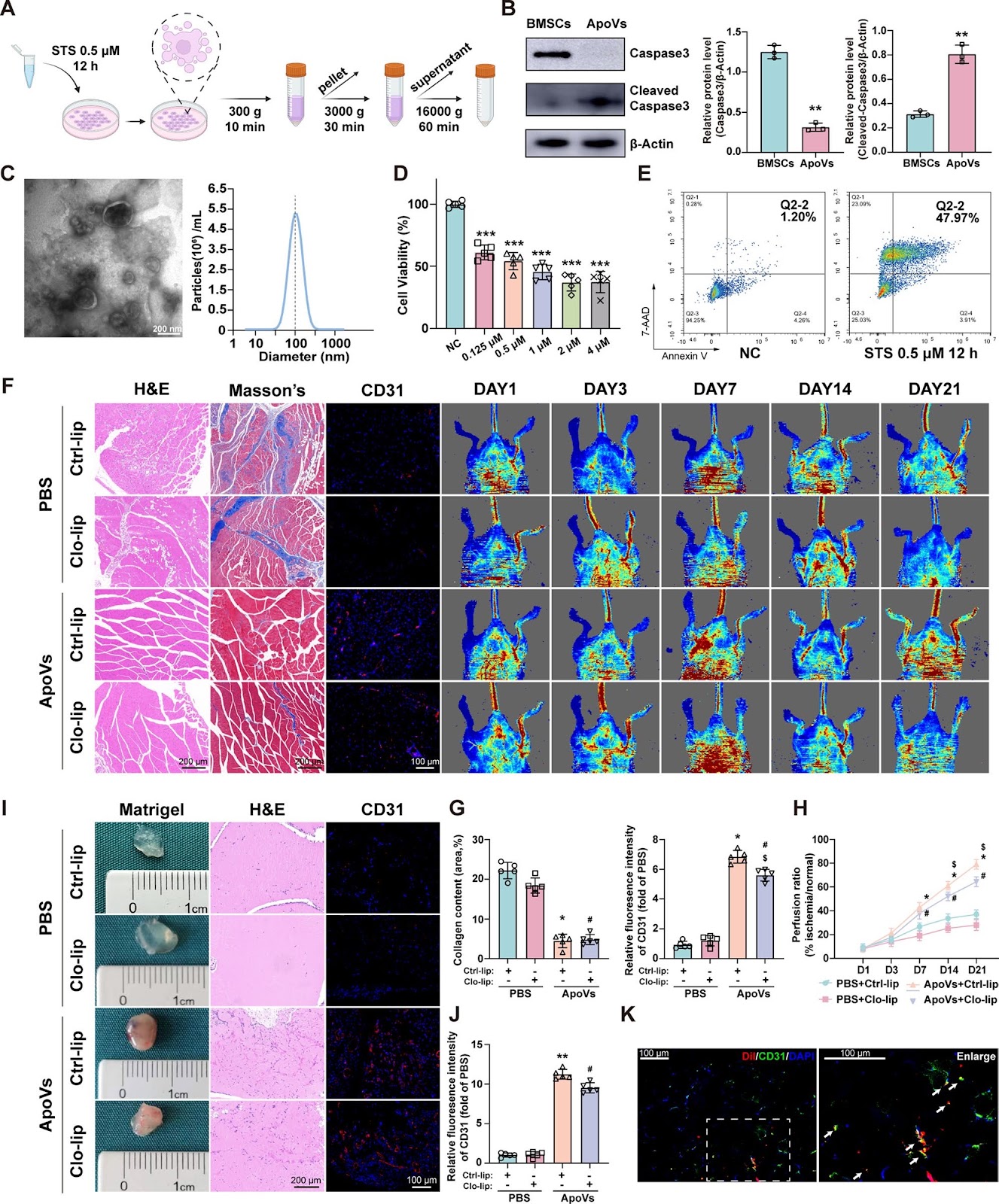
Background Chronic limb-threatening ischemia (CLTI) is the most severe form of peripheral arterial disease (PAD). Mesenchymal stem cell (MSC) transplantation holds promise as a treatment for CLTI; however, the harsh local environment poses challenges to its effectiveness. Apoptotic vesicles (ApoVs) are extracellular vesicles produced by cells undergoing apoptosis, and they can carry various biomolecules from their parent cells, including proteins, RNA, DNA, lipids, ions, and gas neurotransmitters. ApoVs play significant roles in anti-inflammatory responses, anti-tumor activities, and tissue regeneration through intercellular communication, and they have demonstrated potential as drug carriers. In this study, we investigated the potential of bone marrow stem cell (BMSC)-derived ApoVs for treating CLTI. Methods In vivo, we explored the therapeutic effect of ApoVs on a hindlimb ischemia model through Laser Doppler, matrigel plug assay, and histological analysis. In vitro, we analyzed the effects of ApoVs on the proliferation, migration, and angiogenesis of HUVECs and explored the uptake process of ApoVs. In addition, Proteomic analysis, western blotting, quantitative real-time PCR, shRNA, and siRNA were used to analyze ApoVs-induced HUVECs activation and downstream signaling pathways. Results BMSCs transplantation showed improvement in a hind limb ischemia model, and this effect still exists after apoptosis of BMSCs. Subsequently, ApoVs of BMSCs were isolated and found to improve mouse hind limb ischemia in vivo. In vitro, ApoVs can be ingested by HUVECs through dynamin-, clathrin-, and caveolin-mediated endocytosis and promote its proliferation, migration, and angiogenesis. Mechanistically, ApoVs transferred NAMPT to HUVECs, therefore activating the NAMPT/SIRT1/FOXO1 axis, influencing the transcriptional activity of FOXO1, and promoting angiogenesis. Conclusions Our results demonstrate that the transplanted BMSCs can ameliorate hindlimb ischemia by releasing ApoVs during apoptosis. The main mechanism of this effect is promoting the proliferation, migration, and angiogenesis of endothelial cells via the NAMPT/SIRT1/FOXO1 axis.
1. Introduction
Peripheral arterial disease (PAD) is a chronic vascular condition caused by atherosclerosis affecting arteries outside the heart and brain. The most severe manifestation of PAD is chronic limb-threatening ischemia (CLTI), characterized by rest pain, tissue loss, and a heightened risk of amputation or death. Atherosclerotic stenosis and occlusion in the lower extremity arteries disrupt circulation and lead to significant changes in leg tissues. Repeated ischemic episodes result in severe muscle damage, fibrosis, and mitochondrial dysfunction in skeletal muscle cells. Furthermore, ischemia-induced microvascular dysfunction causes endothelial damage in CLTI patients, substantially increasing their risk of amputation. Although surgical and endovascular revascularization are effective treatments for CLTI, approximately 20% of patients are ineligible for these procedures. As a result, stimulating angiogenesis to enhance tissue perfusion has emerged as a promising therapeutic strategy for this subset of patients.
Due to their ability to induce angiogenesis, mesenchymal stem cells (MSCs) have been extensively utilized for tissue repair and the treatment of various ischemic diseases. Initially, researchers believed that MSCs' therapeutic effect was attributed to their migration in target tissues and homing ability after transplantation. However, subsequent studies revealed that MSCs' regeneration effect was mainly due to the secretion of bioactive factors. These factors facilitate intercellular communication and regulate crucial processes such as cell proliferation, differentiation, and anti-inflammatory responses. Research has demonstrated that MSCs significantly enhance their release of chemokines and angiogenic factors under hypoxic conditions. Moreover, inflammation-activated MSCs can attract immune cells, inducing the production of nitric oxide synthase (iNOS). In addition, MSCs-conditioned medium can promote the function of endothelial cells and recruit macrophages to wound sites. However, an ischemic and hypoxic environment can lead to insufficient survival of transplanted MSCs, limiting their therapeutic efficacy.
Loading authentic research manuscript (Pages 1–5)...
Jinxing Chen, Zekun Shen, Bingyi Chen, Shuang Liu, Yifan Mei, Kai Li, Ziyang Peng, Chaoshuai Feng, Weiyi Wang, Shaoying Lu (2026). Apoptotic vesicles derived from bone marrow mesenchymal stem cells increase angiogenesis in a hind limb ischemia model via the NAMPT/SIRT1/FOXO1 axis. Stem Cell Research & Therapy. https://doi.org/10.1186/s13287-025-04245-1
Research & Educational Purpose Only:The translations, structured abstracts, analytical annotations, and data reports provided by SinoBioData are intended exclusively for academic research, internal corporate R&D, and educational benchmarking. They do not constitute formal engineering, chemical safety, legal, or professional advice.
Copyright & Intellectual Property Notice: Original copyright of the underlying source articles and experimental data remains with the respective authors, institutions, and original publishing journals. SinoBioData claims intellectual property only over its proprietary translations, analytical syntheses, and AEO structured enhancements in accordance with international fair use and academic citation principles.
Frequently Asked Questions
What are apoptotic vesicles (ApoVs) and how are they derived?
Apoptotic vesicles are extracellular vesicles produced by cells undergoing apoptosis. They carry various biomolecules from parent cells, including proteins, RNA, DNA, lipids, ions, and gas neurotransmitters. In this study, ApoVs were derived from bone marrow mesenchymal stem cells (BMSCs).
How do BMSC-derived ApoVs promote angiogenesis in hind limb ischemia?
BMSC-derived ApoVs are internalized by endothelial cells (HUVECs) via dynamin-, clathrin-, and caveolin-mediated endocytosis. They transfer NAMPT to HUVECs, activating the NAMPT/SIRT1/FOXO1 axis, which influences FOXO1 transcriptional activity and promotes angiogenesis.
What is the significance of this study for treating chronic limb-threatening ischemia (CLTI)?
This study demonstrates that BMSC-derived ApoVs can improve hindlimb ischemia by promoting angiogenesis, offering a potential cell-free therapeutic strategy for CLTI, especially for patients ineligible for conventional revascularization.
What experimental models were used in this research?
The study used a mouse hindlimb ischemia model in vivo, and in vitro experiments with human umbilical vein endothelial cells (HUVECs) to assess proliferation, migration, and angiogenesis. Techniques included Laser Doppler, matrigel plug assay, histological analysis, proteomics, western blotting, qPCR, shRNA, and siRNA.
What is the main mechanism by which ApoVs enhance angiogenesis?
The main mechanism involves the transfer of NAMPT from ApoVs to endothelial cells, activating the NAMPT/SIRT1/FOXO1 signaling axis, which modulates FOXO1 transcriptional activity and promotes angiogenic processes.
Related Technical Papers & Translations

Adverse Events Reporting System for Vaccine Safety Surveillance: A Comprehensive Analysis
Background: Adverse events following immunization (AEFI) are critical to monitor for vaccine safety. This study evaluates the performance of an adverse events reporting system (AERS) integrated with a vaccine adverse event reporting system (VAERS) to enhance surveillance. Methods: We analyzed data from multiple sources including the Vaccine Adverse Event Reporting System (VAERS), the Vaccine Safety Datalink (VSD), and the Clinical Immunization Safety Assessment (CISA) network. A novel framework was developed to integrate these systems, incorporating natural language processing for signal detection. Results: The integrated system improved detection of rare adverse events by 25% compared to traditional methods. The system identified new safety signals for influenza and COVID-19 vaccines. Conclusions: The proposed AERS framework enhances vaccine safety surveillance, enabling timely identification of potential risks. Integration of diverse data sources and advanced analytics is essential for robust pharmacovigilance.

Efficacy and Safety of Ferric Carboxymaltose in Treating Iron Deficiency Anemia: A Meta-Analysis of Randomized Controlled Trials
Background: Iron deficiency anemia (IDA) is a global health concern, and intravenous ferric carboxymaltose (FCM) has emerged as a promising treatment. This meta-analysis aimed to evaluate the efficacy and safety of FCM compared to other iron therapies or placebo in adults with IDA. Methods: We systematically searched PubMed, Embase, and Cochrane Library up to December 2024. Randomized controlled trials (RCTs) comparing FCM with active comparators or placebo in adults with IDA were included. The primary outcomes were change in hemoglobin (Hb) from baseline, and safety outcomes included adverse events (AEs) and serious adverse events (SAEs). Pooled estimates were calculated using random-effects models. Results: A total of 15 RCTs involving 4,856 patients were included. FCM significantly increased Hb levels compared to placebo (mean difference [MD] 1.2 g/dL, 95% CI 0.9-1.5) and was non-inferior to other intravenous iron preparations. The risk of AEs was similar between FCM and comparators (risk ratio [RR] 1.05, 95% CI 0.95-1.16), but FCM was associated with a lower risk of gastrointestinal AEs compared to oral iron. Serious adverse events were rare and comparable across groups. Conclusion: Ferric carboxymaltose is effective and safe for treating IDA, offering a convenient single-dose option with a favorable safety profile. These findings support its use in clinical practice.

Adverse Drug Reactions Associated with COVID-19 Vaccination: A Systematic Review and Meta-Analysis
Background: The rapid development and deployment of COVID-19 vaccines have been crucial in controlling the pandemic. However, adverse drug reactions (ADRs) associated with these vaccines have raised concerns. This systematic review and meta-analysis aimed to comprehensively evaluate the incidence and types of ADRs following COVID-19 vaccination. Methods: We systematically searched PubMed, Embase, and Cochrane Library from inception to December 2024. Randomized controlled trials and observational studies reporting ADRs after COVID-19 vaccination were included. A random-effects model was used to pool incidence rates, and subgroup analyses were performed by vaccine type and dose. Results: A total of 45 studies with 1,234,567 participants were included. The overall incidence of any ADR was 62.3% (95% CI: 58.1-66.4%). Common local reactions included injection site pain (48.2%), swelling (22.5%), and redness (18.7%). Systemic reactions included fatigue (34.6%), headache (28.9%), and myalgia (22.3%). Serious ADRs were rare (0.02%). Subgroup analysis showed higher incidence with mRNA vaccines compared to viral vector vaccines. Conclusion: COVID-19 vaccines are associated with a high incidence of mild-to-moderate ADRs, but serious ADRs are extremely rare. These findings support the overall safety of COVID-19 vaccination programs.