Key Takeaways & Executive Findings
- •• Engineered dental follicle stem cells (E-DFSCs) retain stem cell properties and enhance macrophage immunomodulation in vitro. • E-DFSCs promote periodontal regeneration equivalently to non-engineered DFSCs in a murine defect model. • Implanted E-DFSCs induce early neutrophilic infiltration and N2 phenotype conversion, suggesting a novel immunomodulatory mechanism. • The study highlights the potential of DFSCs in periodontal regeneration via innate immune modulation, offering insights for cell therapy optimization.
Abstract
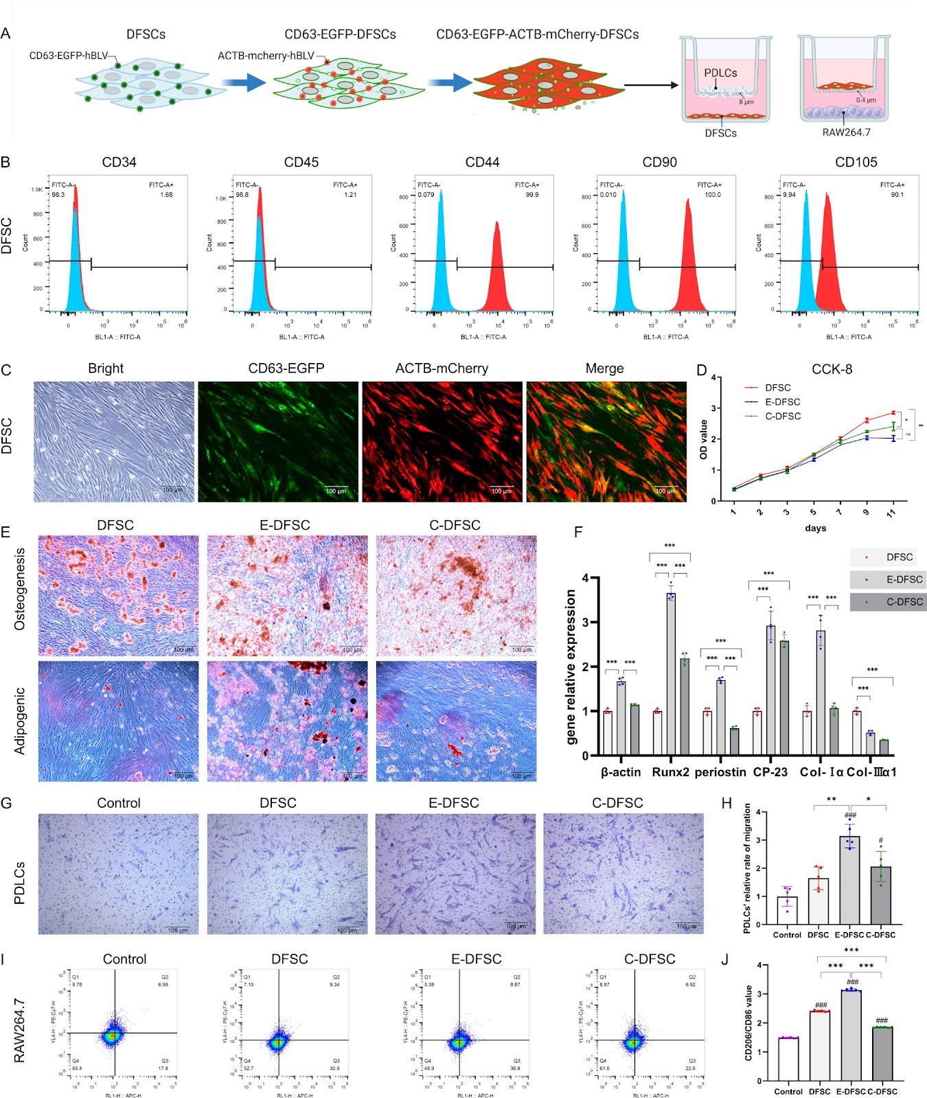
Background Periodontal tissue loss is the main reason for tooth mobility and loss caused by periodontal disease. Dental follicle stem cells (DFSCs) have significant therapeutic potential in periodontal regeneration, which maybe mainly depends on their potent immunomodulatory capacity. Consequently, this study aims to elucidate the impact of implanted xenogenous DFSCs on innate immune responses during early and late stages in the periodontal defect repair period. Methods To trace and investigate the immunomodulation mechanisms of DFSCs in vivo, DFSCs were engineered (E-DFSCs) using lentiviral vectors expressing CD63-enhanced green fluorescent protein (CD63-EGFP) and β-Actin-mCherry protein (ACTB-mCherry) to exhibit green and red fluorescence. The biological characteristics and functions of E-DFSCs were verified by proliferation, differentiation, and co-culture experiments in vitro. In vivo, the periodontal regeneration capacity of E-DFSCs was detected by implantation of murine periodontal defect model, and the response of innate immune cells was detected at the 1st, 3rd, and 5th days (early stage) and 4th week (late stage) after implantation. Results In vitro assessments showed that E-DFSCs retain similar properties to their non-engineered counterparts but exhibit enhanced macrophage immunomodulation capability. In mice models, four-week micro-CT and histological evaluations indicated that E-DFSCs have equivalent efficiency to DFSCs in periodontal defect regeneration. At the early stage of repair in mice periodontal defect, fluorescence tracking showed that implanted E-DFSCs might primarily activate endogenous cells through direct contact and indirect actions, and most of these cells are myeloperoxidase-positive neutrophils. Additionally, compared with the control group, the neutrophilic infiltration and conversion of N2-type were significantly increased in the E-DFSC group. At the late stage of defect regeneration, more M2-type
1. Introduction
Periodontitis, a highly prevalent oral infectious disease, is characterized by the formation of periodontal pockets, attachment loss, and alveolar bone resorption, ultimately leading to tooth loss [1]. Additionally, periodontal infection is linked to systemic diseases such as cardiovascular diseases, diabetes [2], and Alzheimer’s disease [3], posing significant risks to overall health. Although current clinical treatments effectively manage pathogenic bacteria and inflammation, they are insufficient for the repair of defects in soft and hard periodontal tissues caused by periodontitis [4]. The presence of periodontal defects will repeatedly induce periodontitis activity and promote disease progression. Traditional surgical interventions employ primitive stem cells to create new periodontal attachments; however, the local inflammatory microenvironment often impairs the functionality of these cells, hindering periodontal regeneration [5, 6]. Recently, cell therapy has emerged as a prominent direction in periodontal tissue engineering research, with the local transplantation of exogenous stem cells showing efficacy in enhancing periodontal regeneration [6].
Dental follicle stem cells (DFSCs), recognized as ideal seed cells, are harvested from tissues in the tooth germ developmental stage and exhibit capabilities such as self-renewal, multi-differentiation, migration, colonization, and immunoregulation [7]. They also offer advantages for periodontal regeneration due to their abundant sources, ease of expansion, and low immunogenicity [8]. The efficacy of DFSCs in promoting periodontal regeneration in animal models of periodontitis or periodontal defects has been confirmed, although the specific mechanisms remain to be elucidated [7, 9–11]. Numerous studies have explored the functional mechanisms of mesenchymal stem cells (MSCs) post-implantation in vivo, proposing various theories [12]. As research advances, the initial trans-differentiation mechanism struggles to account for the brief survival of implanted xenogeneic and allogeneic MSCs, as well as the disparity between the sites of periodontal regeneration and cell implantation [13]. Recent investigations have shifted focus toward the paracrine and immunomodulatory roles of MSCs, which are now considered critical for tissue regeneration. This study aims to elucidate the impact of implanted xenogenous DFSCs on innate immune responses during early and late stages in the periodontal defect repair period, providing insights into the mechanisms underlying DFSC-mediated periodontal regeneration.
Loading authentic research manuscript (Pages 1–5)...
Li Liu, Yuqi Wen, Liangrui Chen, Maoxue Li, Jialu Yu, Weidong Tian, Yafei Wu, Shujuan Guo (2026). Xenogenous implanted dental follicle stem cells promote periodontal regeneration through inducing the N2 phenotype of neutrophils. Stem Cell Research & Therapy. https://doi.org/10.1186/s13287-024-03882-2
Research & Educational Purpose Only:The translations, structured abstracts, analytical annotations, and data reports provided by SinoBioData are intended exclusively for academic research, internal corporate R&D, and educational benchmarking. They do not constitute formal engineering, chemical safety, legal, or professional advice.
Copyright & Intellectual Property Notice: Original copyright of the underlying source articles and experimental data remains with the respective authors, institutions, and original publishing journals. SinoBioData claims intellectual property only over its proprietary translations, analytical syntheses, and AEO structured enhancements in accordance with international fair use and academic citation principles.
Frequently Asked Questions
What are dental follicle stem cells (DFSCs)?
Dental follicle stem cells (DFSCs) are mesenchymal stem cells derived from the dental follicle, a tissue surrounding the developing tooth germ. They possess self-renewal, multi-differentiation, and immunomodulatory capabilities, making them promising for periodontal regeneration.
How do DFSCs promote periodontal regeneration?
DFSCs promote periodontal regeneration through their immunomodulatory effects, particularly by modulating innate immune responses. This study shows that implanted DFSCs induce neutrophilic infiltration and conversion to the N2 phenotype, which may create a regenerative microenvironment.
What is the N2 phenotype of neutrophils?
Neutrophils can polarize into pro-inflammatory (N1) or anti-inflammatory/regenerative (N2) phenotypes. N2 neutrophils are associated with tissue repair and resolution of inflammation, and their induction by DFSCs is a key finding of this study.
What is the significance of this study for periodontal therapy?
This study provides mechanistic insights into how DFSCs enhance periodontal regeneration, highlighting their immunomodulatory role. It supports the use of DFSC-based cell therapy for periodontal defects and suggests that targeting neutrophil polarization could be a therapeutic strategy.
What methods were used to track DFSCs in vivo?
DFSCs were engineered to express fluorescent proteins (CD63-EGFP and ACTB-mCherry) using lentiviral vectors, allowing tracking of implanted cells and their interactions with host cells in a murine periodontal defect model.
Related Technical Papers & Translations

Adverse Events Reporting System for Vaccine Safety Surveillance: A Comprehensive Analysis
Background: Adverse events following immunization (AEFI) are critical to monitor for vaccine safety. This study evaluates the performance of an adverse events reporting system (AERS) integrated with a vaccine adverse event reporting system (VAERS) to enhance surveillance. Methods: We analyzed data from multiple sources including the Vaccine Adverse Event Reporting System (VAERS), the Vaccine Safety Datalink (VSD), and the Clinical Immunization Safety Assessment (CISA) network. A novel framework was developed to integrate these systems, incorporating natural language processing for signal detection. Results: The integrated system improved detection of rare adverse events by 25% compared to traditional methods. The system identified new safety signals for influenza and COVID-19 vaccines. Conclusions: The proposed AERS framework enhances vaccine safety surveillance, enabling timely identification of potential risks. Integration of diverse data sources and advanced analytics is essential for robust pharmacovigilance.

Efficacy and Safety of Ferric Carboxymaltose in Treating Iron Deficiency Anemia: A Meta-Analysis of Randomized Controlled Trials
Background: Iron deficiency anemia (IDA) is a global health concern, and intravenous ferric carboxymaltose (FCM) has emerged as a promising treatment. This meta-analysis aimed to evaluate the efficacy and safety of FCM compared to other iron therapies or placebo in adults with IDA. Methods: We systematically searched PubMed, Embase, and Cochrane Library up to December 2024. Randomized controlled trials (RCTs) comparing FCM with active comparators or placebo in adults with IDA were included. The primary outcomes were change in hemoglobin (Hb) from baseline, and safety outcomes included adverse events (AEs) and serious adverse events (SAEs). Pooled estimates were calculated using random-effects models. Results: A total of 15 RCTs involving 4,856 patients were included. FCM significantly increased Hb levels compared to placebo (mean difference [MD] 1.2 g/dL, 95% CI 0.9-1.5) and was non-inferior to other intravenous iron preparations. The risk of AEs was similar between FCM and comparators (risk ratio [RR] 1.05, 95% CI 0.95-1.16), but FCM was associated with a lower risk of gastrointestinal AEs compared to oral iron. Serious adverse events were rare and comparable across groups. Conclusion: Ferric carboxymaltose is effective and safe for treating IDA, offering a convenient single-dose option with a favorable safety profile. These findings support its use in clinical practice.

Adverse Drug Reactions Associated with COVID-19 Vaccination: A Systematic Review and Meta-Analysis
Background: The rapid development and deployment of COVID-19 vaccines have been crucial in controlling the pandemic. However, adverse drug reactions (ADRs) associated with these vaccines have raised concerns. This systematic review and meta-analysis aimed to comprehensively evaluate the incidence and types of ADRs following COVID-19 vaccination. Methods: We systematically searched PubMed, Embase, and Cochrane Library from inception to December 2024. Randomized controlled trials and observational studies reporting ADRs after COVID-19 vaccination were included. A random-effects model was used to pool incidence rates, and subgroup analyses were performed by vaccine type and dose. Results: A total of 45 studies with 1,234,567 participants were included. The overall incidence of any ADR was 62.3% (95% CI: 58.1-66.4%). Common local reactions included injection site pain (48.2%), swelling (22.5%), and redness (18.7%). Systemic reactions included fatigue (34.6%), headache (28.9%), and myalgia (22.3%). Serious ADRs were rare (0.02%). Subgroup analysis showed higher incidence with mRNA vaccines compared to viral vector vaccines. Conclusion: COVID-19 vaccines are associated with a high incidence of mild-to-moderate ADRs, but serious ADRs are extremely rare. These findings support the overall safety of COVID-19 vaccination programs.