Key Takeaways & Executive Findings
- •• MDS-derived MSCs exhibit functional impairments including reduced proliferation, impaired differentiation, diminished hematopoietic support, and increased apoptosis. • Upregulation of lipid metabolism in MDS-MSCs contributes to their dysfunction, and can be reversed by etomoxir (ETO), a CPT-1A inhibitor. • MDS-MSCs transmit dysfunction to HSCs via exosomes enriched in CPT-1A, revealing a novel MSCs-metabolism-exosome axis. • Targeting CPT-1A or exosomal CPT-1A may offer new therapeutic strategies for MDS.
Abstract
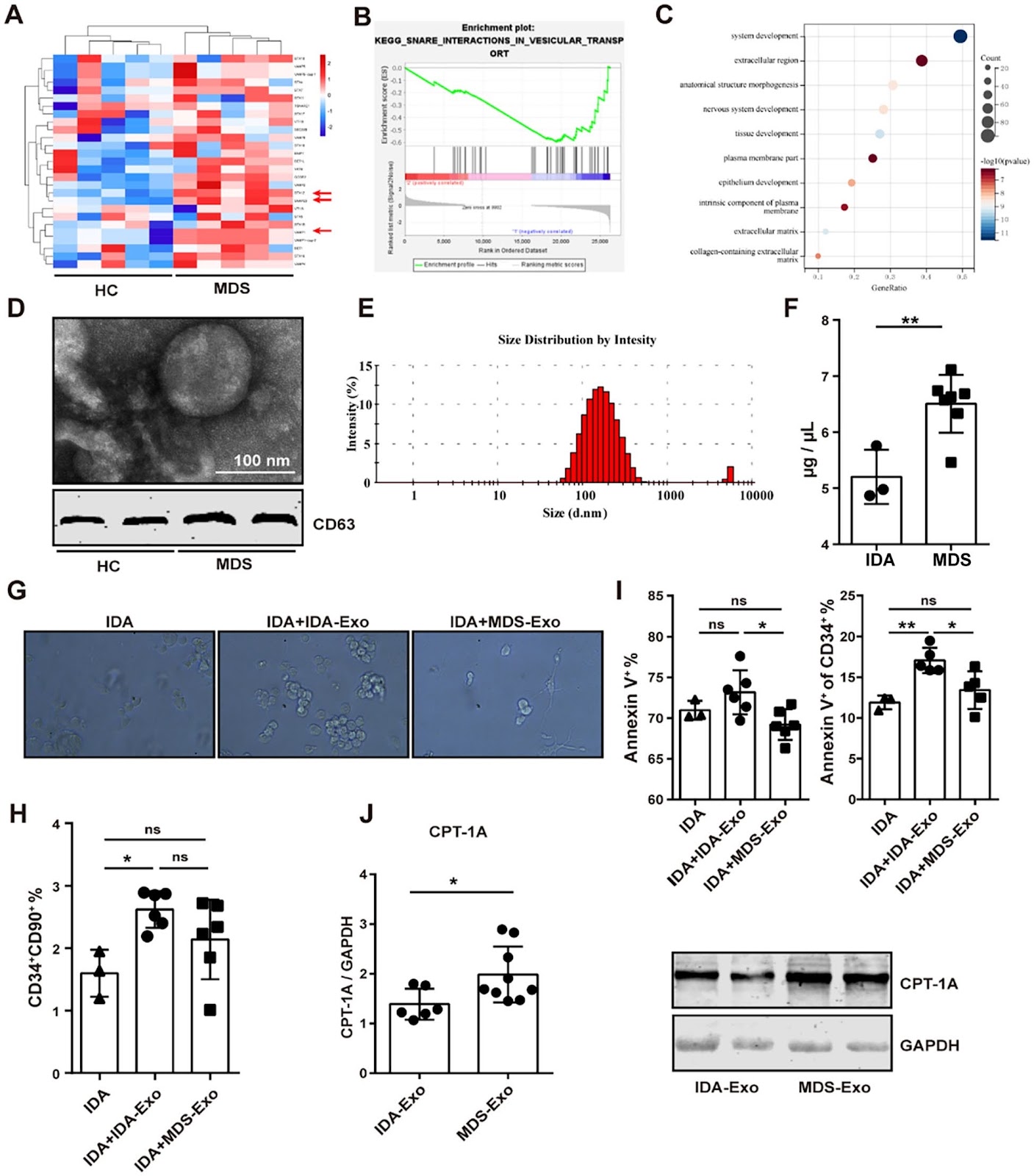
Background Myelodysplastic syndrome (MDS) is a clonal disorder of hematopoietic stem cells (HSCs), characterized by ineffective hematopoiesis and a high risk of progression to acute myeloid leukemia. Elucidating the mechanism underlying the dysfunction of MDS-HSCs is crucial for exploring the pathogenesis of the syndrome. While previous studies have implicated mesenchymal stem cells (MSCs), a principal component of the bone marrow (BM) microenvironment, in the inhibition of normal hematopoiesis, the precise molecular mechanisms have not been fully elucidated. In this study, we investigated the effects of MSCs from MDS patients on hematopoietic functions of HSCs from a metabolic perspective. Methods MSCs were isolated from BM of MDS patients. The proliferation, apoptosis, differentiation and support for hematopoiesis of these cells were analyzed using CCK-8 assay, FC and induction medium and CFU (colony forming units) assay, respectively. Expression levels of metabolic molecules were used as indicators to screen MSCs with different metabolic pathways and were detected by RT-PCR and Western blotting. Exosome derived from MSCs were isolated from the culture supernatant and confirmed by Transmission Electron Microscope, Dynamic Light Scattering and Western blotting. The effects of these exosomes on HSCs were analyzed using the same methods as those used to assess MSCs function. Results Our findings demonstrated that MDS-MSCs exhibited significant functional impairments, including reduced proliferation, impaired differentiation, diminished support for hematopoiesis, and increased apoptosis. Notably, we observed an upregulation of lipid metabolism in these MSCs, which appears to contribute to their dysfunction. Intriguingly, the aberrant lipid metabolic profile can be effectively reversed by the administration of etomoxir (ETO), an inhibitor of carnitine palmitoyltransferase 1A (CPT-1A). Furthermore, MSCs with enhanced lipid metabolism could transmit this dysfunction to HSCs through the secretion of exosomes that are enriched in CPT-1A. Conclusions We suggest that the MDS BM microenvironment disrupts MSCs metabolism by increasing the expression of CPT-1A, which impairs the ability to support normal HSCs. Interestingly, the suppressive effect is mediated by exosomes rich in CPT-1A, which derived from MSCs. These findings provide novel insights into MDS MSCs-metabolism-Exosome axis in ineffective hematopoiesis and offer new strategies for the treatment of MDS.
1. Introduction
Myelodysplastic syndrome (MDS) arise from a small population of clonal disorder hematopoietic stem cells (HSCs) that characterized by aberrant differentiation, peripheral-blood cytopenia, and frequent progression to acute myeloid leukemia[1, 2]. Researchers have found that MDS show an increased incidence with age, often making it challenging to arrive at the appropriate diagnosis[3, 4]. Consequently, it is essential to explore the key mechanism in MDS so that retard the disease process or cure it for an aging society. In clinic practice, the therapy of this disease could be divided into two aspects. One is epigenic therapy, which uses medicine such as azacitidine, decitabine to target the DNA hypermethylation to relieve symptom, but sometimes it is difficult to achieve a proper response for the patients [5]; Another is allogeneic HSCs transplantation, which treated as the only potentially curative option, but some studies indicate that the limited success of HSCs transplantation is attributed to the altered bone marrow (BM) microenvironment in MDS patients[6].
As one of the important components of BM microenvironment, mesenchymal stem cells (MSCs) can form a unique hematopoietic niche with HSCs [7–9], through secreting chemokine (C-X-C motif) CXCL12 to promote the homing of HSCs to the bone marrow [10], and releasing osteopontin to support HSCs expansion [11]. In addition, as multipotent cells, they can also differentiate into osteoblasts secreted chemokine CXCL12 ligand, stem cell factor (SCF), angiogenin, thrombopoietin
Loading authentic research manuscript (Pages 1–5)...
Chunlai Yin, Xue Yan, Jinyi Ren, Cheng Zhang, Jiaqing Liu, Zilong Wang, Jing Liu, Weiping Li, Xia Li (2026). MSCs with upregulated lipid metabolism block hematopoietic stem cell differentiation via exosomal CTP-1A in MDS. Stem Cell Research & Therapy. https://doi.org/10.1186/s13287-025-04154-3
Research & Educational Purpose Only:The translations, structured abstracts, analytical annotations, and data reports provided by SinoBioData are intended exclusively for academic research, internal corporate R&D, and educational benchmarking. They do not constitute formal engineering, chemical safety, legal, or professional advice.
Copyright & Intellectual Property Notice: Original copyright of the underlying source articles and experimental data remains with the respective authors, institutions, and original publishing journals. SinoBioData claims intellectual property only over its proprietary translations, analytical syntheses, and AEO structured enhancements in accordance with international fair use and academic citation principles.
Frequently Asked Questions
What is the role of mesenchymal stem cells (MSCs) in myelodysplastic syndrome (MDS)?
MSCs in the bone marrow microenvironment of MDS patients exhibit functional impairments, including reduced proliferation, impaired differentiation, diminished support for hematopoiesis, and increased apoptosis. They also show upregulated lipid metabolism, which contributes to their dysfunction and affects hematopoietic stem cells.
How do MDS-MSCs affect hematopoietic stem cells (HSCs)?
MDS-MSCs with enhanced lipid metabolism transmit dysfunction to HSCs through the secretion of exosomes enriched in CPT-1A, which block HSC differentiation and impair normal hematopoiesis.
What is the significance of CPT-1A in MDS?
CPT-1A is a key enzyme in lipid metabolism that is upregulated in MDS-MSCs. Its overexpression in MSCs leads to increased lipid metabolism and the release of CPT-1A-enriched exosomes, which suppress HSC function. Inhibiting CPT-1A with etomoxir can reverse the aberrant metabolic profile and restore MSC function.
What are the potential therapeutic implications of this study?
The findings suggest that targeting CPT-1A or exosomal CPT-1A could be a novel therapeutic strategy for MDS, potentially improving hematopoietic support and overcoming the suppressive effects of the MDS microenvironment.
How were exosomes from MSCs characterized in this study?
Exosomes derived from MSCs were isolated from culture supernatant and confirmed using Transmission Electron Microscopy, Dynamic Light Scattering, and Western blotting, ensuring their proper identification and characterization.
Related Technical Papers & Translations

Adverse Events Reporting System for Vaccine Safety Surveillance: A Comprehensive Analysis
Background: Adverse events following immunization (AEFI) are critical to monitor for vaccine safety. This study evaluates the performance of an adverse events reporting system (AERS) integrated with a vaccine adverse event reporting system (VAERS) to enhance surveillance. Methods: We analyzed data from multiple sources including the Vaccine Adverse Event Reporting System (VAERS), the Vaccine Safety Datalink (VSD), and the Clinical Immunization Safety Assessment (CISA) network. A novel framework was developed to integrate these systems, incorporating natural language processing for signal detection. Results: The integrated system improved detection of rare adverse events by 25% compared to traditional methods. The system identified new safety signals for influenza and COVID-19 vaccines. Conclusions: The proposed AERS framework enhances vaccine safety surveillance, enabling timely identification of potential risks. Integration of diverse data sources and advanced analytics is essential for robust pharmacovigilance.

Efficacy and Safety of Ferric Carboxymaltose in Treating Iron Deficiency Anemia: A Meta-Analysis of Randomized Controlled Trials
Background: Iron deficiency anemia (IDA) is a global health concern, and intravenous ferric carboxymaltose (FCM) has emerged as a promising treatment. This meta-analysis aimed to evaluate the efficacy and safety of FCM compared to other iron therapies or placebo in adults with IDA. Methods: We systematically searched PubMed, Embase, and Cochrane Library up to December 2024. Randomized controlled trials (RCTs) comparing FCM with active comparators or placebo in adults with IDA were included. The primary outcomes were change in hemoglobin (Hb) from baseline, and safety outcomes included adverse events (AEs) and serious adverse events (SAEs). Pooled estimates were calculated using random-effects models. Results: A total of 15 RCTs involving 4,856 patients were included. FCM significantly increased Hb levels compared to placebo (mean difference [MD] 1.2 g/dL, 95% CI 0.9-1.5) and was non-inferior to other intravenous iron preparations. The risk of AEs was similar between FCM and comparators (risk ratio [RR] 1.05, 95% CI 0.95-1.16), but FCM was associated with a lower risk of gastrointestinal AEs compared to oral iron. Serious adverse events were rare and comparable across groups. Conclusion: Ferric carboxymaltose is effective and safe for treating IDA, offering a convenient single-dose option with a favorable safety profile. These findings support its use in clinical practice.

Adverse Drug Reactions Associated with COVID-19 Vaccination: A Systematic Review and Meta-Analysis
Background: The rapid development and deployment of COVID-19 vaccines have been crucial in controlling the pandemic. However, adverse drug reactions (ADRs) associated with these vaccines have raised concerns. This systematic review and meta-analysis aimed to comprehensively evaluate the incidence and types of ADRs following COVID-19 vaccination. Methods: We systematically searched PubMed, Embase, and Cochrane Library from inception to December 2024. Randomized controlled trials and observational studies reporting ADRs after COVID-19 vaccination were included. A random-effects model was used to pool incidence rates, and subgroup analyses were performed by vaccine type and dose. Results: A total of 45 studies with 1,234,567 participants were included. The overall incidence of any ADR was 62.3% (95% CI: 58.1-66.4%). Common local reactions included injection site pain (48.2%), swelling (22.5%), and redness (18.7%). Systemic reactions included fatigue (34.6%), headache (28.9%), and myalgia (22.3%). Serious ADRs were rare (0.02%). Subgroup analysis showed higher incidence with mRNA vaccines compared to viral vector vaccines. Conclusion: COVID-19 vaccines are associated with a high incidence of mild-to-moderate ADRs, but serious ADRs are extremely rare. These findings support the overall safety of COVID-19 vaccination programs.