Key Takeaways & Executive Findings
- •• EC d4 are most vulnerable towards Dox-induced cytotoxicity independent of drug transport. • EC d4 and EC d6 display higher levels of drug-induced DSB as compared to mESC. • Dox treatment of EPC causes functional impairments in differentiated progeny, including mitochondrial dysfunction and barrier defects. • Pharmacological protection of EPC from Dox damage may reduce late cardiotoxicity in anticancer regimens.
Abstract
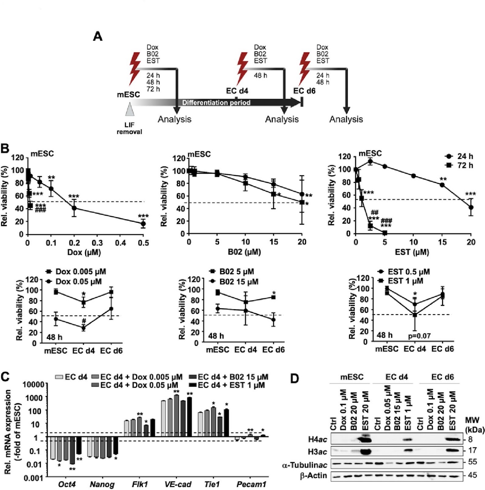
The clinical use of the anticancer drug doxorubicin (Dox) is limited by irreversible cardiotoxicity. The pathophysiological relevance of different cardiac cell types, including endothelial progenitor cells (EPC), in this process is unclear. Since progenitor cells are particularly relevant for tissue regeneration, we hypothesize that residual damage resulting from Dox-based therapeutic regimen may influence their endothelial differentiation accuracy. Therefore, we comparatively investigated the response of murine embryonic stem cells (mESC), endothelial progenitor cells (EC d4) and terminally differentiated endothelial-like cells (EC d6) following exposure to Dox and selected pharmacological inhibitors of DNA repair/DNA damage response (DDR) (RAD51i B02; HDACi entinostat (EST)). We show that EC d4 exhibit enhanced Dox sensitivity as compared to mESC and EC d6. EdU incorporation and replication fork progression analyses revealed pronounced agent-specific differences between mESC, EC d4 and EC d6. Furthermore, DNA damage formation varied in a drug-dependent manner, with mESC showing enhanced residual levels of DNA single-strand breaks (SSB) as compared to EC d4 and EC d6 while EC d6 revealed highest levels of DNA double-strand breaks (DSB). Dox treatment of EC d4 did not prevent their further differentiation into EC d6. However, it caused several functional impairments in the surviving EC d6 progeny, including defects in mitochondrial homeostasis, endothelial barrier function related to cell-cell adhesion factors (ZO1, VE-cadherin), cytokine response and low-density lipoprotein (LDL) uptake. This is accompanied by increased senescence. Summarizing, we demonstrate both overlapping and agent-specific responses of mESC, EC d4 and EC d6 to Dox and DNA repair/DDR inhibitors. Notably, drug treatment of EPC (EC d4) causes multiple dysfunctions in differentiated EC d6. Hence, pharmacological measures aiming to specifically protect EPC from Dox-induced damage are suggested to foster the maintenance of healthy endothelial functionality during regeneration, thereby lowering the risk of detrimental late cardiotoxicity resulting from Dox-based anticancer regimen.
1. Introduction
Anthracycline derivatives such as doxorubicin (Dox) are widely used anticancer drugs [1], which act as type II topoisomerase (Topo II) poisons, thereby leading to the formation of highly cytotoxic DNA double-strand breaks (DSBs) [2]. DSBs are potent triggers of the DNA damage response (DDR), which regulates cell cycle progression, DNA repair and apoptosis, thereby defining the balance between survival and death-related mechanisms [3]. In addition, generation of reactive oxygen species (ROS), DNA intercalation, inhibition of DNA helicases and chromatin damage contribute to Dox-induced cytotoxicity [4, 5]. It is believed that at clinically relevant low Dox concentrations the inhibition of topoisomerase II (TopoII) isoforms and subsequent formation of DSB is particularly relevant for its cytotoxicity. By contrast, using higher Dox concentrations, oxidative stress and DNA intercalation likely become more relevant as toxic mode of action [6].
Cumulative and irreversible cardiotoxicity ultimately leading to cardiomyopathy and congestive heart failure is the clinically most relevant adverse effect of anthracyclines [1]. In view of the low antioxidative capacity of cardiomyocytes, mitochondria-related iron-dependent and iron-independent ROS formation may be of pathophysiological relevance for Dox-induced cardiac damage, with p53-regulated mechanisms of senescence and cell death being involved [7–11]. Hence, chemoprevention of anthracycline-mediated oxidative stress has been considered to achieve cardio-protection [10–12]. Yet, antioxidants failed to demonstrate substantial cardioprotective potency following anthracycline-based anticancer therapy [4, 13, 14]. By contrast, the EDTA analogue dexrazoxane, which is a strong catalytic inhibitor of topoisomerase II, is able to prevent anthracycline-induced cardiac damage [15, 16]. Based on data obtained from the use of dexrazoxane derivatives that are lacking ion-chelating activity but still harboring Topo II inhibitory potency, it is hypothesized that Topo II inhibition is the primary mechanism of cardioprotection.
Loading authentic research manuscript (Pages 1–5)...
Sina Federmann, Michelle Westerhoff, Andreas S. Reichert, Gerhard Fritz (2026). Endothelial progenitor cell susceptibility to DNA damaging and DDR-modulating compounds determines endothelial differentiation accuracy. Stem Cell Research & Therapy. https://doi.org/10.1186/s13287-026-05087-1
Research & Educational Purpose Only:The translations, structured abstracts, analytical annotations, and data reports provided by SinoBioData are intended exclusively for academic research, internal corporate R&D, and educational benchmarking. They do not constitute formal engineering, chemical safety, legal, or professional advice.
Copyright & Intellectual Property Notice: Original copyright of the underlying source articles and experimental data remains with the respective authors, institutions, and original publishing journals. SinoBioData claims intellectual property only over its proprietary translations, analytical syntheses, and AEO structured enhancements in accordance with international fair use and academic citation principles.
Frequently Asked Questions
What is the main finding of this study?
The study shows that endothelial progenitor cells (EPC) are particularly sensitive to doxorubicin-induced DNA damage, and this damage impairs their differentiation into functional endothelial cells, leading to multiple dysfunctions in the progeny.
How does doxorubicin affect endothelial progenitor cells?
Doxorubicin induces DNA double-strand breaks and other damage in EPC, which does not prevent differentiation but causes functional impairments in the resulting endothelial cells, including mitochondrial defects, barrier dysfunction, and increased senescence.
What is the clinical significance of this research?
The findings suggest that protecting endothelial progenitor cells from doxorubicin-induced damage during cancer therapy could help maintain healthy endothelial function and reduce the risk of late cardiotoxicity.
Which cell types were compared in the study?
The study compared murine embryonic stem cells (mESC), endothelial progenitor cells (EC d4), and terminally differentiated endothelial-like cells (EC d6) in response to doxorubicin and DNA repair/DDR inhibitors.
What are the implications for cancer treatment?
The results highlight the need for pharmacological strategies to specifically protect EPC from doxorubicin-induced damage, potentially improving cardiac outcomes in patients receiving anthracycline-based chemotherapy.
Related Technical Papers & Translations

Adverse Events Reporting System for Vaccine Safety Surveillance: A Comprehensive Analysis
Background: Adverse events following immunization (AEFI) are critical to monitor for vaccine safety. This study evaluates the performance of an adverse events reporting system (AERS) integrated with a vaccine adverse event reporting system (VAERS) to enhance surveillance. Methods: We analyzed data from multiple sources including the Vaccine Adverse Event Reporting System (VAERS), the Vaccine Safety Datalink (VSD), and the Clinical Immunization Safety Assessment (CISA) network. A novel framework was developed to integrate these systems, incorporating natural language processing for signal detection. Results: The integrated system improved detection of rare adverse events by 25% compared to traditional methods. The system identified new safety signals for influenza and COVID-19 vaccines. Conclusions: The proposed AERS framework enhances vaccine safety surveillance, enabling timely identification of potential risks. Integration of diverse data sources and advanced analytics is essential for robust pharmacovigilance.

Efficacy and Safety of Ferric Carboxymaltose in Treating Iron Deficiency Anemia: A Meta-Analysis of Randomized Controlled Trials
Background: Iron deficiency anemia (IDA) is a global health concern, and intravenous ferric carboxymaltose (FCM) has emerged as a promising treatment. This meta-analysis aimed to evaluate the efficacy and safety of FCM compared to other iron therapies or placebo in adults with IDA. Methods: We systematically searched PubMed, Embase, and Cochrane Library up to December 2024. Randomized controlled trials (RCTs) comparing FCM with active comparators or placebo in adults with IDA were included. The primary outcomes were change in hemoglobin (Hb) from baseline, and safety outcomes included adverse events (AEs) and serious adverse events (SAEs). Pooled estimates were calculated using random-effects models. Results: A total of 15 RCTs involving 4,856 patients were included. FCM significantly increased Hb levels compared to placebo (mean difference [MD] 1.2 g/dL, 95% CI 0.9-1.5) and was non-inferior to other intravenous iron preparations. The risk of AEs was similar between FCM and comparators (risk ratio [RR] 1.05, 95% CI 0.95-1.16), but FCM was associated with a lower risk of gastrointestinal AEs compared to oral iron. Serious adverse events were rare and comparable across groups. Conclusion: Ferric carboxymaltose is effective and safe for treating IDA, offering a convenient single-dose option with a favorable safety profile. These findings support its use in clinical practice.

Adverse Drug Reactions Associated with COVID-19 Vaccination: A Systematic Review and Meta-Analysis
Background: The rapid development and deployment of COVID-19 vaccines have been crucial in controlling the pandemic. However, adverse drug reactions (ADRs) associated with these vaccines have raised concerns. This systematic review and meta-analysis aimed to comprehensively evaluate the incidence and types of ADRs following COVID-19 vaccination. Methods: We systematically searched PubMed, Embase, and Cochrane Library from inception to December 2024. Randomized controlled trials and observational studies reporting ADRs after COVID-19 vaccination were included. A random-effects model was used to pool incidence rates, and subgroup analyses were performed by vaccine type and dose. Results: A total of 45 studies with 1,234,567 participants were included. The overall incidence of any ADR was 62.3% (95% CI: 58.1-66.4%). Common local reactions included injection site pain (48.2%), swelling (22.5%), and redness (18.7%). Systemic reactions included fatigue (34.6%), headache (28.9%), and myalgia (22.3%). Serious ADRs were rare (0.02%). Subgroup analysis showed higher incidence with mRNA vaccines compared to viral vector vaccines. Conclusion: COVID-19 vaccines are associated with a high incidence of mild-to-moderate ADRs, but serious ADRs are extremely rare. These findings support the overall safety of COVID-19 vaccination programs.