Key Takeaways & Executive Findings
- •• Subchondral injection of UCMSCs significantly improves trabecular microarchitecture (Tb.Sp, Tb.N) in OA rats, outperforming intra-articular injection in these specific parameters. • Both subchondral and intra-articular UCMSC injections effectively preserve cartilage, synovium, and subchondral bone, and promote osteoblastic bone formation while reducing aberrant angiogenesis. • The therapeutic mechanism involves inhibition of excessive TGF-β/pSmad2 signaling, which in turn reduces osteoblast apoptosis and restores bone remodeling balance. • Subchondral delivery of MSCs represents a promising, potentially superior route for treating OA by directly targeting subchondral bone pathology.
Abstract
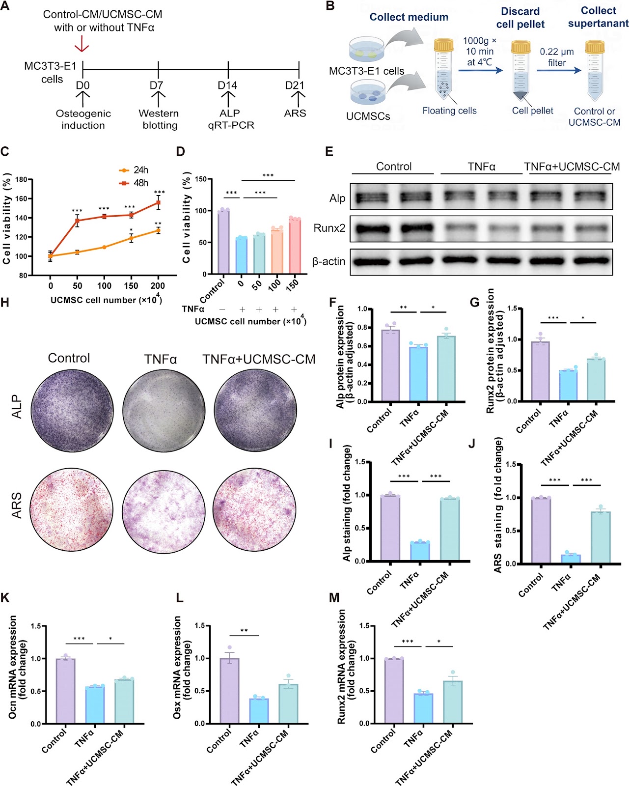
Background Osteoarthritis (OA) is a common degenerative disease caused by multiple pathological mechanisms wherein subchondral bone malfunction plays a substantial role. Recently, subchondral (SC) injection of orthobiologics has been attracting growing interest albeit the mainstream delivery method of mesenchymal stem cells (MSCs) is through intra-articular (IA). This study investigates the effect of SC injection of human umbilical cord mesenchymal stem cells (UCMSCs) on OA and its possible therapeutic mechanism compared to IA injection. Methods Male Sprague-Dawley rats with anterior cruciate ligament transection (ACLT) received saline or UCMSC injections via SC or IA. Consecutive injections once a week for three weeks and withdrawal for another four weeks, followed by Radiographical scanning, histopathological, immunohistochemical, and terminal deoxynucleotidyl transferase (TdT)-mediated dUTP nick-end labelling (TUNEL) staining. Cell counting Kit-8 (CCK-8) assay, alkaline phosphatase (ALP), alizarin red staining (ARS), TUNEL, flow cytometry, quantitative real-time polymerase chain reaction (qRT-PCR) and Western blotting were employed in TNFα-induced MC3T3-E1 cells to illustrate the exact pathogenesis mechanism. Results IA and SC UCMSC injections preserved cartilage, synovium, and subchondral bone parameters like trabecular bone volume fraction (BV/TV). SC injection uniquely improved Trabecular separation (Tb.Sp) and Trabecular number (Tb.N). SC and IA injections of UCMSCs demonstrated equivalent efficacy in promoting osteoblastic bone formation and attenuating aberrant angiogenesis of subchondral bone. In addition, we demonstrated that osteoblast apoptosis and Smad2-dependent TGF-beta (TGF-β) are crucial and interactive subchondral bone pathological features in OA. In vivo and vitro studies further revealed that UCMSCs inhibited excessive TGF-β/pSmad2 signaling to regulate osteoblast apoptosis and bone remodeling, thereby ameliorating OA progression. These findings suggest that SC injection of UCMSCs is a promising therapeutic strategy for OA, potentially offering advantages over IA injection in improving subchondral bone microarchitecture.
1. Introduction
Osteoarthritis (OA) is a common disorder characterized by articular cartilage degeneration, subchondral bone remodeling, meniscal deterioration, synovial inflammation and aberrant vascularization [1, 2]. Although established risk factors including genetic predisposition, mechanical overload, aging, and obesity are well-documented, the precise pathogenic mechanisms driving OA remain unresolved [3]. The last three decades have seen a growing incidence rate of OA as societies age, with estimated annual percentage change of 0.32% [4, 5]. Currently, over 595 million individuals worldwide lived with OA and numbers are predicted to increase up until 2050, placing a larger strain on the health-system burden globally [6]. Unfortunately, the treatments up to now are mostly symptom-relieving, and disease-modified treatment is still lacking because the exact pathogenesis of OA remains unclear [7].
Although commonly described as a wear-and-tear disease, OA is now recognized as a whole-joint pathology involving multiple tissue components [8]. Emerging evidence highlights the pathological significance of meniscal degeneration and infrapatellar fat pad remodeling in OA progression [9, 10]. Notably, subchondral bone microarchitectural alterations and associated histopathological changes have been identified as equally crucial to articular cartilage degeneration in OA pathogenesis, demonstrating comparable etiological importance [11, 12]. The homeostasis of the subchondral bone relies on coupled bone remodeling, namely osteoblast-mediated bone formation and osteoclast-mediated resorption. Accelerated subchondral bone turnover is seen in the early stages, whereas the subchondral bone sclerosis is observed during the advanced and late stages [13]. However, the high bone turnover leads to insufficient bone mineralization, thus compromising mechanical property of the newly formed bone [14]. Additionally, evidence also shown that H-type vessels with high CD31 expression aggravate subchondral angiogenesis and aberrant osteogenesis [15]. Osteoblast plays a critical role in the maintenance of normal bone remodeling. Although apoptosis is essential to physiological bone turn over, overactivated apoptosis will result in osteoblast dysfunctions, reducing osteoblast num
Loading authentic research manuscript (Pages 1–5)...
Congzi Wu, HuiHui Xu, Zhen Wu, Haipeng Huang, Qinwen Ge, Jianbo Xu, Jiali Chen, Pinger Wang, Wenhua Yuan, Hongting Jin, Peijian Tong (2026). Subchondral injection of human umbilical cord mesenchymal stem cells ameliorates knee osteoarthritis by inhibiting osteoblast apoptosis and TGF-beta activity. Stem Cell Research & Therapy. https://doi.org/10.1186/s13287-025-04366-7
Research & Educational Purpose Only:The translations, structured abstracts, analytical annotations, and data reports provided by SinoBioData are intended exclusively for academic research, internal corporate R&D, and educational benchmarking. They do not constitute formal engineering, chemical safety, legal, or professional advice.
Copyright & Intellectual Property Notice: Original copyright of the underlying source articles and experimental data remains with the respective authors, institutions, and original publishing journals. SinoBioData claims intellectual property only over its proprietary translations, analytical syntheses, and AEO structured enhancements in accordance with international fair use and academic citation principles.
Frequently Asked Questions
What is the main finding of this study?
The study demonstrates that subchondral injection of human umbilical cord mesenchymal stem cells (UCMSCs) ameliorates knee osteoarthritis by inhibiting osteoblast apoptosis and TGF-beta activity, with unique improvements in trabecular microarchitecture compared to intra-articular injection.
How does subchondral injection compare to intra-articular injection?
Both routes effectively preserve cartilage and subchondral bone, but subchondral injection uniquely improves trabecular separation and number, suggesting a potential advantage in targeting subchondral bone pathology.
What is the proposed mechanism of UCMSCs in treating OA?
UCMSCs inhibit excessive TGF-β/pSmad2 signaling, which reduces osteoblast apoptosis and restores bone remodeling balance, thereby ameliorating OA progression.
What animal model was used in this study?
The study used male Sprague-Dawley rats with anterior cruciate ligament transection (ACLT) to induce osteoarthritis.
What are the clinical implications of this research?
Subchondral injection of UCMSCs could be a promising therapeutic strategy for OA, potentially offering a more targeted approach to subchondral bone abnormalities compared to conventional intra-articular injections.
Related Technical Papers & Translations

Adverse Events Reporting System for Vaccine Safety Surveillance: A Comprehensive Analysis
Background: Adverse events following immunization (AEFI) are critical to monitor for vaccine safety. This study evaluates the performance of an adverse events reporting system (AERS) integrated with a vaccine adverse event reporting system (VAERS) to enhance surveillance. Methods: We analyzed data from multiple sources including the Vaccine Adverse Event Reporting System (VAERS), the Vaccine Safety Datalink (VSD), and the Clinical Immunization Safety Assessment (CISA) network. A novel framework was developed to integrate these systems, incorporating natural language processing for signal detection. Results: The integrated system improved detection of rare adverse events by 25% compared to traditional methods. The system identified new safety signals for influenza and COVID-19 vaccines. Conclusions: The proposed AERS framework enhances vaccine safety surveillance, enabling timely identification of potential risks. Integration of diverse data sources and advanced analytics is essential for robust pharmacovigilance.

Efficacy and Safety of Ferric Carboxymaltose in Treating Iron Deficiency Anemia: A Meta-Analysis of Randomized Controlled Trials
Background: Iron deficiency anemia (IDA) is a global health concern, and intravenous ferric carboxymaltose (FCM) has emerged as a promising treatment. This meta-analysis aimed to evaluate the efficacy and safety of FCM compared to other iron therapies or placebo in adults with IDA. Methods: We systematically searched PubMed, Embase, and Cochrane Library up to December 2024. Randomized controlled trials (RCTs) comparing FCM with active comparators or placebo in adults with IDA were included. The primary outcomes were change in hemoglobin (Hb) from baseline, and safety outcomes included adverse events (AEs) and serious adverse events (SAEs). Pooled estimates were calculated using random-effects models. Results: A total of 15 RCTs involving 4,856 patients were included. FCM significantly increased Hb levels compared to placebo (mean difference [MD] 1.2 g/dL, 95% CI 0.9-1.5) and was non-inferior to other intravenous iron preparations. The risk of AEs was similar between FCM and comparators (risk ratio [RR] 1.05, 95% CI 0.95-1.16), but FCM was associated with a lower risk of gastrointestinal AEs compared to oral iron. Serious adverse events were rare and comparable across groups. Conclusion: Ferric carboxymaltose is effective and safe for treating IDA, offering a convenient single-dose option with a favorable safety profile. These findings support its use in clinical practice.

Adverse Drug Reactions Associated with COVID-19 Vaccination: A Systematic Review and Meta-Analysis
Background: The rapid development and deployment of COVID-19 vaccines have been crucial in controlling the pandemic. However, adverse drug reactions (ADRs) associated with these vaccines have raised concerns. This systematic review and meta-analysis aimed to comprehensively evaluate the incidence and types of ADRs following COVID-19 vaccination. Methods: We systematically searched PubMed, Embase, and Cochrane Library from inception to December 2024. Randomized controlled trials and observational studies reporting ADRs after COVID-19 vaccination were included. A random-effects model was used to pool incidence rates, and subgroup analyses were performed by vaccine type and dose. Results: A total of 45 studies with 1,234,567 participants were included. The overall incidence of any ADR was 62.3% (95% CI: 58.1-66.4%). Common local reactions included injection site pain (48.2%), swelling (22.5%), and redness (18.7%). Systemic reactions included fatigue (34.6%), headache (28.9%), and myalgia (22.3%). Serious ADRs were rare (0.02%). Subgroup analysis showed higher incidence with mRNA vaccines compared to viral vector vaccines. Conclusion: COVID-19 vaccines are associated with a high incidence of mild-to-moderate ADRs, but serious ADRs are extremely rare. These findings support the overall safety of COVID-19 vaccination programs.