Key Takeaways & Executive Findings
- •• hADSC-Exos effectively reduce UVB-induced skin photoaging by decreasing SA-β-gal-positive cells, p21 expression, ROS levels, and mtDNA common deletion in both HDFs and nude mice. • The anti-photoaging mechanism involves activation of PINK1/Parkin-mediated mitophagy, as evidenced by increased PINK1, Parkin, and LC3bII/I ratio, and decreased p62. • Silencing PINK1 via siRNA abolishes the protective effects of hADSC-Exos, confirming that PINK1/Parkin pathway is essential for their action. • These findings support hADSC-Exos as a promising non-invasive therapeutic strategy for skin rejuvenation and photoaging treatment.
Abstract
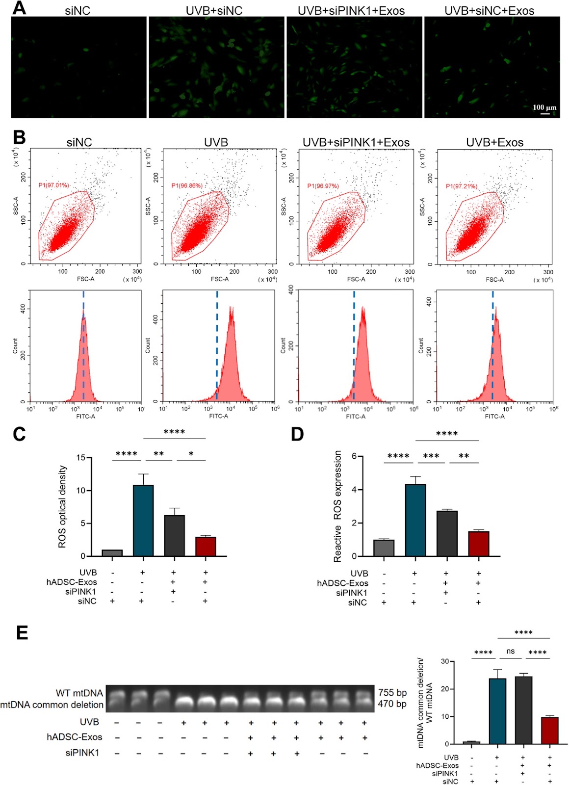
Background: Mitochondrial DNA (mtDNA) deletion and oxidative stress are key contributors to skin photoaging. Mitophagy helps mitigate oxidative stress. Human adipose-derived stem cell exosomes (hADSC-Exos) have been shown to counteract skin photoaging. This study aimed to explore the role and mechanism of hADSC-Exos in addressing skin photoaging. Methods: hADSC-Exos were isolated, and their surface markers were identified. Human dermal fibroblasts (HDFs) and nude mice were exposed to ultraviolet-B (UVB) irradiation, and treated with hADSC-Exos. Oxidative stress and photoaging were assessed through SA-β-gal staining, p21 expression, mtDNA deletion, reactive oxygen species (ROS) levels, and histological analysis. The PINK1, Parkin, LC3b, and p62 protein levels were measured to evaluate mitophagy. The PINK1 small-interfering RNA (siPINK1) was then used in HDFs to investigate the role of hADSC-Exos in mitophagy. Results: In UVB-exposed HDFs and nude mice, the number of SA-β-gal-positive cells, along with levels of p21, ROS, and mtDNA deletion, were significantly increased, but these effects were reduced by hADSC-Exos. Moreover, hADSC-Exos treatment significantly elevated PINK1 and Parkin levels, as well as the LC3bII/I ratio, while reducing p62 expression. In photoaged HDFs treated with hADSC-Exos, PINK1 knockout using siRNA decreased the LC3bII/I ratio and levels of PINK1 and Parkin, while increasing p62, ROS, and mtDNA deletion compared to the negative control (NC) group. Conclusion: hADSC-Exos can mitigate skin photoaging by promoting PINK1/Parkin-mediated mitophagy, thereby reducing mtDNA deletion and oxidative stress.
1. Introduction
Skin aging has become an increasing concern as society and the economy evolve [1]. Various factors contribute to skin aging, which can be categorized into internal and external factors. External factors include air pollution, ultraviolet (UV) radiation, lack of sleep, and smoking [2]. UV radiation, in particular, causes photoaging [3]. Repeated exposure to UV, especially UVB radiation, generates ROS, which accelerate the breakdown of collagen and elastin by upregulating matrix metalloproteinases (MMPs), leading to photoaging symptoms such as wrinkles, dryness, loss of elasticity, and pigmentation [4].
Current treatments for skin photoaging include photodynamic therapy, oral and topical drugs, and stem cell therapies [5]. Human adipose-derived stem cells (hADSCs) are a type of mesenchymal stem cell with self-renewal and multidifferentiation abilities, as well as immunomodulatory effects [6]. Exosomes are extracellular vesicles, ranging from 30 to 200 nm, formed through endocytosis, fusion, and exocytosis [7]. These vesicles are rich in nucleic acids, proteins, cytokines, and other bioactive compounds [8]. As a promising alternative to stem cells, exosomes eliminate the risk of immune rejection associated with stem cell transplants, offering an effective, non-invasive option for anti-aging therapies [9]. This has led to increasing interest in their potential for skin rejuvenation.
ROS include hydrogen peroxide (H2O2), hydroxyl radicals (-OH), and superoxide anions (O2¯) [10]. ROS are natural byproducts of oxygen metabolism. While low levels of ROS are important for cell signaling and maintaining oxygen balance, excessive ROS production can cause significant damage to biological structures, leading to DNA damage and lipid peroxidation [11, 12]. Mitochondria are cellular organelles responsible for energy production through oxidative phosphorylation [13]. They contain their own genetic material, mtDNA, which consists of 16,569 base pairs in a circular, double-stranded form [14]. The electron transport chain in mitochondria is the main source of ROS production, making mtDNA particularly vulnerable to mutations induced by oxidative stress [15]. A common mtDNA mutation is the 4977 bp deletion, known as the “common deletion,” which serves as an indicator of oxidative damage and is associated with photoaging.
Loading authentic research manuscript (Pages 1–5)...
Yihao Wang, Wanxing Liao, Yiping Wang, Junlin Liao, Nian Chen, Chiyu Jia, Li Zeng (2026). Human adipose-derived stem cell exosomes reduce mitochondrial DNA common deletion through PINK1/Parkin-mediated mitophagy to improve skin photoaging. Stem Cell Research & Therapy. https://doi.org/10.1186/s13287-025-04475-3
Research & Educational Purpose Only:The translations, structured abstracts, analytical annotations, and data reports provided by SinoBioData are intended exclusively for academic research, internal corporate R&D, and educational benchmarking. They do not constitute formal engineering, chemical safety, legal, or professional advice.
Copyright & Intellectual Property Notice: Original copyright of the underlying source articles and experimental data remains with the respective authors, institutions, and original publishing journals. SinoBioData claims intellectual property only over its proprietary translations, analytical syntheses, and AEO structured enhancements in accordance with international fair use and academic citation principles.
Frequently Asked Questions
What are human adipose-derived stem cell exosomes (hADSC-Exos)?
hADSC-Exos are extracellular vesicles (30-200 nm) secreted by human adipose-derived stem cells. They contain bioactive molecules like nucleic acids, proteins, and cytokines, and are being explored as a cell-free therapy for skin rejuvenation and anti-aging.
How does UVB radiation cause skin photoaging?
UVB radiation generates reactive oxygen species (ROS) in skin cells, leading to oxidative stress. This upregulates matrix metalloproteinases (MMPs) that degrade collagen and elastin, resulting in wrinkles, dryness, and loss of elasticity. It also causes mitochondrial DNA (mtDNA) deletions, such as the common 4977 bp deletion.
What is the role of mitophagy in skin photoaging?
Mitophagy is a selective autophagy process that removes damaged mitochondria, thereby reducing oxidative stress and preventing the accumulation of mtDNA mutations. Enhancing mitophagy, particularly via the PINK1/Parkin pathway, can protect against UVB-induced photoaging.
How do hADSC-Exos improve skin photoaging?
hADSC-Exos promote PINK1/Parkin-mediated mitophagy in UVB-exposed dermal fibroblasts and skin, which reduces mtDNA common deletion and oxidative stress. This leads to decreased markers of photoaging, such as SA-β-gal positive cells and p21 expression.
What is the significance of the PINK1/Parkin pathway in this study?
The study demonstrates that hADSC-Exos upregulate PINK1 and Parkin, which are key regulators of mitophagy. Silencing PINK1 via siRNA abolishes the protective effects of hADSC-Exos, confirming that this pathway is essential for their anti-photoaging action.
Related Technical Papers & Translations

Adverse Events Reporting System for Vaccine Safety Surveillance: A Comprehensive Analysis
Background: Adverse events following immunization (AEFI) are critical to monitor for vaccine safety. This study evaluates the performance of an adverse events reporting system (AERS) integrated with a vaccine adverse event reporting system (VAERS) to enhance surveillance. Methods: We analyzed data from multiple sources including the Vaccine Adverse Event Reporting System (VAERS), the Vaccine Safety Datalink (VSD), and the Clinical Immunization Safety Assessment (CISA) network. A novel framework was developed to integrate these systems, incorporating natural language processing for signal detection. Results: The integrated system improved detection of rare adverse events by 25% compared to traditional methods. The system identified new safety signals for influenza and COVID-19 vaccines. Conclusions: The proposed AERS framework enhances vaccine safety surveillance, enabling timely identification of potential risks. Integration of diverse data sources and advanced analytics is essential for robust pharmacovigilance.

Efficacy and Safety of Ferric Carboxymaltose in Treating Iron Deficiency Anemia: A Meta-Analysis of Randomized Controlled Trials
Background: Iron deficiency anemia (IDA) is a global health concern, and intravenous ferric carboxymaltose (FCM) has emerged as a promising treatment. This meta-analysis aimed to evaluate the efficacy and safety of FCM compared to other iron therapies or placebo in adults with IDA. Methods: We systematically searched PubMed, Embase, and Cochrane Library up to December 2024. Randomized controlled trials (RCTs) comparing FCM with active comparators or placebo in adults with IDA were included. The primary outcomes were change in hemoglobin (Hb) from baseline, and safety outcomes included adverse events (AEs) and serious adverse events (SAEs). Pooled estimates were calculated using random-effects models. Results: A total of 15 RCTs involving 4,856 patients were included. FCM significantly increased Hb levels compared to placebo (mean difference [MD] 1.2 g/dL, 95% CI 0.9-1.5) and was non-inferior to other intravenous iron preparations. The risk of AEs was similar between FCM and comparators (risk ratio [RR] 1.05, 95% CI 0.95-1.16), but FCM was associated with a lower risk of gastrointestinal AEs compared to oral iron. Serious adverse events were rare and comparable across groups. Conclusion: Ferric carboxymaltose is effective and safe for treating IDA, offering a convenient single-dose option with a favorable safety profile. These findings support its use in clinical practice.

Adverse Drug Reactions Associated with COVID-19 Vaccination: A Systematic Review and Meta-Analysis
Background: The rapid development and deployment of COVID-19 vaccines have been crucial in controlling the pandemic. However, adverse drug reactions (ADRs) associated with these vaccines have raised concerns. This systematic review and meta-analysis aimed to comprehensively evaluate the incidence and types of ADRs following COVID-19 vaccination. Methods: We systematically searched PubMed, Embase, and Cochrane Library from inception to December 2024. Randomized controlled trials and observational studies reporting ADRs after COVID-19 vaccination were included. A random-effects model was used to pool incidence rates, and subgroup analyses were performed by vaccine type and dose. Results: A total of 45 studies with 1,234,567 participants were included. The overall incidence of any ADR was 62.3% (95% CI: 58.1-66.4%). Common local reactions included injection site pain (48.2%), swelling (22.5%), and redness (18.7%). Systemic reactions included fatigue (34.6%), headache (28.9%), and myalgia (22.3%). Serious ADRs were rare (0.02%). Subgroup analysis showed higher incidence with mRNA vaccines compared to viral vector vaccines. Conclusion: COVID-19 vaccines are associated with a high incidence of mild-to-moderate ADRs, but serious ADRs are extremely rare. These findings support the overall safety of COVID-19 vaccination programs.