Key Takeaways & Executive Findings
- •• Autophagy induction with metformin enhances TNT formation and mitochondrial transfer in MSCs, while autophagy inhibition with 3-MA reduces these processes. • Autophagy modulation alters key proteins (Rab8, p-FAK) involved in TNT assembly and vesicle transport, without affecting Miro1/2 levels. • Autophagy stimulation improves mitochondrial membrane integrity and autophagic flux, suggesting a protective role in MSC function. • The study reveals crosstalk between autophagy, Wnt, and apoptosis signaling pathways, providing new insights into MSC-based regenerative therapies.
Abstract
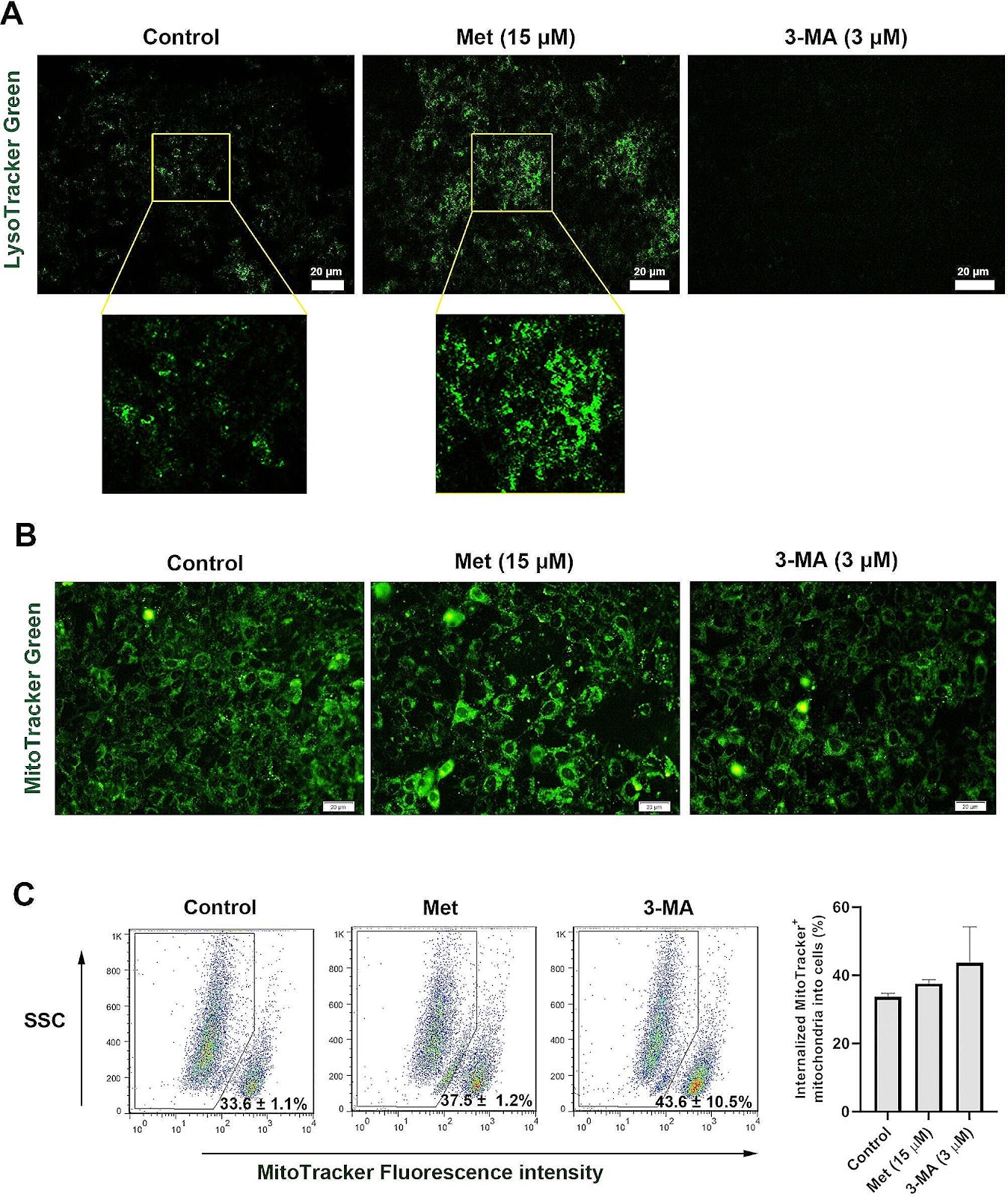
Background Recent studies have proved the role of autophagy in mesenchymal stem cell (MSCs) function and regenerative properties. How and by which mechanism autophagy modulation can affect the juxtacrine interaction of MSCs should be addressed. Here, the role of autophagy was investigated in the formation of tunneling nanotubes (TNTs) and homotypic mitochondrial donation. Methods MSCs were incubated with 15 µM Metformin (Met) and/or 3 µM 3-methyladenine (3-MA) for 48 h. The formation of TNTs was assessed using bright-field and SEM images. The mitochondria density and ΔΨ values were monitored using flow cytometry analysis. Using RT-PCR and protein array, the close interaction and shared mediators between autophagy, apoptosis, and Wnt signaling pathways were also monitored. The total fatty acid profile was assessed using gas chromatography. Result Data indicated the increase of TNT length and number, along with other cell projections after the induction of autophagy while these features were blunted in 3-MA-treated MSCs (p < 0.05). Western blotting revealed the significant reduction of Rab8 and p-FAK in 3-MA-treated MSCs (p < 0.05), indicating the inhibition of TNT assembly and vesicle transport. Likewise, the stimulation of autophagy increased autophagic flux and mitochondrial membrane integrity compared to 3-MA-treated MSCs. Despite these findings, protein levels of mitochondrial membrane Miro1 and 2 were unchanged after autophagy inhibition/stimulation (p > 0.05). We found that the inhibition/stimulation of autophagy can affect the protein, and transcription levels of several mediators related to Wnt and apoptosis signaling
1. Introduction
During the last decades, the discovery and application of various progenitors and stem cell types have revolutionized human medicine [1, 2]. Mesenchymal stem cells (MSCs) are the most commonly used stem cells in several experiments and human clinical trials [3–7]. These cells can use a multitude of mechanisms to exert their regenerative properties in the injured sites. Secretion of chemokines, cytokines, growth factors, metabolic products, and extracellular matrix, commonly referred to as the paracrine effect, is the main underlying mechanism for reparative outcomes [8]. In stem cell secretome, various extracellular vesicle (EVs) types, including exosomes and microvesicles, can transfer signaling molecules and maintain reciprocal communication between cells [9, 10].
Tunneling nanotubes (TNTs) are cytoskeletal protrusions and maintain juxtacrine cell-to-cell communication [11]. TNTs enable the transmission of intracellular organelles in bi- and uni-directional manners, exhibiting distinct structural and functional characteristics in various cell types [12]. These nano-sized structures are supported by the actin polymer backbone and can generated via the elongation of cellular protrusion and dislodgement [13]. Of note, TNTs can facilitate the transfer of diverse cellular cargo such as mitochondria and Golgi vesicles, as well as vesicles, genetic molecules like microRNA and siRNA, proteins, and ions between the cells [14]. It has been found that TNTs can also distribute various microbial pathogens and factors associated with several pathological conditions such as neurological disorders and cancers [15]. MSCs can generate TNTs as intercellular bridges to transport cargo, i.e. mitochondria, for regenerative purposes [16, 17]. Along with cytoskeletal remodeling, several enzymes and mediators such as Miro1 and 2 (mitochondrial Rho-GTPases), Rab8, TRAK1, TRAK2, and Myo19 are required for organelle donation. It has been found that knockdown of Miro1 inhibits TNT production and blunt regenerative potential of MSCs [17, 18].
Autophagy serves as a crucial intracellular degradation system and is regulated by various autophagy-related (ATG) proteins. By the activation of autophagy, the host cells can recycle abnormal cytoplasmic components.
Loading authentic research manuscript (Pages 1–5)...
Fatemeh Sadeghsoltani, Çığır Biray Avci, Parisa Hassanpour, Sanya Haiaty, Mohamad Rahmati, Ali Mota, Reza Rahbarghazi, Maryam Nemati, Mahdi Mahdipour, Mehdi Talebi, Leila Sabour Takanlou, Maryam Sabour Takanlou, Amir Mehdizadeh (2026). Autophagy modulation effect on homotypic transfer of intracellular components via tunneling nanotubes in mesenchymal stem cells. Stem Cell Research & Therapy. https://doi.org/10.1186/s13287-024-03813-1
Research & Educational Purpose Only:The translations, structured abstracts, analytical annotations, and data reports provided by SinoBioData are intended exclusively for academic research, internal corporate R&D, and educational benchmarking. They do not constitute formal engineering, chemical safety, legal, or professional advice.
Copyright & Intellectual Property Notice: Original copyright of the underlying source articles and experimental data remains with the respective authors, institutions, and original publishing journals. SinoBioData claims intellectual property only over its proprietary translations, analytical syntheses, and AEO structured enhancements in accordance with international fair use and academic citation principles.
Frequently Asked Questions
What is the role of autophagy in mesenchymal stem cell communication?
Autophagy modulates the formation of tunneling nanotubes (TNTs) and the transfer of mitochondria between MSCs, enhancing their regenerative potential.
How does metformin affect tunneling nanotubes in MSCs?
Metformin induces autophagy, which increases the number and length of TNTs and promotes mitochondrial donation, while autophagy inhibition with 3-MA reduces these effects.
What are the key proteins involved in TNT-mediated mitochondrial transfer?
Key proteins include Rab8, p-FAK, and Miro1/2, though the study found that Miro1/2 levels were unchanged by autophagy modulation, suggesting other regulatory mechanisms.
What is the clinical significance of this study?
The findings suggest that modulating autophagy in MSCs could enhance their therapeutic efficacy in regenerative medicine by improving intercellular communication and mitochondrial transfer.
How does autophagy interact with Wnt and apoptosis signaling in MSCs?
The study reveals that autophagy modulation affects the expression of mediators in Wnt and apoptosis pathways, indicating a complex crosstalk that may influence MSC function and survival.
Related Technical Papers & Translations

Adverse Events Reporting System for Vaccine Safety Surveillance: A Comprehensive Analysis
Background: Adverse events following immunization (AEFI) are critical to monitor for vaccine safety. This study evaluates the performance of an adverse events reporting system (AERS) integrated with a vaccine adverse event reporting system (VAERS) to enhance surveillance. Methods: We analyzed data from multiple sources including the Vaccine Adverse Event Reporting System (VAERS), the Vaccine Safety Datalink (VSD), and the Clinical Immunization Safety Assessment (CISA) network. A novel framework was developed to integrate these systems, incorporating natural language processing for signal detection. Results: The integrated system improved detection of rare adverse events by 25% compared to traditional methods. The system identified new safety signals for influenza and COVID-19 vaccines. Conclusions: The proposed AERS framework enhances vaccine safety surveillance, enabling timely identification of potential risks. Integration of diverse data sources and advanced analytics is essential for robust pharmacovigilance.

Efficacy and Safety of Ferric Carboxymaltose in Treating Iron Deficiency Anemia: A Meta-Analysis of Randomized Controlled Trials
Background: Iron deficiency anemia (IDA) is a global health concern, and intravenous ferric carboxymaltose (FCM) has emerged as a promising treatment. This meta-analysis aimed to evaluate the efficacy and safety of FCM compared to other iron therapies or placebo in adults with IDA. Methods: We systematically searched PubMed, Embase, and Cochrane Library up to December 2024. Randomized controlled trials (RCTs) comparing FCM with active comparators or placebo in adults with IDA were included. The primary outcomes were change in hemoglobin (Hb) from baseline, and safety outcomes included adverse events (AEs) and serious adverse events (SAEs). Pooled estimates were calculated using random-effects models. Results: A total of 15 RCTs involving 4,856 patients were included. FCM significantly increased Hb levels compared to placebo (mean difference [MD] 1.2 g/dL, 95% CI 0.9-1.5) and was non-inferior to other intravenous iron preparations. The risk of AEs was similar between FCM and comparators (risk ratio [RR] 1.05, 95% CI 0.95-1.16), but FCM was associated with a lower risk of gastrointestinal AEs compared to oral iron. Serious adverse events were rare and comparable across groups. Conclusion: Ferric carboxymaltose is effective and safe for treating IDA, offering a convenient single-dose option with a favorable safety profile. These findings support its use in clinical practice.

Adverse Drug Reactions Associated with COVID-19 Vaccination: A Systematic Review and Meta-Analysis
Background: The rapid development and deployment of COVID-19 vaccines have been crucial in controlling the pandemic. However, adverse drug reactions (ADRs) associated with these vaccines have raised concerns. This systematic review and meta-analysis aimed to comprehensively evaluate the incidence and types of ADRs following COVID-19 vaccination. Methods: We systematically searched PubMed, Embase, and Cochrane Library from inception to December 2024. Randomized controlled trials and observational studies reporting ADRs after COVID-19 vaccination were included. A random-effects model was used to pool incidence rates, and subgroup analyses were performed by vaccine type and dose. Results: A total of 45 studies with 1,234,567 participants were included. The overall incidence of any ADR was 62.3% (95% CI: 58.1-66.4%). Common local reactions included injection site pain (48.2%), swelling (22.5%), and redness (18.7%). Systemic reactions included fatigue (34.6%), headache (28.9%), and myalgia (22.3%). Serious ADRs were rare (0.02%). Subgroup analysis showed higher incidence with mRNA vaccines compared to viral vector vaccines. Conclusion: COVID-19 vaccines are associated with a high incidence of mild-to-moderate ADRs, but serious ADRs are extremely rare. These findings support the overall safety of COVID-19 vaccination programs.