Key Takeaways & Executive Findings
- •• RANKL deficiency in mice increases the frequency of skeletal stem cells (SSCs) but impairs their differentiation along the osteochondrogenic lineage, independent of osteopetrosis. • Pharmacological blockade of RANKL with Denosumab reduces the osteogenic capacity of human bone marrow-derived stromal cells (hBMSCs) in vitro. • RANKL is identified as a novel intrinsic regulatory factor of skeletal stem and progenitor cells, with potential translational implications for regenerative medicine. • The study provides the first characterization of skeletal progenitor cells in severe recessive osteopetrosis mouse models, highlighting RANKL's role beyond osteoclast regulation.
Abstract
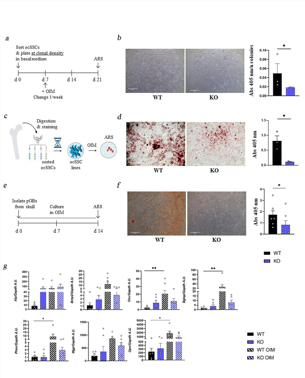
Background Skeletal Stem Cells (SSCs) are required for skeletal development, homeostasis, and repair. The perspective of their wide application in regenerative medicine approaches has supported research in this field, even though so far results in the clinic have not reached expectations, possibly due also to partial knowledge of intrinsic, potentially actionable SSC regulatory factors. Among them, the pleiotropic cytokine RANKL, with essential roles also in bone biology, is a candidate deserving deep investigation. Methods To dissect the role of the RANKL cytokine in SSC biology, we performed ex vivo characterization of SSCs and downstream progenitors (SSPCs) in mice lacking Rankl (Rankl−/−) by means of cytofluorimetric sorting and analysis of SSC populations from different skeletal compartments, gene expression analysis, and in vitro osteogenic differentiation. In addition, we assessed the effect of the pharmacological treatment with the anti-RANKL blocking antibody Denosumab (approved for therapy in patients with pathological bone loss) on the osteogenic potential of bone marrow-derived stromal cells from human healthy subjects (hBMSCs). Results We found that, regardless of the ossification type of bone, osteochondral SSCs had a higher frequency and impaired differentiation along the osteochondrogenic lineage in Rankl−/− mice as compared to wild-type. Rankl−/− mice also had increased frequency of committed osteochondrogenic and adipogenic progenitor cells deriving from perivascular SSCs. These changes were not due to the peculiar bone phenotype of increased density caused by lack of osteoclast resorption (defined osteopetrosis); indeed, they were not found in another osteopetrotic mouse model, i.e., the oc/oc mouse, and were therefore not due to osteopetrosis per se. In addition, Rankl−/− SSCs and primary osteoblasts showed reduced mineralization capacity. Of note, hBMSCs treated in vitro with Denosumab had reduced osteogenic capacity compared to control cultures. Conclusions We provide for the first time the characterization of SSPCs from mouse models of severe recessive osteopetrosis. We demonstrate that Rankl genetic deficiency in murine SSCs and functional blockade in hBMSCs reduce their osteogenic potential. Therefore, we propose that RANKL is an important regulatory factor of SSC features with translational relevance.
1. Introduction
Skeletal stem cells (SSCs) are a bone-specific subtype of somatic stem cells crucial for bone physiology [1]. They are endowed with self-renewal and multipotency capacity, and give rise to osteoblasts, chondrocytes, marrow stromal cells, and adipocytes in different proportions, depending on the compartment of origin (i.e., bone marrow, periosteum, growth plate) [2–6]. Gene expression analysis of SSCs and downstream progenitor populations identified ligands and receptors of signaling pathways that may modulate the activity of the SSCs and their progeny, and suggested a control based also on paracrine and/or autocrine molecular cues [7, 8]. Intrinsic regulatory factors may represent a means to tune the regenerative potential of mesenchymal progenitors, which is a highly pursued (but poorly achieved) goal due to the expected broad application in the field of regenerative medicine [9].
The Receptor Activator of NF-kB Ligand (RANKL) cytokine is recognized as a pleiotropic factor since its discovery; in fact, it was originally cloned by three independent groups and classified as a dendritic cell survival factor [10], a regulator of T cell function [11] and an essential osteoclast differentiation factor [12]. In line with this key function in the bone microenvironment, Rankl deficient (Rankl−/−) mice as well as patients bearing mutations in the TNFSF11 gene (encoding RANKL) display severe osteopetrosis owing to lack of osteoclast formation [13, 14]. Several additional roles have been recognized for RANKL in pathophysiological conditions, making this cytokine an interesting target for therapy [15–19]. Of note, we previously reported that murine bone marrow-derived mesenchymal stromal cells (BMSCs) lacking Rankl (Rankl−/−) displayed a partial osteogenic defect that was corrected
Loading authentic research manuscript (Pages 1–5)...
M. L. Schiavone, L. Crisafulli, C. Camisaschi, G. De Simone, F. R. Liberati, E. Palagano, N. Rucci, F. Ficara, Cristina Sobacchi (2026). Rankl genetic deficiency and functional blockade undermine skeletal stem and progenitor cell differentiation. Stem Cell Research & Therapy. https://doi.org/10.1186/s13287-024-03803-3
Research & Educational Purpose Only:The translations, structured abstracts, analytical annotations, and data reports provided by SinoBioData are intended exclusively for academic research, internal corporate R&D, and educational benchmarking. They do not constitute formal engineering, chemical safety, legal, or professional advice.
Copyright & Intellectual Property Notice: Original copyright of the underlying source articles and experimental data remains with the respective authors, institutions, and original publishing journals. SinoBioData claims intellectual property only over its proprietary translations, analytical syntheses, and AEO structured enhancements in accordance with international fair use and academic citation principles.
Frequently Asked Questions
What is the role of RANKL in skeletal stem cell differentiation?
RANKL is identified as an intrinsic regulatory factor that promotes osteogenic differentiation of skeletal stem cells (SSCs). Genetic deficiency or functional blockade of RANKL impairs the osteogenic potential of SSCs and their progenitors.
How does Denosumab affect human bone marrow stromal cells?
In vitro treatment of human bone marrow-derived stromal cells (hBMSCs) with Denosumab, an anti-RANKL antibody, reduces their osteogenic capacity, indicating that RANKL signaling is important for osteogenic differentiation in humans.
Are the effects of RANKL deficiency on SSCs due to osteopetrosis?
No, the effects are independent of osteopetrosis. The study compared Rankl−/− mice with another osteopetrotic model (oc/oc mice) and found that the changes in SSC frequency and differentiation were specific to RANKL deficiency, not the osteopetrotic phenotype.
What are the implications of this study for regenerative medicine?
The findings suggest that modulating RANKL signaling could be a strategy to enhance the regenerative potential of skeletal stem cells, potentially improving outcomes in bone repair and regenerative therapies.
What is the significance of the study in the context of osteopetrosis?
This study provides the first characterization of skeletal progenitor cells in mouse models of severe recessive osteopetrosis, offering insights into the cellular mechanisms underlying bone defects in this condition and highlighting RANKL's role beyond osteoclast regulation.
Related Technical Papers & Translations

Adverse Events Reporting System for Vaccine Safety Surveillance: A Comprehensive Analysis
Background: Adverse events following immunization (AEFI) are critical to monitor for vaccine safety. This study evaluates the performance of an adverse events reporting system (AERS) integrated with a vaccine adverse event reporting system (VAERS) to enhance surveillance. Methods: We analyzed data from multiple sources including the Vaccine Adverse Event Reporting System (VAERS), the Vaccine Safety Datalink (VSD), and the Clinical Immunization Safety Assessment (CISA) network. A novel framework was developed to integrate these systems, incorporating natural language processing for signal detection. Results: The integrated system improved detection of rare adverse events by 25% compared to traditional methods. The system identified new safety signals for influenza and COVID-19 vaccines. Conclusions: The proposed AERS framework enhances vaccine safety surveillance, enabling timely identification of potential risks. Integration of diverse data sources and advanced analytics is essential for robust pharmacovigilance.

Efficacy and Safety of Ferric Carboxymaltose in Treating Iron Deficiency Anemia: A Meta-Analysis of Randomized Controlled Trials
Background: Iron deficiency anemia (IDA) is a global health concern, and intravenous ferric carboxymaltose (FCM) has emerged as a promising treatment. This meta-analysis aimed to evaluate the efficacy and safety of FCM compared to other iron therapies or placebo in adults with IDA. Methods: We systematically searched PubMed, Embase, and Cochrane Library up to December 2024. Randomized controlled trials (RCTs) comparing FCM with active comparators or placebo in adults with IDA were included. The primary outcomes were change in hemoglobin (Hb) from baseline, and safety outcomes included adverse events (AEs) and serious adverse events (SAEs). Pooled estimates were calculated using random-effects models. Results: A total of 15 RCTs involving 4,856 patients were included. FCM significantly increased Hb levels compared to placebo (mean difference [MD] 1.2 g/dL, 95% CI 0.9-1.5) and was non-inferior to other intravenous iron preparations. The risk of AEs was similar between FCM and comparators (risk ratio [RR] 1.05, 95% CI 0.95-1.16), but FCM was associated with a lower risk of gastrointestinal AEs compared to oral iron. Serious adverse events were rare and comparable across groups. Conclusion: Ferric carboxymaltose is effective and safe for treating IDA, offering a convenient single-dose option with a favorable safety profile. These findings support its use in clinical practice.

Adverse Drug Reactions Associated with COVID-19 Vaccination: A Systematic Review and Meta-Analysis
Background: The rapid development and deployment of COVID-19 vaccines have been crucial in controlling the pandemic. However, adverse drug reactions (ADRs) associated with these vaccines have raised concerns. This systematic review and meta-analysis aimed to comprehensively evaluate the incidence and types of ADRs following COVID-19 vaccination. Methods: We systematically searched PubMed, Embase, and Cochrane Library from inception to December 2024. Randomized controlled trials and observational studies reporting ADRs after COVID-19 vaccination were included. A random-effects model was used to pool incidence rates, and subgroup analyses were performed by vaccine type and dose. Results: A total of 45 studies with 1,234,567 participants were included. The overall incidence of any ADR was 62.3% (95% CI: 58.1-66.4%). Common local reactions included injection site pain (48.2%), swelling (22.5%), and redness (18.7%). Systemic reactions included fatigue (34.6%), headache (28.9%), and myalgia (22.3%). Serious ADRs were rare (0.02%). Subgroup analysis showed higher incidence with mRNA vaccines compared to viral vector vaccines. Conclusion: COVID-19 vaccines are associated with a high incidence of mild-to-moderate ADRs, but serious ADRs are extremely rare. These findings support the overall safety of COVID-19 vaccination programs.