Key Takeaways & Executive Findings
- •• LncRNA SNHG1 enhances chondrogenic differentiation of human jaw bone marrow mesenchymal stem cells (h-JBMMSCs) in vitro, as evidenced by increased Alcian blue, Masson's trichrome, and modified Sirius red staining. • SNHG1 promotes angiogenesis in vitro and microvessel formation in vivo, contributing to improved cartilage repair in a rabbit knee defect model. • Mechanistically, SNHG1 modulates mitochondrial function by reducing reactive oxygen species (ROS) and increasing mitochondrial membrane potential, and it activates the JAK/STAT3 signaling pathway. • These findings suggest that SNHG1 is a promising therapeutic target for cartilage regeneration, offering a potential strategy for treating cartilage defects.
Abstract
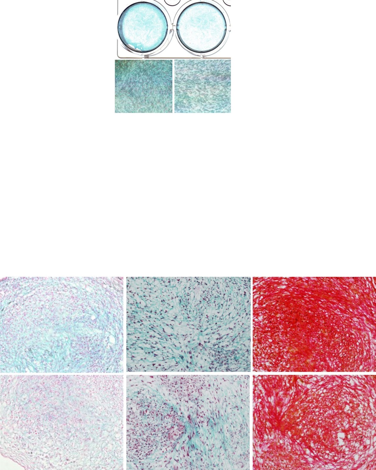
Background Cartilage is a kind of avascular tissue, and it is difficult to repair itself when it is damaged. In this study, we investigated the regulation of chondrogenic differentiation and vascular formation in human jaw bone marrow mesenchymal stem cells (h-JBMMSCs) by the long-chain noncoding RNA small nucleolar RNA host gene 1 (SNHG1) during cartilage tissue regeneration. Methods JBMMSCs were isolated from the jaws via the adherent method. The effects of lncRNA SNHG1 on the chondrogenic differentiation of JBMMSCs in vitro were detected by real-time fluorescence quantitative polymerase chain reaction (RT-qPCR), Pellet experiment, Alcian blue staining, Masson’s trichrome staining, and modified Sirius red staining. RT-qPCR, matrix gel tube formation, and coculture experiments were used to determine the effect of lncRNA SNHG1 on the angiogenesis in JBMMSCs in vitro. A model of knee cartilage defects in New Zealand rabbits and a model of subcutaneous matrix rubber suppositories in nude mice were constructed for in vivo experiments. Changes in mitochondrial function were detected via RT-qPCR, dihydroethidium (DHE) staining, MitoSOX staining, tetramethyl rhodamine methyl ester (TMRM) staining, and adenosine triphosphate (ATP) detection. Western blotting was used to detect the phosphorylation level of signal transducer and activator of transcription 3 (STAT3). Results Alcian blue staining, Masson’s trichrome staining, and modified Sirius Red staining showed that lncRNA SNHG1 promoted chondrogenic differentiation. The lncRNA SNHG1 promoted angiogenesis in vitro and the formation of microvessels in vivo. The lncRNA SNHG1 promoted the repair and regeneration of rabbit knee cartilage tissue. Western blot and alcian blue staining showed that the JAK inhibitor reduced the increase of STAT3 phosphorylation level and staining deepening caused by SNHG1. Mitochondrial correlation analysis revealed that the lncRNA SNHG1 led to a decrease in reactive oxygen species (ROS) levels, an increase in mitochondrial membrane potential
1. Introduction
Human activities are inseparable from joint movement, and articular cartilage plays a role in relieving stress and lubricating joints. Once the cartilage is damaged or degenerated [1, 2], it is difficult to repair itself, because the main components of articular cartilage are collagen fibers and proteoglycans in addition to water, which lack a blood supply and cells cannot migrate to the defect site [3]. At present, the surgical treatment of cartilage defects mainly involves the direct use of growth factors, autologous transplantation, or allogeneic transplantation [4], but there are several problems, such as limited donor sites and immune responses [5]. In addition, synthetic cartilage scaffolds have attracted widespread attention because they are not limited by supply and can be functionalized [6, 7]. However, after implantation, it easily decomposes under joint force, and the production of hyaline cartilage is inefficient and inefficient [8–10].
Mesenchymal stem cells (MSCs) are considered an ideal choice for cartilage regeneration because they can differentiate into chondrocytes with biological functions [11]. In addition, the nutrients in articular cartilage are mainly provided by synovial fluid and subchondral blood vessels. Tissue repair includes inflammation, proliferation, and remodeling [12, 13], and the proliferation stage includes angiogenesis [14, 15]. MSCs not only undergo differentiation but also release cytokines via paracrine signaling to activate dermal fibroblasts, promote the production of collagen, and promote angiogenesis, thus promoting tissue healing [16]. There is evidence that intra-articular or intravascular injection of stem cells is effective [17]. Bone marrow mesenchymal stem cells (BMSCs) can promote osteogenesis, chondrogenesis, and angiogenesis in vivo, and a sufficient number of BMSCs are easily obtained; therefore, they are widely studied [18]. At present, studies have confirmed that BMSCs can be used for cartilage regeneration. BMSCs can not only inhibit the focal death of cartilage through derived exosomes [19] but can also be combined with hydrogels to di
Loading authentic research manuscript (Pages 1–5)...
Hua Liu, Huina Liu, Qiubo Yang, Zhipeng Fan (2026). LncRNA SNHG1 enhances cartilage regeneration by modulating chondrogenic differentiation and angiogenesis potentials of JBMMSCs via mitochondrial function regulation. Stem Cell Research & Therapy. https://doi.org/10.1186/s13287-024-03793-2
Research & Educational Purpose Only:The translations, structured abstracts, analytical annotations, and data reports provided by SinoBioData are intended exclusively for academic research, internal corporate R&D, and educational benchmarking. They do not constitute formal engineering, chemical safety, legal, or professional advice.
Copyright & Intellectual Property Notice: Original copyright of the underlying source articles and experimental data remains with the respective authors, institutions, and original publishing journals. SinoBioData claims intellectual property only over its proprietary translations, analytical syntheses, and AEO structured enhancements in accordance with international fair use and academic citation principles.
Frequently Asked Questions
What is the role of lncRNA SNHG1 in cartilage regeneration?
LncRNA SNHG1 enhances cartilage regeneration by promoting chondrogenic differentiation and angiogenesis of jaw bone marrow mesenchymal stem cells (JBMMSCs), partly through regulation of mitochondrial function and activation of the JAK/STAT3 pathway.
How does SNHG1 affect mitochondrial function in JBMMSCs?
SNHG1 reduces reactive oxygen species (ROS) levels and increases mitochondrial membrane potential, indicating improved mitochondrial function, which contributes to enhanced cell survival and differentiation.
What experimental models were used to study SNHG1?
The study used in vitro assays (e.g., Alcian blue staining, RT-qPCR, tube formation) and in vivo models including knee cartilage defects in New Zealand rabbits and subcutaneous matrix rubber suppositories in nude mice.
What is the clinical significance of this research?
The findings suggest that SNHG1 could be a therapeutic target for enhancing cartilage repair, offering a potential strategy for treating cartilage defects and degenerative joint diseases.
How does SNHG1 regulate angiogenesis?
SNHG1 promotes angiogenesis in vitro and in vivo, likely through paracrine effects and modulation of angiogenic factors, contributing to improved vascularization during cartilage repair.
Related Technical Papers & Translations

Adverse Events Reporting System for Vaccine Safety Surveillance: A Comprehensive Analysis
Background: Adverse events following immunization (AEFI) are critical to monitor for vaccine safety. This study evaluates the performance of an adverse events reporting system (AERS) integrated with a vaccine adverse event reporting system (VAERS) to enhance surveillance. Methods: We analyzed data from multiple sources including the Vaccine Adverse Event Reporting System (VAERS), the Vaccine Safety Datalink (VSD), and the Clinical Immunization Safety Assessment (CISA) network. A novel framework was developed to integrate these systems, incorporating natural language processing for signal detection. Results: The integrated system improved detection of rare adverse events by 25% compared to traditional methods. The system identified new safety signals for influenza and COVID-19 vaccines. Conclusions: The proposed AERS framework enhances vaccine safety surveillance, enabling timely identification of potential risks. Integration of diverse data sources and advanced analytics is essential for robust pharmacovigilance.

Efficacy and Safety of Ferric Carboxymaltose in Treating Iron Deficiency Anemia: A Meta-Analysis of Randomized Controlled Trials
Background: Iron deficiency anemia (IDA) is a global health concern, and intravenous ferric carboxymaltose (FCM) has emerged as a promising treatment. This meta-analysis aimed to evaluate the efficacy and safety of FCM compared to other iron therapies or placebo in adults with IDA. Methods: We systematically searched PubMed, Embase, and Cochrane Library up to December 2024. Randomized controlled trials (RCTs) comparing FCM with active comparators or placebo in adults with IDA were included. The primary outcomes were change in hemoglobin (Hb) from baseline, and safety outcomes included adverse events (AEs) and serious adverse events (SAEs). Pooled estimates were calculated using random-effects models. Results: A total of 15 RCTs involving 4,856 patients were included. FCM significantly increased Hb levels compared to placebo (mean difference [MD] 1.2 g/dL, 95% CI 0.9-1.5) and was non-inferior to other intravenous iron preparations. The risk of AEs was similar between FCM and comparators (risk ratio [RR] 1.05, 95% CI 0.95-1.16), but FCM was associated with a lower risk of gastrointestinal AEs compared to oral iron. Serious adverse events were rare and comparable across groups. Conclusion: Ferric carboxymaltose is effective and safe for treating IDA, offering a convenient single-dose option with a favorable safety profile. These findings support its use in clinical practice.

Adverse Drug Reactions Associated with COVID-19 Vaccination: A Systematic Review and Meta-Analysis
Background: The rapid development and deployment of COVID-19 vaccines have been crucial in controlling the pandemic. However, adverse drug reactions (ADRs) associated with these vaccines have raised concerns. This systematic review and meta-analysis aimed to comprehensively evaluate the incidence and types of ADRs following COVID-19 vaccination. Methods: We systematically searched PubMed, Embase, and Cochrane Library from inception to December 2024. Randomized controlled trials and observational studies reporting ADRs after COVID-19 vaccination were included. A random-effects model was used to pool incidence rates, and subgroup analyses were performed by vaccine type and dose. Results: A total of 45 studies with 1,234,567 participants were included. The overall incidence of any ADR was 62.3% (95% CI: 58.1-66.4%). Common local reactions included injection site pain (48.2%), swelling (22.5%), and redness (18.7%). Systemic reactions included fatigue (34.6%), headache (28.9%), and myalgia (22.3%). Serious ADRs were rare (0.02%). Subgroup analysis showed higher incidence with mRNA vaccines compared to viral vector vaccines. Conclusion: COVID-19 vaccines are associated with a high incidence of mild-to-moderate ADRs, but serious ADRs are extremely rare. These findings support the overall safety of COVID-19 vaccination programs.