Key Takeaways & Executive Findings
- •• Priming induced mesenchymal stem cells (iMSCs) with the pan-PPAR agonist lanifibranor enhances the therapeutic efficacy of their extracellular vesicles (EVs) against acute kidney injury (AKI). • Pan-PPAR-iMSC-EVs significantly reduce inflammation, immune cell infiltration, and apoptosis in a cisplatin-induced AKI mouse model, while increasing renal capillary density. • In vitro, pan-PPAR-iMSC-EVs promote HK-2 cell proliferation and survival under cisplatin-induced apoptosis and suppress inflammatory cytokine expression in M1-polarized THP-1 cells more effectively than unprimed iMSC-EVs. • This study presents a novel cell-free therapeutic strategy for AKI, potentially overcoming limitations of direct MSC use and offering a more potent EV-based treatment.
Abstract
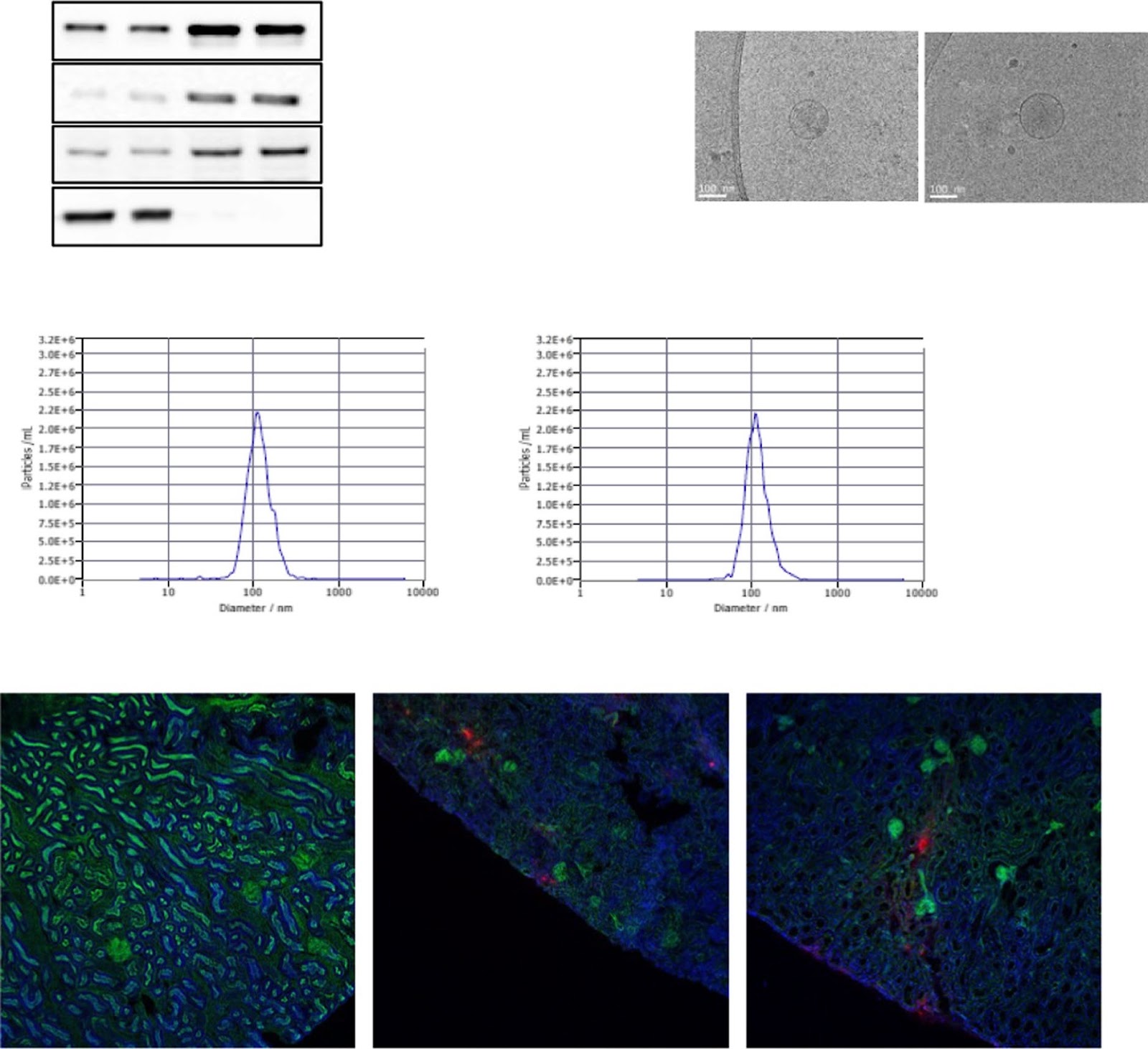
Background Acute kidney injury (AKI) has a complex pathophysiology and imposes serious health concerns worldwide. Extracellular vesicles (EVs) derived from induced mesenchymal stem cells (iMSCs) have been recognized as novel cell-free therapeutics for various inflammatory and degenerative disorders. In this study, we investigated whether iMSCs stimulated with a pan-peroxisome proliferator-activated receptor (PPAR) agonist could enhance the therapeutic efficacy of EVs against AKI. Methods Human iMSCs were primed with or without lanifibranor, a PPAR agonist for 24 h, and EVs were collected after an additional 24 h. The basic characteristics of EVs were evaluated using cryo-transmission electron microscopy imaging, immunoblot detection of EV markers, nanoparticle tracking analysis, and localization in AKI kidneys. In vitro, the potential of the EVs to promote the growth and survival of HK-2 cells undergoing cisplatin-induced apoptosis and anti-inflammatory effects in M1-polarized THP-1 was compared. Subsequently, AKI was induced in BALB/c mice using cisplatin. After 8 and 24 h of cisplatin treatment, iMSC-EVs or pan-PPAR-iMSC-EVs were injected intravascularly. At 96 h after cisplatin administration, the renoprotective effects of iMSC-EVs or pan-PPAR-iMSC-EVs in inhibiting inflammation and apoptosis were compared using serum biochemistry, histology, immunohistochemistry, and gene expression analysis by qPCR. Results Both EV types expressed EV markers and had typical EV morphology, and their localization in the renal tissue was confirmed. The proliferation and survival of HK-2 cells were higher in pan-PPAR-iMSC-EVs than those in iMSC-EVs. In M1-polarized THP-1 cells, the reduction in the mRNA expression of inflammatory cytokines was more significant in pan-PPAR-iMSC-EVs than that in iMSC-EVs. In the mouse model of cisplatin-induced AKI, pan-PPAR-iMSC-EVs markedly enhanced renoprotective effects compared to iMSC-EVs. Specifically, pan-PPAR-iMSC-EVs reduced tissue inflammation, immune cell infiltration, and apoptosis. Pan-PPAR-iMSC-EVs also increased renal capillary density. Conclusion Priming iMSCs with a PPAR agonist significantly improved the therapeutic potential of EVs by reducing inflammation and apoptosis. The reported strategy may contribute to the development of a novel cell-free option for AKI treatment. Trial registration: Not applicable.
1. Introduction
Acute kidney injury (AKI) is a pathological condition in which a sudden loss of renal function occurs within hours to several days [1]. The increased incidence of AKI imposes a high healthcare burden and increased risk of chronic kidney disease (CKD) worldwide. It affects approximately 25% of hospitalized patients, and the risk of AKI in critically ill patients has been reported to be as high as 30 to 60% [2, 3]. AKI, with a complex pathophysiology, generally occurs during major surgery and is primarily caused by nephrotoxic drugs, ischemia, and contrast media [4]. In addition, risk factors, including age, hypertension, diabetes, and chronic renal disease, increase the mortality of patients with AKI [4]. Although several clinical trials have investigated the efficacies of various drugs, including diuretics (mannitol), antioxidants, and DPP4 inhibitors, the results have not been successful [5]. Therefore, innovative therapeutic strategies against AKI are needed [6].
Mesenchymal stem cells (MSCs) have been recognized as promising therapeutic tools for various diseases because they facilitate the recovery of damaged tissue and regulate immune tissue regeneration and responses [7]. Despite meaningful progress, the clinical application of MSCs is limited by several hurdles, including senescence, low survivability in vivo, thrombosis, and tumorigenesis [8]. Furthermore, the direct use of MSCs in vivo is hindered by the trapping of intravascularly administered MSCs in the lungs or liver [9]. To overcome these limitations of using MSCs directly, extracellular vesicles (EVs) secreted by MSCs are recognized as efficient alternatives. EVs are nano-sized vesicles responsible for cell-to-cell communication in vivo, contributing to the maintenance of normal physiology and disease progression [10]. Furthermore, the therapeutic potential of EVs can be enhanced by preconditioning MSCs with various stimuli, such as growth factors, cytokines, or pharmacological agents. In this study, we investigated whether priming iMSCs with a pan-PPAR agonist could enhance the renoprotective effect of their EVs in the context of AKI.
Loading authentic research manuscript (Pages 1–5)...
Hongduk Kim, Seul Ki Lee, Sungok Hong, Tae Sub Park, Jimin Kim, Soo Kim, Tae Min Kim (2026). Pan PPAR agonist stimulation of induced MSCs produces extracellular vesicles with enhanced renoprotective effect for acute kidney injury. Stem Cell Research & Therapy. https://doi.org/10.1186/s13287-023-03577-0
Research & Educational Purpose Only:The translations, structured abstracts, analytical annotations, and data reports provided by SinoBioData are intended exclusively for academic research, internal corporate R&D, and educational benchmarking. They do not constitute formal engineering, chemical safety, legal, or professional advice.
Copyright & Intellectual Property Notice: Original copyright of the underlying source articles and experimental data remains with the respective authors, institutions, and original publishing journals. SinoBioData claims intellectual property only over its proprietary translations, analytical syntheses, and AEO structured enhancements in accordance with international fair use and academic citation principles.
Frequently Asked Questions
What is the main finding of this study?
The study demonstrates that priming induced mesenchymal stem cells (iMSCs) with a pan-PPAR agonist (lanifibranor) enhances the therapeutic efficacy of their extracellular vesicles (EVs) against acute kidney injury (AKI) by reducing inflammation and apoptosis, and increasing renal capillary density.
How were the extracellular vesicles produced and characterized?
Human iMSCs were primed with or without lanifibranor for 24 hours, and EVs were collected after an additional 24 hours. EVs were characterized using cryo-transmission electron microscopy, immunoblotting for EV markers, nanoparticle tracking analysis, and localization studies in AKI kidneys.
What in vitro effects did the EVs show?
In vitro, pan-PPAR-iMSC-EVs promoted the growth and survival of HK-2 cells undergoing cisplatin-induced apoptosis and reduced inflammatory cytokine mRNA expression in M1-polarized THP-1 cells more effectively than unprimed iMSC-EVs.
What were the in vivo outcomes in the mouse model?
In a cisplatin-induced AKI mouse model, pan-PPAR-iMSC-EVs markedly enhanced renoprotective effects compared to iMSC-EVs, including reduced tissue inflammation, immune cell infiltration, and apoptosis, as well as increased renal capillary density.
What is the potential clinical significance of this study?
The study suggests that PPAR agonist-primed iMSC-EVs could serve as a novel cell-free therapeutic option for AKI, potentially overcoming limitations of direct MSC use and offering a more potent treatment strategy.
Related Technical Papers & Translations

Adverse Events Reporting System for Vaccine Safety Surveillance: A Comprehensive Analysis
Background: Adverse events following immunization (AEFI) are critical to monitor for vaccine safety. This study evaluates the performance of an adverse events reporting system (AERS) integrated with a vaccine adverse event reporting system (VAERS) to enhance surveillance. Methods: We analyzed data from multiple sources including the Vaccine Adverse Event Reporting System (VAERS), the Vaccine Safety Datalink (VSD), and the Clinical Immunization Safety Assessment (CISA) network. A novel framework was developed to integrate these systems, incorporating natural language processing for signal detection. Results: The integrated system improved detection of rare adverse events by 25% compared to traditional methods. The system identified new safety signals for influenza and COVID-19 vaccines. Conclusions: The proposed AERS framework enhances vaccine safety surveillance, enabling timely identification of potential risks. Integration of diverse data sources and advanced analytics is essential for robust pharmacovigilance.

Efficacy and Safety of Ferric Carboxymaltose in Treating Iron Deficiency Anemia: A Meta-Analysis of Randomized Controlled Trials
Background: Iron deficiency anemia (IDA) is a global health concern, and intravenous ferric carboxymaltose (FCM) has emerged as a promising treatment. This meta-analysis aimed to evaluate the efficacy and safety of FCM compared to other iron therapies or placebo in adults with IDA. Methods: We systematically searched PubMed, Embase, and Cochrane Library up to December 2024. Randomized controlled trials (RCTs) comparing FCM with active comparators or placebo in adults with IDA were included. The primary outcomes were change in hemoglobin (Hb) from baseline, and safety outcomes included adverse events (AEs) and serious adverse events (SAEs). Pooled estimates were calculated using random-effects models. Results: A total of 15 RCTs involving 4,856 patients were included. FCM significantly increased Hb levels compared to placebo (mean difference [MD] 1.2 g/dL, 95% CI 0.9-1.5) and was non-inferior to other intravenous iron preparations. The risk of AEs was similar between FCM and comparators (risk ratio [RR] 1.05, 95% CI 0.95-1.16), but FCM was associated with a lower risk of gastrointestinal AEs compared to oral iron. Serious adverse events were rare and comparable across groups. Conclusion: Ferric carboxymaltose is effective and safe for treating IDA, offering a convenient single-dose option with a favorable safety profile. These findings support its use in clinical practice.

Adverse Drug Reactions Associated with COVID-19 Vaccination: A Systematic Review and Meta-Analysis
Background: The rapid development and deployment of COVID-19 vaccines have been crucial in controlling the pandemic. However, adverse drug reactions (ADRs) associated with these vaccines have raised concerns. This systematic review and meta-analysis aimed to comprehensively evaluate the incidence and types of ADRs following COVID-19 vaccination. Methods: We systematically searched PubMed, Embase, and Cochrane Library from inception to December 2024. Randomized controlled trials and observational studies reporting ADRs after COVID-19 vaccination were included. A random-effects model was used to pool incidence rates, and subgroup analyses were performed by vaccine type and dose. Results: A total of 45 studies with 1,234,567 participants were included. The overall incidence of any ADR was 62.3% (95% CI: 58.1-66.4%). Common local reactions included injection site pain (48.2%), swelling (22.5%), and redness (18.7%). Systemic reactions included fatigue (34.6%), headache (28.9%), and myalgia (22.3%). Serious ADRs were rare (0.02%). Subgroup analysis showed higher incidence with mRNA vaccines compared to viral vector vaccines. Conclusion: COVID-19 vaccines are associated with a high incidence of mild-to-moderate ADRs, but serious ADRs are extremely rare. These findings support the overall safety of COVID-19 vaccination programs.