Key Takeaways & Executive Findings
- •• TGF-β1-licensed MSC-sEVs (sEVTGFβ) exhibit superior immunomodulatory effects in vitro, reducing macrophage activation and promoting Treg expansion. • Subconjunctival administration of sEVTGFβ ameliorates corneal alkali burn injury, restoring corneal thickness and reducing inflammation and fibrosis. • Licensing MSCs with TGF-β1 alters the surface marker profile of secreted sEVs, enhancing CD44, CD29, and CD73 expression. • sEVTGFβ represents a promising cell-free therapeutic strategy for ocular chemical burns, potentially overcoming limitations of corticosteroid treatments.
Abstract
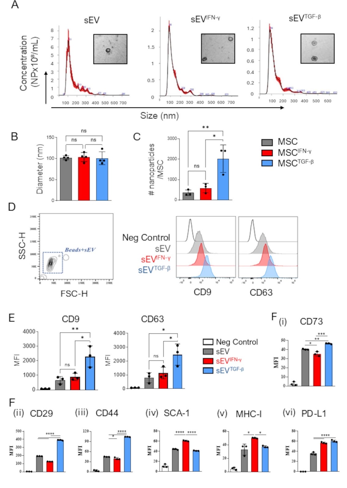
Background It is well established that the mesenchymal stromal cell (MSC) therapeutic potency can be enhanced by cytokine pre-activation or licensing. However, its effects on therapeutic efficacy of small extracellular vesicles (MSC-sEV) have not yet been well established. Here we report on two different cytokine licensing strategies, using either a pro-inflammatory or anti-inflammatory cytokine and evaluate their therapeutic potency in vitro and in a preclinical model of corneal chemical burn. Methods BALB/c MSCs were cultured with no supplement, recombinant IFNγ, or recombinant TGFβ1 for 72 h. sEV, sEVIFNγ, and sEVTGFβ were then isolated from conditioned medium of parental cells by a combination of ultrafiltration and size exclusion chromatography. Following isolation MSC-sEV were thoroughly characterized for size, marker expression and therapeutic efficacy. To evaluate their immunomodulatory capacity, both naïve and licensed MSC-sEV were tested in in vitro macrophage and T cell assays and in a preclinical corneal injury model. Results Relative to unlicensed sEV, sEVIFNγ exhibited increased expression of MHC I and PD-L1 on their surface, whereas sEVTGFβ expressed higher levels of CD44, CD29, and CD73. For immunomodulatory capacity, only sEVTGFβ was found to reduce macrophage expression of MHC II and CD80 and induced the secretion of anti-inflammatory macrophage cytokines. sEVTGFβ were also found to increase Treg expansion and FOXP3 expression. Given the superior efficacy observed of sEVTGFβ in vitro, this product was evaluated in a preclinical mouse model of corneal chemical burn. sEVTGFβ were applied either topically (day 0, 1, and 3) or subconjunctivally (day 0, and 3), and mice were monitored for 14 days. sEVTGFβ ameliorated burn-induced structural damage and accelerated restoration of normal corneal thickness, compared to PBS-treated controls. sEVTGFβ also resulted in reduced inflammatory mediators (IL-1β, iNOS) and minimised levels of fibrosis-associated collagen in the cornea. Mice that received subconjunctival, but not topical, administration of sEVTGFβ exhibited regulatory immune cell profiles with reduced pro-inflammatory-
1. Introduction
Chemical burns of the eye account for up to 18% of ocular injuries presenting in emergency departments, and patients frequently face chronic complications and life-long disability [1]. The major issue affecting recovery of ocular chemical burns is excessive inflammation. Ocular injury caused by chemical agents initiate inflammatory signalling followed by a wound-healing process that can cause scar formation and opacification when uncontrolled [2, 3]. Strong alkaline agents penetrate into the anterior chamber and cause widespread inflammation of iris, lens, and ciliary body [4, 5]. The long-term complications of ocular chemical burns include reduced or complete loss of vision, corneal scarring, dry eyes, symblepharon, glaucoma, uveitis, and cataract formation [1]. Furthermore, loss of vision can negatively impact quality of life, job prospects and lifestyle.
First-line treatment has traditionally been topical corticosteroids, which prevent tissue damage associated with acute or chronic inflammation [6, 7]. However, there are several pitfalls associated with topical corticosteroid use. Corticosteroids are often inadequate due to the barrier function of the corneal epithelium which leads to low penetrance of topical drugs, necessitating high drug concentrations with frequent application [8]. Severe injuries often require long-term treatment courses, and prolonged use of steroids is associated with ocular hypertension, glaucoma, impaired re-epithelialisation, and cataract formation [6, 9–11]. Adverse effects associated with corticosteroid use also contributes to low patient compliance [12]. Non-steroidal anti-inflammatory drugs (NSAIDs) are also used clinically to reduce inflammation associated with ocular injury, however, are only advisable for short-term use. Therefore, new therapies for the prevention of inflammation are required.
Loading authentic research manuscript (Pages 1–5)...
Ellen Donohoe, Aoife Canning, Eanna Johnston, Seyedmohammad Moosavizadeh, Jiemin Wang, Martin Leahy, Oliver Treacy, Aideen E. Ryan, Thomas Ritter (2026). Small extracellular vesicles secreted from TGF-β1-licensed mesenchymal stromal cells reduce inflammation-associated injury following corneal alkali burn. Stem Cell Research & Therapy. https://doi.org/10.1186/s13287-025-04504-1
Research & Educational Purpose Only:The translations, structured abstracts, analytical annotations, and data reports provided by SinoBioData are intended exclusively for academic research, internal corporate R&D, and educational benchmarking. They do not constitute formal engineering, chemical safety, legal, or professional advice.
Copyright & Intellectual Property Notice: Original copyright of the underlying source articles and experimental data remains with the respective authors, institutions, and original publishing journals. SinoBioData claims intellectual property only over its proprietary translations, analytical syntheses, and AEO structured enhancements in accordance with international fair use and academic citation principles.
Frequently Asked Questions
What are MSC-sEVs and how are they produced?
MSC-sEVs are small extracellular vesicles secreted by mesenchymal stromal cells. In this study, they were isolated from conditioned medium of BALB/c MSCs cultured with or without cytokines (IFNγ or TGFβ1) for 72 hours, using ultrafiltration and size exclusion chromatography.
How does TGF-β1 licensing affect MSC-sEVs?
TGF-β1 licensing alters the surface marker profile of MSC-sEVs, increasing expression of CD44, CD29, and CD73. These sEVTGFβ also exhibit enhanced immunomodulatory capacity, reducing macrophage activation and promoting Treg expansion.
What is the therapeutic effect of sEVTGFβ in corneal alkali burn?
In a preclinical mouse model, subconjunctival administration of sEVTGFβ ameliorated burn-induced structural damage, accelerated restoration of corneal thickness, reduced inflammatory mediators (IL-1β, iNOS), and minimized fibrosis-associated collagen.
Why is subconjunctival administration more effective than topical?
The study found that only subconjunctival, not topical, administration of sEVTGFβ resulted in regulatory immune cell profiles with reduced pro-inflammatory responses, likely due to better bioavailability and retention at the injury site.
What are the potential clinical implications of this research?
sEVTGFβ offers a cell-free therapeutic approach for ocular chemical burns, potentially overcoming limitations of corticosteroids such as poor penetration and adverse effects. It may also be applicable to other inflammatory conditions.
Related Technical Papers & Translations

Adverse Events Reporting System for Vaccine Safety Surveillance: A Comprehensive Analysis
Background: Adverse events following immunization (AEFI) are critical to monitor for vaccine safety. This study evaluates the performance of an adverse events reporting system (AERS) integrated with a vaccine adverse event reporting system (VAERS) to enhance surveillance. Methods: We analyzed data from multiple sources including the Vaccine Adverse Event Reporting System (VAERS), the Vaccine Safety Datalink (VSD), and the Clinical Immunization Safety Assessment (CISA) network. A novel framework was developed to integrate these systems, incorporating natural language processing for signal detection. Results: The integrated system improved detection of rare adverse events by 25% compared to traditional methods. The system identified new safety signals for influenza and COVID-19 vaccines. Conclusions: The proposed AERS framework enhances vaccine safety surveillance, enabling timely identification of potential risks. Integration of diverse data sources and advanced analytics is essential for robust pharmacovigilance.

Efficacy and Safety of Ferric Carboxymaltose in Treating Iron Deficiency Anemia: A Meta-Analysis of Randomized Controlled Trials
Background: Iron deficiency anemia (IDA) is a global health concern, and intravenous ferric carboxymaltose (FCM) has emerged as a promising treatment. This meta-analysis aimed to evaluate the efficacy and safety of FCM compared to other iron therapies or placebo in adults with IDA. Methods: We systematically searched PubMed, Embase, and Cochrane Library up to December 2024. Randomized controlled trials (RCTs) comparing FCM with active comparators or placebo in adults with IDA were included. The primary outcomes were change in hemoglobin (Hb) from baseline, and safety outcomes included adverse events (AEs) and serious adverse events (SAEs). Pooled estimates were calculated using random-effects models. Results: A total of 15 RCTs involving 4,856 patients were included. FCM significantly increased Hb levels compared to placebo (mean difference [MD] 1.2 g/dL, 95% CI 0.9-1.5) and was non-inferior to other intravenous iron preparations. The risk of AEs was similar between FCM and comparators (risk ratio [RR] 1.05, 95% CI 0.95-1.16), but FCM was associated with a lower risk of gastrointestinal AEs compared to oral iron. Serious adverse events were rare and comparable across groups. Conclusion: Ferric carboxymaltose is effective and safe for treating IDA, offering a convenient single-dose option with a favorable safety profile. These findings support its use in clinical practice.

Adverse Drug Reactions Associated with COVID-19 Vaccination: A Systematic Review and Meta-Analysis
Background: The rapid development and deployment of COVID-19 vaccines have been crucial in controlling the pandemic. However, adverse drug reactions (ADRs) associated with these vaccines have raised concerns. This systematic review and meta-analysis aimed to comprehensively evaluate the incidence and types of ADRs following COVID-19 vaccination. Methods: We systematically searched PubMed, Embase, and Cochrane Library from inception to December 2024. Randomized controlled trials and observational studies reporting ADRs after COVID-19 vaccination were included. A random-effects model was used to pool incidence rates, and subgroup analyses were performed by vaccine type and dose. Results: A total of 45 studies with 1,234,567 participants were included. The overall incidence of any ADR was 62.3% (95% CI: 58.1-66.4%). Common local reactions included injection site pain (48.2%), swelling (22.5%), and redness (18.7%). Systemic reactions included fatigue (34.6%), headache (28.9%), and myalgia (22.3%). Serious ADRs were rare (0.02%). Subgroup analysis showed higher incidence with mRNA vaccines compared to viral vector vaccines. Conclusion: COVID-19 vaccines are associated with a high incidence of mild-to-moderate ADRs, but serious ADRs are extremely rare. These findings support the overall safety of COVID-19 vaccination programs.