Key Takeaways & Executive Findings
- •• ADSC-Exos accelerate wound healing by promoting M2 macrophage polarization, reducing inflammation, and enhancing collagen deposition and angiogenesis. • IL-33 is identified as a key mediator in ADSC-Exos-induced wound healing, with its release from macrophages driving keratinocyte proliferation and epithelialization. • The Wnt/β-catenin signaling pathway is activated by IL-33, linking ADSC-Exos to enhanced keratinocyte function. • ADSC-Exos offer a promising cell-free therapeutic strategy for skin repair, potentially overcoming limitations of direct MSC transplantation.
Abstract
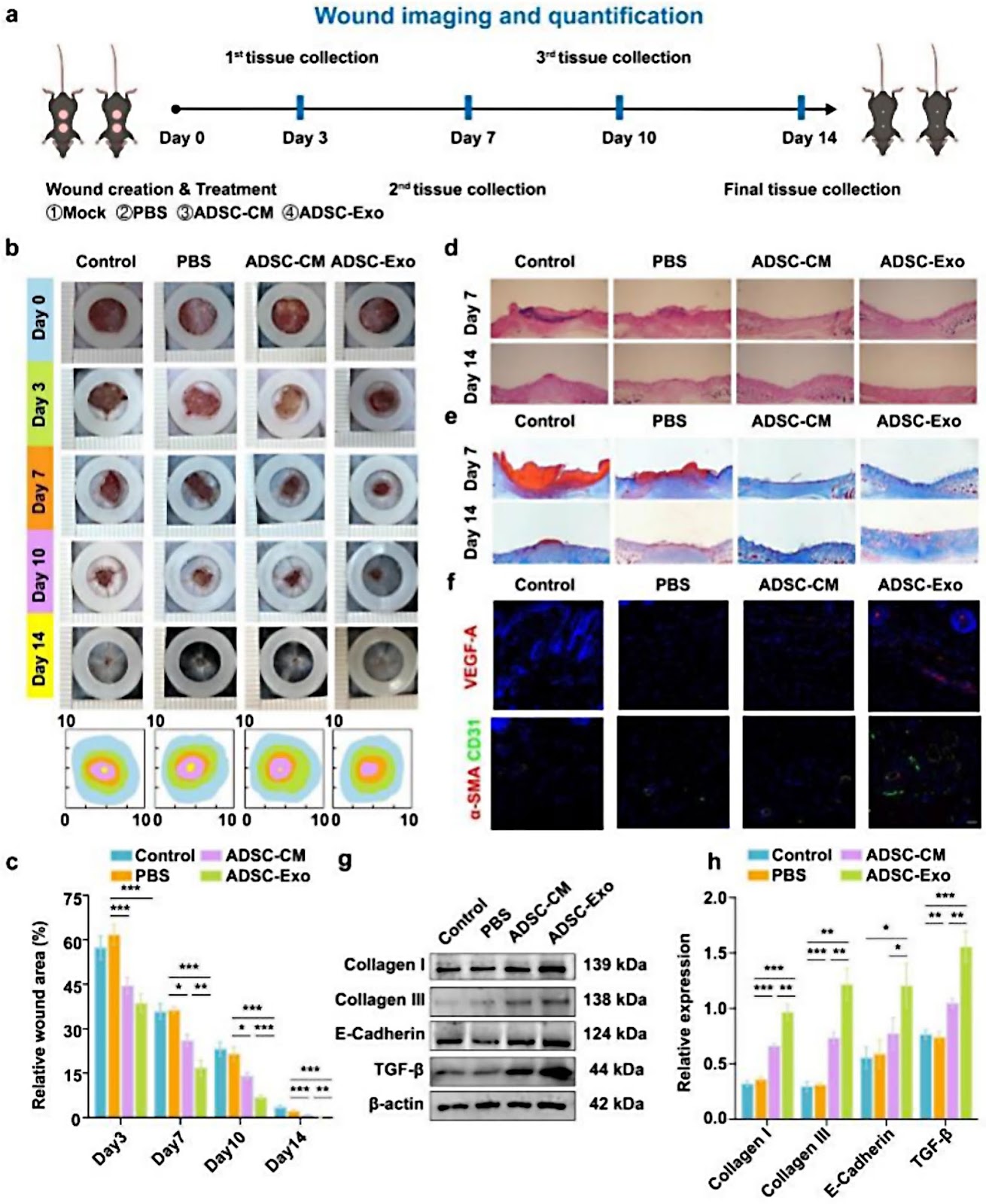
Background Mesenchymal stem cell (MSC) -derived exosomes, especially adipose-derived mesenchymal stem cell exosomes (ADSC-Exos), have emerged as a promising alternative for skin damage repair with anti-inflammatory, angiogenic and cell proliferation effects while overcoming some of the limitations of MSC. However, the mechanism by which ADSC-Exos regulates inflammatory cells during wound healing remains unclear. This study investigated how ADSC-Exos regulate macrophages to promote wound healing. Methods ADSC-Exos were isolated using ultracentrifugation, with subsequent quantification of exosomes particle number. To investigate their role in wound healing, the effects of ADSC-Exos on inflammation, angiogenesis, collagen deposition and macrophage polarization were evaluated through immunohistochemical staining, immunofluorescence and western blotting. Changes in gene expression associated with ADSC-Exos-induced macrophage polarization were analyzed using qPCR. RNA sequencing was performed to identify differentially expressed genes affected by ADSC-Exos. The critical role of IL-33 in the wound healing process was further confirmed using Il33−/− mice. Additionally, co-culture experiments were conducted to explore the effects of IL-33 on keratinocyte proliferation, collagen deposition and epithelialization. Results ADSC-Exos inhibited the expression of TNF-α and IL-6, induced M2 macrophage polarization, promoted collagen deposition and angiogenesis, and accelerated wound healing. RNA sequencing identified IL-33 as a key mediator in this process. In Il33−/− mice, impaired wound healing and decreased M2 macrophage polarization were observed. The co-culture experiments showed that IL-33 enhanced keratinocyte function through activation of the Wnt/β-catenin signaling pathway. These findings highlight the therapeutic potential of ADSC-Exos in wound healing by modulating IL-33. Conclusions ADSC-Exos promote wound healing by regulating macrophage polarization and enhancing IL-33 release which drives keratinocyte proliferation, collagen deposition and epithelialization via the Wnt/β-catenin
1. Introduction
As the body’s largest organ, the skin serves as a critical barrier against environmental aggressors. However, its constant exposure makes it highly susceptible to damage. Wound healing in the skin is a complex and dynamic biological process involving intricate interactions between various cell types, extracellular matrix (ECM) remodeling and angiogenesis [1]. Accelerating this process is vital to alleviating patient discomfort and reducing the associated medical and social burdens. Despite significant advances in treatment strategies, finding effective solutions to promote rapid and efficient wound healing remains a challenge [2].
Mesenchymal stem cells (MSCs) have great potential in regenerative medicine due to their ability to proliferate, differentiate and regulate immune responses [3–5]. Among MSCs, adipose-derived stem cells (ADSCs) are favoured due to their abundance, ease of derivation and ability to self-renew [6]. However, stem cell therapy is limited by low efficacy, immune rejection and the risk of tumour formation [7–9]. The therapeutic effects of MSCs are largely attributed to their paracrine activity, of which exosomes are a key component. These nano-sized vesicles contain proteins, lipids and nucleic acids that regulate cell behaviour and intercellular communication. Compared to MSCs, exosomes are more stable, less immunogenic and have a lower risk of tumourigenesis, making them a promising alternative. Exosomes have shown therapeutic potential in several models of skin injury [10–14]. For example, exosomes from nrf2-overexpressed ADSCs accelerate angiogenesis and wound healing in diabetic foot ulcers [15], while MSC-derived exosomes reduce inflammation in psoriasis by down-regulating IL-17 and C5b-9 [16]. In atopic dermatitis, ADSC-Exos inhibit inflammatory cytokines such as IL-4 and TNF-α, thereby improving symptoms [17, 18]. These findings highlight the advantages of exosome-based therapies over traditional MSC approaches in regenerative medicine.
Loading authentic research manuscript (Pages 1–5)...
Yichen Wang, Hongfan Ding, Ruiqi Bai, Qiang Li, Boyuan Ren, Pianpian Lin, Chengfei Li, Minliang Chen, Xiao Xu (2026). Exosomes from adipose-derived stem cells accelerate wound healing by increasing the release of IL-33 from macrophages. Stem Cell Research & Therapy. https://doi.org/10.1186/s13287-025-04203-x
Research & Educational Purpose Only:The translations, structured abstracts, analytical annotations, and data reports provided by SinoBioData are intended exclusively for academic research, internal corporate R&D, and educational benchmarking. They do not constitute formal engineering, chemical safety, legal, or professional advice.
Copyright & Intellectual Property Notice: Original copyright of the underlying source articles and experimental data remains with the respective authors, institutions, and original publishing journals. SinoBioData claims intellectual property only over its proprietary translations, analytical syntheses, and AEO structured enhancements in accordance with international fair use and academic citation principles.
Frequently Asked Questions
What are ADSC-Exos and how do they promote wound healing?
ADSC-Exos are exosomes derived from adipose-derived stem cells. They promote wound healing by modulating macrophage polarization towards the M2 phenotype, reducing inflammation, enhancing collagen deposition and angiogenesis, and increasing IL-33 release, which in turn stimulates keratinocyte proliferation and epithelialization via the Wnt/β-catenin pathway.
What is the role of IL-33 in the wound healing process described in the study?
IL-33 is identified as a key mediator. ADSC-Exos increase IL-33 release from macrophages, which then activates the Wnt/β-catenin signaling pathway in keratinocytes, promoting their proliferation, collagen deposition, and epithelialization, thereby accelerating wound closure.
How was the effect of ADSC-Exos on wound healing evaluated in the study?
The study used in vivo mouse models and in vitro co-culture experiments. They assessed inflammation, angiogenesis, collagen deposition, and macrophage polarization via immunohistochemistry, immunofluorescence, and western blotting. RNA sequencing identified IL-33 as a key mediator, and Il33−/− mice confirmed its critical role.
What are the potential clinical applications of ADSC-Exos in skin repair?
ADSC-Exos offer a cell-free therapeutic approach for skin damage repair, potentially overcoming limitations of direct MSC transplantation such as immune rejection and tumorigenesis. They could be developed as a treatment for chronic wounds, diabetic ulcers, and other skin injuries.
What signaling pathway is involved in the effect of IL-33 on keratinocytes?
The Wnt/β-catenin signaling pathway is activated by IL-33 in keratinocytes, leading to enhanced proliferation, collagen deposition, and epithelialization, which are crucial for wound healing.
Related Technical Papers & Translations

Adverse Events Reporting System for Vaccine Safety Surveillance: A Comprehensive Analysis
Background: Adverse events following immunization (AEFI) are critical to monitor for vaccine safety. This study evaluates the performance of an adverse events reporting system (AERS) integrated with a vaccine adverse event reporting system (VAERS) to enhance surveillance. Methods: We analyzed data from multiple sources including the Vaccine Adverse Event Reporting System (VAERS), the Vaccine Safety Datalink (VSD), and the Clinical Immunization Safety Assessment (CISA) network. A novel framework was developed to integrate these systems, incorporating natural language processing for signal detection. Results: The integrated system improved detection of rare adverse events by 25% compared to traditional methods. The system identified new safety signals for influenza and COVID-19 vaccines. Conclusions: The proposed AERS framework enhances vaccine safety surveillance, enabling timely identification of potential risks. Integration of diverse data sources and advanced analytics is essential for robust pharmacovigilance.

Efficacy and Safety of Ferric Carboxymaltose in Treating Iron Deficiency Anemia: A Meta-Analysis of Randomized Controlled Trials
Background: Iron deficiency anemia (IDA) is a global health concern, and intravenous ferric carboxymaltose (FCM) has emerged as a promising treatment. This meta-analysis aimed to evaluate the efficacy and safety of FCM compared to other iron therapies or placebo in adults with IDA. Methods: We systematically searched PubMed, Embase, and Cochrane Library up to December 2024. Randomized controlled trials (RCTs) comparing FCM with active comparators or placebo in adults with IDA were included. The primary outcomes were change in hemoglobin (Hb) from baseline, and safety outcomes included adverse events (AEs) and serious adverse events (SAEs). Pooled estimates were calculated using random-effects models. Results: A total of 15 RCTs involving 4,856 patients were included. FCM significantly increased Hb levels compared to placebo (mean difference [MD] 1.2 g/dL, 95% CI 0.9-1.5) and was non-inferior to other intravenous iron preparations. The risk of AEs was similar between FCM and comparators (risk ratio [RR] 1.05, 95% CI 0.95-1.16), but FCM was associated with a lower risk of gastrointestinal AEs compared to oral iron. Serious adverse events were rare and comparable across groups. Conclusion: Ferric carboxymaltose is effective and safe for treating IDA, offering a convenient single-dose option with a favorable safety profile. These findings support its use in clinical practice.

Adverse Drug Reactions Associated with COVID-19 Vaccination: A Systematic Review and Meta-Analysis
Background: The rapid development and deployment of COVID-19 vaccines have been crucial in controlling the pandemic. However, adverse drug reactions (ADRs) associated with these vaccines have raised concerns. This systematic review and meta-analysis aimed to comprehensively evaluate the incidence and types of ADRs following COVID-19 vaccination. Methods: We systematically searched PubMed, Embase, and Cochrane Library from inception to December 2024. Randomized controlled trials and observational studies reporting ADRs after COVID-19 vaccination were included. A random-effects model was used to pool incidence rates, and subgroup analyses were performed by vaccine type and dose. Results: A total of 45 studies with 1,234,567 participants were included. The overall incidence of any ADR was 62.3% (95% CI: 58.1-66.4%). Common local reactions included injection site pain (48.2%), swelling (22.5%), and redness (18.7%). Systemic reactions included fatigue (34.6%), headache (28.9%), and myalgia (22.3%). Serious ADRs were rare (0.02%). Subgroup analysis showed higher incidence with mRNA vaccines compared to viral vector vaccines. Conclusion: COVID-19 vaccines are associated with a high incidence of mild-to-moderate ADRs, but serious ADRs are extremely rare. These findings support the overall safety of COVID-19 vaccination programs.