Key Takeaways & Executive Findings
- •• Fucoxanthin dose-dependently protects placenta-derived mesenchymal stem cells (PL-MSCs) from oxidative stress-induced apoptosis by reducing pro-apoptotic protein expression and increasing anti-apoptotic protein levels. • Transcriptomic analysis reveals that fucoxanthin modulates key pathways including DNA damage repair, ER stress response, p53-induced apoptosis, cell cycle regulation, and PI3K/mTOR and AMPK signaling. • The study provides mechanistic evidence supporting fucoxanthin as a potential adjunct to improve the viability and therapeutic efficacy of MSC-based regenerative therapies. • Findings highlight the importance of oxidative stress mitigation in enhancing MSC survival for clinical applications.
Abstract
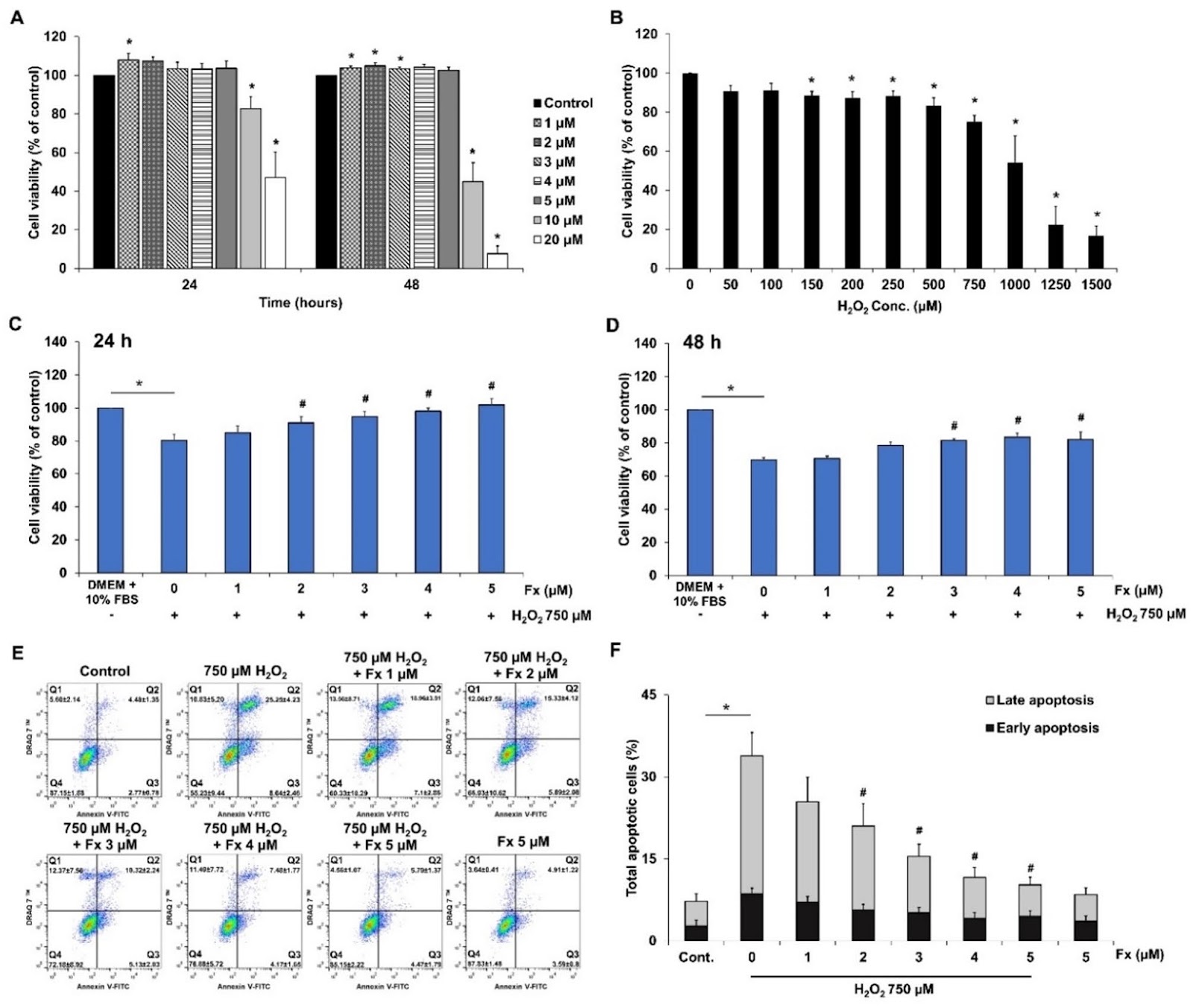
Human mesenchymal stem cells (hMSCs) hold significant promise in regenerative medicine due to their ability to reduce inflammation and promote tissue repair. However, their therapeutic potential is often compromised by their high susceptibility to apoptosis under oxidative stress, prevalent in the microenvironment of the target tissues. Our previous study showed that fucoxanthin, a carotenoid derived from brown algae, can improve the viability of placenta-derived mesenchymal stem cells (PL-MSCs) by reducing intracellular ROS levels through the activation of the PI3K/Akt/Nrf-2 signaling pathway. In this study, we further investigate the mechanisms underlying the protective effect of fucoxanthin against oxidative stress-induced apoptosis in PL-MSCs, using an in vitro model. PL-MSCs were cultured with 750 µM H2O2 to induce oxidative stress and treated with various concentrations of fucoxanthin for 48 h. The effect of fucoxanthin on PL-MSC apoptosis under oxidative stress conditions was determined using CCK-8, Annexin V/DRAQ7™ apoptosis assays, as well as the expression of apoptosis-related genes and proteins. The effect of fucoxanthin on the transcriptome of PL-MSCs under oxidative stress conditions was also assessed by high-throughput Nanostring analysis. The results showed that fucoxanthin significantly decreased the apoptosis of PL-MSCs under oxidative stress in a dose-dependent manner by reducing the expression of pro-apoptotic proteins and inhibiting their activation, while increasing the expression of anti-apoptotic proteins in these cells. Furthermore, fucoxanthin also downregulates the expression of genes associated with the endoplasmic reticulum stress, p53-induced apoptosis, while increasing the expression of genes involved in the regulation of the cell cycle, DNA damage repair, cytokine signaling, nucleotide synthesis, PI3K/mTOR pathway and AMPK pathway in PL-MSCs under oxidative stress conditions. Taken together, the findings provide compelling evidence that fucoxanthin protects PL-MSCs against oxidative stress-induced apoptosis by modulating the expression of various genes involved in DNA damage repair, ER stress response, p53-induced apoptosis in these cells. This suggests that fucoxanthin could be used as a potential supplement to enhance the therapeutic efficacy of MSC-based therapies.
1. Introduction
Human mesenchymal stem cells (hMSCs) could potentially be used in cell-based therapies due to their ability to home in on specific tissues and release various beneficial factors to reduce inflammation and promote tissue repair [1, 2]. However, there is evidence indicating that most MSCs undergo apoptosis shortly after being introduced into the target tissues [3, 4]. The poor viability of grafted MSCs is largely due to oxidative stress caused by an excessive amount of reactive oxygen species (ROS) in target tissues [5]. Oxidative stress has been shown to inhibit proliferation, increase senescence, and induce apoptosis in transplanted MSCs by damaging their cellular components [6].
Fucoxanthin, a marine carotenoid derived from edible brown algae, is known for its strong antioxidant properties that provide various health benefits, including anti-obesity [7], anti-cancer [8] and anti-inflammation [9] effects. Previous studies indicate that fucoxanthin significantly increases glutathione levels, reduces ROS, inhibits DNA damage, restores mitochondrial membrane potential and suppresses apoptosis in human keratinocytes [10, 11], retinal pigment epithelial cells [12, 13] and neurons [14] under oxidative stress conditions. Furthermore, our previous study found that fucoxanthin also protects human placenta-derived mesenchymal stem cells (PL-MSCs) from oxidative stress by reducing intracellular ROS production and improving their viability by activating the PI3K/Akt/Nrf-2 signaling pathway [15].
However, the effect of fucoxanthin on the prevention of MSC apoptosis under oxidative stress conditions has not yet been determined. Therefore, in this study, we further investigate the mechanisms underlying the protective effect of fucoxanthin against oxidative stress-induced apoptosis in PL-MSCs. We use PL-MSCs, which can be harvested in large quantities and have shown promise in regenerative medicine.
Loading authentic research manuscript (Pages 1–5)...
Gunticha Suwanmanee, Pakpoom Kheolamai, Chairat Tantrawatpan, Daniel Grimes, Ioan Valentin Matei, Luminita Paraoan, Sirikul Manochantr (2026). Fucoxanthin protects placenta-derived human mesenchymal stem cells against oxidative stress-induced apoptosis by modulating genes involved in DNA damage repair, ER stress response, p53-induced apoptosis. Stem Cell Research & Therapy. https://doi.org/10.1186/s13287-025-04629-3
Research & Educational Purpose Only:The translations, structured abstracts, analytical annotations, and data reports provided by SinoBioData are intended exclusively for academic research, internal corporate R&D, and educational benchmarking. They do not constitute formal engineering, chemical safety, legal, or professional advice.
Copyright & Intellectual Property Notice: Original copyright of the underlying source articles and experimental data remains with the respective authors, institutions, and original publishing journals. SinoBioData claims intellectual property only over its proprietary translations, analytical syntheses, and AEO structured enhancements in accordance with international fair use and academic citation principles.
Frequently Asked Questions
What is the main finding of this study?
The study demonstrates that fucoxanthin protects placenta-derived mesenchymal stem cells from oxidative stress-induced apoptosis by modulating genes involved in DNA damage repair, ER stress response, and p53-induced apoptosis.
How does fucoxanthin protect stem cells from oxidative stress?
Fucoxanthin reduces intracellular ROS levels, decreases pro-apoptotic protein expression, and increases anti-apoptotic protein expression. It also modulates key signaling pathways such as PI3K/mTOR and AMPK, and enhances DNA damage repair mechanisms.
What are the potential clinical applications of this research?
The findings suggest that fucoxanthin could be used as a supplement to improve the viability and therapeutic efficacy of mesenchymal stem cell-based therapies, particularly in conditions where oxidative stress compromises cell survival.
What experimental model was used in this study?
The study used an in vitro model with placenta-derived mesenchymal stem cells (PL-MSCs) treated with hydrogen peroxide (H2O2) to induce oxidative stress, followed by fucoxanthin treatment at various concentrations.
What techniques were employed to assess the effects?
The researchers used CCK-8 assays, Annexin V/DRAQ7™ apoptosis assays, gene expression analysis, and high-throughput Nanostring transcriptomic analysis to evaluate apoptosis and gene expression changes.
Related Technical Papers & Translations

Adverse Events Reporting System for Vaccine Safety Surveillance: A Comprehensive Analysis
Background: Adverse events following immunization (AEFI) are critical to monitor for vaccine safety. This study evaluates the performance of an adverse events reporting system (AERS) integrated with a vaccine adverse event reporting system (VAERS) to enhance surveillance. Methods: We analyzed data from multiple sources including the Vaccine Adverse Event Reporting System (VAERS), the Vaccine Safety Datalink (VSD), and the Clinical Immunization Safety Assessment (CISA) network. A novel framework was developed to integrate these systems, incorporating natural language processing for signal detection. Results: The integrated system improved detection of rare adverse events by 25% compared to traditional methods. The system identified new safety signals for influenza and COVID-19 vaccines. Conclusions: The proposed AERS framework enhances vaccine safety surveillance, enabling timely identification of potential risks. Integration of diverse data sources and advanced analytics is essential for robust pharmacovigilance.

Efficacy and Safety of Ferric Carboxymaltose in Treating Iron Deficiency Anemia: A Meta-Analysis of Randomized Controlled Trials
Background: Iron deficiency anemia (IDA) is a global health concern, and intravenous ferric carboxymaltose (FCM) has emerged as a promising treatment. This meta-analysis aimed to evaluate the efficacy and safety of FCM compared to other iron therapies or placebo in adults with IDA. Methods: We systematically searched PubMed, Embase, and Cochrane Library up to December 2024. Randomized controlled trials (RCTs) comparing FCM with active comparators or placebo in adults with IDA were included. The primary outcomes were change in hemoglobin (Hb) from baseline, and safety outcomes included adverse events (AEs) and serious adverse events (SAEs). Pooled estimates were calculated using random-effects models. Results: A total of 15 RCTs involving 4,856 patients were included. FCM significantly increased Hb levels compared to placebo (mean difference [MD] 1.2 g/dL, 95% CI 0.9-1.5) and was non-inferior to other intravenous iron preparations. The risk of AEs was similar between FCM and comparators (risk ratio [RR] 1.05, 95% CI 0.95-1.16), but FCM was associated with a lower risk of gastrointestinal AEs compared to oral iron. Serious adverse events were rare and comparable across groups. Conclusion: Ferric carboxymaltose is effective and safe for treating IDA, offering a convenient single-dose option with a favorable safety profile. These findings support its use in clinical practice.

Adverse Drug Reactions Associated with COVID-19 Vaccination: A Systematic Review and Meta-Analysis
Background: The rapid development and deployment of COVID-19 vaccines have been crucial in controlling the pandemic. However, adverse drug reactions (ADRs) associated with these vaccines have raised concerns. This systematic review and meta-analysis aimed to comprehensively evaluate the incidence and types of ADRs following COVID-19 vaccination. Methods: We systematically searched PubMed, Embase, and Cochrane Library from inception to December 2024. Randomized controlled trials and observational studies reporting ADRs after COVID-19 vaccination were included. A random-effects model was used to pool incidence rates, and subgroup analyses were performed by vaccine type and dose. Results: A total of 45 studies with 1,234,567 participants were included. The overall incidence of any ADR was 62.3% (95% CI: 58.1-66.4%). Common local reactions included injection site pain (48.2%), swelling (22.5%), and redness (18.7%). Systemic reactions included fatigue (34.6%), headache (28.9%), and myalgia (22.3%). Serious ADRs were rare (0.02%). Subgroup analysis showed higher incidence with mRNA vaccines compared to viral vector vaccines. Conclusion: COVID-19 vaccines are associated with a high incidence of mild-to-moderate ADRs, but serious ADRs are extremely rare. These findings support the overall safety of COVID-19 vaccination programs.