Key Takeaways & Executive Findings
- •• BM-MSCs enhance oocyte survival and promote follicle growth in reconstituted ovaries. • BM-MSCs do not contribute to follicle formation or proliferate, but localize around growing follicles. • BM-MSCs may direct theca cell differentiation, as indicated by CYP17a1 co-staining. • Artificial ovary with BM-MSCs supports normal folliculogenesis and oocyte maturation after transplantation.
Abstract
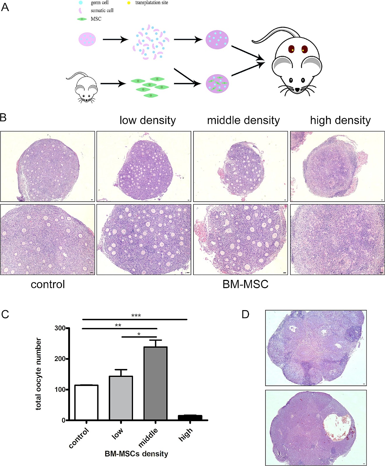
Background Studies have shown that chemotherapy and radiotherapy can cause premature ovarian failure and loss of fertility in female cancer patients. Ovarian cortex cryopreservation is a good choice to preserve female fertility before cancer treatment. Following the remission of the disease, the thawed ovarian tissue can be transplanted back and restore fertility of the patient. However, there is a risk to reintroduce cancer cells in the body and leads to the recurrence of cancer. Given the low success rate of current in vitro culture techniques for obtaining mature oocytes from primordial follicles, an artificial ovary with primordial follicles may be a good way to solve this problem. Methods In the study, we established an artificial ovary model based on the participation of mesenchymal stem cells (MSCs) to evaluate the effect of MSCs on follicular development and oocyte maturation. P2.5 mouse ovaries were digested into single cell suspensions and mixed with bone marrow derived mesenchymal stem cells (BM-MSCs) at a 1:1 ratio. The reconstituted ovarian model was then generated by using phytohemagglutinin. The phenotype and mechanism studies were explored by follicle counting, immunohistochemistry, immunofluorescence, in vitro maturation (IVM), in vitro fertilization (IVF), real-time quantitative polymerase chain reaction (RT-PCR), and Terminal-deoxynucleotidyl transferase mediated nick end labeling(TUNEL) assay. Results Our study found that the addition of BM-MSCs to the reconstituted ovary can enhance the survival of oocytes and promote the growth and development of follicles. After transplanting the reconstituted ovaries under kidney capsules of the recipient mice, we observed normal folliculogenesis and oocyte maturation. Interestingly, we found that BM-MSCs did not contribute to the formation of follicles in ovarian aggregation, nor did they undergo proliferation during follicle growth. Instead, the cells were found to be located around growing follicles in the reconstituted ovary. When theca cells were labeled with CYP17a1, we found some overlapped staining with green fluorescent protein(GFP)-labeled BM-MSCs. The results suggest that BM-MSCs may participate in directing the differentiation of theca layer in the reconstituted ovary. Conclusions The presence of BM-MSCs in the artificial ovary was found to promote the survival of ovarian cells, as well as facilitate follicle formation and development. Since the cells didn’t proliferate in the reconstituted ovary, this
1. Introduction
With rapid advancements in cancer therapy, children and reproductive-age women are benefitting from overall improved survival rates [1]. It is well-documented that treatment of girls and women for cancer with radiation, chemotherapeutic drugs, or a combination of the two therapies can result in significant, and often irreversible, side-effect damage to the reproductive system [2, 3]. Anti-cancer therapy is often a cause of premature ovarian insufficiency (POI) due to the high sensitivity of the ovarian follicle reserve to chemotherapy and radiotherapy [4, 5]. Thus, there is an increased number of patients who received a gonadotoxic treatment and who later face fertility issues [6]. Overall, compared to the general population, women who undergo cancer treatment are 38% less likely to become pregnant. This reduction in the likelihood of subsequent pregnancies has been observed in nearly all types of cancer [7]. Consequently, the preservation of the ovarian reserve and prevention of infertility have become the primary quality of life concerns for patients and their physicians.
Fertility preservation refers to the use of surgical, pharmacologic, or laboratory techniques to provide assistance to women or men at risk of infertility in protecting and preserving their ability to have genetically derived offspring [8]. For women, common fertility preservation methods currently used include egg, embryo, and ovarian tissue freezing, whereas for unmarried or prepubertal women, freezing of the ovarian cortex is more appropriate [9–11]. After the patient’s condition has improved or resolved, the preserved ovarian cortex can be thawed and transplanted back to the p
Loading authentic research manuscript (Pages 1–5)...
Jiazhao Li, Haonan Fan, Wei Liu, Jing Zhang, Yue Xiao, Yue Peng, Weijie Yang, Wenwen Liu, Yuanlin He, Lianju Qin, Xiang Ma, Jing Li (2026). Mesenchymal stem cells promote ovarian reconstruction in mice. Stem Cell Research & Therapy. https://doi.org/10.1186/s13287-024-03718-z
Research & Educational Purpose Only:The translations, structured abstracts, analytical annotations, and data reports provided by SinoBioData are intended exclusively for academic research, internal corporate R&D, and educational benchmarking. They do not constitute formal engineering, chemical safety, legal, or professional advice.
Copyright & Intellectual Property Notice: Original copyright of the underlying source articles and experimental data remains with the respective authors, institutions, and original publishing journals. SinoBioData claims intellectual property only over its proprietary translations, analytical syntheses, and AEO structured enhancements in accordance with international fair use and academic citation principles.
Frequently Asked Questions
What is the role of mesenchymal stem cells in ovarian reconstruction?
The study demonstrates that bone marrow-derived mesenchymal stem cells (BM-MSCs) enhance oocyte survival and promote follicle growth and development in a reconstituted ovary model. They do not proliferate or form follicles themselves but localize around growing follicles and may direct theca cell differentiation.
How was the artificial ovary model constructed?
P2.5 mouse ovaries were digested into single cell suspensions and mixed with BM-MSCs at a 1:1 ratio. The reconstituted ovarian model was generated using phytohemagglutinin, then transplanted under kidney capsules of recipient mice for evaluation.
What are the key findings regarding BM-MSC behavior in the reconstituted ovary?
BM-MSCs did not contribute to follicle formation or proliferate during follicle growth. Instead, they were located around growing follicles and showed overlapping staining with theca cell marker CYP17a1, suggesting a role in directing theca layer differentiation.
What is the clinical significance of this research?
This research provides a potential strategy for fertility preservation in female cancer patients by using an artificial ovary with MSCs, which could restore fertility without the risk of reintroducing cancer cells, addressing the limitations of current ovarian tissue transplantation.
What methods were used to evaluate the effects of BM-MSCs?
The study employed follicle counting, immunohistochemistry, immunofluorescence, in vitro maturation (IVM), in vitro fertilization (IVF), real-time quantitative PCR (RT-PCR), and TUNEL assay to assess follicular development, oocyte maturation, and cell survival.
Related Technical Papers & Translations

Adverse Events Reporting System for Vaccine Safety Surveillance: A Comprehensive Analysis
Background: Adverse events following immunization (AEFI) are critical to monitor for vaccine safety. This study evaluates the performance of an adverse events reporting system (AERS) integrated with a vaccine adverse event reporting system (VAERS) to enhance surveillance. Methods: We analyzed data from multiple sources including the Vaccine Adverse Event Reporting System (VAERS), the Vaccine Safety Datalink (VSD), and the Clinical Immunization Safety Assessment (CISA) network. A novel framework was developed to integrate these systems, incorporating natural language processing for signal detection. Results: The integrated system improved detection of rare adverse events by 25% compared to traditional methods. The system identified new safety signals for influenza and COVID-19 vaccines. Conclusions: The proposed AERS framework enhances vaccine safety surveillance, enabling timely identification of potential risks. Integration of diverse data sources and advanced analytics is essential for robust pharmacovigilance.

Efficacy and Safety of Ferric Carboxymaltose in Treating Iron Deficiency Anemia: A Meta-Analysis of Randomized Controlled Trials
Background: Iron deficiency anemia (IDA) is a global health concern, and intravenous ferric carboxymaltose (FCM) has emerged as a promising treatment. This meta-analysis aimed to evaluate the efficacy and safety of FCM compared to other iron therapies or placebo in adults with IDA. Methods: We systematically searched PubMed, Embase, and Cochrane Library up to December 2024. Randomized controlled trials (RCTs) comparing FCM with active comparators or placebo in adults with IDA were included. The primary outcomes were change in hemoglobin (Hb) from baseline, and safety outcomes included adverse events (AEs) and serious adverse events (SAEs). Pooled estimates were calculated using random-effects models. Results: A total of 15 RCTs involving 4,856 patients were included. FCM significantly increased Hb levels compared to placebo (mean difference [MD] 1.2 g/dL, 95% CI 0.9-1.5) and was non-inferior to other intravenous iron preparations. The risk of AEs was similar between FCM and comparators (risk ratio [RR] 1.05, 95% CI 0.95-1.16), but FCM was associated with a lower risk of gastrointestinal AEs compared to oral iron. Serious adverse events were rare and comparable across groups. Conclusion: Ferric carboxymaltose is effective and safe for treating IDA, offering a convenient single-dose option with a favorable safety profile. These findings support its use in clinical practice.

Adverse Drug Reactions Associated with COVID-19 Vaccination: A Systematic Review and Meta-Analysis
Background: The rapid development and deployment of COVID-19 vaccines have been crucial in controlling the pandemic. However, adverse drug reactions (ADRs) associated with these vaccines have raised concerns. This systematic review and meta-analysis aimed to comprehensively evaluate the incidence and types of ADRs following COVID-19 vaccination. Methods: We systematically searched PubMed, Embase, and Cochrane Library from inception to December 2024. Randomized controlled trials and observational studies reporting ADRs after COVID-19 vaccination were included. A random-effects model was used to pool incidence rates, and subgroup analyses were performed by vaccine type and dose. Results: A total of 45 studies with 1,234,567 participants were included. The overall incidence of any ADR was 62.3% (95% CI: 58.1-66.4%). Common local reactions included injection site pain (48.2%), swelling (22.5%), and redness (18.7%). Systemic reactions included fatigue (34.6%), headache (28.9%), and myalgia (22.3%). Serious ADRs were rare (0.02%). Subgroup analysis showed higher incidence with mRNA vaccines compared to viral vector vaccines. Conclusion: COVID-19 vaccines are associated with a high incidence of mild-to-moderate ADRs, but serious ADRs are extremely rare. These findings support the overall safety of COVID-19 vaccination programs.