Key Takeaways & Executive Findings
- •• PD-L1+ MSCs identified via scRNA-seq exhibit superior immunomodulatory capacity, suppressing T-cell proliferation and reducing TNF-α levels in vitro. • In a curdlan-induced SKG mouse model of ankylosing spondylitis, PD-L1+ MSCs significantly alleviated joint inflammation and reduced pathological ectopic bone formation. • PD-L1+ MSCs modulate immune responses by decreasing Th17 cells and increasing Th2 and Treg populations, along with lowering pro-inflammatory cytokines. • These findings highlight PD-L1+ MSCs as a promising cell-based therapy for AS, potentially surpassing conventional MSC treatments.
Abstract
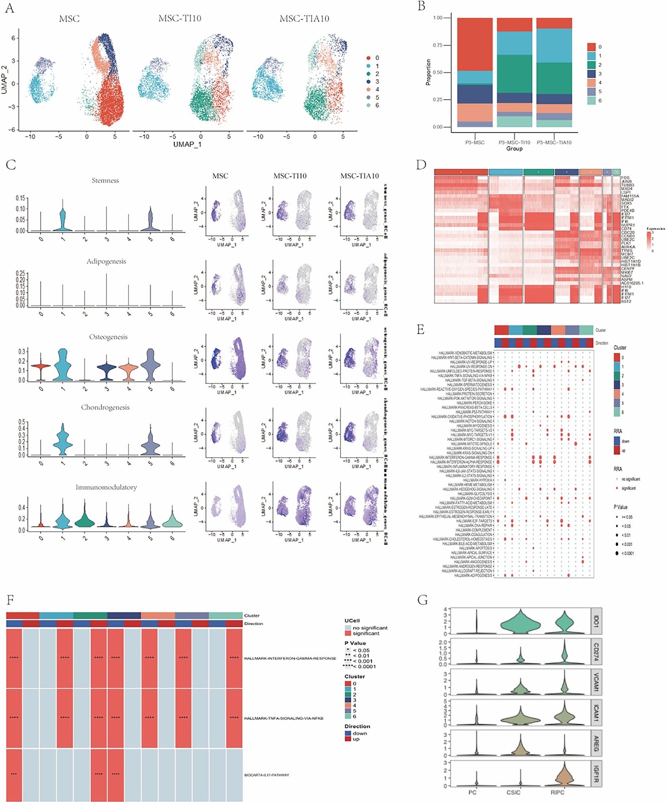
Background This study systematically evaluated the immunomodulatory function of PD-L1-positive mesenchymal stem cells (PD-L1(+) MSCs) using single-cell RNA sequencing (scRNA-seq) and investigated their roles in suppressing inflammation and regulating pathological bone formation in curdlan-induced SKG ankylosing spondylitis (AS) mouse models. Methods scRNA-seq identified MSC subpopulations with high immunomodulatory capacity and key biomarker PD-L1 for subpopulation classification. In vitro co-culture experiments were conducted to evaluate the effects of MSC subpopulations on T-cell proliferation and TNF-α levels. In vivo experiments were performed in forty-eight SKG mouse models to analyze the effects of MSC subpopulations on joint inflammation scores, T-cell subset proportions, inflammatory cytokines, histopathology, and pathological bone formation. Results scRNA-seq revealed significant heterogeneity in MSCs under inflammatory stimulation, with the immunomodulatory subpopulation exhibiting high expression of PD-L1 and IDO. In vitro experiments demonstrated that PD-L1(+) MSCs significantly suppressed T-cell proliferation and reduced TNF-α levels. Joint redness and swelling scores showed that the PD-L1(+) MSC group exhibited the most significant improvement in arthritis, while the IL-17Ai, PD-L1(-) MSC, and MSC groups also effectively reduced inflammation, with significantly lower scores than the model control(MC) group. Histological analysis revealed severe inflammatory cell infiltration in the MC group, while the IL-17Ai, PD-L1(+) MSC, and MSC groups exhibited reduced infiltration. Immunohistochemical analysis further confirmed these findings, with PD-L1(+) MSCs exhibiting a significant reduction in TNF-α and IL-17A-positive cells (P < 0.0001 and P < 0.01, respectively). PD-L1(+) MSCs regulated immune responses by reducing Th17 cell proportions, increasing Th2 and Treg cell proportions, and significantly lowering pro-inflammatory cytokines IFN-γ, IL-17A, and TNF-α. MicroCT analysis indicated that the PD-L1(+) MSC, MSC, and IL-17Ai group effectively suppressed pathological bone formation.
1. Introduction
Ankylosing spondylitis (AS) is an autoimmune disease primarily affecting the axial skeleton and adjacent soft tissues including tendons and ligaments. AS presents with a range of clinical features, the most common of which are chronic back pain and progressive spinal stiffness. In the advanced stages, patients may experience severe symptoms such as impaired spinal mobility and postural deformities, which significantly impact their quality of life. Current treatments for AS fail to reverse structural damage to the spine and other affected joints. Thus, more effective therapeutic strategies are needed to suppress systemic inflammation throughout all stages of disease progression, promote spinal repair, and reduce functional limitations.
Extensive studies have demonstrated the potential of mesenchymal stem cells (MSCs) in immune modulation and tissue repair, highlighting their promise as a therapeutic option for AS. Single-cell sequencing (scRNA-seq) has been utilized to compare MSCs derived from bone marrow, adipose tissue, umbilical cord, and skin, revealing that umbilical cord-derived MSCs exhibit superior immunosuppressive properties [1, 2], thereby making them a promising source for treating autoimmune diseases. Clinical studies have already reported the safety and efficacy of human umbilical cord MSCs (hUC-MSC) in AS patients, although the precise mechanisms underlying their therapeutic effects remain unclear.
Recent scRNA-seq studies on hUC-MSCs have uncovered multiple subpopulations with distinct phenotypes, gene expression profiles, and functional properties. In-depth analyses of these differences enhance our understanding of hUC-MSCs heterogeneity and provide insights into the mechanisms underlying their clinical applications [3]. Zhang et al. identified two distinct subpopulations of Wharton’s jelly cells. One subpopulation exhibited enriched gene expression associated with immunomodulation, cell proliferation, and differentiation, while the other was enriched in genes related to extracellular matrix.
Loading authentic research manuscript (Pages 1–5)...
Xiqing Luo, Liuzhong Zhou, Xianghui Wen, Jinwei Li, Dong Liu, Budian Liu, Shenghui Wen, Jieruo Gu (2026). Single-cell RNA sequencing identifies PD-L1+ mesenchymal stem cells with enhanced immunomodulatory capacity and alleviated the degree of ectopic new bone formation in ankylosing spondylitis. Stem Cell Research & Therapy. https://doi.org/10.1186/s13287-025-04701-y
Research & Educational Purpose Only:The translations, structured abstracts, analytical annotations, and data reports provided by SinoBioData are intended exclusively for academic research, internal corporate R&D, and educational benchmarking. They do not constitute formal engineering, chemical safety, legal, or professional advice.
Copyright & Intellectual Property Notice: Original copyright of the underlying source articles and experimental data remains with the respective authors, institutions, and original publishing journals. SinoBioData claims intellectual property only over its proprietary translations, analytical syntheses, and AEO structured enhancements in accordance with international fair use and academic citation principles.
Frequently Asked Questions
What is the main finding of this study?
The study identifies PD-L1-positive mesenchymal stem cells (PD-L1+ MSCs) as a subpopulation with enhanced immunomodulatory capacity, which significantly suppresses inflammation and reduces pathological bone formation in a mouse model of ankylosing spondylitis.
How were PD-L1+ MSCs identified?
PD-L1+ MSCs were identified using single-cell RNA sequencing (scRNA-seq) to analyze MSC heterogeneity under inflammatory conditions, revealing a subpopulation with high expression of PD-L1 and IDO.
What are the potential clinical implications?
PD-L1+ MSCs could serve as a more effective cell-based therapy for ankylosing spondylitis, potentially improving outcomes by better controlling inflammation and preventing structural damage compared to unselected MSCs.
What methods were used in the study?
The study employed scRNA-seq, in vitro co-culture assays, and in vivo experiments in SKG mouse models to evaluate the effects of PD-L1+ MSCs on T-cell responses, cytokine levels, joint inflammation, and bone formation.
What is the significance of PD-L1 in MSCs?
PD-L1 is a key immune checkpoint molecule that helps MSCs suppress T-cell activation and proliferation, contributing to their immunomodulatory properties. This study highlights its importance in enhancing MSC therapeutic efficacy.
Related Technical Papers & Translations

Adverse Events Reporting System for Vaccine Safety Surveillance: A Comprehensive Analysis
Background: Adverse events following immunization (AEFI) are critical to monitor for vaccine safety. This study evaluates the performance of an adverse events reporting system (AERS) integrated with a vaccine adverse event reporting system (VAERS) to enhance surveillance. Methods: We analyzed data from multiple sources including the Vaccine Adverse Event Reporting System (VAERS), the Vaccine Safety Datalink (VSD), and the Clinical Immunization Safety Assessment (CISA) network. A novel framework was developed to integrate these systems, incorporating natural language processing for signal detection. Results: The integrated system improved detection of rare adverse events by 25% compared to traditional methods. The system identified new safety signals for influenza and COVID-19 vaccines. Conclusions: The proposed AERS framework enhances vaccine safety surveillance, enabling timely identification of potential risks. Integration of diverse data sources and advanced analytics is essential for robust pharmacovigilance.

Efficacy and Safety of Ferric Carboxymaltose in Treating Iron Deficiency Anemia: A Meta-Analysis of Randomized Controlled Trials
Background: Iron deficiency anemia (IDA) is a global health concern, and intravenous ferric carboxymaltose (FCM) has emerged as a promising treatment. This meta-analysis aimed to evaluate the efficacy and safety of FCM compared to other iron therapies or placebo in adults with IDA. Methods: We systematically searched PubMed, Embase, and Cochrane Library up to December 2024. Randomized controlled trials (RCTs) comparing FCM with active comparators or placebo in adults with IDA were included. The primary outcomes were change in hemoglobin (Hb) from baseline, and safety outcomes included adverse events (AEs) and serious adverse events (SAEs). Pooled estimates were calculated using random-effects models. Results: A total of 15 RCTs involving 4,856 patients were included. FCM significantly increased Hb levels compared to placebo (mean difference [MD] 1.2 g/dL, 95% CI 0.9-1.5) and was non-inferior to other intravenous iron preparations. The risk of AEs was similar between FCM and comparators (risk ratio [RR] 1.05, 95% CI 0.95-1.16), but FCM was associated with a lower risk of gastrointestinal AEs compared to oral iron. Serious adverse events were rare and comparable across groups. Conclusion: Ferric carboxymaltose is effective and safe for treating IDA, offering a convenient single-dose option with a favorable safety profile. These findings support its use in clinical practice.

Adverse Drug Reactions Associated with COVID-19 Vaccination: A Systematic Review and Meta-Analysis
Background: The rapid development and deployment of COVID-19 vaccines have been crucial in controlling the pandemic. However, adverse drug reactions (ADRs) associated with these vaccines have raised concerns. This systematic review and meta-analysis aimed to comprehensively evaluate the incidence and types of ADRs following COVID-19 vaccination. Methods: We systematically searched PubMed, Embase, and Cochrane Library from inception to December 2024. Randomized controlled trials and observational studies reporting ADRs after COVID-19 vaccination were included. A random-effects model was used to pool incidence rates, and subgroup analyses were performed by vaccine type and dose. Results: A total of 45 studies with 1,234,567 participants were included. The overall incidence of any ADR was 62.3% (95% CI: 58.1-66.4%). Common local reactions included injection site pain (48.2%), swelling (22.5%), and redness (18.7%). Systemic reactions included fatigue (34.6%), headache (28.9%), and myalgia (22.3%). Serious ADRs were rare (0.02%). Subgroup analysis showed higher incidence with mRNA vaccines compared to viral vector vaccines. Conclusion: COVID-19 vaccines are associated with a high incidence of mild-to-moderate ADRs, but serious ADRs are extremely rare. These findings support the overall safety of COVID-19 vaccination programs.