Key Takeaways & Executive Findings
- •• Osteo-EVs derived from hMSCs during early osteogenesis are enriched with bone-related proteins and exhibit enhanced osteoinductive properties compared to Naïve-EVs. • Both Naïve-EVs and Osteo-EVs promote hMSC proliferation and migration, but Osteo-EVs significantly upregulate osteogenic gene expression and calcium deposition in vitro. • In a rat calvarial defect model, Osteo-EVs combined with collagen membrane scaffold led to superior and more consistent bone regeneration than scaffold alone. • The study highlights the importance of microenvironmental priming of MSCs to produce EVs with enhanced therapeutic potential for bone tissue engineering.
Abstract
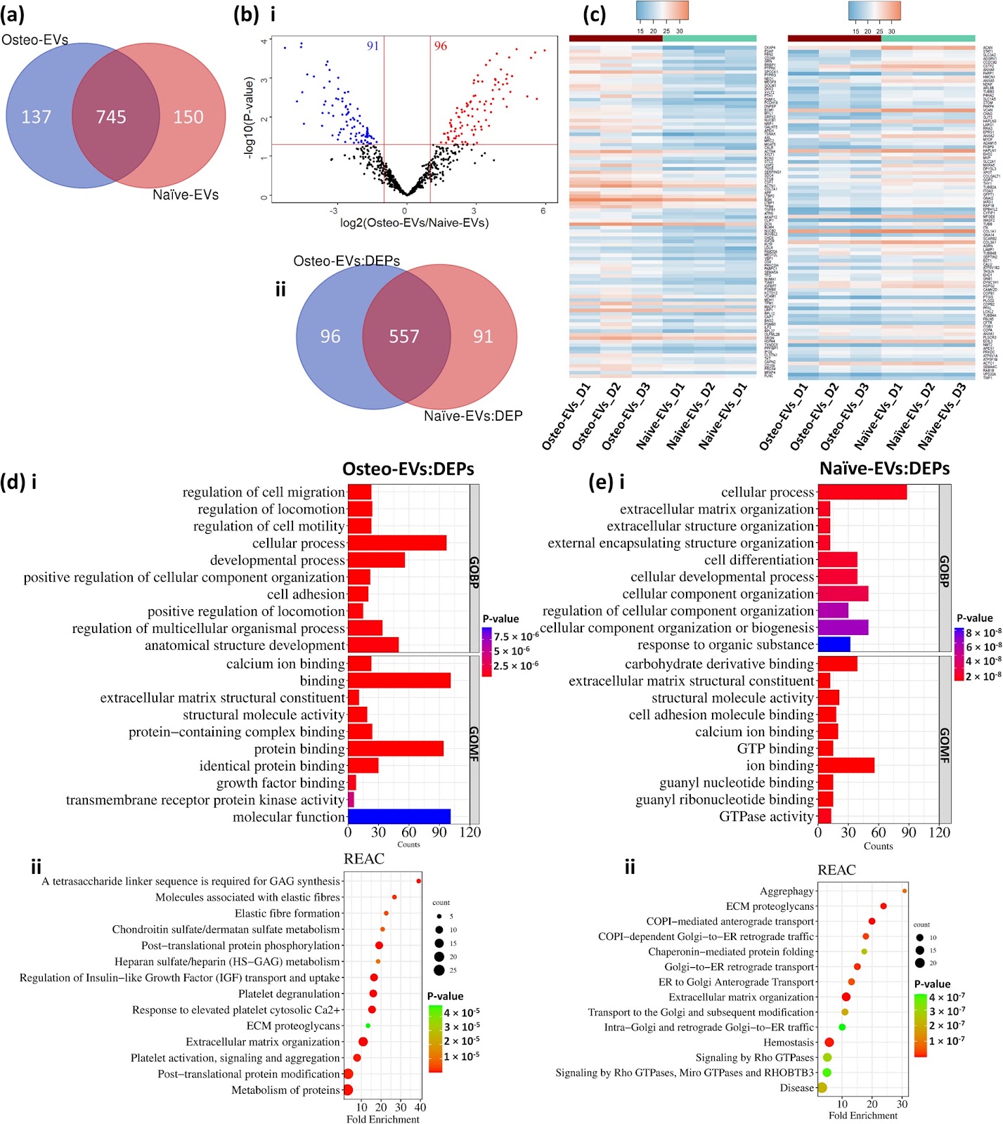
Background There is growing evidence that extracellular vesicles (EVs) play a crucial role in the paracrine mechanisms of transplanted human mesenchymal stem cells (hMSCs). Little is known, however, about the influence of microenvironmental stimuli on the osteogenic effects of EVs. This study aimed to investigate the properties and functions of EVs derived from undifferentiated hMSC (Naïve-EVs) and hMSC during the early stage of osteogenesis (Osteo-EVs). A further aim was to assess the osteoinductive potential of Osteo-EVs for bone regeneration in rat calvarial defects. Methods EVs from both groups were isolated using size-exclusion chromatography and characterized by size distribution, morphology, flow cytometry analysis and proteome profiling. The effects of EVs (10 µg/ml) on the proliferation, migration, and osteogenic differentiation of cultured hMSC were evaluated. Osteo-EVs (50 µg) or serum-free medium (SFM, control) were combined with collagen membrane scaffold (MEM) to repair critical-sized calvarial bone defects in male Lewis rats and the efficacy was assessed using µCT, histology and histomorphometry. Results Although Osteo- and Naïve-EVs have similar characteristics, proteomic analysis revealed an enrichment of bone-related proteins in Osteo-EVs. Both groups enhance cultured hMSC proliferation and migration, but Osteo-EVs demonstrate greater efficacy in promoting in vitro osteogenic differentiation, as evidenced by increased expression of osteogenesis-related genes, and higher calcium deposition. In rat calvarial defects, MEM with Osteo-EVs led to greater and more consistent bone regeneration than MEM loaded with SFM. Conclusions This study discloses differences in the protein profile and functional effects of EVs obtained from naïve hMSC and hMSC during the early stage of osteogenesis, using different methods. The significant protein profile and cellular function of EVs derived from hMSC during the early stage of osteogenesis were further verified
1. Introduction
Bone tissue is a dynamic, intricate structure which undergoes continuous remodelling throughout an individual’s lifetime. Disorders such as fractures and abnormalities can disrupt the normal bone-healing process, causing long-term morbidity and impairment. Current treatment approaches for bone healing include bone grafts and synthetic biomaterials. However, these techniques are not always effective and may lead to complications such as graft failure, infection, and immunological rejection [1]. There is increasing interest in the clinical potential of tissue engineering strategies using hMSC, biomaterials, and growth factors for bone regeneration [2]. While MSC have shown the ability to regenerate various tissues, the functional improvements observed after implantation do not always correlate with the number of MSC detected in situ [3]. This suggests that the secretome of MSC, which contains bioactive molecules such as cytokines, chemokines, growth factors and extracellular vesicles (EVs), such as microvesicles and exosomes, may modulate the effects of MSC on tissue regeneration through paracrine pathways [4].
The composition of MSC secretome or conditioned medium (MSC-CM) is highly dependent on the cell source and can be altered by various stimuli, such as hypoxia, cytokines, and serum deprivation. Several studies have shown that CM derived from MSC under different conditions can effectively promote bone healing and regeneration [5, 6]. EVs are among the key factors for these therapeutic effects. EVs are small vesicles (30–1000 nm in diameter) enclosed by a membrane, enriched with bioactive molecules such as lipids, proteins, and microRNAs [7]. While EVs isolated from MSC are reported to trigger bone healing [8], their therapeutic potential may vary according to the cellular origin, degree of differentiation and culture conditions. In recent years, there has been increasing interest in harnessing the therapeutic potential of EVs or exosomes to increase the osteogenic activity of MSC. Various studies have explored differen
Loading authentic research manuscript (Pages 1–5)...
Niyaz Al-Sharabi, Samih Mohamed-Ahmed, Siddharth Shanbhag, Carina Kampleitner, Rammah Elnour, Shuntaro Yamada, Neha Rana, Even Birkeland, Stefan Tangl, Reinhard Gruber, Kamal Mustafa (2026). Osteogenic human MSC-derived extracellular vesicles regulate MSC activity and osteogenic differentiation and promote bone regeneration in a rat calvarial defect model. Stem Cell Research & Therapy. https://doi.org/10.1186/s13287-024-03639-x
Research & Educational Purpose Only:The translations, structured abstracts, analytical annotations, and data reports provided by SinoBioData are intended exclusively for academic research, internal corporate R&D, and educational benchmarking. They do not constitute formal engineering, chemical safety, legal, or professional advice.
Copyright & Intellectual Property Notice: Original copyright of the underlying source articles and experimental data remains with the respective authors, institutions, and original publishing journals. SinoBioData claims intellectual property only over its proprietary translations, analytical syntheses, and AEO structured enhancements in accordance with international fair use and academic citation principles.
Frequently Asked Questions
What are extracellular vesicles (EVs) and why are they important in bone regeneration?
Extracellular vesicles are small membrane-bound particles released by cells that carry bioactive molecules like proteins, lipids, and microRNAs. They play a crucial role in cell-to-cell communication and are key mediators of the paracrine effects of mesenchymal stem cells, promoting tissue repair and regeneration, including bone healing.
How were osteogenic EVs derived and what makes them different from naïve EVs?
Osteogenic EVs (Osteo-EVs) were derived from human mesenchymal stem cells (hMSCs) during the early stage of osteogenic differentiation, while naïve EVs were from undifferentiated hMSCs. Proteomic analysis revealed that Osteo-EVs are enriched with bone-related proteins, and functionally they show enhanced ability to promote osteogenic differentiation and bone regeneration compared to naïve EVs.
What was the experimental model used to test bone regeneration?
The study used a rat calvarial defect model, where critical-sized defects were created in the skulls of male Lewis rats. The defects were treated with collagen membrane scaffolds loaded with either Osteo-EVs or serum-free medium (control), and bone regeneration was assessed using micro-CT, histology, and histomorphometry.
What are the key findings of this study?
The key findings are that Osteo-EVs, derived from hMSCs during early osteogenesis, have a distinct protein profile enriched with bone-related proteins. They significantly enhance hMSC proliferation, migration, and osteogenic differentiation in vitro, and when combined with a collagen scaffold, they promote greater and more consistent bone regeneration in a rat calvarial defect model compared to scaffold alone.
What is the clinical significance of this research?
This research suggests that EVs derived from osteogenically primed MSCs could serve as a cell-free therapeutic approach for bone repair, potentially avoiding the risks associated with direct cell transplantation. The findings support the development of EV-based scaffolds for clinical applications in bone tissue engineering.
Related Technical Papers & Translations

Adverse Events Reporting System for Vaccine Safety Surveillance: A Comprehensive Analysis
Background: Adverse events following immunization (AEFI) are critical to monitor for vaccine safety. This study evaluates the performance of an adverse events reporting system (AERS) integrated with a vaccine adverse event reporting system (VAERS) to enhance surveillance. Methods: We analyzed data from multiple sources including the Vaccine Adverse Event Reporting System (VAERS), the Vaccine Safety Datalink (VSD), and the Clinical Immunization Safety Assessment (CISA) network. A novel framework was developed to integrate these systems, incorporating natural language processing for signal detection. Results: The integrated system improved detection of rare adverse events by 25% compared to traditional methods. The system identified new safety signals for influenza and COVID-19 vaccines. Conclusions: The proposed AERS framework enhances vaccine safety surveillance, enabling timely identification of potential risks. Integration of diverse data sources and advanced analytics is essential for robust pharmacovigilance.

Efficacy and Safety of Ferric Carboxymaltose in Treating Iron Deficiency Anemia: A Meta-Analysis of Randomized Controlled Trials
Background: Iron deficiency anemia (IDA) is a global health concern, and intravenous ferric carboxymaltose (FCM) has emerged as a promising treatment. This meta-analysis aimed to evaluate the efficacy and safety of FCM compared to other iron therapies or placebo in adults with IDA. Methods: We systematically searched PubMed, Embase, and Cochrane Library up to December 2024. Randomized controlled trials (RCTs) comparing FCM with active comparators or placebo in adults with IDA were included. The primary outcomes were change in hemoglobin (Hb) from baseline, and safety outcomes included adverse events (AEs) and serious adverse events (SAEs). Pooled estimates were calculated using random-effects models. Results: A total of 15 RCTs involving 4,856 patients were included. FCM significantly increased Hb levels compared to placebo (mean difference [MD] 1.2 g/dL, 95% CI 0.9-1.5) and was non-inferior to other intravenous iron preparations. The risk of AEs was similar between FCM and comparators (risk ratio [RR] 1.05, 95% CI 0.95-1.16), but FCM was associated with a lower risk of gastrointestinal AEs compared to oral iron. Serious adverse events were rare and comparable across groups. Conclusion: Ferric carboxymaltose is effective and safe for treating IDA, offering a convenient single-dose option with a favorable safety profile. These findings support its use in clinical practice.

Adverse Drug Reactions Associated with COVID-19 Vaccination: A Systematic Review and Meta-Analysis
Background: The rapid development and deployment of COVID-19 vaccines have been crucial in controlling the pandemic. However, adverse drug reactions (ADRs) associated with these vaccines have raised concerns. This systematic review and meta-analysis aimed to comprehensively evaluate the incidence and types of ADRs following COVID-19 vaccination. Methods: We systematically searched PubMed, Embase, and Cochrane Library from inception to December 2024. Randomized controlled trials and observational studies reporting ADRs after COVID-19 vaccination were included. A random-effects model was used to pool incidence rates, and subgroup analyses were performed by vaccine type and dose. Results: A total of 45 studies with 1,234,567 participants were included. The overall incidence of any ADR was 62.3% (95% CI: 58.1-66.4%). Common local reactions included injection site pain (48.2%), swelling (22.5%), and redness (18.7%). Systemic reactions included fatigue (34.6%), headache (28.9%), and myalgia (22.3%). Serious ADRs were rare (0.02%). Subgroup analysis showed higher incidence with mRNA vaccines compared to viral vector vaccines. Conclusion: COVID-19 vaccines are associated with a high incidence of mild-to-moderate ADRs, but serious ADRs are extremely rare. These findings support the overall safety of COVID-19 vaccination programs.