🧬 SinoBioData Academic Portal
🏛️ Indexed Academic JournalOriginal: 中国组织工程研究

Chinese Journal of Tissue Engineering Research

Premier Chinese Biomedical Journal indexed in SinoBioData: Chinese Journal of Tissue Engineering Research (中国组织工程研究).

Total Research Papers: 200
Access: 100% Free Open Access
Browse by Publication Year & Volume

Published Research Papers

Showing 200 of 200 peer-reviewed papers with full Graphical Abstracts.

Original ResearchVol 1895, Issue 23 • pp. 100-112DOI: 10.12307/2026.21206Jan 15, 2026

Chemokine receptor 7-bone marrow mesenchymal stem cells combined with porcine small intestinal submucosa promote skin repair in rats

Authors: Fan Meirong, Li Guangqi, Song Xumei, Yan Xin, Sui Ruizhi

BACKGROUND: The clinical outcomes of autologous and allogeneic skin transplants, which are commonly used for repairing skin lesions, are often suboptimal. In recent years, advancements in tissue engineering have provided new hope for skin repair. Nevertheless, the regeneration of blood vessels within skin tissue engineering remains a significant challenge. Porcine small intestinal submucosa and bone marrow-derived mesenchymal stem cells are widely utilized natural extracellular matrix biomaterials and seed cells in current tissue engineering research. Chemokine receptor 7 is a cytokine that can promote angiogenesis. OBJECTIVE: To observe the repair effect and angiogenesis ability of chemokine receptor 7-bone marrow mesenchymal stem cells-porcine small intestinal submucosa membrane on rat back skin damage. METHODS: (1) The adenovirus vector overexpressing chemokine receptor 7 was used to transfect bone marrow mesenchymal stem cells. The transfection efficiency was evaluated using western blot assay and RT-qPCR. (2) The bone marrow mesenchymal stem cells overexpressing chemokine receptor 7 were co-cultured with porcine small intestinal submucosa. Cytocompatibility was assessed through scanning electron microscopy and live/dead cell staining. (3) 12 SD rats were utilized to establish a skin defect animal experimental model, and chemokine receptor 7-bone marrow mesenchymal stem cells-porcine small intestinal submucosa (experimental group) and porcine small intestinal submucosa alone (control group) were applied to the skin defects. Wound healing was observed at 1, 3, 7, and 14 days post-modeling. At 7 and 14 days, western blot was used to detect vascular endothelial growth factor protein expression in wound healing tissue, and immunohistochemical staining was used to detect CD31 and proliferating cell nuclear antigen protein expression. RESULTS AND CONCLUSION: (1) Adenovirus-mediated overexpression of chemokine receptor 7 in bone marrow mesenchymal stem cells was successfully constructed. The protein and mRNA expression of chemokine receptor 7 in the transfection group was significantly upregulated compared with the control and empty vector groups (P < 0.001). (2) Scanning electron microscopy and live/dead cell staining showed that bone marrow mesenchymal stem cells overexpressing chemokine receptor 7 grew well on the porcine small intestinal submucosa membrane, indicating good cytocompatibility. (3) Compared with the control group, the wound area in the experimental group was significantly reduced (P < 0.05), and the protein expression of vascular endothelial growth factor, CD31, and proliferating cell nuclear antigen in the wound healing tissue was higher than that in the control group (P < 0.05), indicating that the chemokine receptor 7-bone marrow mesenchymal stem cells-porcine small intestinal submucosa membrane had a strong ability to promote wound angiogenesis.


Chemokine receptor 7-bone marrow mesenchymal stem cells combined with porcine small intestinal submucosa promote skin repair in rats
Graphical Abstract
Original ResearchVol 1895, Issue 23 • pp. 100-112DOI: 10.12307/2026.21207Jan 15, 2026

Function of human amniotic mesenchymal stem cell exosomes in repairing submandibular gland epithelial cells after radiation injury in SD rats

Authors: Zhang Ligang, Liu Tao, Yi Jie, Zhang Nini, Yao Li, Huang Guilin, Hu Xiaohua, Dai Min

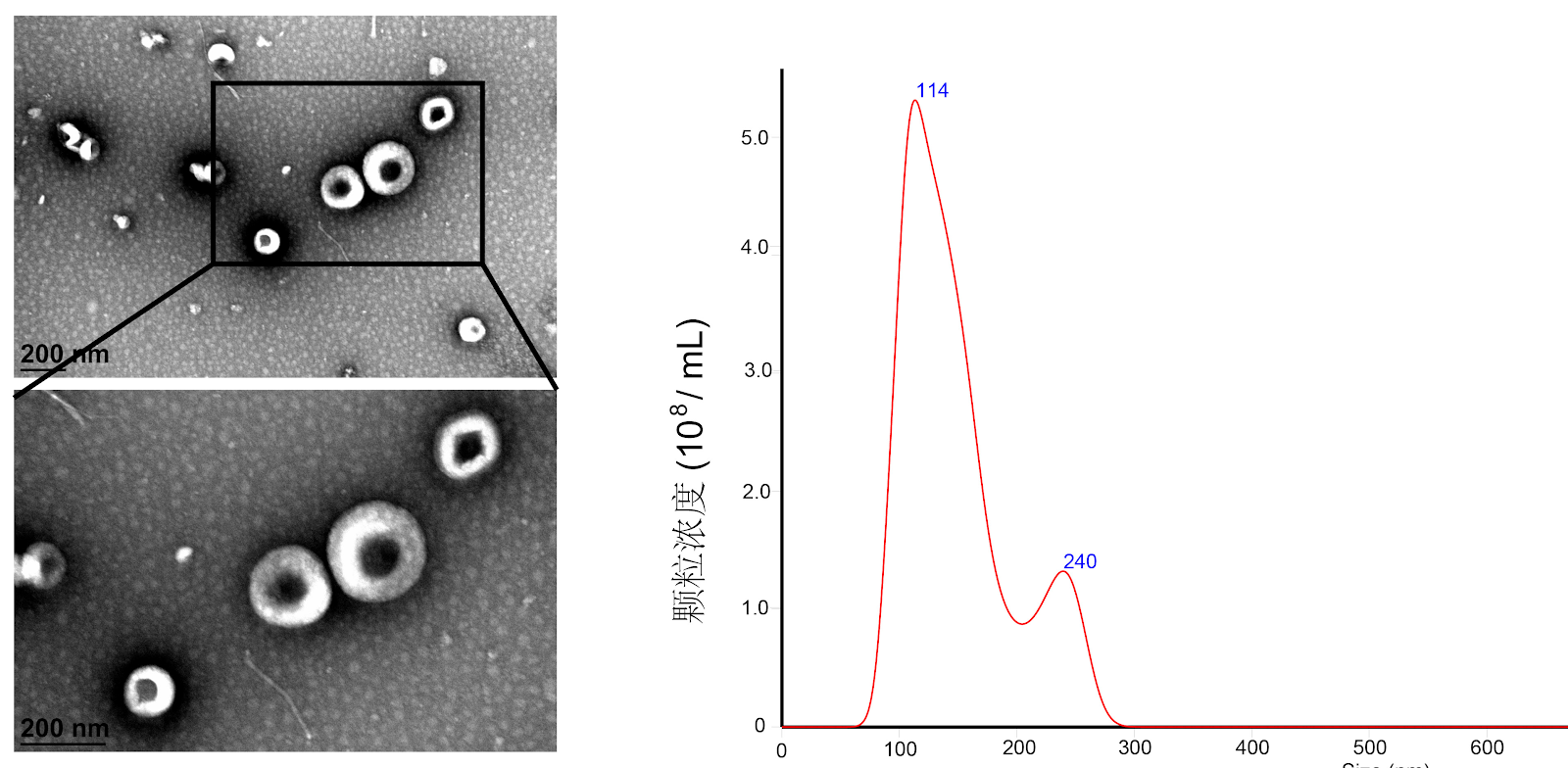
BACKGROUND: Hypoxia preconditioning can increase extracellular vesicles, growth factors, anti-inflammatory and immunosuppressive factors in the parasecretory secretions of mesenchymal stem cells. Exosomes derived from human amniotic mesenchymal stem cells preconditioned with hypoxia are expected to play a better therapeutic role in tissue damage repair. OBJECTIVE: To observe the repair effect of exosomes from human amniotic mesenchymal stem cells preconditioned with hypoxia on radiation-induced submandibular gland epithelial cell damage. METHODS: The passage 3 human amniotic mesenchymal stem cells were divided into two groups: hypoxia and normoxia. They were pretreated with hypoxia (1% O2 by volume) and normoxia (20% O2 by volume) for 48 hours, respectively. Then, normoxic and hypoxic human amniotic mesenchymal stem cell exosomes were extracted from the culture supernatant of human amniotic mesenchymal stem cells by modified ultrahigh-speed centrifugation. The submandibular gland epithelial cells of SD newborn rats were divided into blank control group, radiation control group, normoxia-treated group, and hypoxia-treated group. The submandibular gland epithelial cells were treated with 5 Gy radiation to construct a radiation-induced injury model. The normoxia-treated and hypoxia-treated groups were co-cultured with normoxic or hypoxic exosomes for 3 days. CCK-8 assay was used to detect the proliferation activity of rat submandibular gland epithelial cells; ELISA was used to measure the content of α-amylase in the supernatant; RT-qPCR was used to detect the expression of aquaporin 5 mRNA. RESULTS AND CONCLUSION: The cell proliferation activity in the radiation control group was significantly lower than that in the blank control group (P < 0.05). The cell proliferation activity in the normoxia-treated and hypoxia-treated groups on days 2 and 3 was significantly higher than that in the radiation control group (P < 0.05). The cell proliferation activity in the hypoxia-treated group on day 1 was higher than that in the normoxia-treated group, and showed an increasing trend on days 2 and 3, but there was no statistical difference compared with the normoxia-treated group (P > 0.05). The α-amylase content in the radiation control group was significantly lower than that in the blank control group (P < 0.05). The α-amylase content in the normoxia-treated and hypoxia-treated groups was significantly higher than that in the radiation control group (P < 0.05). Compared with the normoxia-treated group, the α-amylase content in the hypoxia-treated group showed an increasing trend, but there was no statistical difference (P > 0.05). The expression of aquaporin 5 mRNA in the radiation control group was significantly lower than that in the blank control group (P < 0.05). The expression of aquaporin 5 mRNA in the normoxia-treated and hypoxia-treated groups was significantly higher than that in the radiation control group (P < 0.05). The expression of aquaporin 5 mRNA in the hypoxia-treated group was slightly higher than that in the normoxia-treated group, but there was no statistical difference (P > 0.05). The results indicate that both normoxic and hypoxic preconditioned human amniotic mesenchymal stem cell exosomes have a repair effect on radiation-induced submandibular gland epithelial cell damage, but hypoxic exosomes have no obvious advantage.

Function of human amniotic mesenchymal stem cell exosomes in repairing submandibular gland epithelial cells after radiation injury in SD rats
Graphical Abstract
Original ResearchVol 1895, Issue 23 • pp. 100-112DOI: 10.12307/2026.21205Jan 15, 2026

Isolation, cultivation, identification, and induction of M1/M2 polarization in bone marrow-derived macrophages from C57BL/6 mice

Authors: Tan Yuhang, Li Bo, Tang Minghong, Sun Zeyu, Luo Xu

BACKGROUND: Macrophage polarization demonstrates significant potential in disease treatment, particularly in areas such as cancer, inflammation, and autoimmune diseases. Establishing standardized in vitro models can lay the groundwork for in-depth research into the mechanisms of macrophage polarization. OBJECTIVE: To observe the in vitro growth characteristics of bone marrow-derived macrophages from C57BL/6 mice and to establish a standardized in vitro model for M1 and M2 macrophage polarization. METHODS: Femurs and tibias of C57BL/6 mice were aseptically separated, and the contents of the bone marrow cavity were collected. After filtering through a mesh and lysing erythrocytes, the contents were resuspended in high-glucose DMEM containing 20 ng/mL macrophage colony-stimulating factor and inoculated in 6-well plates according to experimental requirements. On day 7, they were differentiated into mature mouse bone marrow-derived macrophages (M0 type). Then, 100 ng/mL lipopolysaccharide was used to induce polarization to M1 type, and 20 ng/mL interleukin-4 was used to induce polarization to M2 type. Flow cytometry and RT-qPCR were used to detect the expression of corresponding markers in macrophages under different polarization states, and Western blot was used to detect the expression of M1 macrophage marker pathway proteins p-STAT1, STAT1 and M2 macrophage marker pathway proteins p-STAT6, STAT6. RESULTS AND CONCLUSION: (1) After stimulation with 20 ng/mL macrophage colony-stimulating factor for 7 days, flow cytometry showed that the positive rate of macrophage surface marker F4/80 reached 98.1%. (2) After stimulation with 100 ng/mL lipopolysaccharide for 6 h, the positive rates of F4/80 and CD86 were about 35%, and RT-qPCR showed that the mRNA expression of M1 macrophage markers inducible nitric oxide synthase, interleukin-6, macrophage inflammatory protein 1α, and monocyte chemoattractant protein 1 were significantly higher than those in the control group (P < 0.01). (3) After stimulation with 20 ng/mL interleukin-4 for 24 h, the mean fluorescence intensity of CD206 was significantly increased, and RT-qPCR showed that the mRNA expression of M2 macrophage markers Chi3l3 (Ym1), interleukin-10, and arginase 1 were significantly higher than those in the control group (P < 0.01). (4) Western blot results showed that lipopolysaccharide-induced M1 macrophage marker pathway protein p-STAT1 was significantly activated; interleukin-4-induced M2 macrophage marker pathway protein p-STAT6 was significantly activated. These results indicate that lipopolysaccharide and interleukin-4 effectively induced polarization of bone marrow-derived macrophages to M1 and M2 types, respectively.

Isolation, cultivation, identification, and induction of M1/M2 polarization in bone marrow-derived macrophages from C57BL/6 mice
Graphical Abstract
Original ResearchVol 1895, Issue 23 • pp. 100-112DOI: 10.12307/2026.21204Jan 15, 2026

Extracellular matrix stiffness affects the proliferation activity of bone marrow stromal stem cells

Authors: GAO Feng, WANG Jiliang, WANG Hongbo, YANG Yongsheng, LIU Yuan, FU Su

BACKGROUND: In tissue engineering bone construction, the physical properties of the scaffold can directly affect the activity and repair effect of seed cells, among which extracellular matrix hardness is a key factor affecting seed cell proliferation activity. Primary cilia and YAP proteins have been shown to be classical mechanoreceptors and downstream transduction factors, which may directly mediate this mechanism. OBJECTIVE: To investigate the regulatory effect of extracellular matrix hardness on the proliferation activity of bone marrow stromal stem cells and the related mechanisms. METHODS: Bone marrow stromal stem cells were passaged and seeded under different hardness of polydimethylsiloxane extracellular matrix conditions (soft, median, and rigid) for culture. Cell proliferation activity was detected using CCK-8 assay. Transcriptional activity of proliferation genes c-myc and CCND1 was measured using qRT-PCR. Activation of Wnt/β-catenin pathway was evaluated using western blot assay. Primary cilia and YAP protein expression levels were evaluated by acetylated α-tubulin and YAP immunofluorescence staining. After passage, bone marrow stromal stem cells were inoculated on polydimethylsiloxane-based membranes of different hardness (soft and hard) for culture. Then siRNA was used to interfere with YAP protein expression. Western blot assay was used to detect YAP, phosphorylated GSK-3β, and β-catenin protein expression. qRT-PCR was used to detect the transcriptional activity of c-myc and CCND1. The length of primary cilia was analyzed after immunofluorescence staining of acetylated α-tubulin. RESULTS AND CONCLUSION: The cell proliferation activity, c-myc and CCND1 transcriptional activity under rigid polydimethylsiloxane conditions were significantly higher than those under soft and median hardness, and the activation of Wnt/β-catenin pathway was stronger. Immunofluorescence staining showed that rigid polydimethylsiloxane induced shortening of primary cilia and increased YAP-positive cells. After siRNA interference of YAP expression, the differences in YAP, phosphorylated GSK-3β, β-catenin protein expression, and c-myc and CCND1 transcriptional activity between groups disappeared, accompanied by the disappearance of primary cilia length differences. The results indicate that extracellular matrix stiffness regulates the proliferation activity of bone marrow stromal stem cells through a novel YAP protein/primary cilia mechanism.

Extracellular matrix stiffness affects the proliferation activity of bone marrow stromal stem cells
Graphical Abstract
Original ResearchVol 1896, Issue 24 • pp. 100-112DOI: 10.12307/2026.21252Jan 15, 2026

Systematic druggable genome-wide Mendelian randomization identifies therapeutic targets for major depressive disorder

Authors: Zhou Menghan, Liu Shuning, Jiang Tao, Sun Zhuangzhuang, Cao Lingling, Su Xin, Yu Cheng, Guo Junpeng

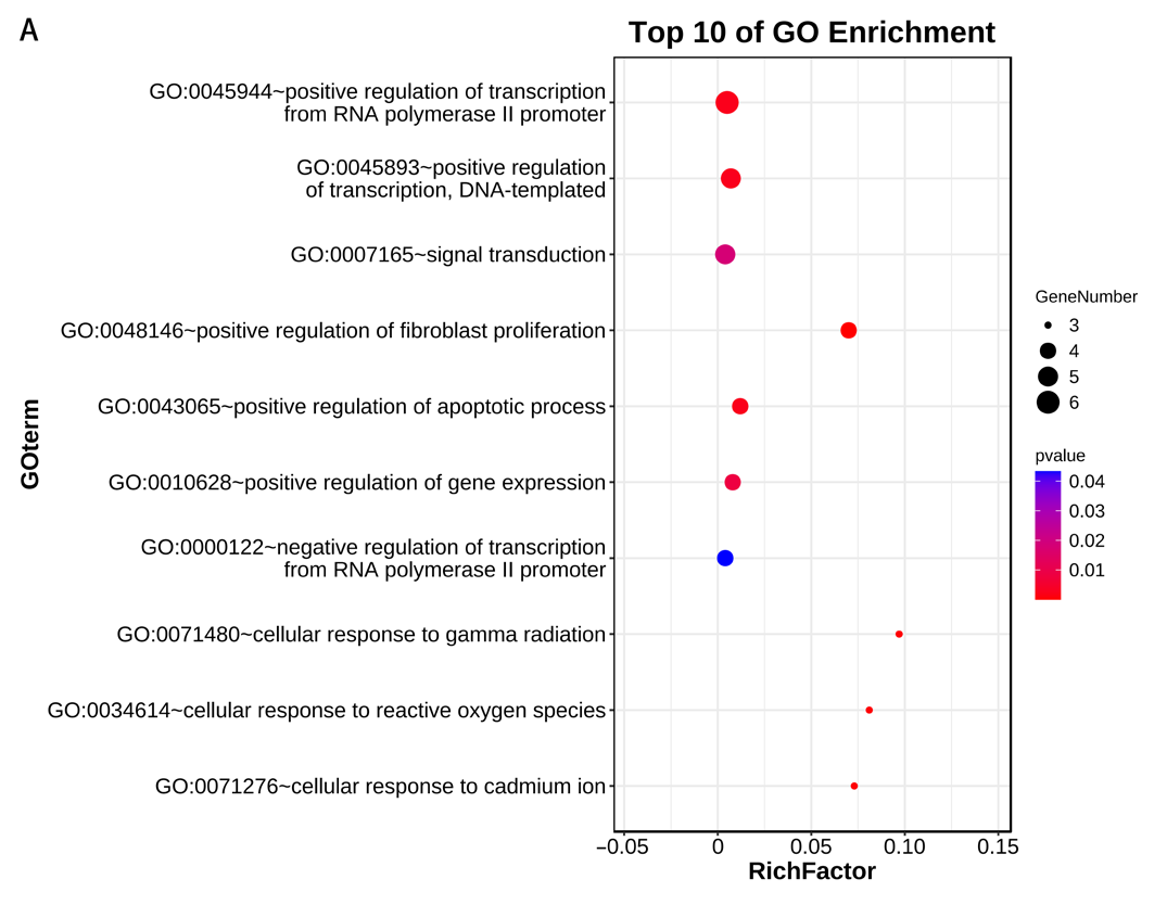
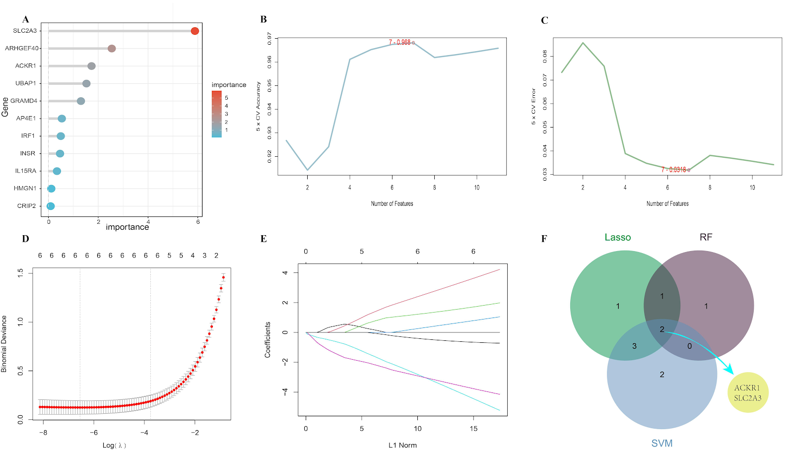
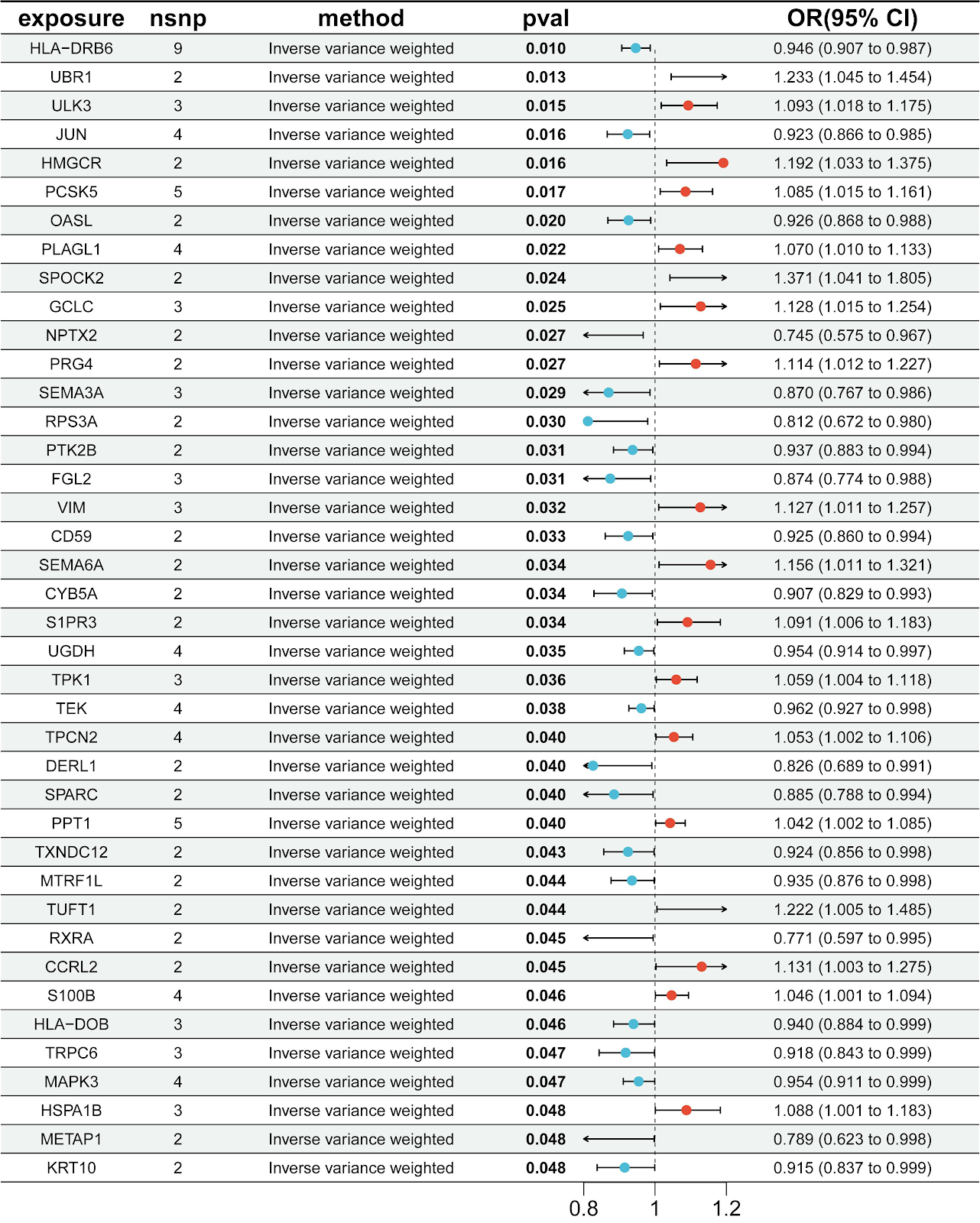
BACKGROUND: The occurrence of major depressive disorder is typically associated with genetic and environmental factors. Currently, the diagnosis of major depressive disorder mainly relies on clinical interviews and symptom assessments, lacking clear and reproducible biological markers. This can lead to misdiagnosis and missed diagnoses, delaying the timing of treatment. OBJECTIVE: To identify druggable genes that may act as potential therapeutic targets for major depressive disorder by conducting comprehensive genome-wide Mendelian randomization analysis. METHODS: By integrating expression quantitative trait locus (eQTL) data and protein quantitative trait locus (pQTL) data from pharmacologically actionable genes with genome-wide association study (GWAS) data on major depressive disorder (including 177 377 cases and 445 321 controls), Mendelian randomization analysis was conducted to identify druggable genes that have a causal relationship with major depressive disorder. Additionally, enrichment analysis, protein-protein interaction network construction, drug target identification, and molecular docking simulations were performed to further explore potential therapeutic strategies. RESULTS AND CONCLUSION: A total of 4 394 druggable genes were analyzed, and 21 druggable genes considerably associated with major depressive disorder were identified. Bayesian colocalization analysis indicated that BTN3A3, CISD1, and PSMB4 had posterior probabilities of hypothesis 4 (H4.abf) > 0.5, supporting the possibility of shared causal variants. GO enrichment analysis mainly involved 'antigen processing and presentation', 'protein degradation and processing', 'mitochondrial outer membrane', and 'immune receptor activity' pathways related to major depression. Protein-protein interaction network analysis showed moderate connectivity among the identified genes (21 nodes, 14 edges). Drug target identification determined gemcitabine (CID 60750), fucose (CID 17106), and isococculidine (CID 2826) as main candidate compounds, which had strong associations with several key genes. Molecular docking analysis revealed stable drug-protein interactions, with isococculidine showing the most stable binding energy (-52.74 kJ/mol) with BTN3A3. In conclusion, Mendelian randomization combined with genomics and structural biology analysis provides valuable decision-making basis for target prioritization and drug repurposing, offering new ideas and directions for efficient utilization of basic research resources and drug development for major depressive disorder.

Systematic druggable genome-wide Mendelian randomization identifies therapeutic targets for major depressive disorder
Graphical Abstract
Original ResearchVol 1896, Issue 24 • pp. 100-112DOI: 10.12307/2026.21243Jan 15, 2026

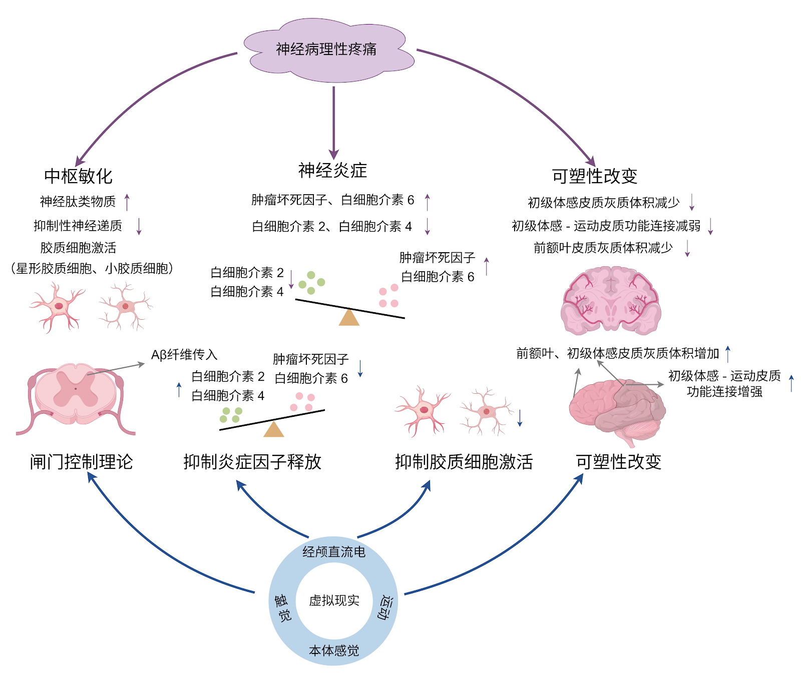
Virtual reality therapy on neuropathic pain following spinal cord injury

Authors: DU Xinyu, ZHAO Donglin, ZHANG Shuyang, LI Shihao, XING Zheng, CHU Xiaolei, LI Qi

BACKGROUND: Virtual reality has demonstrated unique advantages as a novel non-pharmacological intervention in the treatment of neuropathic pain following spinal cord injury. OBJECTIVE: To systematically review the pathogenesis of neuropathic pain following spinal cord injury, summarize the specific application modes and mechanisms of virtual reality therapy for this condition, and explore key factors influencing treatment efficacy, thereby providing novel therapeutic options for patients with neuropathic pain following spinal cord injury. METHODS: A literature search was conducted in the CNKI, PubMed, Web of Science, and Cochrane Library databases up to June 2025. The Chinese and English search terms included “spinal cord injury, central sensitization, cerebral cortex, noninvasive therapy, neuropathic pain, virtual reality.” A total of 1 352 articles were retrieved, and 73 articles that met the inclusion criteria were ultimately included in the analysis. RESULTS AND CONCLUSION: The development of neuropathic pain following spinal cord injury involves complex mechanisms in both the spinal cord and brain, closely associated with central sensitization at the spinal level and abnormal plasticity changes in the brain. Virtual reality, as a novel intervention approach, can be used alone or in combination with other intervention methods, showing unique therapeutic value for the multidimensional pathogenesis of neuropathic pain after spinal cord injury. Standalone virtual reality applications primarily involve virtual walking interventions aimed at restoring abnormal brain plasticity changes, while combined virtual reality applications have shown certain effects in both inhibiting central sensitization and modulating brain plasticity. Although virtual reality intervention shows great potential in the treatment of neuropathic pain following spinal cord injury, current research and applications still have certain limitations. Future efforts should focus on addressing these issues to fully realize the therapeutic value of virtual reality in neuropathic pain following spinal cord injury.

Virtual reality therapy on neuropathic pain following spinal cord injury
Graphical Abstract
Original ResearchVol 1896, Issue 24 • pp. 100-112DOI: 10.12307/2026.21247Jan 15, 2026

The role of exercise-regulated mitophagy in cardiovascular diseases

Authors: JI Long, GONG Guopan, KONG Xiangkui, JIN Pan, CHEN Ziyang, PU Rui

BACKGROUND: Mitophagy plays a crucial regulatory role in the occurrence and development of cardiovascular diseases. Exercise intervention can mediate mitophagy to improve cardiovascular function, which provides new insights for the clinical prevention and treatment of cardiovascular diseases. OBJECTIVE: To summarize the regulatory role of mitophagy in cardiovascular diseases, the influence of exercise on mitophagy, and the mechanism by which exercise-mediated mitophagy improves cardiovascular diseases. METHODS: PubMed and CNKI databases were searched for relevant literature using the search terms of “mitophagy, mitochondrial function, cardiovascular disease, aerobic exercise, resistance training, combined aerobic resistance exercise, high-intensity interval training” in Chinese and English, respectively. Based on the inclusion and exclusion criteria, totally 88 documents were finally included for summary and analysis. RESULTS AND CONCLUSION: (1) Mitophagy plays a crucial role in the regulation of cardiovascular diseases such as heart failure, myocardial hypertrophy, atherosclerosis, and myocardial ischemia-reperfusion injury. Moreover, mitophagy imbalance or disorder can exacerbate the pathological process of cardiovascular diseases. (2) Various exercise modalities can activate mitophagy by regulating the expression of mitophagy-related factors. Among them, aerobic exercise can promote the formation of autophagosomes, thereby enhancing the regulation of mitophagy; resistance exercise can regulate mitochondrial biogenesis; combined aerobic and resistance exercise can further influence mitophagy by promoting lysosomal biogenesis; high-intensity interval training enhances mitophagy function by regulating the expression of mitochondrial dynamics-related proteins. (3) Exercise regulates mitophagy to alleviate myocardial fibrosis, inhibit cardiomyocyte apoptosis, regulate myocardial oxidative stress, and improve endothelial cell function, thereby playing a key role in the prevention and treatment of cardiovascular diseases, providing a new perspective for exercise promoting health and preventing cardiovascular diseases.

The role of exercise-regulated mitophagy in cardiovascular diseases
Graphical Abstract
Original ResearchVol 1896, Issue 24 • pp. 100-112DOI: 10.12307/2026.21242Jan 15, 2026

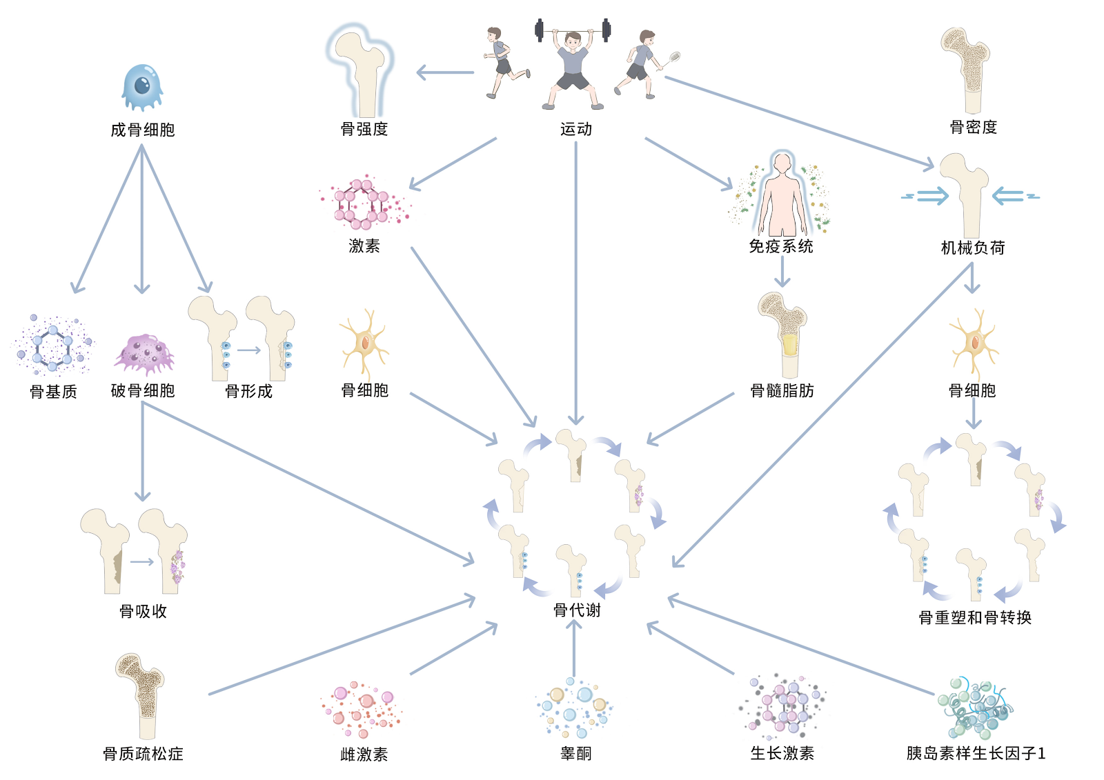
Forkhead box transcription factor O3 affects bone metabolism and participates in the pathological processes of various bone-related diseases

Authors: Han Jie, Hu Tianfa, Wu Yachao, Nong Bin, Yu Kailong

BACKGROUND: Bone metabolism disorders can cause the occurrence of bone-related diseases, and forkhead box transcription factor O3 (FoxO3a) can affect the processes of proliferation, differentiation and apoptosis of bone tissue cells by regulating oxidative stress and autophagy levels, and thereby regulate the bone metabolism. OBJECTIVE: To systematically analyze the relevant research literature on the regulation of bone metabolism by FoxO3a and its mechanism of action in bone diseases and to provide a reference for subsequent studies targeting FoxO3a in the treatment of bone diseases. METHODS: Literature searches were conducted using the following strategies: CNKI (China National Knowledge Infrastructure): SU=FoxO3a OR SU=Foxo3 OR SU=Forkhead box O3 OR SU=AND SU=Forkhead box transcription factor O3) AND SU=bone; WanFang Medical Database: Subject:("FoxO3a") OR Subject:("Foxo3") OR Subject:("Forkhead box O3") OR Subject:("Forkhead box transcription factor O3") AND Subject:("bone"); PubMed: ((FoxO3a) OR (Foxo3) OR (Forkhead box O3))AND ((bone) OR (Skeleton)). Outdated, repetitive, low-quality, and irrelevant studies were excluded, and 56 articles were finally included for review. RESULTS AND CONCLUSION: ①FoxO3a and bone marrow mesenchymal stem cells: FoxO3a can promote the formation of osteogenic lineage and promote early osteogenic differentiation by activating autophagy. Meanwhile, FoxO3a exhibits antioxidant properties in bone marrow mesenchymal stem cells, protecting cells from oxidative stress-induced senescence. ②FoxO3a and osteoblasts: FoxO3a can inhibit osteogenesis by interfering with the Wnt/β-catenin pathway in osteoblasts, while it can activate antioxidant enzymes to protect mature osteoblasts. FoxO3a can promote the proliferation of osteoprogenitor cells and promote osteogenic differentiation by activating autophagy. ③FoxO3a and osteoclasts: FoxO3a expression can resist oxidative stress and activate autophagy to inhibit osteoclastogenesis. ④FoxO3a and osteocytes: FoxO3a can protect osteocytes through antioxidant effects, and can also reduce bone loss by inhibiting p16 and p53 signaling pathways and inhibiting senescence-associated secretory phenotype. ⑤FoxO3a and chondrocytes: FoxO3a plays a protective role in chondrocytes in osteoarthritis, inhibiting chondrocyte breakdown or apoptosis, promoting chondrocyte extracellular matrix synthesis, and inhibiting chondrocyte hypertrophy; however, high co-expression of FoxO3a and Runt-related transcription factor 1 in chondrocytes promotes early chondrogenesis and terminal hypertrophy of chondroprogenitor cells. ⑥FoxO3a affects bone metabolism by participating in processes such as oxidative stress resistance and regulation of autophagy, and participates in the pathological processes of various bone-related diseases.

Forkhead box transcription factor O3 affects bone metabolism and participates in the pathological processes of various bone-related diseases
Graphical Abstract
Original ResearchVol 1896, Issue 24 • pp. 100-112DOI: 10.12307/2026.21248Jan 15, 2026

Transcription factor EB improves Alzheimer’s disease via the autophagy-lysosome pathway

Authors: Hu Yalin, Huang Fengqin, Yang Boyin, Luo Xingmei

BACKGROUND: Studies have confirmed that transcription factor EB and its dependent autophagy-lysosome pathway play a role in the development of various neurodegenerative diseases, including Alzheimer’s disease. OBJECTIVE: To summarize the role of transcription factor EB-mediated autophagy-lysosome pathway in Alzheimer’s disease. METHODS: Electronic databases including PubMed, Web of Science, Cochrane Library, CNKI, WanFang Medical Network, and VIP were searched. The search period was from database inception to January 2025. The search terms were “Alzheimer Disease, AD, Transcription Factor EB, TFEB, Autophagy-lysosome Pathway, Autophagy, Lysosomes, Amyloid beta, Aβ, Tau, Tau protein” in English as well as “Alzheimer’s disease, transcription factor EB, autophagic lysosomes, autophagy, lysosomes, β-amyloid, Tau” in Chinese. Additionally, the references of relevant reviews and grey literature were manually supplemented. A total of 100 articles were finally included for review. RESULTS AND CONCLUSION: Amyloid-β deposition and Tau protein phosphorylation are the key pathological features of Alzheimer’s disease. Abnormalities in the autophagy-lysosome pathway promote the formation of neurotoxic protein aggregates such as amyloid-β and Tau, leading to the clinical manifestations of Alzheimer’s disease characterized by cognitive dysfunction and behavioral abnormalities. Transcription factor EB is a key regulator of the autophagy-lysosome pathway, controlling the transcription of many autophagy-related genes and lysosomal biogenesis. After entering the nucleus, transcription factor EB upregulates the expression of autophagy-lysosome pathway-related genes, significantly increasing the clearance rate of amyloid-β and Tau and significantly reducing neuronal toxicity. Therefore, increasing attention focuses on targeting transcription factor EB to influence autophagy-lysosome biological activity, thereby improving Alzheimer’s disease pathology and behavioral deficits. For example, both pharmacological and non-pharmacological interventions can activate transcription factor EB, reducing amyloid-β deposition and Tau phosphorylation, and improving cognitive function in Alzheimer’s disease. However, sustained activation of transcription factor EB may pose risks such as lysosomal storage disorders, and current intracranial delivery systems have insufficient targeting efficiency. Future development of pathological microenvironment-responsive carriers and combination therapies is needed to achieve precise intervention.

Transcription factor EB improves Alzheimer’s disease via the autophagy-lysosome pathway
Graphical Abstract
Original ResearchVol 1896, Issue 24 • pp. 100-112DOI: 10.12307/2026.21235Jan 15, 2026

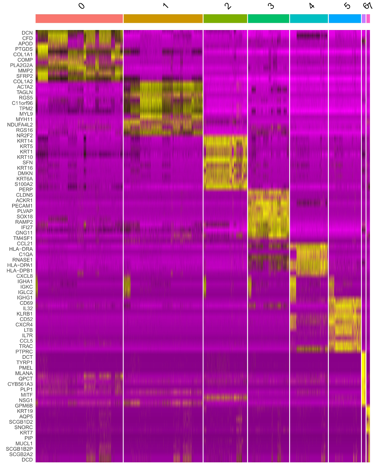
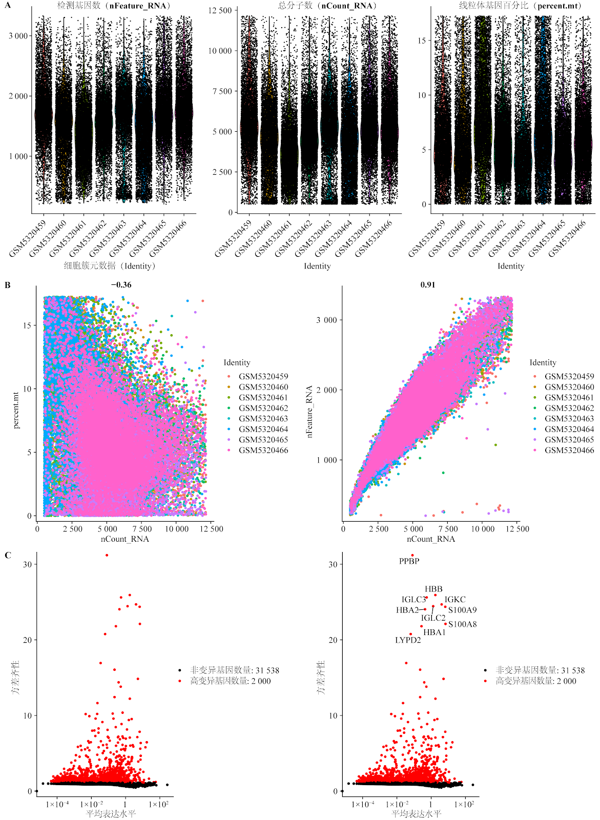
Biomarkers for diabetic foot ulcers: single-cell transcriptomics bioinformatics analysis and experimental validation

Authors: YANG Wenyan, WANG Huayu, YANG Like, PANG Xue, WANG Yutao

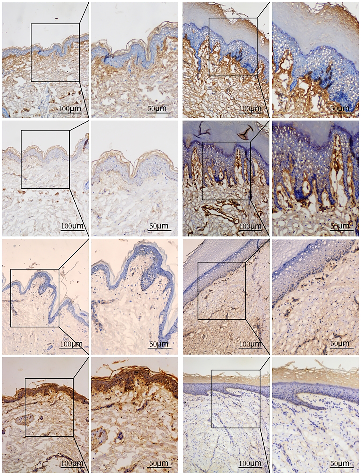
BACKGROUND: Factors such as infection, limb ischemia, and histiocyte activation are involved in diabetic foot ulcers, but the key cell subpopulations influencing diabetic foot ulcer healing remain unclear, and specific biomarkers for diabetic foot ulcers have yet to be identified. Gene Expression Omnibus (GEO) is a publicly accessible database managed by the National Center for Biotechnology Information that stores high-throughput gene expression data, allowing users to freely submit, share, query, and analyze data. Secondary analysis of published data can save research costs and uncover new research targets and ideas. OBJECTIVE: To screen biomarkers for diabetic foot ulcers using single-cell transcriptome and conventional transcriptome bioinformatics analysis, high-dimensional weighted gene co-expression network analysis (hdWGCNA), and weighted gene co-expression network analysis (WGCNA). METHODS: The single-cell transcriptome dataset GSE165816, containing non-healing ulcer tissue samples from diabetic foot ulcer patients and foot skin samples from diabetic patients, was downloaded from GEO. After data quality control, dimensionality reduction, differential analysis, cell type annotation, and pseudotime analysis, cell types spanning the entire course of diabetic foot ulcers were identified, and differentially expressed genes (DEGs) were obtained. hdWGCNA identified gene modules highly correlated with diabetic foot ulcers. Conventional transcriptome datasets GSE68183 and GSE80178, containing non-healing ulcer tissue samples from diabetic foot ulcer patients and foot skin samples from diabetic patients, were downloaded for differential analysis to screen DEGs, and WGCNA was used to identify diabetic foot ulcer-related gene modules. The DEGs from single-cell transcriptome, DEGs from conventional transcriptome samples, and module genes from WGCNA and hdWGCNA were integrated to screen biomarkers for diabetic foot ulcers. The GSE134431 dataset was downloaded as a validation conventional transcriptome dataset, and the expression levels of diabetic foot ulcer biomarkers were compared in single-cell transcriptome and validation conventional transcriptome datasets. Diabetic and diabetic foot ulcer rat models were replicated, wound tissue was collected, and immunohistochemistry and western blot were used to detect biomarker expression levels. RESULTS AND CONCLUSION: Single-cell transcriptome data analysis showed that epithelial cell differentiation spanned the entire pathological process of diabetic foot ulcers. A total of 146 DEGs were obtained from single-cell transcriptome between groups, including 59 upregulated and 87 downregulated DEGs. hdWGCNA identified 19 gene modules related to diabetic foot ulcers, containing 476 core genes. Conventional transcriptome data analysis yielded a total of 913 DEGs, including 343 upregulated and 570 downregulated DEGs. WGCNA obtained 19 diabetic foot ulcer-related gene modules, containing 887 genes. Two biomarkers for diabetic foot ulcers were screened: S100A14 and SFN. The expression levels of these two genes in diabetic foot ulcer samples were higher than those in diabetic foot skin samples in both single-cell transcriptome and validation conventional transcriptome datasets. Animal experiments showed that the expression levels of S100A14 and SFN in wound tissue of diabetic foot ulcer rats were higher than those in back skin tissue of diabetic rats. The results indicate that the pathological process of diabetic foot ulcers involves multiple cell types, among which epithelial cells are the key cell subpopulation. S100A14 and SFN are significantly upregulated in diabetic foot ulcer samples and are potential targets for the treatment of diabetic foot ulcers.

Biomarkers for diabetic foot ulcers: single-cell transcriptomics bioinformatics analysis and experimental validation
Graphical Abstract
Original ResearchVol 1896, Issue 24 • pp. 100-112DOI: 10.12307/2026.21250Jan 15, 2026

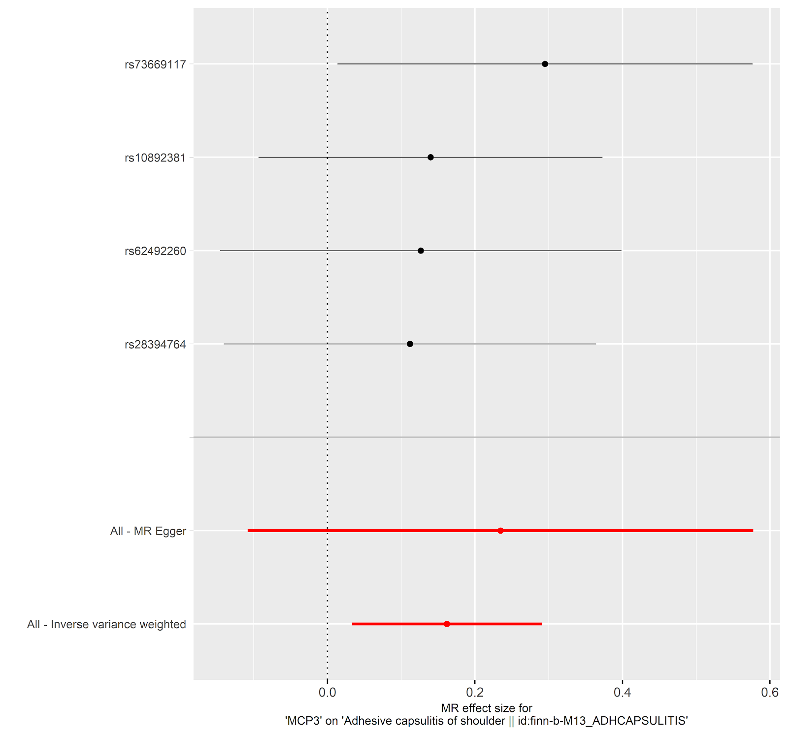
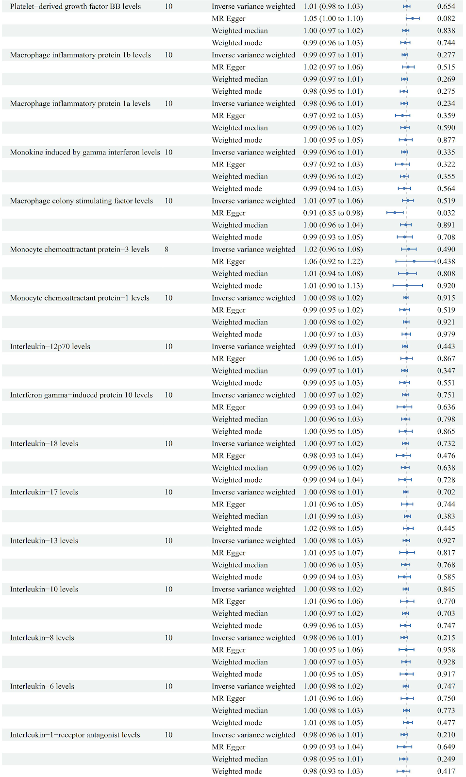
The relationship between inflammatory cytokines and frozen shoulder: a large-sample analysis of the European population based on the FinnGen GWAS database

Authors: Yan Wei, Kong Lingjun, He Tianxiang, Zhu Qingguang, Xi Xiaobing, Fang Min

BACKGROUND: Frozen shoulder is a common disease in orthopedics, but there is no specific clinical indicator for diagnosis. There is a significant association between inflammatory cytokines and frozen shoulder, but the specific causal relationship is not yet clear. This study used summary statistical data from genome-wide association studies (GWAS) for Mendelian randomization analysis. GWAS data are based on large-sample genetic variation information, which can reduce environmental confounding factors and more reliably infer the causal relationship between inflammatory cytokines and frozen shoulder, making up for the limitation that traditional observational studies cannot determine causal associations. OBJECTIVE: To explore the causal relationship between inflammatory cytokines and the onset of frozen shoulder using bidirectional two-sample Mendelian randomization. METHODS: Using summary statistics from GWAS in the FinnGen database, we analyzed the causal relationship between 41 inflammatory cytokines and frozen shoulder. The FinnGen database, jointly initiated by the Finnish National Institute for Health and Welfare (THL), the University of Helsinki, and other Finnish research institutions, includes 2,942 cases and 167,641 European-ancestry controls, integrating genomic, clinical phenotype, and biochemical indicator data from hundreds of thousands to millions of individuals, supporting genetic association studies of diseases. This study is based on publicly available summary statistics databases and does not require ethical approval. Bidirectional Mendelian randomization analyses were performed using inverse variance weighting, weighted median, weighted model, simple model, MR-Egger regression, and sensitivity analyses (including MR-Egger, MR-PRESSO, Cochran's Q test). RESULTS AND CONCLUSION: Monocyte chemoattractant protein-3 (MCP-3) showed significant causal effects in both directions. In the forward analysis, MCP-3 was positively associated with frozen shoulder risk (OR=1.176, 95%CI: 1.034-1.338, P=0.014); in the reverse analysis, frozen shoulder was negatively associated with MCP-3 levels (OR=0.782, 95%CI: 0.625-0.979, P=0.032). Additionally, a significant association was found between tumor necrosis factor beta (TNF-β) and frozen shoulder risk (OR=1.126, 95%CI: 1.002-1.264, P=0.046); in the reverse analysis, stromal cell-derived factor 1 alpha (SDF-1α) was also significantly associated with frozen shoulder risk (OR=1.1, 95%CI: 1.011-1.196, P=0.028), indicating reliable correlations of TNF-β and SDF-1α with frozen shoulder. This bidirectional Mendelian randomization study reveals a complex interaction between MCP-3 and frozen shoulder, suggesting that MCP-3 may serve as a potential therapeutic target. Furthermore, the study indicates that TNF-β is associated with frozen shoulder risk and may be a potential risk factor; while frozen shoulder is also associated with elevated SDF-1α levels, and SDF-1α has the potential to become a diagnostic marker for frozen shoulder. However, further research is needed to elucidate the biological mechanisms underlying these causal relationships. Additionally, the analysis of international databases provides candidate molecules and causal inference paradigms for Chinese research, but it needs to be combined with local data for precise translation.

The relationship between inflammatory cytokines and frozen shoulder: a large-sample analysis of the European population based on the FinnGen GWAS database
Graphical Abstract
Original ResearchVol 1896, Issue 24 • pp. 100-112DOI: 10.12307/2026.21251Jan 15, 2026

Shared genetic basis and causal relationship between nutrition, nutritional status and inflammatory bowel disease

Authors: Liao Guibin, Wu Yixuan, Tang Jing, Huang Jinke, Wang Jun, Yan Ziqi, Liu Shujun, Zhang Haiyan

BACKGROUND: Inflammatory bowel disease, encompassing ulcerative colitis and Crohn’s disease, is a chronic condition linked to malnutrition, sarcopenia, and disease severity, with limited research on their genetic associations. OBJECTIVE: To systematically explore the common genetic basis and causal relationships between nutrition, nutritional status, and inflammatory bowel disease using advanced statistical genetics. METHODS: Single nucleotide polymorphism data for nutritional markers (minerals, vitamins, albumin, hemoglobin, fatty acids) and sarcopenia traits (appendicular lean mass and hand grip strength) were obtained from the GWAS Catalog, and inflammatory bowel disease and its subtypes from the FinnGen database R10. Advanced statistical genetics methods, including linkage disequilibrium score regression, cross-phenotype association analysis, and Mendelian randomization, were used to infer associations. RESULTS AND CONCLUSION: Significant genetic correlations were found: vitamin D with inflammatory bowel disease (rg=-0.080, P=0.029) and ulcerative colitis (rg=-0.087, P=0.027); appendicular lean mass with inflammatory bowel disease (rg=-0.100, P=0.0002), ulcerative colitis (rg=-0.100, P=0.0002), and small intestine Crohn’s disease (rg=-0.081, P=0.035); hand grip strength with small intestine Crohn’s disease (rg=-0.125, P=0.035). Mendelian randomization indicated a positive causal effect of magnesium levels on inflammatory bowel disease (OR=1.41, P=0.036) and small intestine Crohn’s disease (OR=1.78, P=0.035). Cross-phenotype analysis identified shared single nucleotide polymorphisms, particularly in the human leukocyte antigen region, affecting both nutritional status and inflammatory bowel disease. These findings further explain the genetic link between nutrition, sarcopenia, and inflammatory bowel disease, suggesting that targeted nutritional management may be key to slowing disease progression. This study provides new perspectives for personalized treatment and has potential implications for prevention strategies of inflammatory bowel disease.

Shared genetic basis and causal relationship between nutrition, nutritional status and inflammatory bowel disease
Graphical Abstract
Original ResearchVol 1896, Issue 24 • pp. 100-112DOI: 10.12307/2026.21246Jan 15, 2026

Role of fibrosis in tissue injury repair

Authors: LI Feihong, WANG Linrong, CHENG Leping

BACKGROUND: Fibrosis results from dysregulated tissue healing, characterized by abnormal proliferation of fibroblasts and excessive deposition of extracellular matrix, affecting nearly all organs including liver, kidney, heart, lung, and skin. OBJECTIVE: To summarize fibrosis-related diseases such as liver, kidney, cardiac, and pulmonary fibrosis, focusing on the major abnormal cells, signaling pathways, and therapeutic approaches. METHODS: PubMed and CNKI were searched using English terms "fibrosis, fibroblasts, fibrotic organs, extracellular matrix, tissue repair, inflammatory response" and Chinese equivalents. After screening according to inclusion and exclusion criteria, 200 articles were included for review. RESULTS AND CONCLUSION: Key abnormal cells in fibrosis include immune cells (macrophages, neutrophils, lymphocytes), fibroblasts, epithelial cells, and endothelial cells, with fibroblasts playing a central role. Major abnormal pathways include TGF-β, Wnt/β-catenin, Notch, TLR4/MyD88/NF-κB, and Hippo/YAP signaling, whose dysregulation drives fibrosis. Epigenetic modifications (DNA methylation, histone modification, non-coding RNA regulation) modulate fibrosis progression. Anti-fibrotic therapies include pharmacological, cellular, and gene-based approaches, targeting signaling pathways to inhibit persistent fibroblast activation or modulating extracellular matrix deposition to alleviate fibrosis and improve organ function.

Role of fibrosis in tissue injury repair
Graphical Abstract
Original ResearchVol 1895, Issue 23 • pp. 100-112DOI: 10.12307/2026.21203Jan 15, 2026

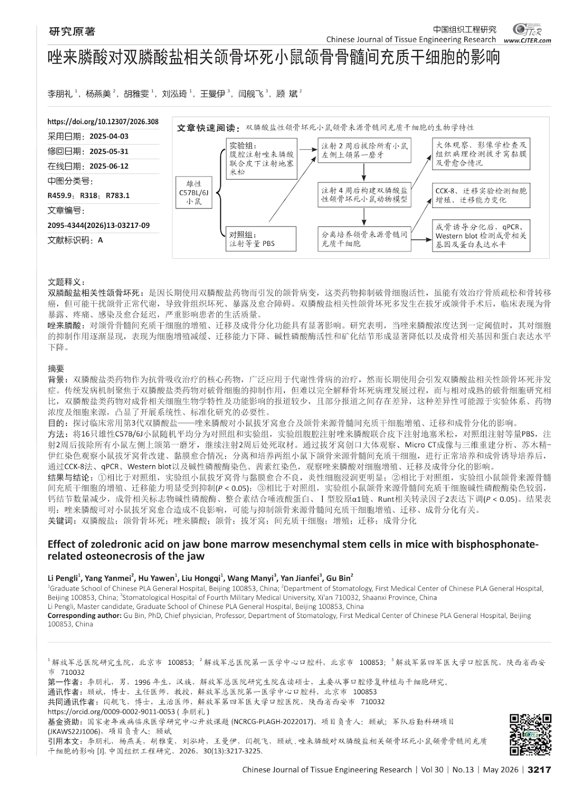
Effect of zoledronic acid on jaw bone marrow mesenchymal stem cells in mice with bisphosphonate-related osteonecrosis of the jaw

Authors: LI Pengli, YANG Yanmei, HU Yawen, LIU Hongqi, WANG Manyi, YAN Jianfei, GU Bin

BACKGROUND: Bisphosphonates, as the core drugs of anti-bone resorption therapy, are widely used in the treatment of metabolic bone diseases. However, long-term use can cause the complications of bisphosphonate related osteonecrosis of the jaw. The traditional pathogenesis focuses on the inhibitory effect of bisphosphonates on osteoclasts, but it is difficult to fully explain the pathological development of osteonecrosis. Compared with the relatively mature osteoclast research, there are fewer reports on the effects of bisphosphonates on the biological characteristics and functions of osteoblast-related cells, and there are differences between some reports. This difference may be due to the experimental system, drug concentration and cell source, highlighting the necessity of conducting systematic and standardized research. OBJECTIVE: To investigate the effect of the third-generation bisphosphonate-zoledronic acid commonly used in clinical practice on the healing of tooth extraction sockets and the proliferation, migration and osteogenic differentiation of bone marrow mesenchymal stem cells derived from the jaw in mice. METHODS: Sixteen male C57BL/6J mice were randomly divided into control and experimental groups. The experimental group received intraperitoneal injection of zoledronic acid combined with subcutaneous injection of dexamethasone, while the control group received an equal volume of PBS. After 2 weeks of injection, the left maxillary first molars of all mice were extracted, and after another 2 weeks of injection, the mice were sacrificed. The healing of extraction sockets was evaluated by gross observation, Micro CT imaging and three-dimensional reconstruction, and hematoxylin-eosin staining. Jaw bone marrow mesenchymal stem cells were isolated and cultured from both groups. After normal culture and osteogenic induction, cell proliferation, migration, and osteogenic differentiation were assessed by CCK-8 assay, qPCR, Western blot, alkaline phosphatase staining, and alizarin red staining. RESULTS AND CONCLUSION: Compared with the control group, the experimental group showed poor healing of extraction sockets with more inflammatory cell infiltration. The proliferation and migration abilities of jaw bone marrow mesenchymal stem cells were significantly inhibited in the experimental group (P < 0.05). Alkaline phosphatase staining was weaker, calcium nodule formation was reduced, and the expression of osteogenic markers (alkaline phosphatase, integrin-binding sialoprotein, collagen type I alpha 1 chain, Runt-related transcription factor 2) was downregulated in the experimental group (P < 0.05). These results indicate that zoledronic acid can adversely affect extraction socket healing, possibly by inhibiting the proliferation, migration, and osteogenic differentiation of jaw bone marrow mesenchymal stem cells.

Effect of zoledronic acid on jaw bone marrow mesenchymal stem cells in mice with bisphosphonate-related osteonecrosis of the jaw
Graphical Abstract
Original ResearchVol 1896, Issue 24 • pp. 100-112DOI: 10.12307/2026.21244Jan 15, 2026

Strategies for the application of miRNA-targeted therapy in the treatment of osteoporosis

Authors: WU Lingjie, ZHENG Kaiyuan, WANG Guangrong, YIN Chong

BACKGROUND: Studies have shown that miRNAs, as important post-transcriptional regulators of genes, play a key role in the onset and progression of osteoporosis. Through in-depth research on the biology of miRNA regulation of osteoporosis, its potential healing mechanisms have been revealed, and this field has become a hot focus of current research. OBJECTIVE: To explore the regulatory role of miRNAs in the development of osteoporosis and their molecular mechanisms, and to provide an overview of the key difficulties encountered in the therapeutic strategies for osteoporosis targeting miRNAs and their solutions. METHODS: We searched PubMed, Web of Science and CNKI databases for relevant literature published up to March 2025. The search terms were “miRNA, osteoporosis, angiogenesis, osteogenesis, gene therapy, drug delivery” in English and “miRNA, osteoporosis, gene therapy, ribonucleic acid drugs, delivery carrier” in Chinese. After reading the titles and abstracts for preliminary screening, we excluded the literature with poor relevance, old information, or repetitive views and lack of authority, and finally included 138 papers for review. RESULTS AND CONCLUSION: (1) miRNAs are highly efficient non-coding RNAs with a wide range of applications that can precisely regulate cellular activities, and they show significant therapeutic potential in regulating osteoblast function and bone angiogenesis. (2) Although miRNA-based targeted drugs have entered preclinical research in other disease areas, clinical translation still faces challenges of insufficient nucleic acid stability in vivo and off-target effects. (3) To address these challenges, researchers have proposed various strategies, including precise targeting of miRNA target genes to reduce off-target effects; chemical modification to improve the stability of nucleic acid drugs in vivo; reducing nucleic acid production costs to advance research; and utilizing viral vectors, exosomes, and various biomaterials to optimize nucleic acid drug delivery routes. (4) Advances in technology continue to innovate in improving the performance of nucleic acid drug carriers, and in the future, precise and efficient drug delivery and targeted therapeutic effects will be achieved.

Strategies for the application of miRNA-targeted therapy in the treatment of osteoporosis
Graphical Abstract
Original ResearchVol 1896, Issue 24 • pp. 100-112DOI: 10.12307/2026.21249Jan 15, 2026

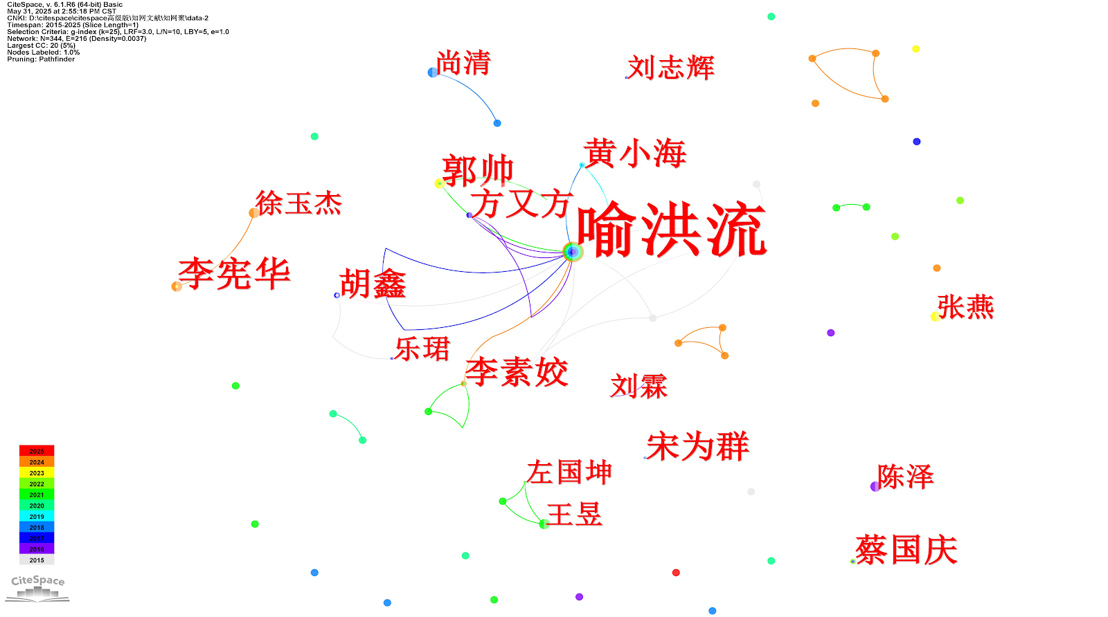
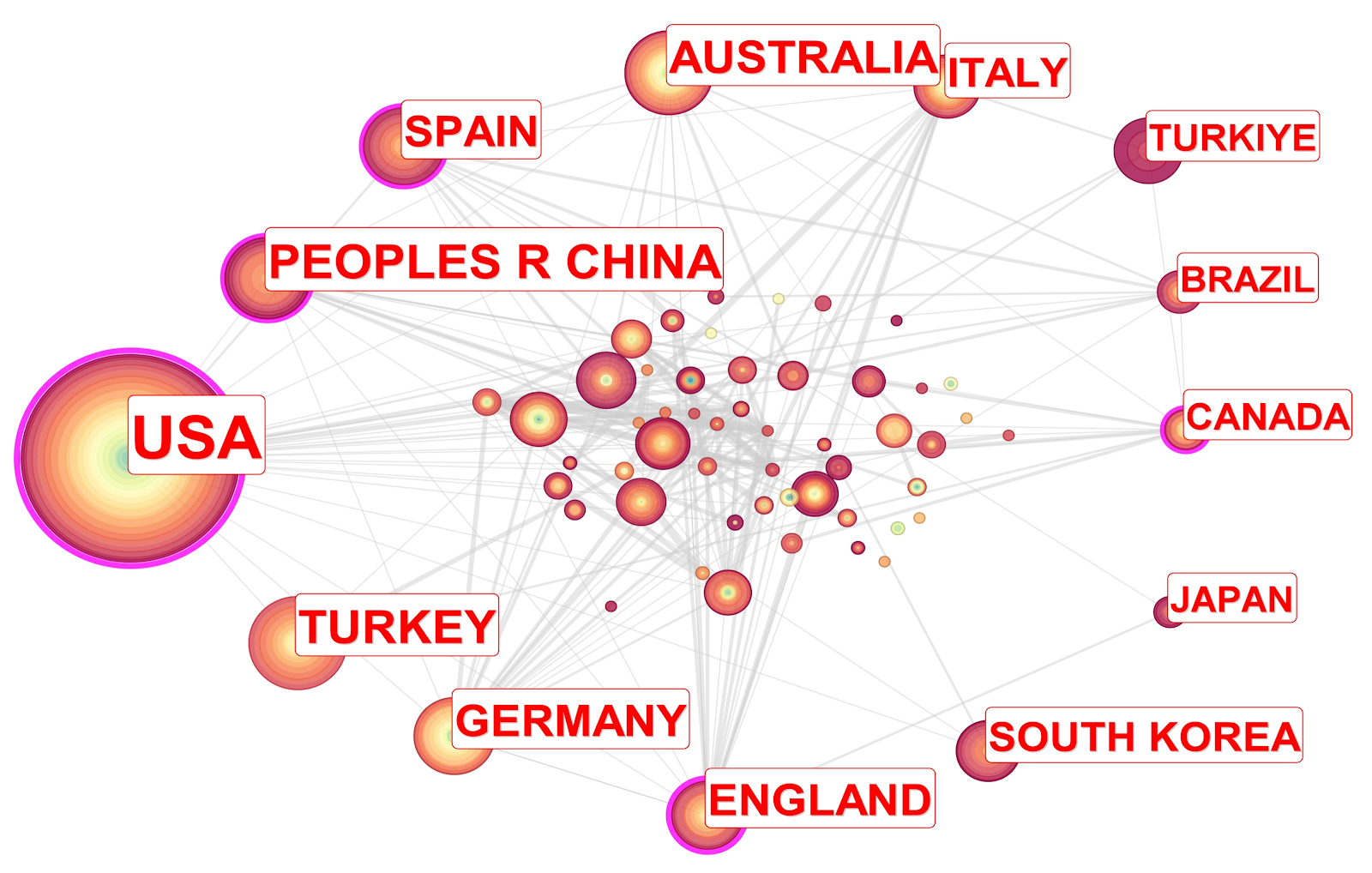
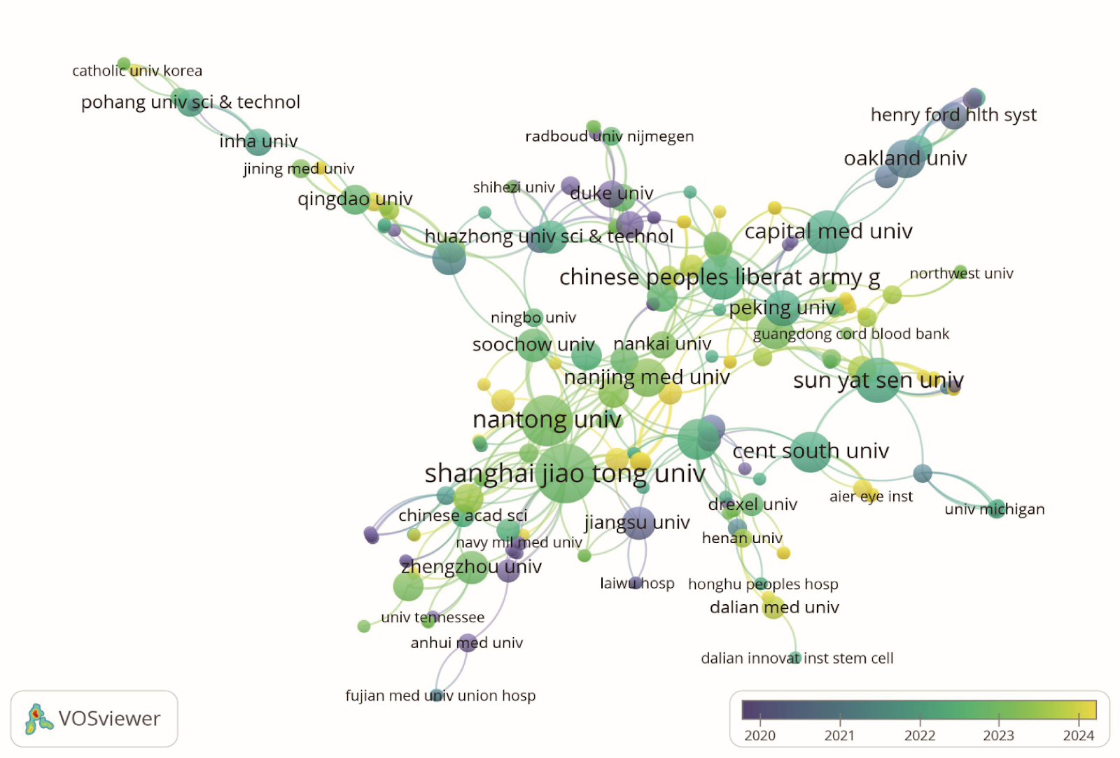
Visualization analysis of dynamic evolution of hot topics in the field of physical activity and neural plasticity

Authors: Deng Qing, Zhang Yeting, Mou Xiangqian, Wang Qingjun

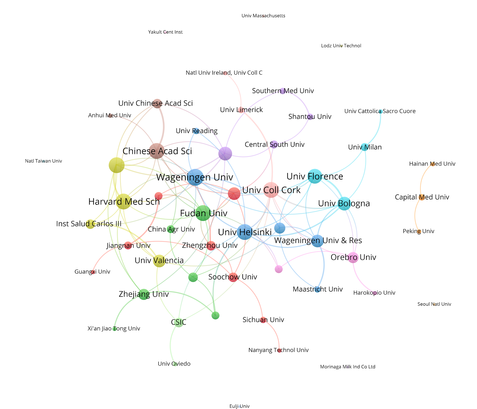
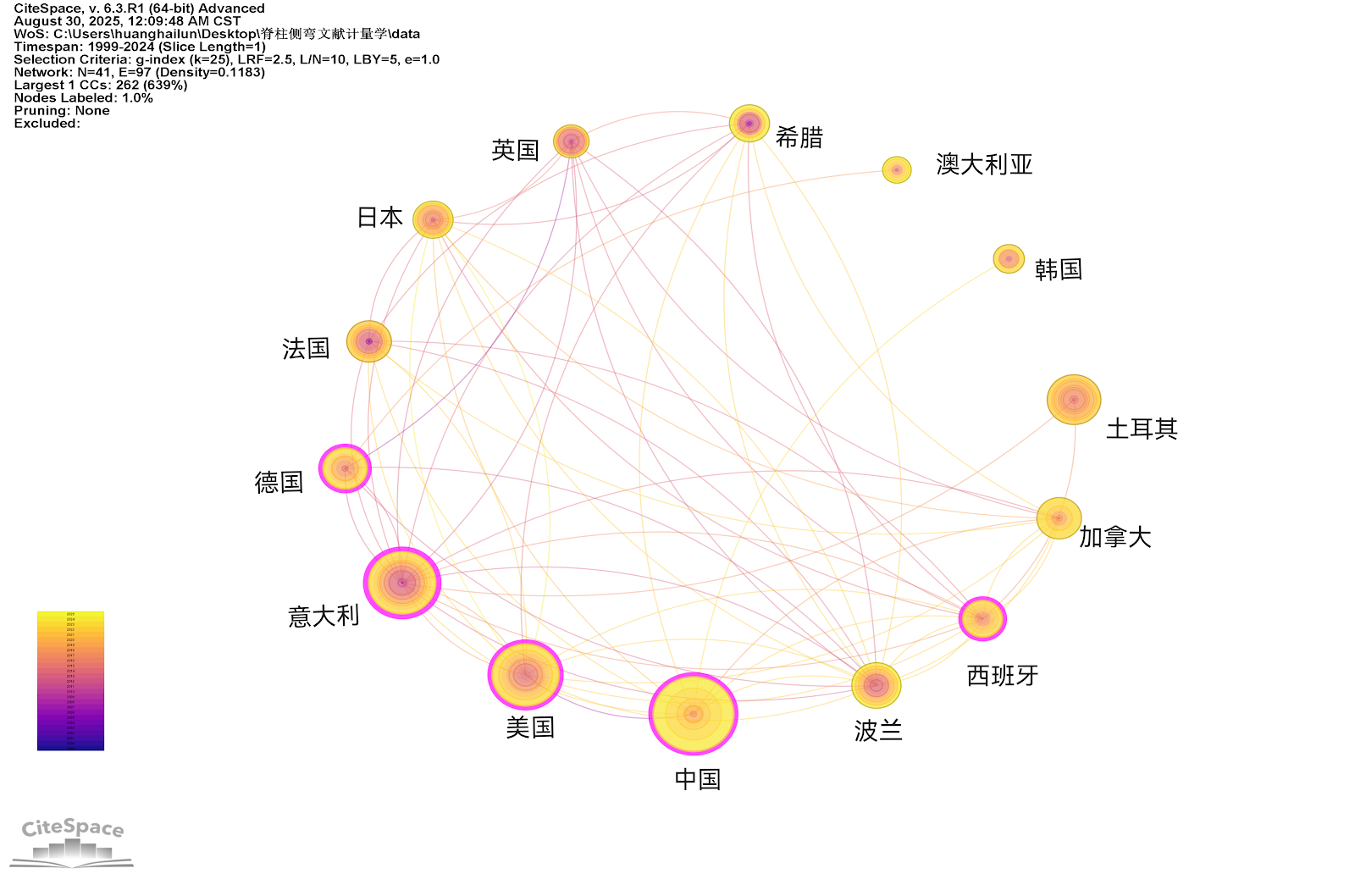
BACKGROUND: In recent years, numerous studies have shown that physical activity significantly promotes neural plasticity, contributing to improvements in cognitive abilities and neurological health. However, the current state and dynamic evolution of the research field on "physical activity and neural plasticity" have not been systematically organized and analyzed. This lack of comprehensive understanding may hinder accurate predictions regarding the future development trends in this area. OBJECTIVE: Using bibliometric methods, we analyzed the dynamic evolution trajectory of research topics in the field of physical activity and neural plasticity, organized the development trends and the evolution of the knowledge framework in this area, and provided directional references for subsequent research. METHODS: Relevant literature was retrieved from the Web of Science Core Collection database (www.webofscience.com) using the following search strategy: TS=(("physical activity" OR exercise OR "motor activity" OR "physical exercise" OR "aerobic exercise" OR "physical training") AND ("neuroplasticity" OR "brain plasticity" OR "neural plasticity" OR "cognitive plasticity" OR "brain adaptability")). A total of 2,098 eligible articles were included. Co-word analysis and visualization were performed using SciMAT software to generate keyword topic overlay maps, strategic coordinate maps, and topic evolution path maps, revealing the dynamic evolution process of research topics in this field. RESULTS AND CONCLUSION: (1) Research in the field of physical activity and neural plasticity is flourishing, with increasingly in-depth studies and ample room for future development. (2) The field comprises five main evolution directions: "physical activity", "adult rats", "stem cells", "individual differences", and "randomized controlled trials", forming 15 sub-evolution paths. (3) Topics such as "older adults", "animal models", and "disease" may become future research focuses. (4) Therefore, it is recommended that future research employ randomized controlled trials and optimized animal model designs to investigate the long-term effects and mechanisms of physical activity on neural plasticity and cognitive function, and focus on clinical validation of multimodal interventions in special populations.

Visualization analysis of dynamic evolution of hot topics in the field of physical activity and neural plasticity
Graphical Abstract
Original ResearchVol 1896, Issue 24 • pp. 100-112DOI: 10.12307/2026.21245Jan 15, 2026

Postbiotic targeting muscle aging: mechanistic insights and application prospects of urolithin A

Authors: YANG Zijiang, GUO Chenggen, DENG Ziao, XUE Xinxuan

BACKGROUND: Urolithin A is a natural active compound produced by the metabolism of dietary polyphenols, which has multiple biological effects such as promoting mitochondrial function, antioxidation and anti-inflammation. In recent years, the development of urolithin A in delaying aging-related diseases has received extensive attention. However, the specific mechanism of its action in improving muscle aging remains unclear and further systematic research is still needed. OBJECTIVE: To systematically explore the mechanism of action of urolithin A in muscle aging, providing a theoretical basis for its potential application value in delaying muscle aging. METHODS: The Web of Science, PubMed, China National Knowledge Infrastructure (CNKI) and WanFang Database were retrieved from January 2000 to April 2025. The search terms were "urolithin A, muscle aging, mitochondrial function, mitophagy, inflammation, oxidative stress, muscle function, skeletal muscle" in English and Chinese. According to the inclusion and exclusion criteria, 80 literatures were finally selected for review. RESULTS AND CONCLUSION: (1) Urolithin A is a metabolite generated by the conversion of dietary polyphenolic compounds ellagic acid and ellagic acid under the action of intestinal microorganisms, and is widely present in pomegranates, berries and nuts. It has a unique α-benzocoumarin structure, with a small molecular weight, strong lipophilicity and easy absorption. The production of urolithin A depends on an individual’s intestinal microbiota and can be classified into different metabolic types. Moreover, this ability weakens with age, reflecting changes in intestinal function and physiological state. (2) Muscle aging is a process driven by mitochondrial dysfunction, chronic inflammation, and neuromuscular degeneration. With age, mitochondrial energy metabolism declines, reactive oxygen species accumulation aggravates cellular damage; chronic low-grade inflammation accelerates protein breakdown, inhibits synthesis, and impairs muscle repair; neuromuscular junction degeneration and signal transduction disorders lead to muscle denervation and atrophy. These factors interact, leading to a continuous decline in muscle mass and function. (3) Urolithin A improves muscle aging through multi-target mechanisms, mainly including: activating PTEN-induced kinase 1/Parkin signaling pathway to clear damaged mitochondria and enhance metabolic functions such as tricarboxylic acid cycle, fatty acid oxidation, and oxidative phosphorylation; inhibiting nuclear factor κB and protein kinase B/mitogen-activated protein kinase signaling pathways, upregulating anti-inflammatory factors such as interleukin-10 and transforming growth factor β1, downregulating pro-inflammatory factors such as interleukin-1β and tumor necrosis factor α, achieving inflammatory regulation; enhancing antioxidant enzyme activities such as catalase, superoxide dismutase, and glutathione peroxidase, inhibiting reactive oxygen species generation, and alleviating oxidative stress. (4) Urolithin A promotes the proliferation and differentiation of muscle stem cells, activates anabolic pathways, enhances protein synthesis, improves muscle strength, and regulates molecular mechanisms related to endurance and anti-fatigue, thereby comprehensively improving muscle performance. (5) Urolithin A shows broad prospects in delaying muscle aging and promoting muscle health, but existing studies are limited by small sample sizes, short durations, and individual differences. Future large-scale, long-term clinical studies are urgently needed to clarify dose-response relationships, explore individualized and combined intervention strategies, and focus on the potential of urolithin A as a sports nutrition supplement in enhancing physical fitness and healthy aging.

Original ResearchVol 1896, Issue 24 • pp. 100-112DOI: 10.12307/2026.21215Jan 15, 2026

Transverse tibial bone transfer accelerates healing of foot ulcers in a rabbit model of type 2 diabetes mellitus: involvement and regulation of circular RNA

Authors: Sun Zuyan, Huang Wenliang, Xu Lin, Li Haojie, Xie Tongliang, Yang Zhihang, Deng Jiang

BACKGROUND: Transverse tibial bone transfer is an emerging surgical technique that enhances local blood circulation and promotes angiogenesis, thereby accelerating the healing of diabetic foot ulcers. Although this technique has demonstrated positive clinical outcomes, its specific molecular mechanisms remain unclear. Recently, the role of circular RNA in angiogenesis and wound healing has gained increasing recognition. Circular RNA may influence the healing process by regulating the expression of related genes; however, its involvement in the treatment of diabetic foot ulcers through transverse tibial bone transfer has yet to be explored. OBJECTIVE: To investigate the therapeutic effects of transverse tibial bone transfer on diabetic foot ulcers in a rabbit model and the mechanism of action. METHODS: Eighteen 3-month-old male New Zealand rabbits, weighing 2.8–3.6 kg, were included in this study. After being fed a high-sugar, high-fat diet for 1 month, type II diabetic rabbit models were induced by intravenous injection of alloxan monohydrate. After successful modeling, the right femoral artery at the mid-upper segment was ligated, and full-thickness skin on the ipsilateral foot dorsum was excised to simulate the pathological features of diabetic foot ulcers. Subsequently, the successfully modeled rabbits were randomly divided into 4 groups (4 rabbits per group): blank group (no additional treatment), dressing change group (routine iodophor disinfection after modeling), sham surgery group (installation of transverse tibial bone transfer scaffold without bone transfer), and surgery group (installation of scaffold and bone transfer). At 7 and 14 days post-surgery, the healing of foot ulcer wounds was observed. At 7, 14, and 21 days post-surgery, serum levels of vascular endothelial growth factor A (VEGF-A) and CD31 were measured by enzyme-linked immunosorbent assay. At 14 days post-surgery, ulcer tissue samples were collected for hematoxylin-eosin staining, CD31 immunofluorescence staining, and western blot analysis of VEGF-A and CD31 protein expression. At 7, 14, and 21 days post-surgery, venous blood from the surgery group was collected for whole-genome sequencing to analyze differential expression of circular RNAs. RESULTS AND CONCLUSION: At 7 and 14 days post-surgery, the surgery group showed significantly better recovery of diabetic foot ulcers compared to the other three groups, with superior promotion of epidermal repair, collagen fiber deposition, and angiogenesis. At 14 and 21 days post-surgery, serum levels of VEGF-A and CD31 in the surgery group were significantly higher than those in the other three groups (P < 0.01). Gene sequencing analysis revealed that the most significant changes in circular RNAs occurred at 21 days post-surgery, especially the expression of circular RNA PDS5B (circPDS5B) adhesion-related factor B, which gradually decreased over time, suggesting that circPDS5B may be closely related to angiogenesis and tissue repair. These results indicate that transverse tibial bone transfer can effectively promote the healing of diabetic foot ulcer wounds in rabbits. Gene sequencing results showed differential expression of circular RNAs, especially significant downregulation of circPDS5B, suggesting that transverse tibial bone transfer may promote wound repair and angiogenesis by activating related molecular pathways.

Transverse tibial bone transfer accelerates healing of foot ulcers in a rabbit model of type 2 diabetes mellitus: involvement and regulation of circular RNA
Graphical Abstract
Original ResearchVol 1896, Issue 24 • pp. 100-112DOI: 10.12307/2026.21228Jan 15, 2026

Potential mechanism by which iroquois homeobox 3 regulates the browning of perivascular adipose tissue in vascular injury

Authors: Hu Xiaoyong, Song Qianhua, Yang Zhaoying, Tang Rui, Li Hongjian

BACKGROUND: Vascular injury-related diseases have garnered significant attention in the medical field, and the browning of perivascular adipose tissue is closely linked to these diseases. However, the regulatory mechanisms of specific genes involved in this process remain unclear. OBJECTIVE: To investigate the potential mechanism by which iroquois homeobox 3 regulates the browning of perivascular adipose tissue in vascular injury. METHODS: The perivascular adipose tissue-related single-cell sequencing data matrix GSE275779 was analyzed to investigate the expression levels and functions of iroquois homeobox 3 in various cell subpopulations. In conjunction with adipocyte-related microarray and sequencing data GSE44059, GSE7032, GSE185518, and GSE168387, differentially expressed genes were identified, and the expression level of iroquois homeobox 3 during the differentiation of browning adipocytes was validated. The downstream target genes of iroquois homeobox 3 were screened using the msigdb database and the ChIP-seq database GTRD. By disrupting iroquois homeobox 3 and overexpressing retinol saturase in adipocyte precursor cells, the mRNA and protein expression levels of browning-related genes were detected by qPCR and western blot. RESULTS AND CONCLUSION: Bioinformatics analysis showed that adipocyte characteristic factors such as PR domain containing 16, cell death-inducing DFFA-like effector A, and uncoupling protein 1 were significantly downregulated in perivascular adipose tissue of diabetic patients, and these genes are involved in adipose browning. Combined with high-throughput sequencing data analysis, it was found that iroquois homeobox 3 is highly expressed in brown adipose tissue and participates in brown adipocyte differentiation. Further screening identified retinol saturase as a downstream target gene of iroquois homeobox 3, and its level was differentially expressed during brown adipocyte differentiation. In mature brown adipocytes, knockdown of iroquois homeobox 3 led to decreased expression of retinol saturase and browning-related markers (uncoupling protein 1, peroxisome proliferator-activated receptor gamma coactivator 1 alpha, PR domain containing 16). In the retinol saturase rescue experiment, overexpression of retinol saturase significantly upregulated the protein levels of browning-related markers but did not affect the expression of iroquois homeobox 3. This study preliminarily reveals the potential mechanism by which iroquois homeobox 3 regulates perivascular adipose tissue browning during vascular injury.

Potential mechanism by which iroquois homeobox 3 regulates the browning of perivascular adipose tissue in vascular injury
Graphical Abstract
Original ResearchVol 1896, Issue 24 • pp. 100-112DOI: 10.12307/2026.21241Jan 15, 2026

Effects of piRNA CFAPIR in doxorubicin-induced ferroptosis models of rat and human cardiomyocytes

Authors: Tao Xiangyu, Wang Shuang, Li Yuhan, Cao Jimin, Sun Teng

BACKGROUND: Ferroptosis plays a critical role in doxorubicin-induced cardiomyopathy; however, its specific regulatory mechanisms require further elucidation. Piwi-interacting RNA 413 (piRNA413) regulates ferroptosis in doxorubicin-induced cardiomyocytes, designated as cardiac ferroptosis-associated piRNA (CFAPIR). However, the specific regulatory mechanism needs to be further elucidated. OBJECTIVE: To investigate the role and regulatory mechanism of piRNA CFAPIR in doxorubicin-induced cardiomyocyte ferroptosis and cardiomyopathy. METHODS: (1) Intraperitoneal injection of doxorubicin was used to induce cardiomyopathy in mice. The myocardium was in situ injected with CFAPIR knocking down lentivirus. The body mass and survival rate of mice were monitored and recorded; cardiac function, heart volume and mass, inflammation, and cardiac fibrosis were assessed. (2) Doxorubicin was used to induce ferroptosis in AC16 cardiomyocytes, and CFAPIR inhibitor was transfected into cells. Cell damage, ferroptosis (expression levels of ferroptosis markers, iron ion, malondialdehyde, and reduced glutathione content), and mitochondrial dysfunction were detected. The effect of CFAPIR on ABCB8 expression was also examined. RESULTS AND CONCLUSION: (1) CFAPIR levels were significantly upregulated in both doxorubicin-induced cardiomyopathy animal models (P < 0.0001) and cardiomyocyte ferroptosis models (P < 0.01). (2) In vivo, knockdown of CFAPIR significantly alleviated doxorubicin-induced cardiotoxicity, including inhibition of body weight loss (P < 0.05), improved survival rate, improved cardiac function (P < 0.01), reduced cardiac atrophy (P < 0.05), inhibited lactate dehydrogenase activity increase (P < 0.05), and reduced cardiac fibrosis (P < 0.0001). (3) In vitro, knockdown of CFAPIR significantly ameliorated doxorubicin-induced cardiomyocyte ferroptosis, manifested by increased cell viability (P < 0.05), decreased lactate dehydrogenase activity (P < 0.01), upregulated expression of ferroptosis markers xCT (P < 0.01) and glutathione peroxidase 4 (P < 0.001), downregulated mRNA level of prostaglandin-endoperoxide synthase 2 (P < 0.05), reduced iron overload (P < 0.05), decreased malondialdehyde content (P < 0.05), increased reduced glutathione content (P < 0.01), reduced reactive oxygen species accumulation (P < 0.01), and increased mitochondrial membrane potential (P < 0.05). (4) Knockdown of CFAPIR significantly attenuated the doxorubicin-induced decrease in iron transporter ABCB8 expression (P < 0.05). (5) These results indicate that CFAPIR levels are significantly upregulated in both animal models of doxorubicin-induced cardiomyopathy and cellular ferroptosis models, and knockdown of CFAPIR significantly improves doxorubicin-induced cardiotoxicity and cardiomyocyte ferroptosis, possibly by targeting mitochondrial iron transporter ABCB8.

Effects of piRNA CFAPIR in doxorubicin-induced ferroptosis models of rat and human cardiomyocytes
Graphical Abstract
Original ResearchVol 1896, Issue 24 • pp. 100-112DOI: 10.12307/2026.21213Jan 15, 2026

U-shaped association between magnesium intake and all-cause and cancer mortality in patients with osteoarthritis

Authors: Zhou Haidong, Lu Yaohong, Fan Shaoyong

BACKGROUND: Clarifying the relationship between dietary magnesium intake and mortality risk in patients with osteoarthritis can provide theoretical basis for optimizing dietary interventions, reducing all-cause mortality and cardiovascular disease mortality, and provide reference value for nutritional management in patients with osteoarthritis. OBJECTIVE: To assess the association between dietary magnesium intake and all-cause and etiological mortality in adults with osteoarthritis in the United States. METHODS: We enrolled data of adults with osteoarthritis from the National Health and Nutrition Examination Survey (conducted by the National Center for Health Statistics under the U.S. Centers for Disease Control and Prevention, it aims to assess the health and nutritional status of the civilian population in the United States) between 2003 and 2020. The association between dietary magnesium intake and mortality in patients with osteoarthritis was evaluated by Cox proportional hazard model and two-stage Cox model. To assess whether the association between dietary magnesium intake and all-cause mortality was consistent across the population and to identify potential high-risk groups, subgroup analyses in terms of age, sex, body mass index, hypertension, diabetes mellitus, physical activity, smoking status, and drinking status were performed and tested for interactions. RESULTS AND CONCLUSION: (1) A total of 2 868 patients with osteoarthritis were included, and 699 all-cause deaths, 281 cardiovascular disease deaths, and 143 cancer deaths were recorded during follow-up. After adjusting for multiple variables, higher dietary magnesium intake was significantly associated with reduced risk of cardiovascular disease mortality; each 1-unit increase in dietary magnesium intake was associated with a 78% reduction in cardiovascular disease mortality (P=0.022 2), and quartile analysis of dietary magnesium intake was consistent with this. Dietary magnesium intake showed a U-shaped association with all-cause mortality and cancer mortality, with thresholds for lowest mortality risk at 0.38 g/d and 0.40 g/d, respectively. When magnesium intake was below the threshold, higher intake was significantly associated with lower all-cause mortality [HR=0.17, 95%CI(0.06, 0.50)] and cancer mortality [HR=0.16, 95%CI(0.01, 1.50)]; but above the threshold, magnesium intake was significantly associated with increased all-cause mortality [HR=2.94, 95%CI(0.55, 15.84)] and cancer mortality [HR=26.30, 95%CI(1.46, 474.73)]. Subgroup analyses further verified the robustness of the results. (2) The results indicate that insufficient dietary magnesium intake may play an adverse role in cardiovascular health, and there is a U-shaped relationship between dietary magnesium intake and all-cause mortality and cancer mortality, with both too low and too high magnesium intake potentially increasing mortality risk. This finding provides new insights into the potential impact of dietary magnesium intake on cardiovascular disease, cancer, and all-cause mortality, provides scientific basis for nutritional intervention in patients with osteoarthritis, and provides theoretical support for the prevention and management of cardiovascular disease and cancer in China, especially in the context of high cardiovascular disease incidence, reasonable magnesium intake helps reduce related mortality.

U-shaped association between magnesium intake and all-cause and cancer mortality in patients with osteoarthritis
Graphical Abstract
Original ResearchVol 1898, Issue 26 • pp. 100-112DOI: 10.12307/2026.21286Jan 15, 2026

Mechanism by which Yougui Pill inhibits pyroptosis of chondrocytes in rats with knee osteoarthritis

Authors: FAN Yuanhe, YANG Yongju, HE Fanyu, QIAO Long, ZHANG Yu, ZHANG Shuai, LI Zhiwen, GUAN Xuefeng

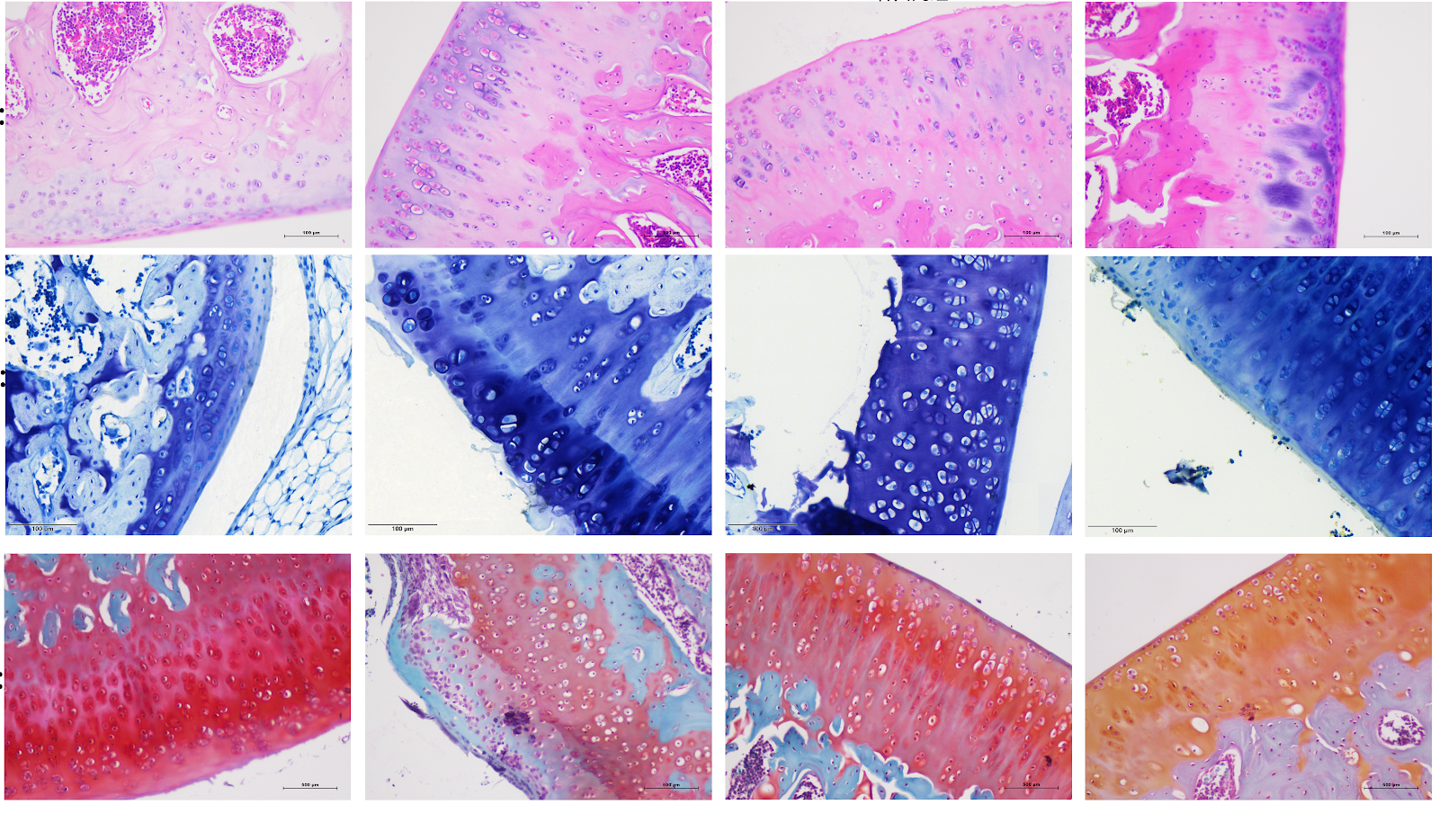
BACKGROUND: Yougui Pill is derived from Jingyue Quanshu. Studies have confirmed that Yougui Pill is highly effective in treating patients with knee osteoarthritis, but its mechanism of action remains unclear. OBJECTIVE: To explore the potential molecular mechanism of Yougui Pill in improving knee osteoarthritis in rats. METHODS: Forty-eight SPF-grade Sprague-Dawley rats were randomly divided into four groups: blank control group, model group, Yougui Pill group and celecoxib group. The latter three groups were subjected to modified Hulth method for surgical modeling of knee osteoarthritis. After wound healing, rats were driven for 8 weeks. After modeling, the celecoxib group was given celecoxib suspension by gavage, the Yougui Pill group was given Yougui Pill decoction by gavage, and the sham operation group and model group were given equal volume of normal saline, once daily for 4 weeks. Hematoxylin-eosin staining, toluidine blue staining, and safranin O-fast green staining were used to observe the pathological changes of rat cartilage tissue; transmission electron microscopy was used to observe the ultrastructure of rat chondrocytes; ELISA was used to detect the levels of interleukin-18, interleukin-1β, and tumor necrosis factor-α in rat serum; western blot was used to detect the protein expression of PI3K, AKT, NF-κB, p-PI3K, p-AKT, p-P65, NLRP3, GSDMD, GSDMD-N, Caspase1, Cleaved-Caspase1, interleukin-18, and interleukin-1β in rat cartilage tissue. RESULTS AND CONCLUSION: Compared with the blank group, the model group showed severe destruction of cartilage edge, cartilage tissue defect, thinning and disordered arrangement of cartilage layer cells, subchondral bone hyperplasia, disordered tide line, severe structural damage of chondrocytes, formation of pyroptotic bodies, significantly increased serum levels of tumor necrosis factor-α, interleukin-18, and interleukin-1β (P < 0.05), and significantly increased protein expression of p-PI3K, p-AKT, p-P65, NLRP3, GSDMD-N, Cleaved-Caspase1, interleukin-18, and interleukin-1β in cartilage tissue (P < 0.01). Compared with the model group, the cartilage structure of rats in the Yougui Pill group and celecoxib group tended to be normal, with deeper cartilage staining, thicker cartilage, more complete chondrocyte membrane, significantly decreased serum levels of tumor necrosis factor-α, interleukin-18, and interleukin-1β (P < 0.05), and significantly decreased protein expression of p-PI3K, p-AKT, p-P65, NLRP3, GSDMD-N, Cleaved-Caspase1, interleukin-18, and interleukin-1β in cartilage tissue (P < 0.01). Compared with the celecoxib group, the Yougui Pill group showed more regular arrangement of chondrocytes, smoother articular cartilage surface, significantly thickened cartilage layer, relatively complete tide line, relatively complete chondrocyte membrane, significantly decreased serum levels of tumor necrosis factor-α, interleukin-18, and interleukin-1β (P < 0.05), and significantly decreased protein expression of p-PI3K, p-AKT, p-P65, NLRP3, GSDMD-N, Cleaved-Caspase1, interleukin-18, and interleukin-1β in cartilage tissue (P < 0.01). These results indicate that Yougui Pill can improve the inflammatory response of chondrocytes in rats with knee osteoarthritis, and the mechanism may be related to inhibiting the activation of PI3K/AKT/NF-κB pathway, thereby regulating NLRP3/Caspase1/GSDMD pathway-mediated pyroptosis.

Mechanism by which Yougui Pill inhibits pyroptosis of chondrocytes in rats with knee osteoarthritis
Graphical Abstract
Original ResearchVol 1896, Issue 24 • pp. 100-112DOI: 10.12307/2026.21222Jan 15, 2026

Cobalt chloride-induced hypoxic environment accelerates knee cartilage degeneration in New Zealand rabbits

Authors: XU Peng, JIANG Wei, YU You, LEI Zhengliang, TIAN Yang, ZHANG Jie, LIU Luchang

BACKGROUND: Cobalt chloride solution is commonly used to induce osteoarthritis cell models in vitro. However, its ability to construct animal models of osteoarthritis by intra-articular injection remains unknown. OBJECTIVE: To investigate the effect of intra-articular injection of different concentrations of cobalt chloride solution on cartilage degeneration in the knee joint. METHODS: Thirty-six healthy adult male New Zealand rabbits were randomly divided into four groups: low, medium and high dose cobalt chloride groups and control group. The right hind knee was intra-articularly injected with 100, 200, and 300 μmol/(L·kg) of cobalt chloride, while the left hind knee served as the control knee and was injected with an equal amount of normal saline. At 4, 8 and 12 weeks after operation, four rabbits were killed respectively. The cartilage on the surface of the femur was exposed for gross morphological observation, and then the cartilage tissues were taken for hematoxylin-eosin staining, safranine O-fast green staining, the Osteoarthritis Research Society International scoring, and immunohistochemical staining of interleukin 1 and tumor necrosis factor α, to determine cartilage degeneration in various aspects. RESULTS AND CONCLUSION: (1) Gross observation: At the same postoperative time point, with the increase of cobalt chloride concentration, cartilage degeneration showed a progressive aggravation trend, and the high-dose cobalt chloride group even involved the deep layer of cartilage and subchondral bone; under the same concentration of cobalt chloride, with the prolongation of modeling time, cartilage degeneration progressed progressively. (2) Hematoxylin-eosin staining, safranine O-fast green staining, and Osteoarthritis Research Society International scoring showed that at the same postoperative time point, with the increase of cobalt chloride concentration, the cartilage surface gradually became rough, the superficial layer became thinner, and the destruction aggravated, and the Osteoarthritis Research Society International score gradually increased (P < 0.05); under the same concentration of cobalt chloride, with the prolongation of modeling time, the arrangement of chondrocytes tended to be disordered, polarity was lost, and the destruction of superficial cartilage and subchondral bone progressively aggravated, and the Osteoarthritis Research Society International score gradually increased (P < 0.05). (3) Immunohistochemistry showed that at the same postoperative time point, with the increase of cobalt chloride concentration, cartilage degeneration aggravated, intracellular brown particles increased, and the positive expression of interleukin 1 and tumor necrosis factor α increased (P < 0.01); under the same concentration of cobalt chloride, with the prolongation of modeling time, cartilage destruction and fissures aggravated, and the positive expression of interleukin 1 and tumor necrosis factor α increased (P < 0.01). This experiment successfully established an osteoarthritis model of New Zealand rabbits induced by intra-articular injection of cobalt chloride solution, preliminarily verified the stability and reliability of the animal model, and also proved that with the increase of modeling concentration and the prolongation of modeling time, cartilage degeneration progressed progressively.

Cobalt chloride-induced hypoxic environment accelerates knee cartilage degeneration in New Zealand rabbits
Graphical Abstract
Original ResearchVol 1896, Issue 24 • pp. 100-112DOI: 10.12307/2026.21239Jan 15, 2026

Effects of high-intensity interval training combined with estrogen on satellite cells, myonuclear domain and ribosome function in ovariectomized rats

Authors: Sun Yuan, Shu Jun, Ren Shuang, Wang Chenyu

BACKGROUND: Estrogen deficiency can lead to a decrease in skeletal muscle mass and muscle strength in postmenopausal women, thereby affecting their quality of life. Muscle mass is maintained by satellite cells, which are regulated by estrogen. Regular exercise, especially high impact exercise (such as resistance training and high-intensity interval training), can induce muscle hypertrophy, but the role and mechanism of estrogen are still unclear. OBJECTIVE: To explore the effects of high-intensity interval training combined with estrogen therapy on skeletal muscle hypertrophy in ovariectomized rats and reveal its possible mechanism. METHODS: Sixty 8-week-old female Sprague-Dawley rats were divided into five groups using a random number table method: sham operation, model sedentary group, model exercise group, model hormone group, or model combined group. Bilateral ovariectomy was used to establish an estrogen deficiency model. Twelve weeks after operation, the model exercise and model combined group performed high-intensity interval training for 8 weeks (3 times/week), and hormone treatment groups received abdominal subcutaneous injection of 17β-estradiol (once a day for 8 weeks). Seventy-two hours after the last training, the grip force of the forelimb was measured by an electronic grip force meter. The gastrocnemius muscle was separated, and muscle mass index was calculated as muscle mass/body mass ratio. Hematoxylin-eosin staining was used to obtain cell cross-sectional area. Immunofluorescence staining was used to classify muscle fiber types and obtain myonuclear number, myonuclear domain size, and activated satellite cell number. BCA method was used to determine total protein concentration. Trizol method was used to extract total RNA. Western blot was used to detect ribosomal protein S6 expression. Real-time quantitative PCR was used to detect ribosomal RNA expression. RESULTS AND CONCLUSION: Compared with the sham operation group, the model sedentary group showed increased body mass and myosin heavy chain type I fiber proportion (P < 0.05), while uterine mass index, gastrocnemius mass index, grip strength, cell cross-sectional area, myosin heavy chain type IIa proportion, satellite cell and myonuclear number, myonuclear domain size, total protein and RNA content, and ribosomal protein S6, 18S rRNA and 28S rRNA expression decreased (P < 0.05). Compared with the model sedentary group, the model exercise group showed decreased body mass and myosin heavy chain type IIb proportion (P < 0.05), while uterine mass index, gastrocnemius mass index, grip strength, cell cross-sectional area, myosin heavy chain type IIa proportion, satellite cell number, myonuclear domain size, total protein and RNA content, and ribosomal protein S6 and 28S rRNA expression increased (P < 0.05). Compared with the model exercise group and model hormone group, the model combined group showed higher gastrocnemius mass index, grip strength, cell cross-sectional area, satellite cell number, myonuclear domain size, total protein and RNA content, and ribosomal protein S6 and 28S rRNA expression (P < 0.05). These results indicate that estrogen can enhance the skeletal muscle hypertrophy response induced by high-intensity interval training in ovariectomized rats, and the mechanism may be related to satellite cell activation, increased myonuclear domain and ribosome biogenesis, and improved ribosome function.

Effects of high-intensity interval training combined with estrogen on satellite cells, myonuclear domain and ribosome function in ovariectomized rats
Graphical Abstract
Original ResearchVol 1896, Issue 24 • pp. 100-112DOI: 10.12307/2026.21233Jan 15, 2026

Gushukang Granule-containing drug serum improves dexamethasone-induced atrophy of C2C12 myotubes via regulating mitochondrial homeostasis

Authors: WEI Wei, LIU Hongfei, QI Xiaonan, LIU Yantong, WANG Deyu, YU Zhitong, QIAO Chunlin, WANG Shixuan, TENG Hai

BACKGROUND: Gushukang Granule is a Chinese herbal compound preparation, commonly used clinically for the treatment of osteoporosis and other bone-related diseases. However, its role in the regulation of muscle metabolism is not clear. OBJECTIVE: To investigate the inhibitory effect of Gushukang Granule-containing serum on dexamethasone-induced C2C12 muscle atrophy. METHODS: (1) In vivo experiment: Thirty-six 3-month-old female Sprague-Dawley rats were randomly divided into model group, blank group and Gushukang group, with 12 rats in each group. The rats in the model group were given 2.5 mg/kg dexamethasone by intragastric administration, once a day, for 1 week, followed by normal feeding for 3 weeks; the Gushukang group was given 0.48 g/kg Gushukang Granule suspension by gavage after modeling, once a day, for 3 weeks; the blank group was not modeled and given equal volume of distilled water by gavage, once a day, for 4 weeks. Two hours after the last administration, left femur and gastrocnemius muscle specimens were taken for hematoxylin-eosin staining, and RNA was extracted from right gastrocnemius and femur tissues; real-time fluorescence quantitative PCR was used to detect mRNA expression levels of mitochondrial function, autophagy and inflammation-related genes. (2) In vitro experiment: C2C12 cells were cultured and induced to differentiate into myotubes with 2% horse serum. Differentiated myotubes were divided into control group, model group and Gushukang-containing serum group. Muscle atrophy model was constructed by incubating with 4 µmol/L dexamethasone for 48 h, and the Gushukang group was treated with 10% Gushukang-containing serum for 24 h after dexamethasone treatment for 24 h. CCK-8 was used to detect cell viability, flow cytometry to detect reactive oxygen species levels, transmission electron microscopy to observe ultrastructural changes, and western blot to detect expression levels of mitochondrial function-related proteins, autophagy-related proteins and antioxidant-related proteins. RESULTS AND CONCLUSION: (1) Hematoxylin-eosin staining showed that compared with the model group, the muscle fiber structure of rats in the Gushukang group recovered better; real-time fluorescence quantitative PCR further verified that Gushukang Granule up-regulated mitochondrial function and autophagy-related genes, supporting its multi-target mechanism to alleviate muscle atrophy. (2) Flow cytometry and CCK-8 results showed that after dexamethasone treatment, reactive oxygen species levels in C2C12 cells were significantly increased (P < 0.05), and cell viability was significantly decreased (P < 0.05); transmission electron microscopy revealed that dexamethasone induced ultrastructural disorder, reduced number of organelles, atrophic and blurred mitochondria, accompanied by a large number of autophagosomes and cytoplasmic vacuoles; western blot results showed that after dexamethasone treatment, the expression of mitochondrial function-related proteins translocase of outer mitochondrial membrane 20 and heat shock protein 60 was significantly decreased, autophagy-related proteins LC3 and Beclin-1 were abnormally expressed (P < 0.05), and silent information regulator 1 expression was significantly decreased (P < 0.05), suggesting that dexamethasone disrupted mitochondrial homeostasis and inhibited autophagy. After treatment with Gushukang Granule-containing serum, reactive oxygen species levels were significantly decreased (P < 0.05), cell viability was significantly increased (P < 0.05); transmission electron microscopy showed that ultrastructure was improved and mitochondrial morphology was relatively restored; western blot results showed that the expression of translocase of outer mitochondrial membrane 20, heat shock protein 60, and silent information regulator 1 was significantly up-regulated (P < 0.05), and LC3 and Beclin-1 expression returned to normal levels (P < 0.05), indicating that Gushukang Granule-containing serum played a positive role in improving mitochondrial function, reducing oxidative stress, and regulating autophagy.

Gushukang Granule-containing drug serum improves dexamethasone-induced atrophy of C2C12 myotubes via regulating mitochondrial homeostasis
Graphical Abstract
Original ResearchVol 1897, Issue 25 • pp. 100-112DOI: 10.12307/2026.21279Jan 15, 2026

Effect of blood flow restriction training on the magnitude and temporal characteristics of post-activation performance enhancement: a systematic review and meta-analysis

Authors: Li Yanfeng, Zhang Yilin, Kong Hao, Zheng Hang, Liu Jiajun, Yin Mingyue, Qiu Bopeng, Huang Kongyun, Liu Hengxian, Zhong Yuming, Chen Jun, Xu Kai

Objective: To systematically compare the acute effects of blood flow restriction combined with preconditioning (to induce post-activation performance enhancement) versus preconditioning alone or sitting, low-intensity preconditioning combined with blood flow restriction versus high-intensity preconditioning, and sitting combined with blood flow restriction versus sitting on sports performance using a multilevel meta-analysis. Methods: Following the PRISMA guidelines, Web of Science, PubMed, SPORTDiscus, and CNKI databases were systematically searched (from inception to May 24, 2025). Inclusion criteria: (1) healthy individuals who were at least physically active; (2) studies with at least one of the following four comparisons: preconditioning + blood flow restriction vs. preconditioning alone; preconditioning + blood flow restriction vs. sitting; low-intensity preconditioning + blood flow restriction vs. high-intensity preconditioning; sitting + blood flow restriction vs. sitting; (3) sports performance (e.g., jump, sprint, bench press throw) as the primary outcome; (4) randomized or non-randomized crossover/parallel designs; (5) published in peer-reviewed Chinese or English journals. Risk of bias was assessed using ROB-2, and evidence quality was evaluated with GRADE. Data were fitted using cluster robust variance estimation and a three-level mixed-effects model, with small-sample corrections. Subgroup analyses and meta-regression explored moderators and sources of heterogeneity. Results: Twelve studies (196 participants, 12 women, 184 men) were included. Main findings: (1) Preconditioning + blood flow restriction was more effective than preconditioning alone in enhancing sports performance (ES=0.21, 95%CI=0.01-0.40, GRADE=low), with the best effect at recovery times of 4-12 min and 50% arterial occlusion pressure (ES=1.49); (2) Preconditioning + blood flow restriction did not significantly differ from sitting (ES=0.52, 95%CI=-0.12-1.15, GRADE=very low), but preconditioning + 140 mmHg blood flow restriction was superior to preconditioning alone (ES=1.21, 95%CI=0.14-2.28); (3) Low-intensity preconditioning + blood flow restriction did not differ from high-intensity preconditioning (ES=-0.10, 95%CI=-0.84-0.64, GRADE=low); (4) Sitting + blood flow restriction did not significantly differ from sitting (ES=0.24, 95%CI=-0.03-0.52, GRADE=very low). Notably, the effects of the latter two comparisons significantly decreased with recovery time (β=-0.04, P < 0.01 and β=-0.04, P=0.02). Conclusion: Preconditioning combined with blood flow restriction is more effective than preconditioning alone in inducing post-activation performance enhancement, preliminarily suggesting the use of 50% arterial occlusion pressure and 4-12 min recovery time. However, preconditioning combined with blood flow restriction does not appear to be more effective than sitting, possibly due to insufficient number of included studies. Additionally, low-intensity preconditioning + blood flow restriction can achieve similar post-activation performance enhancement as high-intensity preconditioning, while the potential benefit of sitting + blood flow restriction on sports performance may diminish over time. Overall, it is preliminarily recommended to use low-intensity preconditioning (e.g., 30% one-repetition maximum squat or bodyweight training) combined with 50% arterial occlusion pressure or 140 mmHg blood flow restriction, with 4-12 min recovery before subsequent performance testing.

Effect of blood flow restriction training on the magnitude and temporal characteristics of post-activation performance enhancement: a systematic review and meta-analysis
Graphical Abstract
Original ResearchVol 1896, Issue 24 • pp. 100-112DOI: 10.12307/2026.21238Jan 15, 2026

Establishing a diagnostic model for recurrent spontaneous abortion based on the levels of autophagy-related genes in the endometrium

Authors: Tang Cen, Hu Wanqin

BACKGROUND: The etiology of recurrent spontaneous abortion is complex. With the development of genetics and other fields, it has been found that the abnormal expression of autophagy-related genes may lead to the imbalance of cell homeostasis, thus triggers pathological processes, such as apoptosis, inflammatory response and immunosuppressive response, and affects the endometrial microenvironment, trophoblastic function and immune cell function, thereby leading to recurrent spontaneous abortion. By using the recurrent spontaneous abortion samples in the Gene Expression Omnibus database, the expression changes and regulatory mechanisms of autophagy-related genes were analyzed, which is helpful to reveal the mechanism of recurrent spontaneous abortion and develop new therapeutic strategies. However, the specific mechanism of autophagy-related genes in recurrent spontaneous abortion and their interaction with other biological processes still need to be further studied. OBJECTIVE: To establish a risk score prognostic model for patients with recurrent spontaneous abortion based on autophagy-related genes. METHODS: Endometrial gene expression matrix of patients with recurrent abortion was obtained from the Gene Expression Omnibus database, autophagy-related genes were obtained from the Human Autophagy Database (HADb), and 30 differentially co-expressed autophagy-related genes were identified. The biological functions of autophagy-related genes were analyzed by gene ontology (GO), Kyoto Encyclopedia of Genes and Genomes (KEGG) and DisGeNET enrichment. LASSO and logistic regression analysis identified 16 autophagy-related genes as potential biomarkers. A nomogram model was then constructed, and the predictive accuracy was evaluated using receiver operating characteristic curves. Six machine learning models (random forest, support vector machine, generalized linear model, etc.) were compared to select the optimal model. Nomogram, calibration curves, and decision curve analysis were used to validate the predictive efficacy. RESULTS AND CONCLUSION: GO analysis showed that autophagy-related genes in recurrent spontaneous abortion patients were mainly enriched in autophagy regulation, catabolic processes, and formation of mitochondrial or other organelle membranes. KEGG analysis showed enrichment in autophagy regulation, phosphatidylinositol 3-kinase/protein kinase B signaling pathway, human papillomavirus infection, and neurodegeneration pathways. GSEA analysis indicated involvement in copper detoxification, proton transmembrane transport, sarcoplasmic reticulum components, and regulation of nucleoside diphosphate phosphatase activity. A predictive risk model was constructed, identifying 16 specific autophagy-related genes as predictive targets. Based on the detection efficacy of machine models, the optimal neural network model was selected, and five most important autophagy-related gene variables (MAP2K7, CALCOCO2, SAR1A, TUSC1, and STK11) were identified. Using recurrent spontaneous abortion samples from the European population in the GEO database, the gene expression patterns, differentially expressed genes, and signaling pathways in the endometrium were analyzed from the genetic single nucleotide polymorphism level. The predictive model and machine learning model with good predictive efficacy can screen new therapeutic targets and potential biomarkers for recurrent spontaneous abortion, which has certain guiding significance for clinical work and mechanism research.

Establishing a diagnostic model for recurrent spontaneous abortion based on the levels of autophagy-related genes in the endometrium
Graphical Abstract
Original ResearchVol 1896, Issue 24 • pp. 100-112DOI: 10.12307/2026.21240Jan 15, 2026

Effects of protein kinase C on the expression of myocardial SarcKATP channels in model rats during exercise preconditioning

Authors: Wang Kai, Zhou Yuehui

BACKGROUND: Exercise preconditioning produces early and late myocardial protective effects, in which protein kinase C and myocardial ATP-sensitive potassium channels (SarcKATP) are mediators and effectors, respectively. Protein kinase C regulates the expression of myocardial SarcKATP channels. OBJECTIVE: To compare the effects of protein kinase C on the expression of myocardial SarcKATP channel subunits, inward recirculating potassium channel 6.2 (Kir6.2) and sulfonylurea receptor 2A (SUR2A), in exercise preconditioning. METHODS: Forty-eight Sprague-Dawley rats were randomly divided into five groups: control group (no intervention), early exercise preconditioning group, protein kinase C inhibitor (pre-exercise intraperitoneal injection) + early exercise preconditioning group, late exercise preconditioning group, and protein kinase C inhibitor + late exercise preconditioning group. After preconditioning, the distribution and expression changes of Kir6.2 and SUR2A mRNAs in the rat myocardium were observed and detected using real-time fluorescent quantitative PCR. The distribution and expression changes of Kir6.2 and SUR2A proteins were observed and detected using western blot. RESULTS AND CONCLUSION: (1) Compared with the control group, the mRNA expression of Kir6.2 and SUR2A showed no significant difference in the early and late exercise preconditioning group. (2) Compared with the early exercise preconditioning group, the protein kinase C inhibitor + early exercise preconditioning group showed decreased Kir6.2 mRNA expression but increased Kir6.2 protein expression; both SUR2A mRNA and protein expression decreased. (3) Compared with the late exercise preconditioning group, the protein kinase C inhibitor + late exercise preconditioning group showed decreased Kir6.2 mRNA and SUR2A mRNA expression, decreased Kir6.2 protein expression, but increased SUR2A protein expression. (4) These results indicate that for the same subunit (Kir6.2 or SUR2A), protein kinase C exerts coordinated and complementary regulatory effects in early and late exercise preconditioning; for different subunits (Kir6.2 and SUR2A), protein kinase C also exerts coordinated and complementary regulatory effects on their expression in early and late exercise preconditioning.

Effects of protein kinase C on the expression of myocardial SarcKATP channels in model rats during exercise preconditioning
Graphical Abstract
Original ResearchVol 1896, Issue 24 • pp. 100-112DOI: 10.12307/2026.21225Jan 15, 2026

Articular cartilage lesions at different stages of steroid-induced osteonecrosis of the femoral head: characteristics and mechanisms of crescent sign formation

Authors: Wan Ziyi, Jiang Mengyu, Zhou Yuehui, Xue Yuxuan, Wei Yangwenxiang, Zhou Chi

BACKGROUND: The crescent sign is a significant radiological feature in the progression of steroid-induced osteonecrosis of the femoral head (SIONFH), indicating the separation and defect of articular cartilage and subchondral bone. The appearance of the crescent sign is associated with the mid-to-late stages of the disease and poor prognosis. However, studies on the specific pathological characteristics and progression patterns of articular cartilage in SIONFH remain unclear. OBJECTIVE: To observe the pathological features of articular cartilage in specimens from different stages of SIONFH, explore the progression and pathological mechanisms, and elucidate the formation mechanism of the crescent sign, providing a theoretical basis for optimizing hip-preserving strategies. METHODS: Femoral head specimens were collected from patients with SIONFH who underwent total hip arthroplasty at the First Affiliated Hospital of Guangzhou University of Chinese Medicine from 2021 to 2024. According to the ARCO staging, they were divided into mild, moderate, and severe collapse groups, with fresh femoral neck fracture specimens as controls. All specimens were cut coronally, and the folded cartilage surface in the necrotic area was taken; control group took corresponding area. Hematoxylin-eosin staining and Safranin O-fast green staining were used for morphological observation, immunohistochemistry and western blot for biomarker expression, and apoptosis kit for apoptosis level. RESULTS AND CONCLUSION: (1) Gross observation: The control group showed smooth cartilage surface without folds or hyperplasia, no separation or defect between articular cartilage and subchondral bone, and tough texture. In SIONFH specimens, obvious folds were visible on the cartilage surface, with separation and defects between articular cartilage and subchondral bone, and a loose sensation on pressing. (2) Pathological observation: In the control group, chondrocytes in each layer were arranged neatly, cartilage matrix stained uniformly, tidemark was intact and continuous, calcified cartilage layer and subchondral bone connection was clear and complete, and bone trabeculae were arranged neatly. In SIONFH specimens, chondrocytes were disorganized, empty lacunae increased, matrix staining loss of varying degrees, tidemark duplication and loss, calcified cartilage layer showed numerous cavities and sclerosis, with granulation tissue invasion into cavities, separation and defects between calcified cartilage and subchondral bone, and abundant proliferative granulation tissue in subchondral bone trabecular spaces. (3) Immunohistochemistry: In SIONFH specimens, positive staining of Runt-related transcription factor 2, matrix metalloproteinase 13, matrix metalloproteinase 3, and collagen type I alpha 2 chain increased in calcified cartilage layer and deep cartilage; vascular endothelial growth factor A, hypoxia-inducible factor 1 alpha, interleukin-1 beta, and tumor necrosis factor alpha positive staining increased in subchondral bone trabecular spaces and deep cartilage granulation and scar tissue. (4) Western blot results showed decreased expression of collagen type II alpha 1 chain and SOX9, and increased expression of Runt-related transcription factor 2, matrix metalloproteinase 13, hypoxia-inducible factor 1 alpha, and vascular endothelial growth factor A in SIONFH specimens. (5) Caspase3/7 activity in SIONFH samples was significantly higher than that in the control group, positively correlated with the degree of collapse. (6) These results indicate that articular cartilage lesions in SIONFH mainly concentrate in the deep cartilage and calcified cartilage around the necrotic area. Necrosis of subchondral bone leads to changes in local microenvironment and elastic modulus, causing sclerosis of calcified cartilage. With continued weight-bearing, stress concentration at the necrosis-sclerosis junction leads to brittle fracture, which is the starting point of fracture. Bone and cartilage fracture leads to destruction of the subchondral cortical bone barrier, and invasion of granulation tissue from subchondral bone trabecular spaces directly stimulates calcified cartilage and deep cartilage, resulting in terminal differentiation, apoptosis, matrix degradation, and cavity formation of chondrocytes, leading to decreased repair capacity of articular cartilage. The diseased cartilage cannot properly interlock with subchondral bone, and with disease progression, extensive separation and defects eventually appear between bone and cartilage, manifesting as the crescent sign on imaging.

Articular cartilage lesions at different stages of steroid-induced osteonecrosis of the femoral head: characteristics and mechanisms of crescent sign formation
Graphical Abstract
Original ResearchVol 1896, Issue 24 • pp. 100-112DOI: 10.12307/2026.21237Jan 15, 2026

Prostaglandin E1 pretreatment inhibits ferroptosis in endothelial cells in a rat model of spinal cord ischemia-reperfusion injury

Authors: Huang Yushan, Wang Rongrong, Li Xiangmiao, Bai Jinzhu

BACKGROUND: Ferroptosis is an important pathological mechanism in spinal cord ischemia-reperfusion injury. Although studies have confirmed that prostaglandin E1 attenuates cerebral microvascular endothelial cell injury in the hippocampus induced by chronic cerebral hypoperfusion, its effect on ferroptosis of endothelial cells after spinal cord ischemia-reperfusion injury remains poorly studied. OBJECTIVE: To investigate whether prostaglandin E1 pretreatment attenuates spinal cord ischemia-reperfusion injury by inhibiting ferroptosis in endothelial cells and to elucidate possible mechanisms. METHODS: (1) Cell experiment: Rat spinal cord microvascular endothelial cells were divided into four groups. Control group was cultured under normoxia (20% O2) with complete medium. Model group was subjected to oxygen-glucose deprivation (OGD) for 3 hours (hypoxia chamber with 95% N2 and 5% CO2, glucose-free serum-free medium) followed by reoxygenation for 12 hours (normoxia, complete medium) to simulate spinal cord ischemia-reperfusion injury. Pretreatment group received prostaglandin E1 for 2 hours after OGD and before reoxygenation. Inhibitor group received ML385 (Nrf2 inhibitor) for 2 hours after OGD, then prostaglandin E1 for 2 hours, followed by reoxygenation for 12 hours. After treatment, intracellular malondialdehyde, glutathione, and Fe2+ levels were measured; cell viability was assessed by CCK-8; immunofluorescence staining and western blot were used to detect ACSL4 and GPX4 expression; flow cytometry measured reactive oxygen species; western blot detected Nrf2 and HO-1 protein expression. (2) Animal experiment: 45 rats were randomly divided into three groups: sham group (n=15) underwent laparotomy without aortic occlusion; model group (n=15) underwent occlusion of abdominal aorta for 30 minutes followed by tail vein injection of saline, then reperfusion; pretreatment group (n=15) underwent occlusion for 30 minutes followed by tail vein injection of prostaglandin E1, then reperfusion. At 24 hours after reperfusion, motor function and neuronal injury were assessed by BBB score, inclined plane test, and Nissl staining; blood-spinal cord barrier integrity and microvascular density were evaluated by spinal cord water content, immunofluorescence staining of ZO-1, and CD34 immunohistochemistry; ferroptosis in spinal cord tissue was assessed by immunofluorescence, Prussian blue staining, western blot, and biochemical assays. RESULTS AND CONCLUSION: (1) Cell experiment: OGD/reoxygenation reduced cell viability, induced ferroptosis, and downregulated Nrf2 and HO-1 protein expression in rat spinal cord microvascular endothelial cells. Prostaglandin E1 pretreatment inhibited these effects; ML385 partially reversed the protective effect of prostaglandin E1. (2) Animal experiment: Prostaglandin E1 pretreatment alleviated motor dysfunction, neuronal injury, and blood-spinal cord barrier damage, improved microvascular density, and inhibited ferroptosis in spinal cord tissue after spinal cord ischemia-reperfusion injury. (3) These results indicate that prostaglandin E1 pretreatment protects against spinal cord ischemia-reperfusion injury by activating the Nrf2/HO-1 signaling pathway to inhibit ferroptosis in endothelial cells.

Prostaglandin E1 pretreatment inhibits ferroptosis in endothelial cells in a rat model of spinal cord ischemia-reperfusion injury
Graphical Abstract
Original ResearchVol 1896, Issue 24 • pp. 100-112DOI: 10.12307/2026.21236Jan 15, 2026

Mechanism by which the paraventricular nucleus of the hypothalamus is involved in chronic pain and anxiety in mice with lumbar disc herniation

Authors: Shi Gaolong, Ge Caijun, Chen Jianpeng, Wang Yuanbin, Fan Zelin, Yan Jun, Wang Qianliang

BACKGROUND: Patients with lumbar disc herniation (LDH) often experience comorbid anxiety due to chronic pain and functional limitations, significantly affecting their quality of life. However, the mechanisms underlying the pain-anxiety comorbidity remain unclear. OBJECTIVE: To investigate the neural regulatory mechanisms of the paraventricular nucleus in the hypothalamus in a mouse model of lumbar disc herniation with chronic pain-anxiety comorbidity. METHODS: A total of 100 C57BL/6 mice were randomly divided into a normal group (24 mice) and a model group (76 mice). The lumbar disc herniation model was established in the model group using a needle puncture method. Seventy-two successfully modeled mice were randomly divided into the model group, oxytocin group, and oxytocin+Vasotocin group, with 24 mice in each group. Mice in the oxytocin group received a 200 nL injection of oxytocin (0.5 μg/μL) into the paraventricular nucleus of the hypothalamus. Mice in the oxytocin+Vasotocin group received a 200 nL injection of oxytocin into the paraventricular nucleus and a 20 μL intraperitoneal injection of Vasotocin (an oxytocin antagonist, 0.15 μg/μL). Anxiety-like behavioral changes were evaluated via the elevated plus maze and open field tests on day 20 after modeling. Mechanical paw withdrawal threshold and thermal paw withdrawal latency experiments were conducted for all groups before modeling and 21 days after modeling. On day 21 post-modeling, immunofluorescence staining was used to observe c-FOS expression in the paraventricular nucleus of the hypothalamus; qPCR was used to detect mRNA expression of inflammatory factors prostaglandin E2, tumor necrosis factor α, and interleukin-1β in dorsal root ganglion tissue; Western blot was used to detect oxytocin receptor and p-ERK1/2 protein expression in the paraventricular nucleus. RESULTS AND CONCLUSION: Compared with the normal group, the model group showed significantly decreased mechanical and thermal pain thresholds (P < 0.05), significantly reduced time and entries in the open arms of the elevated plus maze (P < 0.05), significantly reduced time and entries in the open field (P < 0.05), significantly increased mRNA expression of prostaglandin E2, tumor necrosis factor α, and interleukin-1β in dorsal root ganglion tissue (P < 0.05), significantly increased c-FOS expression in the paraventricular nucleus (P < 0.05), significantly decreased oxytocin receptor protein expression, and significantly increased p-ERK1/2 protein expression (P < 0.05). Compared with the model group, the oxytocin group showed significantly increased mechanical and thermal pain thresholds (P < 0.05), significantly increased time and entries in the open arms of the elevated plus maze (P < 0.05), significantly increased time and entries in the open field (P < 0.05), significantly decreased mRNA expression of prostaglandin E2, tumor necrosis factor α, and interleukin-1β in dorsal root ganglion tissue (P < 0.05), significantly decreased c-FOS expression in the paraventricular nucleus (P < 0.05), significantly increased oxytocin receptor protein expression, and significantly decreased p-ERK1/2 protein expression (P < 0.05). Compared with the oxytocin group, the use of Vasotocin reversed the beneficial effects of oxytocin on pain and anxiety, increased inflammatory factor expression, significantly decreased oxytocin receptor protein expression, and significantly increased p-ERK1/2 protein expression. These results indicate that oxytocin can significantly improve chronic pain and anxiety-like behavior in mice with lumbar disc herniation, inhibit dorsal root ganglion inflammation, and the mechanism may be related to activation of the ERK signaling pathway in the paraventricular nucleus and downregulation of inflammatory factor expression.

Mechanism by which the paraventricular nucleus of the hypothalamus is involved in chronic pain and anxiety in mice with lumbar disc herniation
Graphical Abstract
Original ResearchVol 1897, Issue 25 • pp. 100-112DOI: 10.12307/2026.21275Jan 15, 2026

Mechanisms by which mitochondria-endoplasmic reticulum interaction stress mediates activation of inflammatory vesicles in nerve roots of lumbar intervertebral disc herniation rabbits modulated by acupotomy

Authors: Jiang Qiang, Ding Yu, Ding Zhili, Han Jiaheng

BACKGROUND: Acupotomy, as one of the representative therapies of minimally invasive interventional therapy, has been applied to the clinical treatment of lumbar disc herniation, which can antagonize nerve root inflammatory response in lumbar disc herniation with precise curative effects, but its potential mechanism of action remains to be explored. OBJECTIVE: To investigate how acupotomy intervention affects the mitochondrial-mitochondria-associated endoplasmic reticulum membranes-endoplasmic reticulum interaction stress-mediated NLRP3 inflammasome activation in the microenvironment of nerve roots in the model rabbits of lumbar disc herniation. METHODS: Forty healthy adult New Zealand white rabbits were randomly divided into a blank control group (10 rabbits) and a model group (30 rabbits). Animal models were established in the model group through a standard autologous nucleus pulposus transplantation method. Once the model was successfully established, the model rabbits were randomly divided into model control group (10 rabbits), electroacupuncture intervention group (10 rabbits), and acupotomy intervention group (10 rabbits). One week after modeling, acupotomy and electroacupuncture interventions were performed. After 3 weeks of intervention, rabbit dorsal root ganglion cells were isolated and cultured. TUNEL staining was used to detect cell apoptosis. Western blot was used to detect the expression levels of NLRP3, MAMs-related proteins, endoplasmic reticulum stress marker protein GRP78, specific marker protein CHOP, and TXNIP protein in the key PERK axis. Mito Tracker and ER Tracker fluorescent probe staining and transmission electron microscopy were used to observe the structural coupling of mitochondria and endoplasmic reticulum. Flow cytometry was used to analyze Ca2+ levels. Reactive oxygen species probe staining was used to detect reactive oxygen species content. RESULTS AND CONCLUSION: Compared with the model control group, the apoptosis rate in the acupotomy intervention group was significantly decreased (P=0.000 2); the protein expressions of NLRP3 (P=0.014 4), IP3R (P=0.013 2), GRP75 (P=0.009 9), VDAC1 (P=0.000 3), GRP78 (P=0.006 5), CHOP (P=0.008 5), and TXNIP (P=0.001 5) were significantly downregulated, while MFN2 (P=0.010 8) protein expression was significantly upregulated; Ca2+ level (P < 0.000 1) and reactive oxygen species level (P=0.039 2) were significantly decreased. Mito Tracker and ER Tracker fluorescent probe staining results showed that the co-localization level of mitochondria-endoplasmic reticulum was highest in the model control group, lowest in the normal control group, and higher in the acupotomy intervention group than in the normal control group but lower than in the model control group. Transmission electron microscopy observation showed that the contact/association between mitochondria and endoplasmic reticulum was enhanced in the model control group, less in the normal control group, and enhanced in the acupotomy intervention group compared with the normal control group but lower than in the model control group. These results indicate that acupotomy intervention can regulate the interaction stress between mitochondria and endoplasmic reticulum, modulate mitochondria-associated endoplasmic reticulum membranes, reduce Ca2+ influx, reactive oxygen species generation, and mitochondrial dysfunction, thereby inhibiting the formation of NLRP3 inflammasome. This elucidates the upstream mechanism by which acupotomy inhibits NLRP3 inflammasome assembly by regulating mitochondria-endoplasmic reticulum interaction stress, reveals the deep-level therapeutic targets of acupotomy for lumbar disc herniation, and provides a theoretical basis for acupotomy treatment of lumbar disc herniation.

Mechanisms by which mitochondria-endoplasmic reticulum interaction stress mediates activation of inflammatory vesicles in nerve roots of lumbar intervertebral disc herniation rabbits modulated by acupotomy
Graphical Abstract
Original ResearchVol 1897, Issue 25 • pp. 100-112DOI: 10.12307/2026.21271Jan 15, 2026

Visual analysis of research hotspots in the field of gut microbiota in the elderly at home and abroad

Authors: Li Yiguang, Guo Haonan, Ding Xiaotao, Yuan Mengyao, Jiang Lijin, Fan Xinfeng, Feng Yan

BACKGROUND: As the population ages, research on gut microbiota in the elderly is gaining attention. However, bibliometric analysis in this field is still lacking. OBJECTIVE: To comprehensively analyze literature on gut microbiota in older adults from multiple databases, identify current research hotspots, predict future trends, and provide potential directions for subsequent research. METHODS: CNKI was searched using the subject terms “gut microbiota in older adults,” “gut microecology in older adults,” and “intestinal flora in older adults.” The Web of Science database was searched using the search strategy of “TS=(elderly gut microbe OR elderly gut microbiome OR elderly gut microbiota OR elderly intestinal microbiome OR elderly intestinal microbiota).” Bibliometric tools VOSviewer and CiteSpace were employed to systematically analyze publication years, country distribution, research institutions, authors, and keywords in the retrieved literature. RESULTS AND CONCLUSION: A total of 250 and 604 eligible articles were obtained from the CNKI and Web of Science databases, respectively. From 2014 to 2023, the global publication volume in the field of gut microbiota in older adults showed a steady upward trend. Research interest and discussions in this field have increased significantly worldwide, with expanding depth and breadth through interdisciplinary collaboration. Notably, COVID-19, oxidative stress, depression, and cognitive impairment emerged as prominent keywords in the past 2 years. This bibliometric analysis visually demonstrated the research status and development trends in the field of gut microbiota in the elderly over the past decade. The field is currently in a rising phase, and further exploration of the mechanisms of gut microbiota and intervention strategies for related diseases is still needed.

Visual analysis of research hotspots in the field of gut microbiota in the elderly at home and abroad
Graphical Abstract
Original ResearchVol 1897, Issue 25 • pp. 100-112DOI: 10.12307/2026.21272Jan 15, 2026

Feng's spinal manipulation for cervical spondylosis: kinematic changes

Authors: GU Jiang-peng, GUO Wei, CHEN Xu-jing, LIU Xiao-min, LIU Hong-bo, SUN Peng, YE Chao-qun, FENG Wei, WANG Fei

BACKGROUND: Patients with cervical spondylosis often exhibit varying kinematic abnormalities due to degenerative structural changes and biomechanical imbalances in the cervical spine. Although previous studies have compared specific kinematic parameters between healthy individuals and cervical spondylosis patients, research on coupled motions and their associated ratios remains limited. OBJECTIVE: To investigate changes in kinematic parameters in cervical spondylosis patients before and after Feng's spinal manipulation therapy. METHODS: Thirty patients with cervical spondylosis and 30 healthy controls were enrolled. Participants completed three standardized motion tasks: lateral flexion, flexion-extension, and axial rotation. Three-dimensional cervical spine kinematics were quantified using stereophotogrammetry upon admission and discharge. The following kinematic parameters were analyzed: primary range of motion, coupled motion range, coupled motion patterns, motion symmetry, motion smoothness, and motion velocity. RESULTS AND CONCLUSION: Compared with healthy controls, patients with cervical spondylosis showed significantly reduced maximal angles in lateral flexion, flexion-extension, and axial rotation (P < 0.05), and significantly increased ratios of coupled flexion-extension during lateral flexion, coupled rotation during lateral flexion, coupled lateral flexion during extension, and coupled lateral flexion during rotation (P < 0.05). After treatment, patients showed significant improvements in visual analog scale score and cervical dysfunction index (P < 0.05). Significant differences were found in maximal lateral flexion angle, lateral flexion symmetry, maximum and average lateral flexion velocity, maximal flexion-extension angle, maximum and average flexion-extension velocity, maximal rotation angle, rotation symmetry, maximum rotation velocity, and average left rotation velocity before and after treatment (P < 0.05). No significant differences were observed in coupled motion patterns before and after treatment (P > 0.05). Significant differences were found in the ratios of coupled flexion-extension during right lateral flexion, coupled rotation during lateral flexion, coupled flexion-extension during right rotation, and coupled lateral flexion during rotation before and after treatment (P < 0.05). In conclusion, patients with cervical spondylosis exhibit increased ratios of some coupled motions relative to primary motions. Feng's spinal manipulation can significantly improve clinical symptoms and effectively restore cervical motor function.

Feng's spinal manipulation for cervical spondylosis: kinematic changes
Graphical Abstract
Original ResearchVol 1897, Issue 25 • pp. 100-112DOI: 10.12307/2026.21270Jan 15, 2026

Burn and multi-omic biomarkers: causal relationships with 41 inflammatory factors and 35 blood and urine markers

Authors: Gao Minyi, Liu Pinghong, Lin Haixiong

BACKGROUND: Conventional observational studies are inadequate to reveal the potential causal relationship of biomarkers in burns patients. Mendelian randomization, leveraging genetic variation as an instrumental variable to mimic the advantages of randomized controlled trials, has emerged as a crucial tool for dissecting causal associations in complex diseases. OBJECTIVE: To explore the relationship of burn injury with 41 inflammatory cytokines and 35 blood and urinary biomarkers using Mendelian Randomization. METHODS: (1) Burn-related data of genome-wide association studies were obtained from the IEU open GWAS project database, constructed by The University of Bristol, UK, including 218 131 samples and 16 380 465 single nucleotide polymorphisms were included in the study. (2) Data for 41 types of inflammatory cytokines were derived from a study involving 8 293 participants in the Finnish Young Cardiovascular Risk Study database, which is constructed by the Research Centre for Applied and Preventive Cardiovascular Medicine, University of Turku. (3) Data for 35 types of blood and urinary biomarkers were derived from a study involving 363 228 participants from the UK Biobank, which is a large biomedical database project jointly initiated by the UK government, the Wellcome Trust, and the Medical Research Council of the UK. Single nucleotide polymorphisms were employed as instrumental variables, and analyses were conducted using inverse variance weighting, MR Egger, weighted median, and weighted mode methods. Cochrane's Q test was used to identify heterogeneity, and MR Egger intercept test, MR-PRESSO test, and leave-one-out analysis were used to assess the reliability of exposure-outcome associations. RESULTS AND CONCLUSION: Burn injury reduced levels of interleukin-9 (OR=0.97; 95%CI, 0.949 to 0.997; P=0.030) and testosterone (OR=0.997; 95%CI, 0.995 to 0.999; P=0.025), with no heterogeneity or horizontal pleiotropy, demonstrating robustness. The Mendelian randomization analysis indicates that burn injury leads to decreased levels of interleukin-9 and testosterone, suggesting that increasing these levels may aid in tissue repair and improve protein breakdown rate after burn.

Burn and multi-omic biomarkers: causal relationships with 41 inflammatory factors and 35 blood and urine markers
Graphical Abstract
Original ResearchVol 1897, Issue 25 • pp. 100-112DOI: 10.12307/2026.21274Jan 15, 2026

Performance of unstable barbell bench press and changes in electromyographic activity after transcranial direct current stimulation

Authors: Wang Lejun, Chi Wenxin, Song Xiaoqian, Li Qian, Qiao Minjie, Tao Haifeng

BACKGROUND: Transcranial direct current stimulation can enhance human motor performance by modulating cortical excitability, but its impact on the performance of unstable resistance exercise remains unclear. OBJECTIVE: To investigate the effects of transcranial direct current stimulation on unstable barbell bench press performance and electromyographic activity. METHODS: A randomized, self-controlled crossover design was employed. Twenty-two male college students were randomly assigned to receive either active anodal transcranial direct current stimulation or sham stimulation. After transcranial direct current stimulation interventions, subjects performed a fatigue test involving the unstable barbell bench press. Performance during the unstable barbell bench press task was assessed by recording both the number of completed repetitions and barbell acceleration, serving as indicators of load capacity and movement stability control. In addition, surface electromyographic signals were collected from the right biceps brachii, triceps brachii, anterior deltoid, posterior deltoid, and pectoralis major during the exercise. A paired t-test was used to examine differences in repetition counts between true and sham stimulation conditions. Repeated measures analysis of variance was applied to analyze differences in triaxial acceleration, agonist muscle activation levels, and antagonist co-activation levels. RESULTS AND CONCLUSION: (1) No significant difference in the number of barbell bench press repetitions was observed between the true and sham stimulation conditions. However, the mean amplitude of Y-axis acceleration was substantially lower in the true stimulation group than the sham stimulation group. (2) True transcranial direct current stimulation markedly increased the activation level of the anterior deltoid and the co-activation level of the posterior deltoid. (3) There was no significant interaction between transcranial direct current stimulation and exercise phase on any measured variable. (4) These findings suggest that transcranial direct current stimulation does not significantly affect muscle endurance performance during unstable barbell bench press, but it can improve movement stability, possibly by increasing anterior deltoid activation and posterior deltoid co-activation, thereby enhancing shoulder joint stiffness and stability. In competitive sports and clinical rehabilitation, transcranial direct current stimulation may be considered to modulate performance in unstable load-bearing tasks according to training objectives.

Performance of unstable barbell bench press and changes in electromyographic activity after transcranial direct current stimulation
Graphical Abstract
Original ResearchVol 1897, Issue 25 • pp. 100-112DOI: 10.12307/2026.21266Jan 15, 2026

Visual analysis of shear wave elastography in skeletal muscle research

Authors: Li Qian, Li Zhenxing, Qiao Pengyan, Wang Pingzhi

BACKGROUND: Shear-wave elastography is valuable for rehabilitation diagnosis and treatment, but it has not been sufficiently promoted in clinical practice. OBJECTIVE: To explore the trends and hotspots of ultrasound shear wave elastography in skeletal muscle research by visualizing and analyzing the international literature from the past 10 years, thereby providing a reference for clinical diagnosis and follow-up research. METHODS: Based on the Web of Science Core Collection database (2015-2024), the number of publications, countries/regions, institutions, authors, journals, cited literature, and key words from the 978 included articles were visualized and analyzed using CiteSpace software. RESULTS AND CONCLUSION: (1) With a 16.2% average annual growth in global publications, China has the highest number of publications worldwide (197), but its international collaborative network is relatively weak. The University of Nantes in France has the highest number of publications (50), and the University of Queensland has the most influential collaborative network. (2) Ultrasound in Medicine and Biology is the journal with the most publications (33). Noriaki Ichihashi is the most prolific author. (3) The gastrocnemius muscle is one of the most frequently examined sites. Shear wave elastography shows significant clinical potential in central nervous system diseases and sports injuries. (4) The research focus has shifted from basic biomechanics to dynamic clinical assessment and therapeutic interventions. (5) Future diagnostic techniques should be more standardized and refined, establishing normative data ranges for muscle tissue, while considering individual biological variability in elasticity values.

Visual analysis of shear wave elastography in skeletal muscle research
Graphical Abstract
Original ResearchVol 1897, Issue 25 • pp. 100-112DOI: 10.12307/2026.21267Jan 15, 2026

Association between sarcopenia and osteoporosis: a genome-wide data analysis in European populations

Authors: Yin Xingxiao, Jiang Yang, Song Yanping, Yao Na, Shen Zhen, Li Yanqi, Song Yueyu, Peng Hao, Chen Qigang

BACKGROUND: Sarcopenia and osteoporosis have attracted significant attention in the academic community due to their high prevalence and severe adverse outcomes. Although existing studies have suggested a potential causal relationship between sarcopenia and osteoporosis, the evidence remains insufficient. OBJECTIVE: Based on large-scale genome-wide data, to explore the causal relationship between genetically predicted sarcopenia and osteoporosis through a bidirectional Mendelian randomization approach. METHODS: Genome-wide significant loci (P < 5×10⁻⁸) associated with sarcopenia-related traits were selected from the UK Biobank database (an open database jointly developed by the UK government, the Medical Research Council, and the Wellcome Trust), followed by linkage disequilibrium analysis. Osteoporosis data were obtained from the GEnetic Factors for OSteoporosis Consortium (GEFOS; an open database funded by the EU Framework Program for Research and Development, jointly led by Erasmus University Medical Center in the Netherlands), including 28,498 European ancestry subjects, with a focus on data from osteoporosis-prone fracture sites. The study used inverse variance weighting as the primary analysis method, supplemented by MR-Egger regression, weighted median method, and MR-RAPS for multiple validation. To ensure the reliability of the results, multiple sensitivity analyses were performed. RESULTS AND CONCLUSION: Inverse variance weighting analysis showed a bidirectional causal relationship between whole-body fat-free mass and bone mineral density (P < 0.05). Forward causal analysis indicated that whole-body fat-free mass was positively associated with lumbar spine bone mineral density (OR=1.124, 95%CI: 1.008-1.253, P=0.035) and negatively associated with forearm bone mineral density (OR=0.821, 95%CI: 0.699-0.966, P=0.017). Reverse causal analysis showed that forearm bone mineral density (OR=1.033, 95%CI: 1.002-1.066, P=0.036), lumbar spine bone mineral density (OR=1.054, 95%CI: 1.025-1.084, P < 0.001), and femoral neck bone mineral density (OR=1.059, 95%CI: 1.008-1.113, P=0.021) were all positively associated with whole-body fat-free mass. A reduction in whole-body fat-free mass can lead to decreased lumbar spine bone mineral density, and a decrease in bone mineral density at various sites further exacerbates the loss of whole-body fat-free mass. Although the data in this study mainly come from European populations, due to the universality of genome-wide association analysis methods and the commonality of genetic backgrounds, the results still have important reference value for exploring the pathogenesis of sarcopenia and osteoporosis in the Chinese population, formulating clinical intervention strategies, and assessing genetic risk.

Association between sarcopenia and osteoporosis: a genome-wide data analysis in European populations
Graphical Abstract
Original ResearchVol 1897, Issue 25 • pp. 100-112DOI: 10.12307/2026.21265Jan 15, 2026

Application of patch-clamp technique in traditional Chinese medicine: a visual analysis of relevant literature

Authors: YANG Jun, LI Bin, XING Guogang, CAI Jie, LIU Lu, CHEN Peng, ZHANG Tao, FU Yuanbo, LIU Huilin

BACKGROUND: In recent years, the interdisciplinary application potential of patch-clamp technique in traditional Chinese medicine research has gradually emerged, but a systematic summary of its applications in this field has not yet been conducted. OBJECTIVE: To visualize the application of patch-clamp technology in traditional Chinese medicine field through CiteSpace knowledge map analysis, and to reveal the progress and trends of this technology in the modernization research of traditional Chinese medicine. METHODS: The literature sources included CNKI, VIP, WanFang, PubMed and Web of Science Core Collection database. The computer-assisted literature search was conducted to build a database of patch-clamp technology applications in traditional Chinese medicine field from database inception to September 2024. The authors, institutions, and keywords were subjected to visual analysis and knowledge map drawing using CiteSpace 6.3.R1 software and bibliometric methods. RESULTS AND CONCLUSION: (1) A total of 819 articles were included, with 968 authors. The First Affiliated Hospital of Henan University of Chinese Medicine was the institution with the most publications in the Chinese database, while Harbin Medical University was the institution with the most publications in the English database. (2) The research directions, keyword clustering, and emergence analysis of each institution showed that the application of patch-clamp technique in traditional Chinese medicine mainly focused on cardiovascular electrophysiology, pharmacology of Chinese materia medica, and nervous system electrophysiology. (3) The development of patch-clamp technique in traditional Chinese medicine generally presented an evolutionary path of 'basic mechanism → target deepening → clinical translation', reflecting a transformation from a single technical tool to a multidisciplinary intersection platform. (4) It is suggested that the core influence of authors needs further improvement, and cross-regional cooperation among research institutions is insufficient, so cross-regional cooperation should be strengthened. Current research techniques are single and research content is thin; future research should integrate multiple technologies, cross disciplines, enrich research content, and expand research directions, providing evidence support for in-depth exploration of the internal mechanisms of traditional Chinese medicine.

Application of patch-clamp technique in traditional Chinese medicine: a visual analysis of relevant literature
Graphical Abstract
Original ResearchVol 1897, Issue 25 • pp. 100-112DOI: 10.12307/2026.21278Jan 15, 2026

Intervention with Compound Kidney-Invigorating Granules in a mouse model of osteoporosis: role of the TRIB3/beta-catenin axis

Authors: WANG Yan, LYU Hao, HU Zhimu, ZHOU Yao, LIU Qiang, YANG Yuxiang, YI Hairu, WANG Jiuxiang, JIANG Ting

BACKGROUND: Previous studies have shown that knockdown of β-catenin can inhibit the osteogenic differentiation of human bone marrow mesenchymal stem cells and reduce the expression of TRIB3. Serum containing Compound Kidney-Invigorating Granules can promote the expression of β-catenin and TRIB3 in human bone marrow mesenchymal stem cells, and induce human bone marrow mesenchymal stem cells to differentiate into osteogenic cells. OBJECTIVE: To further explore the mechanism of Compound Kidney-Invigorating Granules in a mouse model of osteoporosis based on the TRIB3/β-catenin axis. METHODS: 8-week-old female C57BL/6 mice were randomly divided into the following experimental groups: blank control, sham operation, model, and low-, medium-, and high-dose Compound Kidney-Invigorating Granules groups, and positive drug group. Except for the blank control and sham operation groups, bilateral ovariectomy was performed to establish an osteoporosis mouse model. One week after modeling, mice in the low-, medium-, and high-dose groups were intragastrically administered 7.05, 14.1, and 28.2 g/kg Compound Kidney-Invigorating Granules, respectively; the blank control, sham operation, and model groups received an equal volume of normal saline once daily; the positive control group received 1.53 mg/kg alendronate sodium once weekly. After 12 weeks of administration, Micro-CT was used to detect changes in femoral bone microarchitecture; hematoxylin-eosin staining and Masson staining were used to detect pathological changes in the femur; Western blot was used to detect the expression levels of TRIB3, β-catenin, alkaline phosphatase, and osteopontin in bone tissue. RESULTS AND CONCLUSION: Compared with the blank control and sham operation groups, the model group showed sparse bone trabeculae and significantly increased empty lacunae; the protein expression levels of TRIB3, β-catenin, alkaline phosphatase, and osteopontin were significantly decreased (P < 0.05). Compared with the model group, the medium- and high-dose Compound Kidney-Invigorating Granules groups showed more complete and regular bone trabeculae, and the protein expression levels of TRIB3, β-catenin, alkaline phosphatase, and osteopontin were significantly upregulated (P < 0.05). These results indicate that Compound Kidney-Invigorating Granules exert a therapeutic effect on osteoporosis model mice, suggesting that the formula may act through the TRIB3/β-catenin axis.

Intervention with Compound Kidney-Invigorating Granules in a mouse model of osteoporosis: role of the TRIB3/beta-catenin axis
Graphical Abstract
Original ResearchVol 1897, Issue 25 • pp. 100-112DOI: 10.12307/2026.21268Jan 15, 2026

Osteoporotic vertebral compression fractures: a visual analysis of current status and emerging trends

Authors: Qu Bolin, Ciren Lunzhu, Guo Jinyang, Meng Hanlu, Ding Guanxiang, Sang Hongpeng

BACKGROUND: Current literature primarily focuses on comparing the clinical efficacy and surgical safety of various operative approaches for osteoporotic vertebral compression fractures, while there is a notable lack of research addressing the current status, hotspots, and future trends in the treatment and prognosis of osteoporotic vertebral compression fractures. OBJECTIVE: To analyze the current status, hotspots and trends of research on osteoporotic vertebral compression fractures by bibliometric methods. METHODS: English articles related to the treatment and prognosis of osteoporotic vertebral compression fractures published between 1994 and 2023 were retrieved from the Web of Science Core Collection. Bibliometric and visualization analyses were conducted using CiteSpace and VOSviewer to evaluate publication volume, geographic distribution, institutional contributions, journal sources, author contributions, keyword frequency, research hotspots, and the most influential literature. RESULTS AND CONCLUSION: A total of 2,275 articles were included. Global research output on osteoporotic vertebral compression fractures increased annually, with the highest number of publications in 2022 (215 articles). The literature spanned 68 countries/regions, with China leading with 644 articles. Soochow University was the most productive institution (90 articles). European Spine Journal had the highest number of publications (n=146), while Spine had the highest citation count (n=1,923), average citations per article (58.5), and H-index (45), ranking second in publication volume (n=106). Yang, Huilin was the most prolific author (n=65), and French physician Galibert, P had the highest citations (n=300). Early research hotspots focused on 'vertebroplasty', 'balloon kyphoplasty', and 'polymethylmethacrylate', which later shifted to 'surgical management' and 'nonoperative treatment', and more recently expanded to 'safety' and 'risk factors', which are expected to remain future research trends.

Osteoporotic vertebral compression fractures: a visual analysis of current status and emerging trends
Graphical Abstract
Original ResearchVol 1897, Issue 25 • pp. 100-112DOI: 10.12307/2026.21269Jan 15, 2026

Molecular mechanism and natural drug screening for ferroptosis-targeted therapy in rheumatoid arthritis

Authors: Zhou Wen, Yang Hongwei

BACKGROUND: Current research in rheumatoid arthritis focuses on iron metabolism-related proteins and the effects of ferroptosis on immune cells. This study proposes new approaches to target ferroptosis in the treatment of rheumatoid arthritis from the perspective of traditional Chinese medicine. These approaches include developing new traditional Chinese medicine therapies, creating individualized treatment plans based on patients’ genes and biomarkers, optimizing therapeutic strategies, and improving symptoms. These strategies aim to facilitate early treatment and improve prognosis. OBJECTIVE: Bioinformatics was applied to investigate the molecular mechanism of treating rheumatoid arthritis from the perspective of ferroptosis, and to screen potential traditional Chinese medicines and active ingredients, opening up a new way for the treatment of rheumatoid arthritis. METHODS: The Gene Expression Omnibus database, maintained by the National Center for Biotechnology Information, is primarily used to store and share high-throughput gene expression, microarray, and sequencing data. The Gene Expression Omnibus database enables researchers to search and analyze genomic data related to various diseases. This study is based on publicly available summary statistics databases and does not require ethical approval. The Gene Expression Omnibus database was searched for datasets related to rheumatoid arthritis that met the screening criteria. The Sanger sequencing platform was then used to obtain the transcriptome data of rheumatoid arthritis. Finally, the limma algorithm was applied to screen the differentially expressed genes. Meanwhile, the ferroptosis-related gene set was extracted from the FerrDb database. Through integration analysis, we obtained the intersection of the differentially expressed genes and the ferroptosis-related genes. We constructed a protein interaction network and performed network topology analysis. Then, using DAVID Bioinformatics Resources 6.8, we conducted Gene Ontology and Kyoto Encyclopedia of Genes and Genomes pathway enrichment analyses to explore biological functions and signaling pathways. Finally, the SymMap platform was used to identify natural medicines, and the Traditional Chinese Medicine Systems Pharmacology Database and Analysis Platform was used to find small molecule compounds and corresponding targets for molecular docking analysis. RESULTS AND CONCLUSION: Two datasets, GSE55457 and GSE55235, were identified from the GEO database, and 340 common differentially expressed targets were obtained via limma analysis. A total of 487 ferroptosis-related targets were collected from the FerrDb platform, and 17 common targets related to ferroptosis and rheumatoid arthritis were obtained after intersection. A protein interaction network was constructed using the 17 common targets, containing 16 target proteins and 33 interactions. Core targets such as EGFR, AR, MAPK8, CDKN1A, JUN, ATM, and EGR1 occupied important positions in the network. Five core targets (EGFR, AR, MAPK8, CDKN1A, and JUN) were identified as key ferroptosis-related targets in rheumatoid arthritis. GO and KEGG enrichment analyses showed that treating rheumatoid arthritis via the ferroptosis pathway may be related to DNA binding processes and SMAD2/3 signaling pathways. Molecular docking results showed that natural small molecule compounds such as progesterone, estradiol, and quercetin could form good binding with core targets, providing new directions for drug development and research in rheumatoid arthritis.

Molecular mechanism and natural drug screening for ferroptosis-targeted therapy in rheumatoid arthritis
Graphical Abstract
Original ResearchVol 1897, Issue 25 • pp. 100-112DOI: 10.12307/2026.21277Jan 15, 2026

Tetramethylpyrazine improves iron metabolism disorders in a rat model of spinal cord injury via the Keap-1/Nrf2 signaling pathway

Authors: ZHENG Peng, JIA Xiaoning, TAO Jingwei, FAN Xiao

BACKGROUND: The clinical management of spinal cord injury remains a global medical challenge, with no currently available ideal treatment. Traditional Chinese medicine has therapeutic advantages for spinal cord injury. Notably, tetramethylpyrazine, an active component of Chuanxiong rhizome, has been shown to significantly suppress pathological responses including neuroinflammation and apoptosis after spinal cord injury, exhibiting promising therapeutic potential. However, its mechanisms require further elucidation. OBJECTIVE: To investigate the regulatory effects of tetramethylpyrazine on the Kelch-like ECH-associated protein 1/nuclear factor-erythroid 2-related factor 2 (Keap-1/Nrf2) signaling pathway and iron metabolism following spinal cord injury, and to elucidate its neuroprotective mechanisms. METHODS: Thirty-six Sprague-Dawley rats were randomly allocated into: sham group (laminectomy+saline, n=12), model group (spinal cord injury+saline, n=12), and tetramethylpyrazine group (spinal cord injury+tetramethylpyrazine, n=12). After 4 weeks, neuronal morphology was assessed by Nissl staining; iron deposition by Prussian blue staining; iron content by iron assay kit; expression of Keap-1, ferritin heavy chain 1 (FTH1), and ferritin light chain (FTL) by immunohistochemistry; Nrf2 expression by immunofluorescence; protein levels of Nrf2, FTH1, and FTL by western blot; and mRNA levels of Nrf2, Keap-1, FTH1, and FTL by RT-PCR. RESULTS AND CONCLUSION: Compared with the sham group, the model group showed disrupted neuronal structure, increased iron deposition and iron content, increased Keap-1 expression, and decreased Nrf2, FTH1, and FTL expression at both protein and mRNA levels (P < 0.01). Tetramethylpyrazine treatment significantly ameliorated these changes, as evidenced by improved neuronal structure, reduced iron deposition and content, decreased Keap-1 expression, and increased Nrf2, FTH1, and FTL expression compared with the model group (P < 0.01). These findings indicate that tetramethylpyrazine can regulate the Keap-1/Nrf2 signaling pathway and its downstream targets FTH1 and FTL, thereby improving iron metabolism disorders and facilitating spinal cord injury repair.

Tetramethylpyrazine improves iron metabolism disorders in a rat model of spinal cord injury via the Keap-1/Nrf2 signaling pathway
Graphical Abstract
Original ResearchVol 1897, Issue 25 • pp. 100-112DOI: 10.12307/2026.21276Jan 15, 2026

Potential targets of glucagon-like peptide 1 receptor agonist ticagrelor in the treatment of Alzheimer’s disease

Authors: ZHANG Xiaomin, DU Pengyang, ZHANG Xiuping, XUE Guofang

BACKGROUND: Glucagon-like peptide 1 receptor agonists, as novel drug candidates for the treatment of neurodegenerative diseases, have achieved breakthrough progress in clinical research on Alzheimer’s disease, with drugs such as Semaglutide advancing to phase III clinical trials. However, there remains a significant knowledge gap regarding the molecular mechanism of neuroprotective effects of these drugs. OBJECTIVE: To innovatively integrate multi-omics analysis techniques and network pharmacology methods, to systematically analyze the intersection network between the gene lineage related to Alzheimer’s disease pathology and the potential targets of ticagrelor, to identify key regulatory genes, and to verify their molecular mechanisms through in vitro and in vivo experiments. METHODS: A multi-dimensional research strategy was adopted: (1) Constructing the differential expression gene profile of Alzheimer’s disease using the DisGeNET database that covers various disease-related genomics. (2) Obtaining the structure of Tirzepatide from PubChem database with bioactive molecules and screening potential targets. (3) Conducting Gene Ontology (GO) functional annotation and Kyoto Encyclopedia of Genes and Genomes (KEGG) pathway enrichment analysis using DAVID database. (4) Constructing protein-protein interaction network using STRING database and Cytoscape 3.9.1, and screening key genes via topological network analysis. (5) Cell-level verification: HT22 cells were divided into control group, model group (treated with β-amyloid 1-42 oligomers for 36 h to establish an in vitro AD model), and treatment group (pretreated with β-amyloid 1-42 oligomers for 24 h, then co-treated with ticagrelor for 12 h). Western blot was used to analyze the protein expression of angiotensin II type 2 receptor (AGTR2), and ELISA was used to detect the expression levels of synaptic markers such as synaptophysin 1 and postsynaptic density protein 95. (6) Animal experiments: Three groups were used: control group (WT C57BL/6 mice, intraperitoneal injection of saline), model group (3xTg mice, intraperitoneal injection of saline), and treatment group (3xTg mice, intraperitoneal injection of 20 nmol/L ticagrelor), all administered every other day for a total of 15 doses. Morris water maze was used to analyze cognitive behavioral improvements in AD model mice; Western blot was used to quantitatively analyze the expression of β-amyloid (6E10) and phosphorylated Tau protein (P-tau-181). RESULTS AND CONCLUSION: (1) A total of 3,397 AD-related genes were screened from DisGeNET database; 10 key genes with the highest connectivity were identified based on protein association: AGTR2, NTSR1, NTSR2, GHSR, C5AR1, C3AR1, OPRM1, SSTR2, OPRD1, STAT3. GO enrichment and KEGG pathway analysis suggested that ticagrelor may improve AD by enhancing neuroreceptor-ligand function. (2) Cell experiments suggested that ticagrelor may exert therapeutic effects by improving synaptic function in AD, and AGTR2 may be a potential target of ticagrelor in treating AD. (3) Animal experiments indicated that ticagrelor improved cognitive ability in 3xTg mice, and ameliorated abnormal β-amyloid deposition and Tau protein phosphorylation in the brain of 3xTg mice. (4) Conclusion: The study reveals that AGTR2 is a key molecular target of ticagrelor in the pathological process of AD, and ticagrelor may treat AD by regulating AGTR2-mediated synaptic function improvement.

Potential targets of glucagon-like peptide 1 receptor agonist ticagrelor in the treatment of Alzheimer’s disease
Graphical Abstract
Original ResearchVol 1897, Issue 25 • pp. 100-112DOI: 10.12307/2026.21273Jan 15, 2026

Analysis of influencing factors and risk prediction model for spasticity severity in stroke patients with hemiplegia

Authors: Cao Xinyan, Leng Xiaoxuan, Gao Shiai, Chen Jinhui, Liu Xihua

BACKGROUND: Spastic hemiplegia remains a challenging clinical problem that urgently needs to be addressed. Currently, most research primarily focuses on discussing the influencing factors of spasticity onset. This study, however, utilizes binary logistic regression to primarily explore the key factors affecting the severity of spasticity in stroke patients, providing a reliable basis for personalized treatment plans for patients. OBJECTIVE: To identify the influencing factors of spasticity severity in stroke patients with hemiplegia through univariate and multivariate logistic regression analyses, and to construct a risk prediction model. METHODS: A total of 120 patients with post-stroke spasticity hospitalized at the Affiliated Hospital of Shandong University of Traditional Chinese Medicine from November 2024 to March 2025 were enrolled. A self-designed questionnaire was used for data collection. Logistic regression analysis was performed to screen the influencing factors of spasticity severity in hemiplegic patients, and a risk prediction model was constructed. The predictive performance of the model was evaluated via a receiver operating characteristic curve analysis. RESULTS AND CONCLUSION: Among 120 stroke spasticity patients, 66 had mild spasticity with Modified Ashworth Scale < 2 and 54 had severe spasticity with Modified Ashworth Scale ≥ 2. The results of logistic regression analysis showed that for stroke spasticity patients, advanced age, normal sensory function, higher Barthel index, and Fugl-Meyer motor function score were protective factors for spasticity severity; while depression and anxiety, poor sleep quality, and pain were risk factors. The area under the receiver operating characteristic curve of the logistic regression model for spasticity severity in stroke hemiplegic patients was 0.969 [95%CI (0.944, 0.994)], indicating that the prediction model based on these factors has high predictive efficacy. Clinicians should adopt multidimensional and individualized intervention strategies to actively prevent and reduce spasticity, thereby improving functional prognosis and quality of life.

Analysis of influencing factors and risk prediction model for spasticity severity in stroke patients with hemiplegia
Graphical Abstract
Original ResearchVol 1897, Issue 25 • pp. 100-112DOI: 10.12307/2026.21264Jan 15, 2026

Sarcopenia and non-alcoholic fatty liver disease: analysis of the gut microbiota

Authors: ZHANG Zheng, ZHANG Yibo, XU Bin, YAN Shichao, GUO Hui

BACKGROUND: Previous studies have established a correlation between non-alcoholic fatty liver disease and sarcopenia; however, their causal relationship remains uncertain. The gut-muscle-liver axis hypothesis posits intricate interactions between the gut microbiota and both sarcopenia and non-alcoholic fatty liver disease, yet the precise pathogenic mechanisms underlying these interactions remain poorly elucidated. OBJECTIVE: To investigate the potential causal relationship between sarcopenia and non-alcoholic fatty liver disease using Mendelian randomization analysis and to delve into the potential role of the gut microbiota in mediating or influencing the interplay between non-alcoholic fatty liver disease and sarcopenia. METHODS: Sarcopenia data were sourced from the UK Biobank (the UK National-Level Biomedical Database, supported by the UK government and developed in 2006 in collaboration with institutions such as the University of Oxford and the University of Manchester, which encompasses multidimensional data including genes, imaging, and health records from 500 000 participants), with relevant traits including appendicular muscle mass, grip strength, and walking speed. The non-alcoholic fatty liver disease dataset was derived from a publicly accessible GWAS summary dataset compiled by Ghodsian et al., comprising aggregated statistics from GWAS cohorts including eMERGE and FinnGen, updated GWAS data of non-alcoholic fatty liver disease from the UK Biobank, and newly conducted GWAS data from the Estonian Biobank. The 211 gut microbiota data were obtained from a large-scale human gut microbiome composition study conducted by the MiBioGen consortium. Inverse variance weighting, weighted median, MR-Egger, weighted model, and simple model methods were used to assess the mutual influences among non-alcoholic fatty liver disease, sarcopenia, and gut microbiota-related traits. RESULTS AND CONCLUSION: The inverse variance weighting analysis indicated that walking speed and appendicular muscle mass were negatively correlated with non-alcoholic fatty liver disease, while left and right hand grip strength showed no significant correlation with non-alcoholic fatty liver disease risk. Reverse Mendelian randomization analysis showed that non-alcoholic fatty liver disease was negatively correlated with appendicular muscle mass, but no significant correlation was found between non-alcoholic fatty liver disease and walking speed or left and right hand grip strength. Thirty-nine gut microbiota taxa were significantly associated with sarcopenia onset, and six gut microbiota taxa had a causal relationship with non-alcoholic fatty liver disease. The study suggests that gut microbiota may regulate the 'gut-liver-muscle axis' through short-chain fatty acid metabolism, providing a new direction for cross-organ mechanism research for Chinese scholars. Combined with the unique genetic background of the Chinese population (such as ALDH2 mutations and genes related to high-salt diet), it can further analyze the race-specific pathways of metabolic-muscle comorbidity, providing a scientific basis for formulating dietary recommendations that conform to the Chinese dietary structure (such as high grain intake).

Sarcopenia and non-alcoholic fatty liver disease: analysis of the gut microbiota
Graphical Abstract
Original ResearchVol 1897, Issue 25 • pp. 100-112DOI: 10.12307/2026.21258Jan 15, 2026

Traditional Chinese sports in the treatment of osteoporosis: potential biological mechanisms and clinical application progress

Authors: DU Xingbin, JIANG Fugao, KONG Jianda

BACKGROUND: Although traditional drug therapy has achieved good results in the treatment of osteoporosis, long-term use often leads to adverse events. Currently, exercise intervention (including traditional Chinese sports) has gradually become a hot topic and a new choice for the prevention and treatment of osteoporosis. OBJECTIVE: To analyze the biological mechanisms and related clinical application progress of traditional Chinese sports (such as Tai Chi Chuan, Baduanjin, Wuqinxi, Yijinjing) in the prevention and treatment of osteoporosis, and to explore their potential mechanisms and effects in improving bone health in multiple aspects. METHODS: Relevant literature search was retrieved in the Web of Science, PubMed, CNKI, WanFang Database, and VIP Journal Database. The search terms included "Osteoporosis, Traditional Chinese Exercises, Tai Chi Chuan, Baduanjin, Wuqinxi, Yijinjing, Bone Mineral Density, Bone Metabolism, Blood Circulation, Anti-inflammatory, Antioxidant, Clinical Application" in English and Chinese. Finally, 59 articles were included for review. RESULTS AND CONCLUSION: Traditional Chinese exercises significantly improve bone mineral density and mitigate bone loss through the combined effects of multiple pathways, including mechanical loading to regulate bone metabolism, modulating the gut microbiota-bone axis, enhancing anti-inflammatory and antioxidant effects, and endocrine regulation. Clinical evidence has indicated that Tai Chi Chuan has particularly significant effects on improving lumbar spine and femoral neck bone mineral density in postmenopausal women; Baduanjin combined with medication can significantly regulate glucose metabolism and bone turnover; Wuqinxi indirectly reduces fracture risk by improving joint function; Yijinjing shows advantages in gender-related bone mineral density maintenance. Notably, traditional exercises regulate immune cells (such as Th1 cells, regulatory T cells) and oxidative stress pathways, providing a theoretical basis for developing novel exercise intervention programs.

Traditional Chinese sports in the treatment of osteoporosis: potential biological mechanisms and clinical application progress
Graphical Abstract
Original ResearchVol 1898, Issue 26 • pp. 100-112DOI: 10.12307/2026.21311Jan 15, 2026

m6A-related ferroptosis gene expression and its association with immune infiltration in Alzheimer’s disease: machine learning and molecular biology validation

Authors: XU Dongfang, ZHAO Kun, LU Changzhu, WANG Yuge, BAI Lianjie, MENG Fanmou, WANG Yang, YAO Hongbo

BACKGROUND: Alzheimer’s disease (AD) is a neurodegenerative disorder. Although β-amyloid and Tau proteins are core biomarkers for AD diagnosis, their heterogeneity and diagnostic limitations necessitate the exploration of novel biomarkers for disease diagnosis and treatment. OBJECTIVE: To analyze the interaction between N6-methyladenosine (m6A) epitranscriptomic modifications and ferroptosis genes in AD using machine learning, bioinformatics analysis, and experimental validation, to identify characteristic genes for AD pathogenesis, and to reveal their association with immune microenvironment regulation, thereby providing novel biomarkers for early diagnosis and precise treatment of AD. METHODS: Genomic data of human hippocampal tissues from GSE5281, GSE48350 (training sets), and GSE33000 (validation set) in the GEO database were integrated. Differentially expressed m6A regulators in AD were screened in the training sets, and the correlation between m6A and ferroptosis genes was assessed to identify ferroptosis-related differentially expressed genes associated with m6A. Support vector machine recursive feature elimination combined with Boruta feature selection was used to determine AD characteristic genes. Gene set enrichment analysis was performed to dissect functional modules of characteristic genes. A logistic regression model combined with receiver operating characteristic curves was constructed to evaluate the diagnostic efficacy of characteristic genes in the validation set. Single-sample gene set enrichment analysis was applied to quantify immune cell infiltration levels and analyze their regulatory association with characteristic genes. Transcription factor/miRNA-mRNA regulatory networks were predicted using ENCORI, miRWalk 3.0, and NetworkAnalyst databases. Potential therapeutic compounds were screened via the CTD database. qRT-PCR and western blotting were used to validate characteristic genes in hippocampal tissues of APP/PS1 double-transgenic mice. RESULTS AND CONCLUSION: (1) Two significantly differentially expressed m6A regulators, Wilms tumor 1 associated protein (WTAP) and methyltransferase-like protein 14 (METTL14), were identified, with 16 ferroptosis-related genes associated with them. (2) Machine learning identified five core characteristic genes: fumarate hydratase (FH), aspartate aminotransferase (GOT1), HRas proto-oncogene (HRAS), metallothionein 3 (MT3), and SET domain containing 1B (SETD1B). (3) Characteristic genes were functionally enriched in oxidative phosphorylation, Huntington disease, Parkinson disease, fatty acid degradation and metabolism, and proteasome signaling pathways. (4) The logistic regression diagnostic model achieved area under the curve values of 0.873 and 0.904 in the training and validation sets, respectively, indicating excellent diagnostic efficacy. (5) Immune microenvironment analysis showed that HRAS was significantly correlated with chemokine receptor family and plasmacytoid dendritic cell infiltration levels. (6) A regulatory network comprising 5 mRNAs, 37 miRNAs, and 142 transcription factors was constructed, and 71 potential therapeutic drugs were predicted. (7) Experimental validation showed that mRNA and protein expression of GOT1, HRAS, and SETD1B in the hippocampus of APP/PS1 mice were significantly different (P < 0.05 or P < 0.01), consistent with bioinformatics analysis. (8) The results reveal that FH, GOT1, HRAS, MT3, and SETD1B can serve as characteristic genes for AD; immune infiltration correlation analysis suggests that HRAS may serve as a potential immunotherapeutic marker for AD, providing a theoretical basis for early diagnosis and targeted therapy.

m6A-related ferroptosis gene expression and its association with immune infiltration in Alzheimer’s disease: machine learning and molecular biology validation
Graphical Abstract
Original ResearchVol 1898, Issue 26 • pp. 100-112DOI: 10.12307/2026.21309Jan 15, 2026

Scientometric deconstruction of developmental dynamics in upper-limb rehabilitation robotics: evidence network analysis via CiteSpace

Authors: Wang Feifei, Wang Zhennan

BACKGROUND: Upper limb rehabilitation robots have emerged as an indispensable component in global healthcare systems. Despite the extensive body of research in this field, which primarily focuses on areas such as development and application, there remains a significant need for comprehensive, systematic literature analysis to thoroughly examine the current research landscape, identify emerging hotspots, and predict future trends in this domain. OBJECTIVE: To conduct a visual analysis of research status, hotspots, and trends in the field of upper limb rehabilitation robots over the past decade using CiteSpace software, with the aim of identifying key research directions and providing intuitive references for researchers. METHODS: This study systematically retrieved literature related to upper-limb rehabilitation robotics from CNKI and the Web of Science Core Collection published between January 1, 2015, and March 13, 2025. CiteSpace 6.1.R1 software was employed for visualized analysis of included literature, covering key dimensions such as publication volume, authors, institutions, keywords, clusters, and bursts. RESULTS AND CONCLUSION: (1) A total of 1 054 articles were included, involving 659 authors. The United States held the highest overall ranking in terms of both research publication volume and centrality in this field. Among the contributing institutions, Northeast University and Univ Shanghai Sci&Technol ranked the highest in publication volume. Visualization analysis indicated an overall upward trend in upper limb rehabilitation robotics research, yet revealed limited collaboration among researchers and institutions. Main research directions included core technical methods, robot design and optimization, control strategies and algorithms, clinical application and evaluation, and emerging technologies and interdisciplinary integration. (2) Future research could focus on technological integration and intelligent upgrading, interdisciplinary collaborative innovation, and improvement of clinical applications to promote the continuous development and refinement of the upper limb rehabilitation robot field.

Scientometric deconstruction of developmental dynamics in upper-limb rehabilitation robotics: evidence network analysis via CiteSpace
Graphical Abstract
Original ResearchVol 1897, Issue 25 • pp. 100-112DOI: 10.12307/2026.21263Jan 15, 2026

Association between immune cells and cardiovascular disease risk: a genome-wide association study in European populations

Authors: Huang Zhe, Shang Baoling, Yao Gengzhen, Pan Guangming

BACKGROUND: Previous studies have linked immune cells to cardiovascular disease risk. As confounding factors are incompletely addressed, the causal relationship between them remains unclear. OBJECTIVE: To evaluate the potential causal relationship between immune cells and cardiovascular disease. METHODS: The source of research data mainly involves three databases: Genome-Wide Association Study (GWAS) database (GWAS Catalog, jointly maintained by the National Institute of Human Genomics and the European Institute of Bioinformatics), UK biobank (a database of British population genomics, health, and disease phenotypes supported by the UK government and the Wellcome Trust), and IEU OpenGWAS (a GWAS database developed by the MRC Epidemiology Unit at the University of Bristol, UK, primarily for European populations). All are open databases, and the study has been approved by the relevant institutional review boards. Using 731 immune cell phenotypes as exposures and 7 cardiovascular diseases (atrial fibrillation, dilated cardiomyopathy, coronary atherosclerotic heart disease, heart failure, hypertrophic cardiomyopathy, hypertension, and valvular heart disease) as outcomes, a two-sample Mendelian randomization analysis was performed. Inverse variance weighting and weighted median methods were mainly used for Mendelian randomization analysis and sensitivity analysis to assess heterogeneity and pleiotropy. RESULTS AND CONCLUSION: (1) After false discovery rate correction, immune phenotypes had statistically significant effects on atrial fibrillation and hypertension. Five cell types were associated with atrial fibrillation risk, including CD11c on monocytes (OR=0.917, 95%CI: 0.876-0.960), FSC-A on myeloid dendritic cells (OR=0.942, 95%CI: 0.910-0.974), CX3CR1 on CD14+ CD16- monocytes (OR=1.045, 95%CI: 1.022-1.070), CX3CR1 on monocytes (OR=1.050, 95%CI: 1.024-1.076), and CX3CR1 on CD14+ CD16+ monocytes (OR=1.050, 95%CI: 1.024-1.077). Three immune phenotypes with protective effects on hypertension were identified: CD19 on switched memory B cells (OR=0.986, 95%CI: 0.980-0.993), CD25++CD8+ T cells (OR=0.993, 95%CI: 0.990-0.997), and CD25++CD8+ T cells absolute count (OR=0.993, 95%CI: 0.989-0.996). No potential heterogeneity or horizontal pleiotropy was observed in sensitivity analyses. (2) The study found causal relationships between 4 monocyte types and 1 myeloid dendritic cell type and atrial fibrillation, and potential causal relationships between 1 memory B cell type and 2 T cell types and hypertension, suggesting the necessity of considering immune cell phenotypes when monitoring and treating atrial fibrillation and hypertension. This study used public databases for analysis, providing a reference for research on immune cell subsets and cardiovascular disease in the Chinese population, and offering insights for further prevention and treatment of atrial fibrillation and hypertension in Chinese people.

Association between immune cells and cardiovascular disease risk: a genome-wide association study in European populations
Graphical Abstract
Original ResearchVol 1897, Issue 25 • pp. 100-112DOI: 10.12307/2026.21260Jan 15, 2026

Association between environmental exposure to endocrine disrupting chemicals and the risk of type 1 diabetes

Authors: WANG Ting, YANG Yang, LI Yuping, YANG Lin

BACKGROUND: As a group of ubiquitous exogenous compounds in the environment, endocrine disrupting chemicals can interfere with endocrine system function and contribute to various diseases. In recent years, the correlation between exposure to endocrine disrupting chemicals and type 1 diabetes risk has become a research hotspot, but the exact underlying mechanisms remain unclear. OBJECTIVE: To review the research progress on the association between endocrine disrupting chemicals and type 1 diabetes in terms of epidemiological studies, animal experiments, and related mechanism studies. METHODS: The literature retrieval was conducted on CNKI and PubMed databases from January 2000 to January 2025 with the keywords of “endocrine disrupting chemicals; EDCs; type 1 diabetes; T1DM” in Chinese and English, respectively. A total of 55 articles were selected for the review. RESULTS AND CONCLUSION: Typical endocrine disrupting chemicals such as bisphenol A, pesticides and heavy metals can promote the development of type 1 diabetes through various pathways, including inducing immune dysregulation, activating oxidative stress, and epigenetic regulation. However, existing studies are limited by issues such as non-unified exposure assessment methods, an unclear dose-response relationship, and population heterogeneity. Future research should focus on identifying critical exposure windows and integrating multi-omics approaches to provide new strategies for the prevention of type 1 diabetes.

Association between environmental exposure to endocrine disrupting chemicals and the risk of type 1 diabetes
Graphical Abstract
Original ResearchVol 1897, Issue 25 • pp. 100-112DOI: 10.12307/2026.21254Jan 15, 2026

Roles and mechanisms of mitochondrial dynamics in bone defect repair

Authors: Zhou Fada, Long Zhisheng

BACKGROUND: Mitochondrial dynamic changes, such as fusion, fission and autophagy, are particularly important for maintaining mitochondrial health homeostasis and cellular balance. Increasing studies have shown that these mitochondrial dynamic changes play a significant role in the healing process of bone defects. In-depth research on mitochondrial dynamics creates new possibilities for the treatment of bone defects. OBJECTIVE: To explore the mechanism and principles of mitochondrial dynamics and its research and development in bone defect repair. METHODS: Relevant literature was retrieved from databases such as CNKI, WanFang Data, and PubMed published from 1990 to 2024 using the keywords of “mitochondrial dynamics, bone defect repair, mitochondrial fusion and fission, osteocytes” in Chinese and “mitochondrial dynamics, bone defect repair, mitochondrial dysfunction” in English. All retrieved documents were strictly screened, analyzed, and sorted one by one according to the inclusion criteria. A total of 77 documents were included for comprehensive analysis, consisting of 15 Chinese documents and 62 English documents. RESULTS AND CONCLUSION: (1) The repair of bone defects is finely regulated by a variety of cells and molecular signaling pathways, which is a highly complex process. Mitochondrial dynamics play a particularly critical role in this process, as they can significantly influence bone cell function and bone metabolism, thereby further promoting the repair of bone defects. (2) Future research could focus on in-depth exploration of the molecular mechanisms of mitochondrial dynamics, development of novel nano-targeted particles and mitochondrial clinical drugs, and creating more possibilities for the clinical application of mitochondrial dynamics in bone defect repair.

Roles and mechanisms of mitochondrial dynamics in bone defect repair
Graphical Abstract
Original ResearchVol 1897, Issue 25 • pp. 100-112DOI: 10.12307/2026.21257Jan 15, 2026

Animal experimental study on the treatment of lumbar intervertebral disc degeneration with Chinese herbal compound: species selection, modeling method and drug administration

Authors: XIE Ziying, LI Songbo, LI Jianwen, YIN Yuchao, ZHENG Baichuan, HU Chengshang

BACKGROUND: Chinese herbal compound has a unique curative effect on lumbar disc degeneration. In order to further understand the mechanism of its action, researchers have carried out many animal experiments in vivo, but there is no agreement on the animal model, the way of administration and the choice of observation indicators. OBJECTIVE: To review the progress in animal experimental studies on the treatment of lumbar disc degeneration with Chinese herbal compounds, focusing on the preparation of animal models of lumbar disc degeneration, the administration methods of Chinese herbal compounds and the evaluation indexes of efficacy, in order to provide a reference for related studies. METHODS: CNKI, WanFang, VIP, PubMed and Web of Science were retrieved by computer for relevant literature published from database inception to November 2024. The Chinese terms were “Chinese medicine, compound, Chinese medicine, herbal, drug combination, soup, formula, low back pain, degenerative disc disease, disc degeneration, intervertebral disc injury, animal, rat, mouse, rabbit, dog, pig, sheep, monkey, primate, model, modeling, experiment, research, progress, review”; the English terms were “traditional Chinese medicine, Chinese herbal medicine, Chinese medicine formula, herbal formula, intervertebral disc degeneration, degenerative disc disease, disc degeneration, intervertebral disc injury, animal model, animal experiment, preclinical study, rat, mouse, rabbit, sheep, dog, pig, non-human primate, monkey, primate, treatment, therapy, effect, intervention”. A total of 789 relevant articles were retrieved, and 139 articles met the inclusion criteria. RESULTS AND CONCLUSION: (1) Researchers using Chinese herbal compounds to treat intervertebral disc degeneration preferred rats, gerbils, guinea pigs, mice, or New Zealand rabbits as experimental animals. (2) Various modeling methods for intervertebral disc degeneration have their own advantages and disadvantages; the annulus fibrosus puncture model is the most widely used model in such studies, and combined modeling methods can better simulate the process of intervertebral disc degeneration. (3) The administration routes of Chinese herbal compounds in animal models are consistent with those in humans, with oral administration being the main route. (4) Chinese herbal compounds have good therapeutic effects on intervertebral disc degeneration, often verified by imaging, behavioral, molecular biological, and histological methods.

Animal experimental study on the treatment of lumbar intervertebral disc degeneration with Chinese herbal compound: species selection, modeling method and drug administration
Graphical Abstract
Original ResearchVol 1897, Issue 25 • pp. 100-112DOI: 10.12307/2026.21261Jan 15, 2026

Glycocalyx: the new link between exercise and disease

Authors: Ma Zhennan, Wang Yinfeng, Yao Lijuan, Chen Leqin

BACKGROUND: The glycocalyx serves as a selective permeability barrier that enables the controlled exchange of substances and maintains fluid balance between within and outside the blood vessels. It is also involved in various pathological processes, including inflammation, thrombus formation, and microcirculation disorders, and is significantly associated with the development and progression of diseases such as atherosclerosis, diabetes, and cancer. OBJECTIVE: To correlate glycocalyx with exercise and disease. METHODS: A literature search was conducted across international databases (MedReading, PubMed, and Web of Science) and Chinese databases (CNKI, WanFang, and VIP) to identify academic articles. The search terms used were “glycocalyx, physical exercise, disease” in Chinese and “glycocalyx, physical exercise, exercises, physical activity, acute exercise, isometric exercises, aerobic exercise, resistance training, exercise training, disease, diseases” in English. A total of 81 publications were included in the final analysis. RESULTS AND CONCLUSION: As a biological barrier of the vascular endothelium, the glycocalyx plays a key role in regulating vascular permeability, mediating inflammatory responses, sensing blood shear stress, and facilitating anticoagulation. The integrity of the glycocalyx is essential for maintaining stable normal blood circulation and ensuring the physiological functions of various organs in the body. Shedding of the glycocalyx can induce structural changes in the endothelial barrier, leading to an abnormal increase in endothelial permeability and accelerating the pathological processes associated with atherosclerosis. Research has confirmed that the extensive thickening and shedding of the glycocalyx on the surface of cancer cells promote tumor proliferation, metastasis, and disease progression. In traumatic diseases, the severity can be assessed by measuring the levels of debris resulting from glycocalyx injury. The glycocalyx is influenced by factors such as the duration of exercise, changes in exercise mode, and exercise intensity. Acute exercise can induce microvascular changes and increase glycocalyx thickness. Aerobic exercise-induced shedding sensitivity of glycocalyx components varies by sex, age, and body mass index. Resistance exercise has positive acute effects on endothelial glycocalyx. Long-term exercise training can protect the glycocalyx. The glycocalyx serves as an intervention target for atherosclerosis, sepsis, cancer, and other diseases, providing theoretical support for developing non-pharmacological therapeutic strategies. However, clinical application of glycocalyx damage markers is not yet standardized, and the mechanisms among glycocalyx, exercise, and disease require further investigation.

Glycocalyx: the new link between exercise and disease
Graphical Abstract
Original ResearchVol 1897, Issue 25 • pp. 100-112DOI: 10.12307/2026.21262Jan 15, 2026

Mitophagy regulates osteoclasts: a new perspective for osteoporosis treatment

Authors: Gao Jiabin, Li Tianqi, Xu Kun, Zhu Hanmin, Zhou Xi, Li Wei

BACKGROUND: The development of osteoporosis is closely associated with the disruption of bone homeostasis, particularly due to the enhanced bone resorption activity of osteoclasts. Mitophagy, an autophagic pathway that selectively degrades damaged mitochondria, has recently been identified as being intricately linked to the pathogenesis and progression of osteoporosis. OBJECTIVE: To elucidate the mechanisms of mitophagy and its regulatory roles in osteoclasts and to investigate the potential mechanisms by which mitophagy influences bone homeostasis through the regulation of osteoclastogenesis and apoptosis. METHODS: The databases searched included PubMed, CNKI, WanFang Data, and VIP databases. The search terms were “mitophagy, bone metabolism, osteoclasts, bone homeostasis, bone resorption, bone loss” in Chinese and English. The search time frame was from January 2008 to April 2025. Based on the inclusion criteria, the search results were screened and excluded, and 101 articles were finally included for review and analysis. RESULTS AND CONCLUTION: Mitophagy is a crucial mitochondrial quality control mechanism within cells, primarily responsible for the selective elimination of damaged or dysfunctional mitochondria. The main pathways of mitophagy include the phosphatase and tensin homolog-induced kinase 1/E3 ubiquitin ligase pathway and the receptor-mediated mitophagy pathway. Mitophagy often exhibits dual beneficial and detrimental properties in the body. Excessive mitophagy can lead to imbalance of bone metabolism and subsequent osteoporosis by maintaining intracellular reactive oxygen species homeostasis, regulating energy metabolism, promoting osteoclast differentiation, inhibiting osteoclast apoptosis, and increasing bone resorption.

Mitophagy regulates osteoclasts: a new perspective for osteoporosis treatment
Graphical Abstract
Original ResearchVol 1897, Issue 25 • pp. 100-112DOI: 10.12307/2026.21256Jan 15, 2026

Application and progress of transcriptomics and proteomics techniques in the study of intervertebral disc degeneration

Authors: ZHOU Tianle, WANG Wei, ZHANG Zhiwen, LIU Ximing

BACKGROUND: Intervertebral disc degeneration is a chronic spinal disease characterized by accelerated apoptosis of nucleus pulposus cells and decomposition of the extracellular matrix, which often leads to low back pain and spinal dysfunction. The molecular mechanism has not been fully understood, hindering the development of precision treatment strategies. Transcriptomics technology can be used to deeply analyze gene expression patterns, and proteomics technology can be used to identify protein function dynamics. The combined application of these two technologies provides an important means to elucidate the pathological mechanism of intervertebral disc degeneration. OBJECTIVE: To review the research progress of transcriptomics and proteomics technologies in intervertebral disc degeneration, and discuss the key roles of these two omics technologies in analyzing molecular mechanisms, screening diagnostic markers, and exploring therapeutic targets. METHODS: A computerized search of relevant literature published from January 1995 to April 2025 in PubMed, Web of Science, CNKI, and Sinomed databases was performed. Chinese search terms were "transcriptomics, ribonucleic acid sequencing, proteomics, intervertebral disc degeneration, nucleus pulposus, annulus fibrosus, cartilage endplate" and English search terms were "transcriptomics, ribonucleic acid sequencing, proteomics, intervertebral disc degeneration, nucleus pulposus, annulus fibrosus, cartilage endplate". After excluding duplicate and irrelevant literature, 24 articles were included for review. RESULTS AND CONCLUSION: Transcriptomics studies have elucidated the dynamic evolution of gene expression in intervertebral disc degeneration, revealing key regulatory networks involved in apoptosis, inflammatory response, and matrix degradation. Proteomics has deeply analyzed the dynamic changes in protein composition in the nucleus pulposus and annulus fibrosus, emphasizing the key functions of matrix metalloproteinases and cytokines in matrix degradation. The combination of the two provides a multi-dimensional perspective on the molecular mechanisms of intervertebral disc degeneration, enhancing the depth of mechanistic research. Comprehensive analysis indicates that the synergistic application of transcriptomics and proteomics shows significant potential in revealing the molecular mechanisms and potential therapeutic targets of intervertebral disc degeneration.

Application and progress of transcriptomics and proteomics techniques in the study of intervertebral disc degeneration
Graphical Abstract
Original ResearchVol 1897, Issue 25 • pp. 100-112DOI: 10.12307/2026.21259Jan 15, 2026

Exercise interventions regulate thyroid hormones: effects on the liver, skeleton, muscle, heart, and brain

Authors: CHENG Yang, HUANG Qingqiang, BU Shumin, YI Yue

BACKGROUND: Thyroid hormones play a critical role in regulating growth, development, energy metabolism, and maintaining homeostasis in mammals. Exercise interventions can modulate thyroid hormones levels through various mechanisms. However, the regulatory effects of exercise on multiple organs via thyroid hormones have not yet been fully elucidated. OBJECTIVE: To summarize thyroid hormones synthesis, metabolism, and its regulation through exercise, and explore its dynamic regulatory functions in the liver, bone, muscle, heart, and brain. METHODS: A comprehensive search was conducted in databases including China National Knowledge Infrastructure (CNKI), WanFang database, VIP, Web of Science, and PubMed for relevant articles published from database inception until February 2025. The search terms were “thyroid hormones, thyroxine, triiodothyronine, thyroid, hypothyroidism, hyperthyroidism, exercise, training, physical activity, liver, hepatic, muscle, bone, osteoporosis, osteoblasts, osteoclasts, heart, myocardium, cardiomyopathy, cardiac, myocardial infarction, brain, cognition, nervous” in Chinese and English. A total of 81 studies were included in this review. RESULTS AND CONCLUSION: Exercise can regulate thyroid hormones levels, reshape tissue-specific expression of deiodinases, or alter target organ thyroid hormone receptor sensitivity, forming dynamic regulation of multiple organs. Exercise interventions have the potential to reverse multi-organ pathological phenotypes caused by thyroid dysfunction, such as metabolic dysfunction-associated fatty liver disease, bone homeostasis imbalance, muscle function decline, cardiac function decline, and cognitive dysfunction. Although existing evidence reveals the regulatory effects of exercise on thyroid hormones, its molecular mechanisms and regulatory effects on multiple organs still need further analysis. In addition, current related studies are mostly based on rodents and mostly aerobic exercise; the extent to which they apply to human metabolic diseases requires more clinical evidence support.

Exercise interventions regulate thyroid hormones: effects on the liver, skeleton, muscle, heart, and brain
Graphical Abstract
Original ResearchVol 1897, Issue 25 • pp. 100-112DOI: 10.12307/2026.21253Jan 15, 2026

Mitophagy impairment mediated muscular atrophy: insights from the Drosophila model

Authors: Li Zijing, Chen Xuwu, Ouyang Xinye, Wang Maoyuan

BACKGROUND: Muscular atrophy is a pathological process characterized by the progressive decline in muscle mass and function, which severely affects patients' quality of life. In recent years, the role of mitophagy, as an important mitochondrial quality control mechanism for maintaining intracellular homeostasis, has attracted significant attention in the context of muscle atrophy. Drosophila, as a classical model organism, has become a crucial tool for studying the connection between muscle atrophy and mitophagy mechanism due to its conserved muscle functional structure and straightforward genetic manipulation. OBJECTIVE: To review the molecular mechanism of mitophagy dysfunction in muscular atrophy and to summarize the research progress of relevant Drosophila models in this field, with the aim of providing new insights and directions for the study of the pathological mechanism and the development of therapeutic strategies for muscular atrophy. METHODS: PubMed and China National Knowledge Infrastructure databases were searched using keywords including 'skeletal muscle, muscle regenerate, denervation muscle atrophy, muscle atrophy, sarcopenia, drosophila, drosophila melanogaster, mitophagy, mitochondrial dysfunction' and 'muscle atrophy, skeletal muscle, muscle regeneration, sarcopenia, denervation muscle atrophy, Drosophila, mitophagy, mitochondrial dysfunction'. The search period was from January 2001 to February 2025. After screening, 68 articles were included for review. RESULTS AND CONCLUSION: Studies using Drosophila models indicate that mitophagy plays a critical role in the development of muscle atrophy. Mitophagy dysfunction leads to the accumulation of damaged mitochondria in muscle cells, triggering oxidative stress, energy metabolism disorders, and inducing myocyte apoptosis, thereby exacerbating muscle atrophy. Furthermore, Drosophila models have shown great advantages in screening potential therapeutic targets and identifying intervention strategies, providing new avenues for mechanistic research and therapeutic development for muscle atrophy. By summarizing the findings from Drosophila models, this review emphasizes the strategy of treating muscle atrophy by modulating mitophagy mechanisms, highlights the unique advantages of Drosophila models in studying the molecular mechanisms of mitophagy and muscle atrophy, and suggests that future research should integrate translational medicine, high-throughput molecular screening, and multi-omics approaches to further explore unknown molecular mechanisms and new therapeutic targets.

Mitophagy impairment mediated muscular atrophy: insights from the Drosophila model
Graphical Abstract
Original ResearchVol 1897, Issue 25 • pp. 100-112DOI: 10.12307/2026.21255Jan 15, 2026

Biological mechanisms and future research trends of cartilaginous endplate degeneration

Authors: Jiang Chao, Che Yanjun

BACKGROUND: The cartilaginous endplate is the bridge connecting the intervertebral disc to the vertebral body. Degeneration of the cartilaginous endplate is a central link in various degenerative spinal disorders. OBJECTIVE: To conduct an in-depth analysis of the biological mechanisms of cartilaginous endplate degeneration and to reveal the key role of cartilaginous endplate degeneration in intervertebral disc degeneration. METHODS: Computerized searches were conducted in Web of Science, PubMed, CNKI, WanFang, and VIP databases. The search terms included “cartilaginous endplate degeneration, inflammatory factors, biomechanics, extracellular matrix” in Chinese and English. The literature search spanned from the inception of each database to August 2024. Based on predefined inclusion and exclusion criteria, a total of 68 articles were ultimately selected for review. RESULTS AND CONCLUSION: Intervertebral disc degeneration is a cascade reaction triggered by changes in the mechanical environment, leading to cell-mediated biochemical, mechanical, and structural changes. The cartilaginous endplate, as a dynamic mechanical barrier of the intervertebral disc, has biomechanical properties determined by the components of the extracellular matrix. The degeneration of the cartilaginous endplate is characterized by changes in the extracellular matrix components, including the degradation of type II collagen and proteoglycans, and the increase in type I collagen and type X collagen. These changes directly impact the attachment, proliferation, and differentiation of cartilaginous endplate cells. Maintaining and restoring a healthy extracellular matrix is the main direction for the treatment of intervertebral disc degeneration. Cartilaginous endplate degeneration is a complex process involving multiple factors and mechanisms, spanning biomechanics, biochemistry, and cell biology. Through multidisciplinary collaboration, such as the combination of biomechanics and molecular biology, applying controlled fixation-traction under a comprehensive understanding of the mechanism of traction promoting intervertebral disc regeneration may provide more effective treatment for patients with intervertebral disc degeneration.

Biological mechanisms and future research trends of cartilaginous endplate degeneration
Graphical Abstract
Original ResearchVol 1898, Issue 26 • pp. 100-112DOI: 10.12307/2026.21308Jan 15, 2026

Keloid pathogenesis is correlated with fibroblast heterogeneity genes: single-cell transcriptomic analysis based on GEO database

Authors: Guo Tao, Liu Yuxin, Yan Meirong, Wang Xiaoni

BACKGROUND: Keloid is a chronic fibrotic skin disorder driven by abnormal fibroblast activation and dysregulated immune responses. However, its underlying molecular mechanisms remain largely unclear. With the advancement of single-cell transcriptomic technologies, integrating public databases with systematic bioinformatics analyses offers new opportunities to identify diagnostic biomarkers and therapeutic targets. OBJECTIVE: To identify key biomarkers associated with fibroblast heterogeneity and immune cell interactions in the pathogenesis of keloids. METHODS: Single-cell transcriptomic dataset GSE181297 and bulk transcriptomic dataset GSE14572 were retrieved from the Gene Expression Omnibus (GEO) public database. Cell subtypes were annotated and analyzed for changes in cellular composition. Pseudotime analysis was applied to infer differentiation trajectories of various cell populations. Weighted gene co-expression network analysis and differential gene expression analysis were conducted to identify fibroblast-related differentially expressed genes. Functional enrichment analyses, including Gene Ontology and Kyoto Encyclopedia of Genes and Genomes, were used to determine the involved biological processes and pathways. Protein-protein interaction network analysis, combined with three machine learning algorithms, was employed to identify hub genes. Receiver operating characteristic curve analysis was conducted to assess the diagnostic value of the candidate biomarkers. The expression patterns and correlation of hub genes in immune cells were evaluated. RESULTS AND CONCLUSION: Single-cell transcriptomic analysis revealed significantly increased proportions of endothelial cells, fibroblasts, smooth muscle cells, T cells, mast cells, macrophages, and lymphatic endothelial cells in keloid tissues, with fibroblasts being the predominant cell type. Pseudotime analysis showed that fibroblasts were mainly distributed in states 1, 2, and 3, at the initial stage of differentiation, with high developmental potential. Differential analysis identified 80 fibroblast-related differentially expressed genes, mainly enriched in regionalization, skeletal system morphogenesis, and embryonic skeletal development pathways. Integrating protein-protein interaction network and multiple machine learning models, HOXC4 was finally identified as a key biomarker. Receiver operating characteristic curve analysis indicated that HOXC4 had good diagnostic performance and was highly expressed in keloid tissues. Correlation analysis showed that HOXC4 was significantly positively correlated with resting natural killer cells, while negatively correlated with activated dendritic cells and activated natural killer cells. This study systematically revealed the heterogeneity of keloid fibroblasts and their association with the immune microenvironment. HOXC4 was identified as a key biomarker related to fibroblast functional status and immune regulation, providing a potential new target for early diagnosis and targeted therapy of keloids.

Keloid pathogenesis is correlated with fibroblast heterogeneity genes: single-cell transcriptomic analysis based on GEO database
Graphical Abstract
Original ResearchVol 1898, Issue 26 • pp. 100-112DOI: 10.12307/2026.21307Jan 15, 2026

Multi-omics approach unveils novel therapeutic targets for osteoporosis: integrated analysis of Asian and European gene-tissue expression consortium data

Authors: Chen Yongxi

BACKGROUND: With the acceleration of China's aging population, the number of osteoporosis patients has been increasing significantly. Recent advancements in genome-wide association studies and single-cell transcriptomic sequencing have empowered researchers to identify novel osteoporosis-associated genes through integrative multi-omics analyses. OBJECTIVE: To identify potential therapeutic targets for osteoporosis using summary data-based Mendelian randomization approaches that integrate genome-wide association studies and transcriptomic data from Asian and European populations. METHODS: By integrating cis-expression quantitative trait loci (cis-eQTL) and protein quantitative trait loci (pQTL) datasets from multiple tissues (blood and muscle-bone) with osteoporosis genome-wide association study data (the 2021 European population osteoporosis GWAS data from FinnGen and the 2020 East Asian population GWAS from Biobank Japan), we employed summary data-based Mendelian randomization (SMR) to identify osteoporosis-associated genes. Colocalization analysis, single-cell sequencing, and enrichment analysis were performed for further validation. All data were obtained from published studies or publicly available databases with ethical approval and informed consent. RESULTS AND CONCLUSION: SMR analysis identified 64 genes significantly associated with osteoporosis (after removing duplicates), among which HLA-DQA1, HLA-DQA2, HLA-DQB1, HLA-DQB2, and HLA-DRB5 were validated in both outcome datasets. Colocalization analysis provided evidence for HLA-DQA2 and HLA-DQB1 (posterior probability PPH4 > 0.8). Plasma levels of HLA-DQA2 were associated with reduced osteoporosis risk. Single-cell analysis revealed increased abundance of dendritic cells, B cells, macrophages, and neutrophils in the osteoporotic immune microenvironment. Enrichment analysis showed that identified genes were enriched in MHC class II antigen presentation pathway. This study identified several previously unreported osteoporosis-associated genes through bioinformatics integration of Asian and European GWAS data, warranting further exploration as potential therapeutic targets.

Multi-omics approach unveils novel therapeutic targets for osteoporosis: integrated analysis of Asian and European gene-tissue expression consortium data
Graphical Abstract
Original ResearchVol 1898, Issue 26 • pp. 100-112DOI: 10.12307/2026.21310Jan 15, 2026

Visual analysis of the research literature on plantar fasciitis

Authors: Wang Fei, Wang Hangping, Dai Rongjuan, Gong Lingxuan

BACKGROUND: Existing research on plantar fasciitis predominantly focuses on single techniques, short-term efficacy, pathogenesis, and diagnostic/evaluation studies. Systematic integration of global research trends and core hotspots is lacking. OBJECTIVE: To analyze the current status, hotspots, and trends in research on plantar fasciitis. METHODS: Search terms and free-text keywords related to plantar fasciitis were retrieved from PubMed to construct a search strategy. Relevant literature published between 1996 and 2025 was retrieved from the Web of Science Core Collection database. CiteSpace software, Excel, and Sci Explorer were used to perform co-occurrence analysis, salience analysis, and clustering analysis on countries, authors, institutions, disciplines, journals, keywords, and cited literature within this field. RESULTS AND CONCLUSION: A total of 1 606 articles were included in this visual analysis. The number of publications in plantar fasciitis research showed a fluctuating upward trend from 1996 to 2025. The United States was the most prolific country, contributing 423 articles (27.10% of the total). The institution with the highest output was Harvard University (USA) with 36 publications. The most prolific author was Landorf KB from La Trobe University with 33 publications. The most influential journal was Foot & Ankle International, with 112 articles and 950 total citations. The most influential paper was 'Shock Wave Therapy for Chronic Proximal Plantar Fasciitis'. In the field of plantar fasciitis, major treatment hotspots include extracorporeal shock wave therapy, platelet-rich plasma, and corticosteroid injections, but the long-term effectiveness, safety, cost, and standardized treatment protocols remain controversial and are current clinical research hotspots. In diagnosis, shear wave elastography is a hotspot for diagnostic evaluation, and more objective and quantitative diagnostic criteria are current research focuses. In mechanism exploration, research includes the biomechanical chain of vertical pressure transmission from hip-knee-ankle-foot and its influence on the occurrence of plantar fasciitis.

Visual analysis of the research literature on plantar fasciitis
Graphical Abstract
Original ResearchVol 1898, Issue 26 • pp. 100-112DOI: 10.12307/2026.21300Jan 15, 2026

Traditional Chinese Medicine compound formula and effective ingredients for treating intervertebral disc degeneration: a multi-targeted and holistic regulation concept

Authors: ZHONG Haotian, LI Jianwen, HAN Weichao, LI Songbo

BACKGROUND: Traditional Chinese Medicine (TCM) has significant potential and benefits in the treatment of intervertebral disc degeneration. However, reviews on the mechanisms by which TCM ameliorates disc degeneration are relatively limited. OBJECTIVE: To review how TCM compounds and their active ingredients improve intervertebral disc degeneration through mechanisms such as anti-inflammation, antioxidative stress, maintenance of extracellular matrix balance and regulation of programmed cell death. METHODS: CNKI, WanFang, PubMed, and Web of Science were retrieved for relevant literature published from January 2001 to June 2025. The Chinese search terms were “Chinese herb, compound, traditional Chinese medicine, herbal medicine, soup, formula, degenerative disc disease, disc degeneration, disc injury, annulus fibrosus, cartilage endplates, nucleus pulposus, research, progress, review, experiment, inflammation, oxidative stress, extracellular matrix, programmed cell death, apoptosis, pyroptosis, ferroptosis, autophagy, immune cells, mechanism”, and English search terms were “Traditional Chinese medicine, herbal medicine, herbal formula, Intervertebral Disc Degeneration, Intervertebral Disc, disc degeneration, nucleus pulposus, annulus fibrosus, cartilage endplate, Oxidative Stress, Extracellular Matrix, Inflammation, Programmed Cell Death, Apoptosis, Pyroptosis, Ferroptosis, Autophagy, review, progress, experiment”. According to inclusion and exclusion criteria, 104 articles were finally included for review. RESULTS AND CONCLUSION: TCM can effectively inhibit the release of inflammatory factors and activation of inflammatory pathways through multiple mechanisms, improving the local inflammatory state of the intervertebral disc, but the specific mechanisms in immune cell-mediated inflammatory responses remain unclear. TCM shows strong multi-target regulatory ability in alleviating oxidative stress in the intervertebral disc, especially in enhancing antioxidant enzyme activity, stabilizing mitochondrial function, and inhibiting reactive oxygen species accumulation. However, systematic comparisons of antioxidant effects of different active ingredients in different disc cell types are lacking, and the crosstalk between related signaling pathways needs further exploration. TCM can effectively alleviate disc structural disruption by regulating extracellular matrix synthesis and degradative enzyme activity. Apoptosis, pyroptosis, ferroptosis, and autophagy collectively participate in the development of intervertebral disc degeneration; they are not isolated but interact through complex signaling networks. Therefore, the multi-target and holistic regulatory advantages of TCM are expected to simultaneously regulate multiple programmed cell death processes, protecting disc cells from multiple levels and delaying degeneration.

Traditional Chinese Medicine compound formula and effective ingredients for treating intervertebral disc degeneration: a multi-targeted and holistic regulation concept
Graphical Abstract
Original ResearchVol 1898, Issue 26 • pp. 100-112DOI: 10.12307/2026.21303Jan 15, 2026

Effects of antioxidant pretreatment on skeletal muscle damage and oxidative stress following acute high-intensity exercise: a meta-analysis

Authors: Xia Caigui, Li Wei, Su Yuying, Shi Yu, Yang Zhonghe

OBJECTIVE: Current evidence indicates that exercise-induced oxidative stress involves a dual role of reactive oxygen species, which participate in exercise adaptation while potentially causing tissue damage, highlighting the necessity for precise regulation of antioxidant dosage and timing. This study employs a Meta-analytic approach to systematically evaluate the effects of antioxidant pretreatment on biomarkers of skeletal muscle oxidative stress injury following acute strenuous exercise, and to explore the moderating effects of dosage, intervention duration, and training status. METHODS: A systematic search was conducted for randomized controlled trials that investigated the effects of antioxidant pretreatment on exercise-induced oxidative stress in PubMed, Web of Science, EBSCO, CNKI, VIP and WanFang databases from inception to February 2025. Literature quality was assessed using the physiotherapy evidence database scale. Data analysis was performed using RevMan 5.4 and Stata statistical software. RESULTS: (1) This meta-analysis included 16 studies from 12 publications, comprising 264 athletes and regularly exercising individuals. (2) The physiotherapy evidence database scale scores ranged from 6-8 (7 studies) to 9 (5 studies), indicating overall high methodological quality. (3) Meta analysis results showed that antioxidant pretreatment significantly decreased post-exercise serum creatine kinase [standardized mean difference (SMD)=-0.31, 95% confidence interval (CI) (-0.63, 0.00), P=0.05], interleukin-6 [SMD=-0.66, 95%CI (-1.03, -0.29), P=0.0005], and malondialdehyde levels [SMD=-1.10, 95%CI (-1.96, -0.23), P=0.01], and increased glutathione peroxidase activity [SMD=1.33, 95%CI (0.87, 1.78), P < 0.00001] and total antioxidant capacity [MD=4.77, 95%CI (3.87, 5.67), P < 0.00001]. Subgroup analysis showed that low-dose (≤500 mg/d) short-term (≤14 d) intervention had a more significant inhibitory effect on malondialdehyde levels (SMD=-1.15), while high-dose long-term intervention may inhibit exercise adaptation. Training status significantly moderated the effect size, with general athletes showing greater reduction in malondialdehyde levels than elite athletes (P < 0.05). CONCLUSION: Antioxidant pretreatment can effectively alleviate oxidative stress damage induced by acute strenuous exercise, but its effect is influenced by dosage, intervention duration, and training status. Short-term high-dose supplementation is suitable for rapid recovery during competition periods, while long-term application requires weighing antioxidant benefits against the risk of adaptation inhibition.

Effects of antioxidant pretreatment on skeletal muscle damage and oxidative stress following acute high-intensity exercise: a meta-analysis
Graphical Abstract
Original ResearchVol 1898, Issue 26 • pp. 100-112DOI: 10.12307/2026.21304Jan 15, 2026

Traditional Chinese sports intervene in sarcopenia and its complications in the elderly: a meta-analysis on improving muscle strength, mass, and physical function

Authors: Chu Rui, Li Mingming, Xie Yeshou, Ni Tao, Du Yinuo

OBJECTIVE: Muscle function decline and physical impairment caused by sarcopenia and its complications increase the risk of adverse health outcomes in the elderly. However, systematic evidence on the efficacy of traditional Chinese sport interventions for these conditions remains insufficient. Therefore, this study employs meta-analysis to systematically evaluate the effectiveness of traditional Chinese sports in treating sarcopenia and its complications. METHODS: Following the Preferred Reporting Items for Systematic reviews and Meta-Analyses (PRISMA) guidelines, we systematically searched PubMed, Web of Science, Cochrane Library, CNKI, and Wanfang databases to choose randomized controlled trials evaluating traditional Chinese sport interventions for sarcopenia and its complications. Experimental group received one or more traditional Chinese sports, including Yijinjing, Tai Chi, and Baduanjin, while control group received health education, usual care, or remote Tai Chi instruction. Outcome measures included handgrip strength, knee muscle strength, appendicular skeletal muscle mass index, sit-to-stand test, gait speed, timed up-and-go test, and short physical performance battery. Meta-analysis was performed using Revman 5.4 and Stata 15.1 software, and subgroup analyses were conducted to identify optimal intervention parameters. RESULTS: A total of 21 studies (n=1,313) were included. Meta-analysis showed that traditional Chinese sports significantly improved handgrip strength (MD=1.83, 95%CI: 1.65, 2.01, P < 0.00001), knee muscle strength (MD=5.98, 95%CI: 3.85, 8.11, P < 0.00001), appendicular skeletal muscle mass index (MD=0.22, 95%CI: 0.10, 0.34, P=0.0004), gait speed (MD=0.12, 95%CI: 0.09, 0.16, P < 0.00001), sit-to-stand test performance (MD=1.92, 95%CI: 1.41, 2.43, P < 0.00001), timed up-and-go test performance (SMD=-1.00, 95%CI: -1.25, -0.74, P < 0.00001), and short physical performance battery score (MD=1.12, 95%CI: 0.77, 1.46, P < 0.00001). Subgroup analyses revealed that for improving handgrip strength, Yijinjing (intervention >12 weeks, >3 times/week, ≤30 min/session) was most effective; for improving appendicular skeletal muscle mass index, Baduanjin (12 weeks, ≤3 times/week, >30 min/session) was optimal; for improving gait speed, both Tai Chi (>12 weeks, >3 times/week, ≤30 min/session) and Baduanjin (12 weeks, 3 times/week, >30 min/session) were effective; for improving sit-to-stand test performance, Yijinjing (8 weeks, 5 times/week, ≤40 min/session) was best; and for improving timed up-and-go test performance, Baduanjin (>12 weeks, ≤3 times/week, >30 and ≤40 min/session) was most significant. CONCLUSION: Traditional Chinese sports can effectively improve handgrip strength, knee muscle strength, appendicular skeletal muscle mass, and physical function in elderly patients with sarcopenia and its complications, with specific effects under different intervention parameters. It is recommended to prescribe personalized exercise programs based on target outcomes.

Traditional Chinese sports intervene in sarcopenia and its complications in the elderly: a meta-analysis on improving muscle strength, mass, and physical function
Graphical Abstract
Original ResearchVol 1898, Issue 26 • pp. 100-112DOI: 10.12307/2026.21305Jan 15, 2026

Network meta-analysis of different virtual reality devices for treating upper limb motor dysfunction after stroke

Authors: NIE Yue, SONG Shuhua, ZHAO Shengting, DONG Yangyang, YANG Bingxin

OBJECTIVE: Upper limb dysfunction after stroke is a common complication that seriously affects the quality of life and daily activity of patients. Virtual reality technology, as an emerging rehabilitation method, can effectively promote neural remodeling and functional recovery. This study will systematically evaluate the therapeutic effects of different virtual reality devices on upper limb motor dysfunction in patients with stroke. METHODS: The China National Knowledge Infrastructure (CNKI), WanFang Database, VIP website, PubMed, Web of Science, Embase, and the Cochrane Library were searched to retrieve relevant literature. Relevant data were extracted, and their quality was assessed. The control group received conventional rehabilitation treatment, while the experimental group received virtual reality rehabilitation training in addition to the treatment provided to the control group. Statistical analysis was performed using RevMan 5.4 and Stata 18.0 software. RESULTS: (1) A total of 12 articles and 571 patients were included in the meta-analysis. (2) Meta-analysis results showed that the Fugl-Meyer score of the upper limb in the virtual reality group was 7.29 times that of the conventional group (MD=7.29, 95%CI: 5.60-8.98, P < 0.05); the Action Research Arm Test score in the virtual reality group was 10.69 times that of the conventional group (MD=10.69, 95%CI: 4.96-16.43, P < 0.05); the modified Barthel index score in the virtual reality group was 8.25 times that of the conventional group (MD=8.25, 95%CI: 3.38-13.12, P < 0.05). (3) Subgroup analysis showed that patients aged 50-59 years had better improvement in upper limb Fugl-Meyer score; patients with disease duration within 3 months had better improvement; intervention duration ≥4 weeks had the best improvement. (4) Network meta-analysis showed that smart glove intervention [MD=-1.05, 95%CI(-1.85, -0.24), P < 0.05] was most effective for improving upper limb motor function; Armeo Spring intervention [MD=-1.19, 95%CI(-1.87, -0.51), P < 0.05] was most effective for improving upper limb coordination; Kinect intervention [MD=-0.59, 95%CI(-1.13, -0.06), P < 0.05] was most effective for improving hand dexterity; VREX intervention [MD=-0.76, 95%CI(-1.28, -0.23), P < 0.05] was most effective for improving activities of daily living. CONCLUSION: For improving upper limb motor function, the smart glove system is the first choice; for improving upper limb coordination, the Armeo Spring system is the first choice; for improving hand dexterity, the Kinect system is the first choice; for improving activities of daily living, the VREX system is the first choice. This study has certain limitations, and the above conclusions should be interpreted with caution.

Network meta-analysis of different virtual reality devices for treating upper limb motor dysfunction after stroke
Graphical Abstract
Original ResearchVol 1898, Issue 26 • pp. 100-112DOI: 10.12307/2026.21302Jan 15, 2026

Exercise-intestinal flora and aging

Authors: WANG Yinfeng, YAO Lijuan, MA Zhennan, CHEN Leqin

BACKGROUND: The benefits of exercise as a classical intervention for aging have been widely recognized. The homeostatic balance of a wide range of microorganisms in the intestinal flora indirectly regulates aging, and the bidirectional association between exercise and the intestinal flora can collectively influence the process of aging. OBJECTIVE: To sort out the effects of exercise, intestinal flora and their interactions on aging, and to explore the specific physiological mechanisms involved. METHODS: A computer-based search in CNKI, WanFang, VIP, PubMed, MedReading, and Web of Science, with the time limit of 1976-01-01/2025-02-28, was conducted to collect the relevant studies on the effects of exercise and intestinal flora on aging. The search terms were “intestinal flora, gut microbiota, physical exercise, age, aerobic exercise, resistance exercise, low intensity exercise, moderate intensity exercise, high intensity exercise” in Chinese and English. RESULTS AND CONCLUSION: (1) Exercise and intestinal flora are both means to intervene in aging, and the combined benefits of exercise and gut microbiota in intervening in aging are even more pronounced. (2) Exercise changes the composition and function of intestinal flora, stimulates intestinal production of short-chain fatty acids, regulates host metabolism and immune function, reduces inflammatory response, and promotes the synthesis of vitamins and neurotransmitters. (3) The specific manifestations of aging, when the intestinal flora is regulated via different exercise modes, are different. (4) Different gut-organ axis regulated by exercise has different effects on aging.

Exercise-intestinal flora and aging
Graphical Abstract
Original ResearchVol 1898, Issue 26 • pp. 100-112DOI: 10.12307/2026.21297Jan 15, 2026

The interaction and balance between cellular senescence and tissue repair

Authors: Kan Weiyi, Wang Linrong, Cheng Leping

Background: The role of cellular senescence in tissue injury repair has attracted increasing attention. The accumulation of senescent cells not only affects normal physiological functions but also exacerbates inflammatory responses and fibrosis, thereby accelerating the aging process. In recent years, the potential therapeutic value of clearing senescent cells in tissue repair has become a research hotspot. Objective: To review the dual role of cellular senescence in tissue injury repair and explore the application prospects of senescent cell clearance strategies in delaying aging and promoting tissue repair. Methods: The authors searched PubMed for relevant literature up to May 2025 using English search terms "Senolytics, aging, senescence, treatment, clearance, tissue injury". After initial screening by reading titles and abstracts, and excluding irrelevant, outdated, or repetitive studies, 137 articles were selected for analysis. Results and Conclusion: Cellular senescence is an important hallmark of organismal aging. Senescent cells secrete senescence-associated secretory phenotype (SASP) factors, affecting local tissue repair and regeneration. Clearing senescent cells can alleviate chronic inflammation, promote tissue functional recovery, and delay the onset of aging-related diseases. Current strategies for clearing senescent cells include non-targeted and targeted approaches. Non-targeted strategies delay aging through lifestyle modifications, while targeted strategies selectively eliminate senescent cells via drugs, transgenic techniques, gene editing, reprogramming, and immunotherapy, thereby delaying the aging process.

The interaction and balance between cellular senescence and tissue repair
Graphical Abstract
Original ResearchVol 1898, Issue 26 • pp. 100-112DOI: 10.12307/2026.21301Jan 15, 2026

Mechanisms by which exercise regulates gut microbiota in the prevention and treatment of non-alcoholic fatty liver disease

Authors: WANG Wei, CHEN Jun, JIA Shaohui, XUE Xinxuan, DONG Kunwei

BACKGROUND: The pathogenesis of non-alcoholic fatty liver disease is closely associated with gut microbiota dysbiosis. In recent years, accumulating evidence has indicated that exercise may exert beneficial effects on host metabolic homeostasis by modulating the composition and function of the gut microbiota, thereby playing a positive role in the prevention and treatment of non-alcoholic fatty liver disease. OBJECTIVE: To systematically summarize current research progress on the interplay between gut microbiota and non-alcoholic fatty liver disease, to further elucidate the regulatory effects of exercise on gut microbiota, and to explore in depth the potential mechanisms by which exercise intervention may prevent or ameliorate non-alcoholic fatty liver disease via the "gut–liver axis." METHODS: Search terms included "non-alcoholic fatty liver disease," "gut microbiota," "exercise," "bile acid," "short-chain fatty acid," "lipopolysaccharide," "trimethylamine oxide," and "indole" in Chinese and English, respectively. China National Knowledge Infrastructure (CNKI), and WanFang Database were searched for relevant studies published up to March 2025. A total of 85 core studies were identified based on the inclusion and exclusion criteria. RESULTS AND CONCLUSION: (1) The composition of gut microbiota in patients with non-alcoholic fatty liver disease is significantly abnormal, with increased abundance of pro-inflammatory bacteria (such as Proteobacteria, Escherichia coli, and Streptococcus) and pathogenic bacteria (such as Enterobacteriaceae), while the abundance of anti-inflammatory and homeostatic bacteria (such as Ruminococcus and Faecalibacterium) is decreased. These adverse changes in microbial composition may promote the entry of metabolites into the liver by increasing intestinal permeability, activating inflammatory pathways, and increasing endogenous ethanol production, thereby driving the pathological progression of non-alcoholic fatty liver disease. (2) Modulating gut microbiota through probiotic supplementation or fecal microbiota transplantation can effectively reduce transaminase levels and chronic inflammation in patients with non-alcoholic fatty liver disease, suggesting that gut microbiota may be an important target for the prevention and treatment of non-alcoholic fatty liver disease. (3) Exercise can regulate gut microbiota composition, increase the abundance of beneficial bacteria, reduce the abundance of pro-inflammatory bacteria, and promote the activation of key metabolic pathways, thereby improving host metabolic health. However, current research on the effects of exercise on gut microbiota in patients with non-alcoholic fatty liver disease remains relatively limited, especially the effects and mechanisms of different exercise types, intensities, and durations on gut microbiota and host metabolism are still unclear. (4) Exercise may regulate gut microbiota and increase short-chain fatty acid production to activate G protein-coupled receptors 41/43 and AMP-activated protein kinase pathways, inhibit histone deacetylase activity, thereby reducing hepatic fat accumulation, alleviating liver inflammation, and decreasing insulin resistance; exercise may regulate the gut microbiota-bile acid axis to improve bile acid metabolism, thereby mediating the farnesoid X receptor/G protein-coupled bile acid receptor 5 signaling pathway to prevent and treat non-alcoholic fatty liver disease; exercise may reshape gut microbiota to reduce the abundance of lipopolysaccharide-containing Gram-negative bacteria and improve intestinal barrier function to reduce lipopolysaccharide production and translocation, preventing and treating non-alcoholic fatty liver disease; exercise may regulate gut microbiota composition to enhance the synthesis of indole and its derivatives, while inhibiting the production of ethanol and trimethylamine oxide, thereby enhancing liver metabolic capacity, improving intestinal barrier function, and reducing liver inflammation, playing a positive role in the prevention and treatment of non-alcoholic fatty liver disease.

Mechanisms by which exercise regulates gut microbiota in the prevention and treatment of non-alcoholic fatty liver disease
Graphical Abstract
Original ResearchVol 1898, Issue 26 • pp. 100-112DOI: 10.12307/2026.21306Jan 15, 2026

Non-invasive brain stimulation for core symptoms in children with autism spectrum disorder: a network meta-analysis

Authors: Fang Enhui, Guan Hui, Ma Lihong

OBJECTIVE: Non-invasive brain stimulation has been shown to improve restricted, repetitive behaviors and social deficits in children with autism; however, the efficacy of different stimulation protocols varies. This study systematically evaluated the efficacy of non-invasive brain stimulation on core symptoms in children with autism and compared the efficacy of different stimulation protocols. METHODS: Comprehensive electronic searches were conducted across CNKI, VIP, WanFang, CBM, PubMed, Embase, Cochrane Library, and Web of Science from database inception through March 2025 to identify randomized controlled trials evaluating non-invasive brain stimulation protocols targeting core symptoms in children with autism spectrum disorder. Two independent reviewers performed dual-phase screening, data extraction, and methodological quality assessment using the Cochrane Risk of Bias Tool version 2.0. Both conventional and network meta-analyses were implemented through Revman 5.4 and Stata 17.0. RESULTS: A total of 27 studies were finally included for review, involving 10 stimulation protocols of non-invasive brain stimulation and including 1 701 children with autism. (1) The results of conventional Meta-analysis showed that non-invasive brain stimulation was more effective than conventional rehabilitation in lowering the scores of Childhood Autism Rating Scale, Autism Behavior Checklist, Autism Treatment Evaluation Checklist, and Repetitive Behavior Scale-Revised. (2) The network meta-analysis showed that compared with conventional rehabilitation, high-frequency repetitive transcranial magnetic stimulation over the dorsolateral prefrontal cortex [MD=-6.00, 95%CI(-8.68, -3.33), P < 0.05, SUCRA=89.5%] was most effective in improving Childhood Autism Rating Scale scores, while high-frequency repetitive transcranial magnetic stimulation over Broca's area [MD=-15.11, 95%CI(-18.28, -11.95), P < 0.05, SUCRA=91.1%] was most effective in improving Autism Behavior Checklist scores. CONCLUSION: Current evidence indicates that high-frequency repetitive transcranial magnetic stimulation is most effective in improving core symptoms in children with autism. Due to the dual limitations of methodological heterogeneity and limited sample size in the included studies, large-sample, methodologically rigorous randomized controlled trials are urgently needed to further verify the reliability of these conclusions.

Non-invasive brain stimulation for core symptoms in children with autism spectrum disorder: a network meta-analysis
Graphical Abstract
Original ResearchVol 1898, Issue 26 • pp. 100-112DOI: 10.12307/2026.21299Jan 15, 2026

Animal models of neurogenic heterotopic ossification: key disease progression and pathogenesis

Authors: Ren Qingsong, Xie Yulei, Liu Jingjing, Lin Jingyi, Long Danlei, Zhang Chunyu, Xie Liang, Zheng Kaiyuan, Wang Yinxu

BACKGROUND: Neurogenic heterotopic ossification frequently occurs within 1 to 3 months following spinal cord injury or traumatic brain injury, characterized by abnormal bone formation in periarticular soft tissues. The precise pathogenesis remains unclear, underscoring the urgent need for systematic research to inform clinical management. OBJECTIVE: To summarize recent advances in animal models of neurogenic heterotopic ossification and elucidate its underlying mechanisms, with a particular focus on the pathological differentiation of osteogenic precursor cells, remodeling of the local tissue microenvironment, and the interplay between neural regulation and neurogenic heterotopic ossification formation. METHODS: PubMed, CNKI, and SinoMed were searched from inception to January 2025. Chinese search terms included 'neurogenic heterotopic ossification, spinal cord injury, traumatic brain injury, heterotopic ossification'; English search terms included 'Neurogenic Heterotopic Ossification, spinal cord injury, Traumatic brain injury, ossification, heterotopic, Central nervous system'. Literature related to animal models and mechanisms of neurogenic heterotopic ossification was included to summarize key pathogenic processes. RESULTS AND CONCLUSION: The recruitment and aberrant osteogenic differentiation of osteogenic precursor cells (mainly fibro-adipogenic progenitors) are regulated by local microenvironmental factors such as hypoxia, inflammation, and angiogenesis. Neurotrophic factors, calcitonin gene-related peptide, and substance P promote aberrant ossification through neuro-immune interactions. Future research should construct a systematic molecular map, explore core signaling pathways, and develop novel targeted interventions to achieve early identification and individualized treatment of neurogenic heterotopic ossification, thereby improving patient outcomes.

Original ResearchVol 1898, Issue 26 • pp. 100-112DOI: 10.12307/2026.21296Jan 15, 2026

Mechanisms of miRNAs involved in cartilage development: new strategies and targets

Authors: WANG Zhengye, LIU Wanlin, ZHAO Zhenqun

BACKGROUND: The molecular regulation of cartilage development is one of the key scientific issues in the field of orthopedics. MicroRNAs (miRNAs), as important regulators of gene expression, regulate post-transcriptional silencing or translation inhibition by binding to the 3' untranslated region (UTR) of target mRNAs, and are involved in the regulation of cell differentiation, proliferation, and metabolic homeostasis. In recent years, studies have found that miRNAs play an important role in cartilage development and various cartilage-related diseases, and their abnormal expression is closely related to diseases such as skeletal dysplasia and osteoarthritis. In-depth research on the mechanisms of miRNAs in cartilage development is of great significance for understanding the fate determination of chondrocytes and the pathogenesis of related diseases. OBJECTIVE: To comprehensively summarize the mechanisms of miRNAs in cartilage development, explore their regulatory roles in chondrocyte differentiation, proliferation, and apoptosis, and their functions in disease states such as osteoarthritis and chondrodysplasia, providing a theoretical basis for the prevention and treatment of related diseases. METHODS: PubMed, CNKI, Wanfang, and VIP databases were searched from inception to June 2025, supplemented by manual retrieval of relevant books. High-quality literature on miRNA regulation of cartilage development was screened, prioritizing studies published within the last 10 years. A total of 99 articles (95 in English, 4 in Chinese) were included for systematic analysis and summary. RESULTS AND CONCLUSION: Studies have shown that miRNAs precisely regulate key transcription factors, signaling pathways, and epigenetic modifications, thereby influencing chondrocyte differentiation, proliferation, and apoptosis. In disease states, abnormal miRNA expression is closely related to various cartilage diseases. These findings not only reveal the direct regulatory role of miRNAs in cartilage development but also provide new strategies and targets for the diagnosis, prognosis, and treatment of related diseases. With the continuous development of cutting-edge technologies such as gene editing, single-cell sequencing, and bioinformatics, the application of miRNA research in cartilage development and diseases has made significant progress, promising new means for early diagnosis, precise treatment, and prognosis assessment of skeletal diseases.

Mechanisms of miRNAs involved in cartilage development: new strategies and targets
Graphical Abstract
Original ResearchVol 1898, Issue 26 • pp. 100-112DOI: 10.12307/2026.21284Jan 15, 2026

Molecular dynamic characteristics of rat gastrocnemius muscle under acute and short-term exercise intervention during the subacute phase of spinal cord injury

Authors: Wei Xinyi, Zheng Yan, Chen Qian, Ren Jiajia, Li Jian

BACKGROUND: Spinal cord injury triggers a cascade of neuro-muscular system damage, involving central pattern generator dysfunction and peripheral muscle molecular network disruptions. Exercise can regulate key genes and promote the recovery of spinal cord injury. OBJECTIVE: To identify exercise-regulated key genes through bioinformatics analysis and explore the mechanisms by which exercise intervention facilitates the recovery of spinal cord injury. METHODS: The GSE45550 dataset based on the GPL1355 platform was obtained from the Gene Expression Omnibus (GEO) database. Differentially expressed genes regulated by acute and short-term exercise interventions during the subacute phase of spinal cord injury in rats were identified. Gene Ontology functional enrichment analysis, Kyoto Encyclopedia of Genes and Genomes pathway analysis, and Gene Set Enrichment Analysis were performed on these differentially expressed genes, and a protein-protein interaction network was constructed. RESULTS AND CONCLUSION: (1) After acute exercise intervention in the subacute phase of spinal cord injury, 106 genes were upregulated and 97 genes were downregulated in the gastrocnemius muscle, whereas short-term exercise intervention resulted in 138 upregulated and 105 downregulated genes. (2) Gene Ontology analysis showed that acute exercise mainly enriched genes related to chromosome segregation, while short-term exercise mainly promoted signal transduction processes. (3) KEGG analysis indicated that acute exercise was mainly associated with gastric acid secretion and upregulation of motor protein pathways, whereas short-term exercise mainly involved upregulation of neuroactive ligand-receptor interaction and downregulation of some inflammatory signaling pathways. (4) GSEA analysis revealed that acute exercise mainly upregulated cell cycle and DNA separation, while short-term exercise mainly downregulated interleukin-17 signaling and tumor necrosis factor signaling pathways. (5) Protein-protein interaction network showed that acute and short-term exercise interventions formed 2 and 3 central modules, respectively. In summary, acute exercise intervention significantly activates cell proliferation-related pathways (such as cell cycle and mitosis), upregulating pro-proliferative genes like Top2a and Sele; whereas short-term intervention exerts anti-inflammatory effects by downregulating inflammatory pathways such as interleukin-17 and tumor necrosis factor, and downregulating factors like COMP. These findings provide a reference for the molecular mechanism research of spinal cord injury rehabilitation.

Molecular dynamic characteristics of rat gastrocnemius muscle under acute and short-term exercise intervention during the subacute phase of spinal cord injury
Graphical Abstract
Original ResearchVol 1898, Issue 26 • pp. 100-112DOI: 10.12307/2026.21287Jan 15, 2026

Correlation between cervical instability and neck muscle changes in middle-aged and young adults

Authors: Lu Guangqi, Li Jing, Sun Xinyue, Liang Long, Liu Guangwei, Zhou Shuaiqi, Mao Hanze, Ma Mingming, Cui Ying, Liu Yakun, Hu Jiaming, Zhu Liguo, Yu Jie, Zhuang Minghui

BACKGROUND: The onset of cervical instability in middle-aged and young adults often begins with neck muscle injuries. A deeper understanding of changes in neck muscles during cervical instability and their correlation can provide valuable data to support the prevention and treatment of cervical instability in this population. OBJECTIVE: To explore the correlation between cervical instability and neck muscle changes in middle-aged and young adults. METHODS: A total of 98 patients with cervical C4/5 instability and 88 healthy subjects, aged 18-45 years, were enrolled through recruitment advertisements and the Department of Spine, Wangjing Hospital, China Academy of Traditional Chinese Medicine. Cervical X-rays were collected to measure cervical curvature and C4/5 vertebral angular displacement. Cervical magnetic resonance imaging was taken to obtain data on C4/5 intervertebral disc signal intensity, as well as the relative cross-sectional area and fat ratio of neck muscles, including prevertebral muscles, deep posterior cervical muscles, and superficial muscles. A univariate intergroup comparison of X-ray and magnetic resonance imaging data was conducted between cervical instability subjects and healthy controls, along with Spearman correlation analysis between C4/5 angular displacement and disc signal intensity, relative cross-sectional area of neck muscles and fat percentage at the C4/5 level in cervical instability patients. RESULTS AND CONCLUSION: The cervical instability group had significantly greater age, C4/5 horizontal displacement, C4/5 angular displacement, and fat ratio of deep posterior cervical muscles than the healthy group (P < 0.05), while cervical curvature and relative cross-sectional area of deep posterior cervical muscles were significantly smaller (P < 0.05). Spearman correlation analysis showed a negative correlation between C4/5 angular displacement and relative cross-sectional area of deep posterior cervical muscles (P < 0.05). These findings suggest that changes in deep posterior cervical muscles may be closely related to the occurrence of cervical instability in middle-aged and young adults.

Correlation between cervical instability and neck muscle changes in middle-aged and young adults
Graphical Abstract
Original ResearchVol 1898, Issue 26 • pp. 100-112DOI: 10.12307/2026.21292Jan 15, 2026

Stroboscopic visual interference combined with balance training improves the balance ability of older adults

Authors: HU Zhe, SUN Yuxiang, HAN Xiao, LIU Yabin, SI Luyao, LI Zhouyu, JIA Yi

BACKGROUND: Numerous studies have indicated that stroboscopic visual interference combined with balance training can promote postural stability by reducing visual compensation in the central nervous system and increasing residual proprioceptive and vestibular input during training. OBJECTIVE: To clarify the effects of stroboscopic visual interference combined with balance training on improving balance ability of older adults by comparing the effects of balance training under different visual conditions. METHODS: Forty-three older adults were recruited and randomized into a normal balance training group (n=23) and a strobe vision training group (n=20). Among them, the balance training content was the same, and the training was conducted 3 times per week for 8 weeks. For the strobe vision training group, strobe glasses were worn during the balance training, and the strobe difficulty level (levels 1-8) was adjusted adaptively. Indicator tests were conducted at weeks 0, 4, 8, and 10 to assess the dynamic and static postural stability and the scores on the Berg Balance Scale of all subjects. RESULTS AND CONCLUSION: (1) Static postural stability: In the flat-surface eyes-closed single-leg stance test, both groups showed a significant time main effect (P < 0.001), but the group effect (P=0.530) and group×time interaction effect (P=0.780) were not significant. In the foam-surface eyes-closed single-leg stance test, both groups showed significant time (P < 0.001) and group (P=0.024) effects, but no group×time interaction effect (P=0.063). Compared with before training, both groups showed significant improvements in static postural stability after 8 weeks (P=0.034, P < 0.001) and 10 weeks (P=0.003, P < 0.001). (2) Dynamic postural stability: There was a significant group×time interaction effect (P < 0.001), while the main effects of group (Timed Up and Go and 3-m heel-to-toe walk: P=0.461, P=0.926) and time (P=0.120, P=0.937) were not statistically significant. Compared with before training, both groups showed significant improvements in dynamic postural stability after 4, 8, and 10 weeks of training (all P < 0.05). (3) Berg Balance Scale scores: There was a significant time effect (P < 0.001); group (P=0.420) and group×time (P=0.239) factors had no statistical significance on Berg Balance Scale scores. Compared with before training, both groups had higher Berg Balance Scale scores after 4 weeks (P=0.025), 8 weeks (P < 0.001), and 10 weeks (P=0.003). (4) It is suggested that both traditional balance training and strobe training can significantly improve the dynamic and static stability of older adults. Compared with traditional balance training, balance training based on stroboscopic visual interference can significantly improve the dynamic balance ability of older adults.

Stroboscopic visual interference combined with balance training improves the balance ability of older adults
Graphical Abstract
Original ResearchVol 1898, Issue 26 • pp. 100-112DOI: 10.12307/2026.21285Jan 15, 2026

Differences in angiogenesis and osteogenic effects between autogenous bone and mixed bone in guided bone regeneration

Authors: YANG Ning, HAN Zekui, WANG Xinyu, HUANG Yiping, HAN Jiaqi, WANG Yu, DUAN Feng

BACKGROUND: Angiogenesis is a key factor in the success of guided bone regeneration, but the impact of different bone graft materials on angiogenesis and osteogenic effects remains unclear. OBJECTIVE: To compare the angiogenesis and osteogenic effects of autologous bone powder and the mixture of autologous bone powder and artificial bone powder during guided bone regeneration. METHODS: Preoperative scanning of the jawbone data of New Zealand white rabbits using cone beam CT was performed to extract a rabbit jawbone model, which was then 3D printed to create a bone cutting guide plate. Twenty-one New Zealand white rabbits were used to construct a rabbit mandibular bone defect model. Each rabbit had two bone defect areas on both sides, one side was the autologous bone powder + artificial bone powder group, implanted with a mixture of autologous bone powder and artificial bone powder at a volume ratio of 1:1; the other side was the autologous bone powder group, implanted with autologous bone powder only. Angiogenesis and osteogenesis in the bone defect area were detected at 2, 5, 7, 14 days and 4, 8, 12 weeks after modeling. RESULTS AND CONCLUSION: (1) At 12 weeks postoperatively, the tissue morphology and angiogenesis of the autologous bone powder group and the autologous bone powder + artificial bone powder group were similar; (2) The number of vascular cross-sections in the autologous bone powder group showed a more significant upward trend at 2-7 days, while the trends of the two groups were similar in other periods; (3) At 8 weeks postoperatively, the distribution range of new bone in the autologous bone powder + artificial bone powder group was not as large as that in the autologous bone powder group. At 12 weeks, the continuity and density of bone tissue in both groups were better, close to normal bone tissue morphology; (4) At 8 weeks, the bone volume fraction of the autologous bone powder group was significantly higher than that of the autologous bone powder + artificial bone powder group (P < 0.05). At 12 weeks, there was no significant difference in bone volume parameters between the two groups (P > 0.05); (5) The results indicate that early angiogenesis and bone regeneration were more obvious with autologous bone powder implantation, but by 12 weeks postoperatively, the effect of mixed implantation of autologous bone powder and artificial bone powder was similar to that of autologous bone powder alone in repairing rabbit mandibular defects, indicating that with the progress of bone remodeling, the comprehensive effects of the two materials tend to be consistent.

Differences in angiogenesis and osteogenic effects between autogenous bone and mixed bone in guided bone regeneration
Graphical Abstract
Original ResearchVol 1898, Issue 26 • pp. 100-112DOI: 10.12307/2026.21283Jan 15, 2026

Serum remnant cholesterol reduces bone quality in obese mice

Authors: HOU Xiao-li, CAO Fu-yuan, GAO Jing-yuan, XING Lei, LIU Ning, ZHANG Nan, FAN Xin-hao, CAO Guo-long, TIAN Fa-ming

BACKGROUND: High cumulative remnant cholesterol levels are associated with the risk of various metabolic diseases, but their impact on bone quality remains to be explored. OBJECTIVE: To investigate the effects of high cumulative remnant cholesterol levels on bone mass, microstructure, and biomechanical properties in high-fat diet treated mice. METHODS: Ten healthy male SPF-grade C57BL6 mice were randomly allocated into normal control group and high-fat diet group. The normal control group was fed a normal diet for 20 weeks, while the high-fat diet group was fed a high-fat, high-cholesterol diet for 20 weeks. Mouse body mass was detected every week. After 20 weeks of feeding, serum levels of total cholesterol, remnant cholesterol, low-density lipoprotein cholesterol, high-density lipoprotein cholesterol, type I collagen carboxy-terminal cross-linked telopeptide, and type I procollagen amino-terminal propeptide were measured. Micro-CT was used to assess the microstructure of femoral cancellous and cortical bone. Three-point bending test was performed to measure the elastic modulus and maximum stress of the femur. RT-qPCR was used to detect the mRNA expression of RUNT-related transcription factor 2, type I collagen, osteocalcin, alkaline phosphatase, osteoprotegerin, receptor activator of nuclear factor-κB ligand, nuclear factor of activated T cells 1, and cathepsin K in the tibia. Pearson correlation analysis was used to analyze the correlation between remnant cholesterol, total cholesterol, high-density lipoprotein cholesterol, and low-density lipoprotein cholesterol levels and bone mineral density, as well as the correlation between remnant cholesterol levels and bone volume fraction, trabecular number, structure model index, and trabecular separation. RESULTS AND CONCLUSION: (1) From the 6th week of feeding, the body mass of mice in the high-fat diet group was higher than that in the control group (P < 0.05). The serum levels of total cholesterol, remnant cholesterol, and type I collagen carboxy-terminal cross-linked telopeptide in the high-fat diet group were higher than those in the control group (P < 0.05). (2) Micro-CT detection showed that compared with the control group, the bone microstructure of mice in the high-fat diet group underwent obvious degeneration, specifically manifested as significantly decreased bone mineral density, bone volume fraction, trabecular connectivity density, and trabecular number in cancellous bone, and significantly increased trabecular separation, structure model index, and trabecular pattern factor (P < 0.05). There were no significant changes in cortical bone thickness, volume, and area (P > 0.05). There was no significant difference in elastic modulus and maximum stress of the femur between the two groups (P > 0.05). (3) RT-qPCR detection showed that the mRNA expression of RUNT-related transcription factor 2, type I collagen, osteocalcin, alkaline phosphatase, and osteoprotegerin in the high-fat diet group was lower than that in the control group (P < 0.05), while the mRNA expression of nuclear factor of activated T cells 1 and cathepsin K was higher than that in the control group (P < 0.05). (4) Pearson analysis showed that remnant cholesterol and total cholesterol levels were significantly negatively correlated with bone mineral density (P < 0.05), and remnant cholesterol was significantly negatively correlated with bone volume fraction and trabecular number (P < 0.05), and significantly positively correlated with structure model index and trabecular separation (P < 0.05). These results indicate that remnant cholesterol may reduce bone quality by affecting the balance of bone turnover.

Serum remnant cholesterol reduces bone quality in obese mice
Graphical Abstract
Original ResearchVol 1898, Issue 26 • pp. 100-112DOI: 10.12307/2026.21290Jan 15, 2026

Methylation alterations of Fbln1 gene in the hippocampus of PSEN1/PSEN2 double knockout and APP/PS1 transgenic mice

Authors: Ruan Sibei, Li Li, Jian Yue, Ling Feng, Tang Mingxi

BACKGROUND: Alzheimer's disease (AD) is a progressive neurodegenerative disorder characterized by abnormal deposition of β-amyloid (Aβ) and neurofibrillary tangles of tau protein. Current medications only alleviate some symptoms, and despite extensive efforts to develop new therapies, such as anti-Aβ immunotherapy and β-secretase inhibitors, clinical trials have not been successful. OBJECTIVE: To investigate the methylation changes of the Fbln1 gene in the hippocampus of PSEN1/PSEN2 double knockout (dKO) mice, which lack Aβ deposition, and APP/PS1 double transgenic (DTG) mice, which exhibit Aβ deposition, to explore non-Aβ-related mechanisms and potential targets in AD. METHODS: Hippocampal tissues were collected from female dKO mice at 7 months (early AD) and 12 months (mid-stage AD) of age, with age-matched wild-type (WT) mice as controls. Epigenetic reduced representation bisulfite sequencing (RRBS) was used to screen for aberrantly methylated genes, identifying Fbln1. Bisulfite sequencing PCR (BSP) was performed to validate the methylation status of Fbln1 in mid-stage dKO mice. RT-PCR and western blot were used to measure Fbln1 mRNA and protein expression in early and mid-stage dKO mice, as well as in 12-month-old DTG mice. Finally, the expression levels of Fbln1 and Aβ were compared between dKO and DTG mice, with age-matched WT mice as controls. RESULTS AND CONCLUSION: RRBS showed that Fbln1 was hypomethylated in the hippocampus of mid-stage dKO mice (P < 0.05), while early-stage dKO mice showed a trend of hypomethylation but without statistical significance (P > 0.05). BSP confirmed the abnormal hypomethylation of Fbln1 in mid-stage dKO mice. In early-stage dKO mice, Fbln1 mRNA and protein levels were not significantly different from WT (P > 0.05). In mid-stage dKO mice, Fbln1 mRNA and protein levels were significantly higher than in WT (t=5.336, P < 0.01; t=8.985, P < 0.01). Similarly, mid-stage DTG mice showed significantly higher Fbln1 mRNA and protein levels than WT (t=4.151, P < 0.01; t=8.392, P < 0.01), but there was no significant difference between the two AD models (P > 0.05). In mid-stage dKO mice, there was no significant difference between Fbln1 and Aβ protein levels (P > 0.05), whereas in DTG mice, the difference was significant (t=6.348, P < 0.01), indicating that Fbln1 plays a role in both Aβ-dependent and Aβ-independent mechanisms. These findings suggest that Fbln1 methylation changes may contribute to age-dependent neurodegeneration in dKO mice and may be involved in both Aβ and non-Aβ pathways in AD, providing new insights and potential targets for non-Aβ-related mechanisms. Fbln1, as an aging-related factor, holds promise as a novel target.

Methylation alterations of Fbln1 gene in the hippocampus of PSEN1/PSEN2 double knockout and APP/PS1 transgenic mice
Graphical Abstract
Original ResearchVol 1898, Issue 26 • pp. 100-112DOI: 10.12307/2026.21293Jan 15, 2026

Roles of pregnane X receptor in sodium arsenite-induced oxidative stress and inflammatory injury in human normal hepatocytes

Authors: ZHANG Xiaoxu, TIAN Zhenli, XIE Tingting

BACKGROUND: As the primary organ for arsenic metabolism in the body, the liver has become a focal point for research on the mechanisms of arsenic toxicity. OBJECTIVE: To investigate the role of pregnane X receptor in sodium arsenite-induced oxidative stress and inflammatory injury in human normal hepatocytes. METHODS: Human normal hepatocyte MIHA cells were exposed to 0 (control), 10, 20, 30 μmol/L sodium arsenite for 48 hours. Changes in cell morphology were observed. Cell viability was measured via the cell counting kit-8 assay. Intracellular reactive oxygen species levels were detected using fluorescence probe staining combined with a microplate reader. Malondialdehyde levels were measured by thiobarbituric acid method. Glutathione reductase activity was detected by NADPH method. Total superoxide dismutase activity was measured by WST-8 method. Levels of interleukin-6, interleukin-1β, and tumor necrosis factor-α in cell supernatant were detected by ELISA. mRNA expression of pregnane X receptor and cytochrome P450 3A4 enzyme was detected by qRT-PCR. Protein expression of pregnane X receptor, cytochrome P450 3A4, nuclear factor-κB p65, nuclear factor-κB p-p65, proliferating cell nuclear antigen, interleukin-6, interleukin-1β, tumor necrosis factor-α, nuclear factor-κB inhibitor protein α, cyclooxygenase-2, p-nuclear factor-κB inhibitor protein α, nuclear factor erythroid 2-related factor 2, Keap1, and p-nuclear factor erythroid 2-related factor 2 was detected by western blot. RESULTS AND CONCLUSION: Compared with the control group, cells in all sodium arsenite groups showed unclear cell membrane boundaries, reduced cytoplasm, decreased cell fusion rate, and widened intercellular spaces. Compared with the control group, intracellular reactive oxygen species and malondialdehyde levels were increased (P < 0.05), levels of interleukin-6, interleukin-1β, and tumor necrosis factor-α in cell supernatant were increased (P < 0.05), protein expression of p-nuclear factor-κB inhibitor protein α, nuclear factor-κB p-p65, nuclear factor-κB p65, tumor necrosis factor-α, and interleukin-1β were increased (P < 0.05), cell viability was decreased (P < 0.05), protein expression of proliferating cell nuclear antigen, nuclear factor erythroid 2-related factor 2, p-nuclear factor erythroid 2-related factor 2 and total superoxide dismutase activity were decreased (P < 0.05), and mRNA and protein expression of pregnane X receptor and cytochrome P450 3A4 enzyme were decreased (P < 0.05). Compared with the control group, glutathione reductase activity was decreased in 20 and 30 μmol/L sodium arsenite groups (P < 0.05), and protein expression of Keap1, interleukin-6, and cyclooxygenase-2 was increased (P < 0.05). These results indicate that sodium arsenite may induce oxidative stress and inflammatory injury in hepatocytes by downregulating pregnane X receptor expression, inhibiting the nuclear factor erythroid 2-related factor 2 antioxidant pathway, and activating the nuclear factor-κB inflammatory pathway, while also inhibiting the expression of the drug-metabolizing enzyme cytochrome P450 3A4.

Roles of pregnane X receptor in sodium arsenite-induced oxidative stress and inflammatory injury in human normal hepatocytes
Graphical Abstract
Original ResearchVol 1898, Issue 26 • pp. 100-112DOI: 10.12307/2026.21282Jan 15, 2026

Mechanical loading of the patellofemoral joint and its correlation with various body mass indices in older adults practicing Tai Chi exercises

Authors: Li Guangmin, Wang Jiangna

BACKGROUND: As the aging population grows in China, overweight and obesity are increasingly leading to lower limb joint injuries in older adults. OBJECTIVE: To compare the biomechanical data of the lower limb patellofemoral joint during Tai Chi movements and normal walking among older adults with different body mass indices, analyze the correlation between body mass index and biomechanical data of the lower limb patellofemoral joint, and investigate the impact of different body mass indices on the mechanical loading of the patellofemoral joint in older adults. METHODS: The older adults with obesity (n=17), overweight (n=17), and normal body mass index (n=19) who had been practicing Tai Chi for an extended period were recruited based on body mass index classification. The VCION 3D infrared motion capture system and Kistler force measurement system were used to collect kinematic and dynamic data during typical Tai Chi movements and normal walking. A biomechanical model of the patellofemoral joint was established to calculate biomechanical parameters of the patellofemoral joint. The effects of body mass index on the mechanical characteristics of the patellofemoral joint during walking and Tai Chi in the older adults were quantified using analysis of variance and correlation analysis. RESULTS AND CONCLUSION: (1) Compared with the normal group, patellofemoral joint force and contact area peak during normal walking were significantly lower in the overweight and obese groups (P < 0.05); during Tai Chi movements, knee flexion moment, patellofemoral joint force, and quadriceps muscle force were significantly lower (P < 0.05). (2) Body mass index was significantly negatively correlated with the maximum knee flexion angle, maximum and minimum patellofemoral joint contact area during the Tai Chi movement 'Reversing the Wind' (P < 0.05); and with the maximum knee extension moment, maximum and minimum patellofemoral joint reaction force, maximum quadriceps muscle force, maximum knee angle, and maximum and minimum patellofemoral joint contact area during the 'Brush Knee and Twist Step' movement (P < 0.05). These results indicate that as body mass index increases, the dynamic load on the patellofemoral joint during Tai Chi movements decreases; compared with normal-weight individuals, overweight and obese older adults show significantly lower maximum patellofemoral joint force during the 'Brush Knee and Twist Step' movement, indicating lower dynamic load. Therefore, Tai Chi is recommended as a fitness exercise for overweight and obese older adults to avoid sports injuries caused by high joint loads.

Mechanical loading of the patellofemoral joint and its correlation with various body mass indices in older adults practicing Tai Chi exercises
Graphical Abstract
Original ResearchVol 1898, Issue 26 • pp. 100-112DOI: 10.12307/2026.21291Jan 15, 2026

Role and mechanism of ABL1 in myocardial necroptosis and cardiac ischemia/reperfusion injury

Authors: Yuan Min, Han Yu, Liu Jinhong, Zhang Jingyu, Cao Jimin, Sun Teng

BACKGROUND: ABL1 is involved in the regulation of multiple cellular processes, yet its functions within the cardiovascular system remains largely unexplored. In particular, its role in cardiac ischemia/reperfusion injury and necroptosis has not been reported. OBJECTIVE: To investigate the role of ABL1 in cardiac ischemia/reperfusion injury and myocardial necroptosis, as well as the underlying molecular mechanisms. METHODS: (1) Animal experiment: C57BL/6J mice were randomly divided into four groups: sham surgery group, ischemia/reperfusion group, ABL1 knockdown + ischemia/reperfusion group, and ABL1 negative control + ischemia/reperfusion group. Lentiviral vectors targeting ABL1 were injected in situ into the myocardium. One week later, ischemia/reperfusion injury was induced by ligation of the left anterior descending coronary artery followed by reperfusion. ABL1 protein expression, cardiac function, myocardial fibrosis, and cardiomyocyte surface area were assessed. (2) Cell experiment: H9c2 cells were divided into four groups: negative control cell line + PBS, ABL1 knockdown cell line + PBS, negative control cell line + H2O2 500 µmol/L, and ABL1 knockdown cell line + H2O2 500 µmol/L. Additionally, H9c2 cells were divided into five groups: negative control cell line + PBS, negative control cell line + H2O2 500 µmol/L, ABL1 knockdown cell line + H2O2 500 µmol/L, ABL1 knockdown cell line + Parkin overexpression adenovirus + H2O2 500 µmol/L, and ABL1 knockdown cell line + Parkin negative control adenovirus + H2O2 500 µmol/L. Cell viability, necroptosis, reactive oxygen species levels, and mitochondrial membrane potential were measured. Expression of ABL1, Parkin, and cyclophilin D was detected, and the interaction between ABL1 and Parkin was examined. RESULTS AND CONCLUSION: (1) ABL1 protein expression was significantly downregulated in the mouse cardiac ischemia/reperfusion model. (2) Knockdown of ABL1 exacerbated ischemia/reperfusion-induced cardiac dysfunction, as evidenced by decreased left ventricular ejection fraction and fractional shortening, and increased left ventricular end-systolic and end-diastolic diameters. (3) Knockdown of ABL1 promoted ischemia/reperfusion-induced myocardial fibrosis and aggravated ventricular remodeling. (4) ABL1 protein expression was significantly downregulated in the cardiomyocyte oxidative stress model. (5) Knockdown of ABL1 exacerbated oxidative stress-induced cell viability loss, necroptosis, and reactive oxygen species accumulation. (6) ABL1 regulated mitochondrial membrane permeability, modulated the expression of Parkin and cyclophilin D, and regulated cellular oxidative stress levels by targeting Parkin. (7) These results indicate that ABL1 expression is significantly downregulated in both in vivo ischemia/reperfusion and in vitro oxidative stress models, and knockdown of ABL1 aggravates cardiac ischemia/reperfusion injury and cardiomyocyte oxidative stress injury, acting through the Parkin-CypD pathway.

Role and mechanism of ABL1 in myocardial necroptosis and cardiac ischemia/reperfusion injury
Graphical Abstract
Original ResearchVol 1898, Issue 26 • pp. 100-112DOI: 10.12307/2026.21295Jan 15, 2026

Role of chondrocyte ferroptosis in the pathogenesis of osteoarthritis

Authors: Su Jiemao, Qi Yansong, Kong Keyu, Zhai Zanjing, Xu Yongsheng

BACKGROUND: As a programmed cell death, ferroptosis relies on lipid peroxidation triggered by iron overload, leading to the death of chondrocytes and the exacerbation of joint degeneration. OBJECTIVE: To summarize the molecular mechanism of chondrocyte ferroptosis in the progression of osteoarthritis, and intervention strategies for this mechanism. METHODS: A literature search was conducted in CNKI, PubMed, and Web of Science databases using Chinese and English search terms including 'cartilage, ferroptosis, Nrf2, NF-E2-related factor 2, SIRT, Sirtuin, PINK1, PTEN induced putative kinase 1, stromal cell-derived factor 1, SDF1, nanoparticle, mitophagy, hydrogel' etc. According to inclusion criteria, 69 articles were finally included for review. RESULTS AND CONCLUSION: The mechanism of ferroptosis involves iron deposition, lipid peroxidation, and abnormal amino acid metabolism, among which reactive oxygen species accumulation and lipid peroxidation are necessary conditions for generating ferroptosis signals. In osteoarthritis, chondrocyte ferroptosis is one of the key pathological mechanisms leading to cartilage degeneration. Current research on the mechanism of chondrocyte ferroptosis and related materials mainly focuses on antioxidant pathways. Multiple studies have attempted to construct materials with antioxidant properties or regulate metal ion distribution to alleviate cartilage damage caused by osteoarthritis and improve disease progression. In addition, developing materials that interfere with ferroptosis through other pathways is also a focus of current research on osteoarthritis treatment. However, these research results have only produced effective therapeutic effects in cell and animal experiments, and clinical studies have not yet been conducted. Future research needs to further explore ferroptosis-related signaling networks and the interaction between materials and biological interfaces to develop more efficient and safe treatment strategies.

Role of chondrocyte ferroptosis in the pathogenesis of osteoarthritis
Graphical Abstract
Original ResearchVol 1898, Issue 26 • pp. 100-112DOI: 10.12307/2026.21288Jan 15, 2026

Mechanical differences between medial collateral ligament and lateral collateral ligament and influence of elastin degradation

Authors: Xu Hongzhang, Huang Bo, Zhao Dongliang, Hu Ying, Qiao Dan, Deng Yuping

BACKGROUND: As crucial stabilizers of the knee joint, the medial collateral ligament and lateral collateral ligament play essential roles in restricting valgus and varus movements, respectively. However, the mechanical differences between the medial collateral ligament and lateral collateral ligament, the microstructure characteristics, and the effect of elastin degradation on their mechanical properties remain poorly understood. OBJECTIVE: To compare the mechanical differences between the medial collateral ligament and lateral collateral ligament, quantify the structural characteristics of the collagen fiber alignment, and investigate the effect of elastin degradation on the mechanical properties of both ligaments. METHODS: Left medial collateral ligaments and lateral collateral ligaments were harvested from adult pigs, frozen, and thawed. Quasi-static uniaxial tensile tests were performed to measure the mechanical properties of the medial collateral ligament and lateral collateral ligament, and the effects of repeated stretching on their mechanical properties were compared. Second harmonic generation imaging using a two-photon microscope was used to quantify the collagen fiber structure of the medial collateral ligament and lateral collateral ligament. After repeated stretching, the medial collateral ligament and lateral collateral ligament were incubated in elastase solution for 12 hours, followed by uniaxial tensile tests to determine the effect of elastin treatment on ligament mechanical properties. RESULTS AND CONCLUSION: (1) Quasi-static uniaxial tensile tests showed that the high-tension elastic modulus of the medial collateral ligament was higher than that of the lateral collateral ligament (P < 0.05), while there was no significant difference in the low-tension elastic modulus between the two groups (P > 0.05). Repeated stretching significantly reduced the low-tension elastic modulus of both the medial collateral ligament and lateral collateral ligament. (2) Elastase treatment significantly reduced the low-tension and high-tension elastic moduli of both the medial collateral ligament and lateral collateral ligament, and the decrease in the high-tension elastic modulus of the lateral collateral ligament was greater than that of the medial collateral ligament. After elastase treatment, both the low-tension and high-tension elastic moduli of the medial collateral ligament were higher than those of the lateral collateral ligament (P < 0.05). (3) Two-photon imaging showed that the collagen fibers of the medial collateral ligament maintained a crimped structure, and its fiber waviness was significantly higher than that of the lateral collateral ligament. (4) These results indicate that the medial collateral ligament has stronger elastic properties than the lateral collateral ligament, and elastase treatment has a greater effect on the mechanical properties of the lateral collateral ligament. These mechanical results may be related to the more crimped collagen fiber arrangement in the medial collateral ligament.

Mechanical differences between medial collateral ligament and lateral collateral ligament and influence of elastin degradation
Graphical Abstract
Original ResearchVol 1898, Issue 26 • pp. 100-112DOI: 10.12307/2026.21289Jan 15, 2026

Constructing an in vitro model of ulcerative colitis in mice based on organoid technology

Authors: Zhou Li, Li Rui, Chen Hao, Chen Jiaqi, Liu Yuhong, Wu Na

BACKGROUND: The pathogenesis of ulcerative colitis is highly complex, necessitating the development of models that more closely mimic human physiological and pathological responses to study the mechanisms underlying its onset and progression. OBJECTIVE: To establish a mouse ulcerative colitis organoid model. METHODS: Colon organoids of C57BL/6J mice were extracted, cultured and passaged in vitro. Colon organoids from mice after three generations of passage were taken and incubated in lipopolysaccharide at varying concentrations [0 (control), 150, 175, 200, 225, 250, 275, 300, 325, and 350 μg/mL] to induce inflammation for 24 hours. The morphology of mouse colon organoids was observed under a microscope, and changes in proliferation viability were assessed using the cell counting kit-8 assay. After 24 hours of incubation with 0, 225, 250, 275 μg/mL lipopolysaccharide, the levels of tumor necrosis factor α, interleukin-6, interleukin-9, and interleukin-10 were measured by ELISA. After 24 hours of incubation with 0 and 275 μg/mL lipopolysaccharide, the expression of occludin and zonula occludens-1 was detected by immunofluorescence staining, and the mRNA expression of tumor necrosis factor α, interleukin-6, interleukin-9, occludin, and zonula occludens-1 was detected by q-PCR. RESULTS AND CONCLUSION: (1) Under the microscope, colon organoids in the 150-275 μg/mL lipopolysaccharide group showed varying degrees of swelling, while those in the 300-350 μg/mL lipopolysaccharide group had inhibited growth and swelling. CCK-8 assay showed that 150-350 μg/mL lipopolysaccharide reduced the proliferation viability of mouse colon organoids, with 225-350 μg/mL having a more pronounced effect. Based on cell morphology and proliferation viability results, 225, 250, and 275 μg/mL lipopolysaccharide were selected for ELISA. (2) Compared with the control group, the levels of interleukin-6 and tumor necrosis factor α were increased in the 225, 250, and 275 μg/mL lipopolysaccharide groups (P < 0.05), and the level of interleukin-9 was increased in the 275 μg/mL lipopolysaccharide group (P < 0.05). (3) Immunofluorescence staining showed that compared with the control group, the expression of occludin and zonula occludens-1 was decreased in the 275 μg/mL lipopolysaccharide group. q-PCR detection showed that compared with the control group, the mRNA expression of interleukin-6 and tumor necrosis factor α was increased (P < 0.05), occludin mRNA expression was decreased (P < 0.05), and there was no significant difference in the expression of interleukin-9 and zonula occludens-1 (P > 0.05). (4) These results indicate that an in vitro mouse ulcerative colitis model based on organoids was successfully constructed, providing a powerful tool for studying the mechanisms of ulcerative colitis and screening effective drugs.

Constructing an in vitro model of ulcerative colitis in mice based on organoid technology
Graphical Abstract
Original ResearchVol 1898, Issue 26 • pp. 100-112DOI: 10.12307/2026.21294Jan 15, 2026

Mechanism of cuproptosis in the diagnosis and treatment of orthopedic-related diseases

Authors: Li Huaying, Li Hao, Peng Wuxun, Dong Wentao

BACKGROUND: Studies have shown that cuproptosis plays a critical role in the pathogenesis and treatment of orthopedic diseases. However, the regulatory roles and mechanisms of cuproptosis in orthopedic-related diseases remain unclear. OBJECTIVE: To review the roles and mechanisms of cuproptosis in orthopedic-related diseases. METHODS: A literature search was conducted in the PubMed database using the following English keywords: "cuproptosis," "copper steady state," "osteoarthritis," "osteoporosis," "rheumatoid arthritis," "osteosarcoma," and "oxidative stress." The search included publications up to March 2025. According to the inclusion criteria, 55 articles were finally included for review. RESULTS AND CONCLUSION: In osteoarthritis, excessive copper ions induce the expression of metal-regulatory transcription factor 1, indirectly activating matrix metalloproteinases and leading to cartilage degradation. Cuproptosis disrupts the tricarboxylic acid cycle and inhibits glutamine metabolism, triggering oxidative stress and accelerating chondrocyte death. In osteoporosis, cuproptosis suppresses glutamine metabolism and mineralization in osteoblasts while promoting osteoclast differentiation, thereby disrupting the balance between bone formation and resorption. In rheumatoid arthritis, copper ions activate the phosphatidylinositol 3-kinase/protein kinase B/mitogen-activated protein kinase signaling pathway, promoting synovial cell abnormal activation and inflammatory factor release, exacerbating joint destruction. In osteosarcoma, high concentrations of copper ions selectively kill tumor cells by targeting ferredoxin 1, inducing mitochondrial dysfunction and proteotoxic stress. The mechanisms of cuproptosis in orthopedic diseases are complex and diverse, involving multiple cell types and signaling pathways. Targeting cuproptosis or its related pathways may provide new strategies for the treatment of orthopedic diseases, such as the application of copper chelators or copper ion carriers, which hold significant clinical potential. Future research should further explore the specific regulatory mechanisms of cuproptosis and its therapeutic value in disease treatment.

Mechanism of cuproptosis in the diagnosis and treatment of orthopedic-related diseases
Graphical Abstract
Original ResearchVol 1899, Issue 27 • pp. 100-112DOI: 10.12307/2026.21415Jan 15, 2026

X-ray imaging and finite element analysis of the L6-S1 intervertebral disc in rats under abnormal forward-flexed posture

Authors: HE Miao, WU Gang, ZHANG Xuxing

BACKGROUND: Our group developed a rat lumbar spine model inducing L6-S1 segmental degeneration by prolonged fixation in an abnormal forward-bending posture through a specific device. However, biomechanical evaluation of this model remains lacking. OBJECTIVE: To evaluate the biomechanical properties of L6-S1 motion segment in rats with abnormal forward bending posture through X-ray verification and finite element analysis. METHODS: This study utilized a previously established SD rat model of abnormal forward-flexed posture. Lateral X-ray images of three healthy female SD rats were taken in both restrained (unanesthetized) and relaxed (anesthetized) states to measure the L6-S1 disc angle and analyze its changes under different postures. Micro-CT data from one healthy female SD rat were used to reconstruct a 3D L6-S1 model with Mimics, Geomagic Wrap, and SolidWorks. The model was then meshed, assigned material properties, and subjected to forward flexion loading simulation in ANSYS Workbench to calculate stress distribution in L6-S1 disc structures. RESULTS AND CONCLUSION: (1) The mean L6-S1 intervertebral disc angle was (12.16±0.57)° in relaxed posture and (1.26±0.26)° in restrained posture. (2) Under 10° forward flexion, the maximum von Mises stresses in the upper endplate, lower endplate, annulus fibrosus, and nucleus pulposus were 10.398, 19.928, 6.819, and 0.104 MPa, respectively, with endplates showing significantly higher stresses. (3) The forward-flexed posture reduced the L6-S1 disc angle, altering disc morphology and load distribution. The finite element model simulated the biomechanical environment under abnormal posture, indicating that endplates may be the earliest structures to undergo degenerative changes.

X-ray imaging and finite element analysis of the L6-S1 intervertebral disc in rats under abnormal forward-flexed posture
Graphical Abstract
Original ResearchVol 1898, Issue 26 • pp. 100-112DOI: 10.12307/2026.21280Jan 15, 2026

Bushen Tongshi Pills improves osteogenic disorders in alcoholic femoral head necrosis rats

Authors: WANG Weiwei, DING Qiang, RONG Xiangbin, GUO Liang, ZHAO Canbin, TAO Hongcheng, NIU Chicheng, LIU Jinfu, ZENG Ping

BACKGROUND: Bushen Tongshi Pills has been proven to delay the progression of collapse in alcoholic femoral head necrosis, but the mechanism is still unclear. OBJECTIVE: To explore the mechanism by which Bushen Tongshi Pills improve osteogenic disorders of alcoholic femoral head necrosis. METHODS: Fifty male Sprague-Dawley rats were randomly divided into a control group, a model group, and low, medium, and high dose groups of Bushen Tongshi Pills, with 10 rats in each group. Except for the control group, other groups were fed with ethanol-containing Lieber-DeCarli liquid diet for 8 weeks to construct alcoholic femoral head necrosis models, while the low, medium, and high dose groups were given 1.05, 2.1, 4.2 g/kg Bushen Tongshi Pills by gavage daily. After 8 weeks, Micro-CT was used to observe the gross morphology of the femoral head, hematoxylin-eosin staining was used to observe the pathological morphology of the femoral head, ELISA was used to detect serum levels of interleukin-1β and interleukin-18, immunohistochemical staining and western blot were used to detect the expression of NOD-like receptor protein 3, Caspase-1, gasdermin D, Runt-related transcription factor 2, osteocalcin, and type I collagen in rat femoral head tissues, and RT-qPCR was used to detect the mRNA expression of these genes. RESULTS AND CONCLUSION: (1) Micro-CT and hematoxylin-eosin staining results showed that the medium and high dose groups of Bushen Tongshi Pills significantly improved bone loss and pathological morphological development in the femoral head of alcoholic femoral head necrosis model rats. (2) ELISA results showed that compared with the control group, serum levels of interleukin-1β and interleukin-18 in the model group were significantly increased (P < 0.05); compared with the model group, serum levels of interleukin-1β and interleukin-18 in all Bushen Tongshi Pills dose groups were significantly decreased (P < 0.05), in a dose-dependent manner, with the most significant decrease in the high dose group (P < 0.05). (3) Immunohistochemistry, western blot, and RT-qPCR results showed that compared with the control group, the protein and mRNA expression of NOD-like receptor protein 3, Caspase-1, and gasdermin D in the femoral head of model rats were significantly upregulated (P < 0.05), while the protein and mRNA expression of osteogenic factors Runt-related transcription factor 2, osteocalcin, and type I collagen were significantly downregulated (P < 0.05); compared with the model group, the protein and mRNA expression of NOD-like receptor protein 3, Caspase-1, and gasdermin D in the femoral head of rats in all Bushen Tongshi Pills dose groups were downregulated in a dose-dependent manner, while the protein and mRNA expression of Runt-related transcription factor 2, osteocalcin, and type I collagen were upregulated in a dose-dependent manner, with the most significant upregulation in the high dose group (P < 0.05). (4) Western blot results showed that compared with the control group, the expression of cleaved-Caspase-1, gasdermin D-N protein, cleaved-Caspase-1/Caspase-1 ratio, and gasdermin D-N/gasdermin D ratio in the femoral head of model rats were significantly upregulated (P < 0.05); compared with the model group, the expression of cleaved-Caspase-1, gasdermin D-N protein, and gasdermin D-N/gasdermin D ratio in all Bushen Tongshi Pills dose groups were significantly downregulated (P < 0.05), and the cleaved-Caspase-1/Caspase-1 ratio in the medium and high dose groups was significantly downregulated (P < 0.05). (5) These results indicate that Bushen Tongshi Pills may inhibit the release of inflammatory factors in alcoholic femoral head necrosis model rats through the NOD-like receptor protein 3/Caspase-1/gasdermin D pyroptosis pathway, promote osteogenic differentiation, and repair necrotic bone tissue.

Bushen Tongshi Pills improves osteogenic disorders in alcoholic femoral head necrosis rats
Graphical Abstract
Original ResearchVol 1899, Issue 27 • pp. 100-112DOI: 10.12307/2026.21417Jan 15, 2026

Morphological measurement of anterior cervical pedicle screw placement assisted by Mimics three-dimensional CT reconstruction

Authors: HU Yin, XING Le, HAN Kangen, LI Junchao, QIAN Mengran, GU Hongwen, YU Hailong, WANG Hongwei

BACKGROUND: The anterior transpedicular screw fixation system can achieve adequate decompression and strong fixation in a single anterior surgery. However, due to its unique anatomical structure, the risk of screw placement is relatively high. Previous domestic and international scholars have confirmed the feasibility of this approach through anatomical measurements, but there are some drawbacks such as relatively small sample sizes, difficulties in locating the pedicle axis, and limitations in measurement methods. OBJECTIVE: To perform morphological measurements of the adult cervical spine based on imaging to provide anatomical guidance for anterior transpedicular pedicle screw fixation. METHODS: 3D CT scan data of 50 adult cervical vertebrae were imported into the Mimics system for 3D reconstruction. Morphological data were measured, including pedicle axis distance, pedicle width, pedicle height, pedicle horizontal axial angle, pedicle sagittal angle, distance from the entry point in the transverse plane, distance to the entry point in the sagittal plane, axial vertebral length and axial pedicle length. RESULTS AND CONCLUSION: (1) Positioning of nail entry points: C3 and C4 were located on the opposite side of the median sagittal plane of the vertebral body, with distances from the median sagittal line of approximately 2.060 mm and 2.310 mm. C5 could be located on the same side of the median sagittal line as or opposite to the median sagittal line, with an average value of approximately 1.224 mm. C6-C7 were located on the same side of the body, with distances of 1.132 mm and 2.538 mm from the midline; the distance from the upper endplate increased gradually from C3 to C7, with average values ranging from 2.362 to 7.350 mm. (2) Direction of nail entry: the transverse angle increased gradually from C3 to C4 (46.32°-47.36°) and decreased from C5 to C7 (44.03° to 37.80°); the sagittal angle required caudal deviation for C3-C4 (95.75° and 100.93°) and cephalad deviation for C5-C7 (104.38°, 110.34°, and 104.86°). (3) There were no significant differences in entry point location and direction between genders or sides (P > 0.05). For screw selection, except for individual patients with cervical developmental abnormalities, for most subaxial cervical pedicle screws, it is safe and reliable to choose screws at least 30 mm long and 4.0 mm in diameter for males, and at least 28 mm long and 3.5 mm in diameter for females. (4) Morphological measurements confirmed that anterior transpedicular screw fixation of the subaxial cervical spine is feasible, but individualized principles should be followed to formulate personalized fixation plans.

Morphological measurement of anterior cervical pedicle screw placement assisted by Mimics three-dimensional CT reconstruction
Graphical Abstract
Original ResearchVol 1899, Issue 27 • pp. 100-112DOI: 10.12307/2026.21414Jan 15, 2026

Perioperative hidden blood loss and risk factors in transforaminal lumbar interbody fusion calculated by a new method

Authors: Peng Yujian, Xie Yu, Wang Qianliang, Jiang Fengxian

BACKGROUND: Transforaminal lumbar interbody fusion is one of the main surgical methods for treating degenerative lumbar diseases such as lumbar disc herniation, lumbar spinal stenosis, and lumbar spondylolisthesis. Hidden blood loss refers to the concealed loss of blood volume in patients, which is often overlooked by people. OBJECTIVE: To evaluate the perioperative blood loss during transforaminal lumbar interbody fusion using a new method, calculate the hidden blood loss based on the new method, and analyze its risk factors. METHODS: The medical records of 93 patients with lumbar degenerative diseases (lumbar spinal stenosis, lumbar disc herniation, and lumbar spondylolisthesis) who were hospitalized in the Department of Spine Surgery of Second Affiliated Hospital of Soochow University from October 2023 to October 2024 were retrospectively analyzed. The general data of patients were collected, such as age, gender, height, body mass, body mass index, and whether they had hypertension and diabetes; surgical data, such as the number of surgical segments, operation time, and American Society of Anesthesiologists anesthesia grade; laboratory tests, such as prothrombin time, activated partial thromboplastin time, international normalized ratio, platelet count, fibrinogen, and D-dimer level. Pearson or Spearman correlation analysis was used to explore the correlation between patient characteristics and postoperative hidden blood loss, and multivariate linear regression analysis was utilized to determine the independent risk factors for postoperative hidden blood loss. RESULTS AND CONCLUSION: (1) The average hidden blood loss calculated by the new method for transforaminal lumbar interbody fusion was (284.24±352.76) mL, accounting for 58.6% of total blood loss; while the traditional method calculated an average hidden blood loss of (165.77±339.89) mL, accounting for 34.15% of total blood loss, with a significant difference between the two (P < 0.05). (2) In univariate analysis, hidden blood loss was positively correlated with the number of segments (r=0.213, P=0.040) and operation time (r=0.210, P=0.043), and negatively correlated with platelet count (r=-0.324, P=0.018). (3) In multivariate linear regression analysis, decreased platelet count was an independent risk factor for hidden blood loss (P=0.016). (4) These findings suggest that the new method provides a more accurate estimation of hidden blood loss, and hidden blood loss is an important component of perioperative total blood loss; increased number of segments, prolonged operation time, and decreased platelet count are risk factors for hidden blood loss in transforaminal lumbar interbody fusion, with decreased platelet count being an independent risk factor.
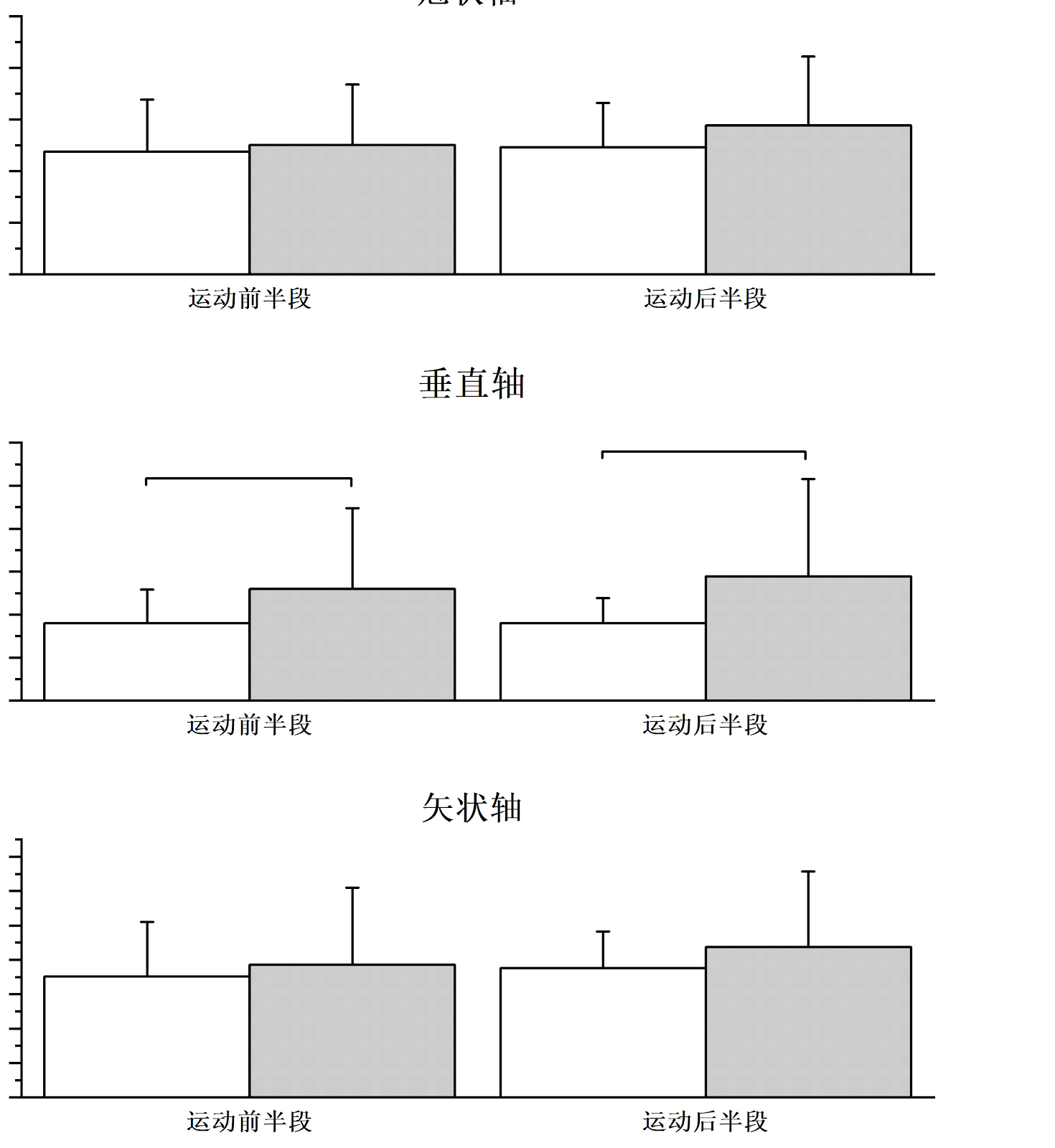
Perioperative hidden blood loss and risk factors in transforaminal lumbar interbody fusion calculated by a new method
Graphical Abstract
Original ResearchVol 1899, Issue 27 • pp. 100-112DOI: 10.12307/2026.21428Jan 15, 2026

Meta-analysis of application effect of 3D-printed artificial vertebral bodies in anterior cervical corpectomy and fusion

Authors: WU Ronghai, ZHENG Zhouhang, CHEN Huan, YOU Dongchun, GUO Weifeng, LIU Xingming, ZHANG Yu

OBJECTIVE: In recent years, many scholars have applied 3D-printed artificial vertebrae to anterior cervical vertebral subtotal vertebral resection and bone grafting fusion, but whether it is more effective than traditional titanium cages remains controversial. This study aims to systematically evaluate the effectiveness and safety of 3D-printed artificial vertebrae compared with traditional titanium cages as implants for anterior cervical corpectomy and fusion in the treatment of spondylosis. METHODS: Databases such as CNKI, WangFang, CBM, VIP, PubMed, EMBASE, and The Cochrane Library were searched to collect the clinical research on the application of 3D-printed artificial vertebrae in anterior cervical corpectomy and fusion from the establishment of each database to February 2025. After screening the literature, extracting the data and evaluating the methodological quality of the included studies, the meta-analysis was performed using Rev Man 5.4 software. RESULTS: A total of 10 studies were included, comprising 2 prospective randomized controlled studies, 6 retrospective cohort studies, and 2 prospective cohort studies, all of high quality. The included studies involved 534 patients, with 273 in the 3D-printed group and 261 in the control group. Meta-analysis results showed that the 3D-printed group was superior to the control group in terms of operation time [SMD=-1.13, 95%CI(-1.87, -0.39), P=0.003], loss of intervertebral disc height at last follow-up [SMD=-3.01, 95%CI(-5.74, -0.29), P=0.03], neck disability index at 3 months postoperatively [SMD=-0.34, 95%CI(-0.66, -0.03), P=0.03], prosthesis subsidence rate [OR=0.19, 95%CI(0.11, 0.32), P < 0.000 01], and postoperative dysphagia incidence [OR=0.43, 95%CI(0.21, 0.90), P=0.03], with significant differences. There were no significant differences in blood loss, hospital stay, postoperative Japanese Orthopaedic Association score, postoperative visual analogue scale score, postoperative neck disability index (at 6 months and last follow-up), and fusion rate between the two groups (P > 0.05). CONCLUSION: Compared with traditional titanium cages, 3D-printed artificial vertebral bodies have significant advantages in improving surgical efficiency, maintaining postoperative intervertebral disc height, reducing postoperative dysphagia incidence, and reducing prosthesis subsidence rate.
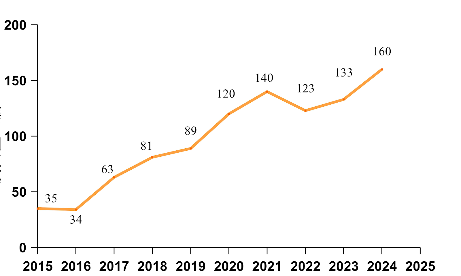
Meta-analysis of application effect of 3D-printed artificial vertebral bodies in anterior cervical corpectomy and fusion
Graphical Abstract
Original ResearchVol 1898, Issue 26 • pp. 100-112DOI: 10.12307/2026.21281Jan 15, 2026

Characteristics of lower limb muscle motor activation in patients with unilateral knee osteoarthritis

Authors: Shao Yunbo, Guo Jiayi, Li Feng

BACKGROUND: There are mechanical changes in the lower limb muscles in patients with knee osteoarthritis. These changes can be objectively reflected through surface electromyography. Currently, there is limited research on abnormal electrophysiological signals of lower limb muscles, especially of calf muscles, during walking in patients with unilateral knee osteoarthritis. OBJECTIVE: To investigate the motor activation status and activation mode of the medial thigh muscle, lateral thigh muscle, tibialis anterior muscle, and gastrocnemius muscle during daily walking in patients with unilateral knee osteoarthritis using surface electromyography. METHODS: Fifty subjects with unilateral knee osteoarthritis who met the inclusion criteria underwent surface electromyography walking tests. The target muscles were the medial thigh muscle, lateral thigh muscle, anterior tibialis muscle, and gastrocnemius muscle. The walking time was 60 seconds. Surface electromyography signals were collected from both healthy and affected sides. The main observation indicators were root mean square value, integrated electromyography value, median frequency, and muscle contraction ratio. Whether there is an abnormal activation status and its characteristics of lower limb muscles in unilateral knee osteoarthritis patients were investigated by surface electromyography. The abnormal movement activation of lower limb muscles in unilateral knee osteoarthritis patients was explored from an electrophysiological perspective. RESULTS AND CONCLUSION: (1) The integrated electromyography value and root mean square value of the medial and lateral thigh muscles on the affected side were significantly lower than those on the healthy side (P < 0.001); the median frequency values of the medial and lateral thigh muscles on the affected side were significantly lower than those on the healthy side (P < 0.001), with a more significant shift to low frequency, indicating a higher fatigue risk. (2) The integrated electromyography value and root mean square value of the tibialis anterior muscle on the affected side were significantly higher than those on the healthy side (P < 0.001); the median frequency value of the tibialis anterior muscle on the affected side was significantly lower than that on the healthy side (P < 0.001), with a more significant shift to low frequency, indicating a significantly increased fatigue risk. (3) The integrated electromyography value and root mean square value of the gastrocnemius muscle on the affected side were significantly higher than those on the healthy side (P < 0.001); the median frequency value of the gastrocnemius muscle on the affected side did not show a significant excessive shift compared with the healthy side (P > 0.05), indicating similar fatigue levels. (4) The muscle activation ratio on the affected side was significantly lower than that on the healthy side (P < 0.001), and the activation of the medial and lateral thigh muscles on the affected side was significantly unbalanced compared with the healthy side. These results indicate that during daily walking, patients with unilateral knee osteoarthritis exhibit insufficient activation and reduced activation efficiency of the medial and lateral thigh muscles, with high fatigue levels and risk, and unbalanced activation of these muscles, which is detrimental to knee joint stability and balance. The tibialis anterior and gastrocnemius muscles on the affected side show compensatory activation and abnormal co-activation levels, which are important factors aggravating joint stiffness, with the most obvious compensation in the tibialis anterior muscle, and there is a risk of fatigue-induced muscle atrophy.



Characteristics of lower limb muscle motor activation in patients with unilateral knee osteoarthritis
Graphical Abstract
Original ResearchVol 1899, Issue 27 • pp. 100-112DOI: 10.12307/2026.21420Jan 15, 2026

AI Algorithm Analysis for CT Three-Dimensional Diagnosis and Accurate Assessment of Posterior Cruciate Ligament Tibial Avulsion Fractures

Authors: CHENG Yongzhong, LI Rui, LUO Xiangli, WANG Fan, CHEN Yang, YAN Wei

BACKGROUND: Surgical decision-making for posterior cruciate ligament avulsion fractures is highly dependent on imaging evaluation. Traditional methods rely on subjective interpretation of CT images, which suffer from limitations such as difficulties in quantifying three-dimensional spatial displacement parameters and insufficient precision in assessing rotational angles. Given the advancements in artificial intelligence (AI) technology, there is a need to develop automated, intelligent image recognition software based on AI algorithms. OBJECTIVE: To investigate the intelligent diagnostic capabilities of AI algorithms for posterior cruciate ligament tibial avulsion fractures in 3D CT images and their effectiveness in accurately assessing 3D parameters of fracture fragments. METHODS: Knee CT data from 24 patients with posterior cruciate ligament tibial avulsion fractures who were treated at the Wangjing Hospital of the China Academy of Chinese Medical Sciences between December 1, 2022, and August 30, 2024, were retrospectively collected. Three-dimensional reconstruction, intelligent fracture point recognition, and simulated reduction were performed using self-developed AI image recognition software. Translational and rotational parameters of the fracture fragments along the X, Y, and Z axes were obtained. These measurements were compared with those from traditional radiology reading software (PACS system) using rank-sum tests, Bland-Altman analysis, and linear regression models to assess consistency, and coefficients of variation were calculated to verify software stability. RESULTS AND CONCLUSION: ①There were no significant differences between AI software and traditional methods in measuring fracture fragment displacement (X/Y/Z axis translation and rotation) (P > 0.05). ②Bland-Altman analysis showed good consistency between the two methods, with no significant differences (P > 0.05). ③Linear regression models for X, Y, Z axis displacement and angles showed R² values > 0.99. ④The coefficients of variation for three repeated fracture point identifications by the AI software showed that for total fracture identification, 21 cases had coefficients of variation < 20%, and for articular surface fracture points, 18 cases had coefficients of variation < 20%. ⑤These findings indicate that the AI image recognition software can accurately quantify three-dimensional parameters of posterior cruciate ligament avulsion fracture fragments, with measurement results consistent with traditional methods and good stability. It can assist doctors in judging the degree of displacement and provide precise data support for preoperative planning. The software has good application prospects in avulsion fractures, and future studies should expand the sample size and further verify its impact on surgical outcomes.

AI Algorithm Analysis for CT Three-Dimensional Diagnosis and Accurate Assessment of Posterior Cruciate Ligament Tibial Avulsion Fractures
Graphical Abstract
Original ResearchVol 1899, Issue 27 • pp. 100-112DOI: 10.12307/2026.21416Jan 15, 2026

Degree of paraspinal muscle fat infiltration predicts non-infectious poor wound healing following lumbar surgery

Authors: Xiong Shishuo, Xiang Rong, Zhang Yukai, Lan Qing, Guo Haiwei, Tian Han, Zhong Yelin, Zhang Yinyin, Guo Zehua, Lu Wenhao, Li Ying

BACKGROUND: Non-infectious poor wound healing following lumbar surgery is a significant clinical complication that prolongs hospitalization and increases the risk of reoperation. However, its predictive indicators remain unclear. Based on the hypothesis that paraspinal muscle degeneration may impede tissue repair by altering the local microenvironment, this study aimed to investigate the predictive value of preoperative paraspinal muscle fatty infiltration for non-infectious poor wound healing and its association with osteoporosis. OBJECTIVE: To quantify the degree of paraspinal muscle fatty infiltration using preoperative MRI and evaluate its predictive value for non-infectious poor wound healing after lumbar surgery. METHODS: A retrospective analysis was conducted on medical records of 4,368 patients who underwent traditional open posterior lumbar surgery at Third Affiliated Hospital of Guangzhou University of Chinese Medicine between 2019 and 2024. We screened 190 patients with a postoperative hospital stay of 15 days or longer. Based on postoperative wound healing and infection indicators, 41 patients with non-infectious poor healing were selected as the poor healing group. From the remaining 4,178 patients, 40 patients with good healing were selected as the good healing group. The poor healing group was further subdivided into osteoporosis and non-osteoporosis subgroups. Preoperative lumbar MRI images were collected, and Image J software was used to measure the cross-sectional area of the psoas major muscle and the percentage of fat infiltration in the erector spinae and multifidus muscles. RESULTS AND CONCLUSION: (1) There were no significant differences in gender, age, or diabetes between the poor healing and good healing groups (P > 0.05). (2) The functional cross-sectional area and fat infiltration percentage of the psoas major, erector spinae, and multifidus muscles were significantly different between the two groups (P < 0.05). (3) Logistic regression analysis showed that fat infiltration percentage was an independent risk factor for poor wound healing. (4) Receiver operating characteristic curve analysis showed that fat infiltration percentage had high predictive value for poor wound healing (area under the curve > 0.7). (5) One-way ANOVA indicated that osteoporosis was a risk factor for fat infiltration in the L4 multifidus muscle (P < 0.05). (6) The results indicate that paraspinal muscle fat infiltration percentage is an important predictor of non-infectious poor wound healing after traditional open posterior lumbar surgery, providing clinical reference. Osteoporosis was also confirmed as a risk factor for L4 multifidus fat infiltration, but due to the small subgroup sample size, whether osteoporosis affects non-infectious poor healing after lumbar surgery requires further clinical trials.

Degree of paraspinal muscle fat infiltration predicts non-infectious poor wound healing following lumbar surgery
Graphical Abstract
Original ResearchVol 1899, Issue 27 • pp. 100-112DOI: 10.12307/2026.21412Jan 15, 2026

Impact of disease duration on lumbar curvature correction in patients with rigid post-traumatic thoracolumbar kyphosis

Authors: Yang Junjie, Zhang Hao, Chen Zhike, Chen Yao, Jia Bingxu, Wang Qing, Li Guangzhou, Wang Gaoju

BACKGROUND: Currently, most studies on rigid post-traumatic thoracolumbar kyphosis focus on overall sagittal balance and surgical intervention, while the effect of the disease duration on the change of lumbar compensatory curvature and degeneration is still unclear. OBJECTIVE: To explore the effect of the disease duration on lumbar degeneration and the potential mechanism of rigid post-traumatic thoracolumbar kyphosis in patients with rigid post-traumatic thoracolumbar kyphosis, and provide a basis for optimizing treatment strategies. METHODS: Clinical and imaging data from 79 rigid post-traumatic thoracolumbar kyphosis patients were retrospectively analyzed. The patients were divided into two groups according to the disease duration: Patients with a disease duration of ≤ 5 years were categorized as group A (n=40), and those with > 5 years as group B (n=39). X-ray images were used to measure the local kyphosis angle of the injured vertebra, the height of the posterior walls of the injured vertebra and adjacent vertebrae, lumbar lordosis, the intervertebral space angle for each lumbar segment, and sacral slope. The Weishaupt-CT classification system was employed to assess lumbar facet joint degeneration. Pfirrmann-MRI grading was used to evaluate intervertebral disc degeneration. Clinical outcomes including visual analog scale for back pain, Oswestry Disability Index, SRS-22 score, and American Spinal Injury Association impairment scale were compared between groups. The influence of disease duration on clinical symptoms and imaging features was analyzed. RESULTS AND CONCLUSION: (1) There were no significant differences in age, sex, visual analog scale score, fracture site, fracture morphology, or American Spinal Injury Association grade between the two groups (P > 0.05). The SRS-22 subscore was significantly higher in group A than in group B (P < 0.05), while the Oswestry Disability Index was significantly higher in group B than in group A (P < 0.05). (2) The local kyphosis angle, lumbar lordosis, and L4/5 intervertebral space angle were significantly greater in group B than in group A (P < 0.05). (3) There were no significant differences in L1/2, L2/3, L3/4, L5/S1 intervertebral space angles and sacral slope between groups (P > 0.05). (4) The degree of facet joint degeneration at L3/4, L4/5, and L5/S1 was significantly more severe in group B than in group A (P < 0.05). The degree of intervertebral disc degeneration at L2/3, L3/4, L4/5, and L5/S1 was significantly more severe in group B than in group A (P < 0.05). (5) Pearson correlation analysis showed that within group B, disease duration was positively correlated with local kyphosis angle and lumbar lordosis (r=0.335, 0.418, P < 0.05). (6) In patients with rigid post-traumatic thoracolumbar kyphosis, long-term compensation leads to increased lumbar lordosis and accelerated lumbar degeneration. The L4/5 segment is the main compensatory segment in lumbar curvature compensation, and special attention should be paid to the correction of lower lumbar curvature during surgical correction.

Impact of disease duration on lumbar curvature correction in patients with rigid post-traumatic thoracolumbar kyphosis
Graphical Abstract
Original ResearchVol 1899, Issue 27 • pp. 100-112DOI: 10.12307/2026.21413Jan 15, 2026

Comparison of stability of percutaneous minimally invasive pedicle screw insertion in thoracolumbar fractures through and across the injured vertebra under navigation

Authors: JIA Yingao, QIAO Lina, ZHANG Chao, LIANG Jiawen, XUE Liang, WANG Fei

BACKGROUND: Recently, percutaneous minimally invasive pedicle screw fixation systems have become a popular treatment option for thoracolumbar fractures due to ongoing innovation in orthopedic internal fixation devices. Currently, while many studies compare the effectiveness of open pedicle screw fixation through or across the fractured vertebra, fewer studies compare these two methods when using navigation-assisted percutaneous minimally invasive techniques. OBJECTIVE: To compare the clinical efficacy of percutaneous minimally invasive pedicle screw fixation in the treatment of thoracolumbar fracture through and across the injured vertebra. METHODS: A retrospective analysis was performed on 67 patients with single level thoracolumbar fracture without spinal cord nerve injury who were treated in the Department of Spine Surgery, Affiliated Hospital of Yan'an University from October 2021 to June 2023. All of them were treated with percutaneous pedicle screw fixation with the assistance of computer navigation, and were followed up to 6 months after surgery. The injured vertebrae were divided into transinjured vertebrae group (n=35) and cross-injured vertebrae group (n=32). RESULTS AND CONCLUSION: ① There were no significant differences in preoperative general data between the two groups (P > 0.05), indicating comparability. ② Intragroup comparison: In both groups, the anterior vertebral height ratio, Cobb angle, visual analog scale score, and Oswestry disability index at 7 days and 6 months after surgery were significantly improved compared with preoperative values (P < 0.05). ③ Intergroup comparison: The operation time, intraoperative blood loss, incision length, and intraoperative fluoroscopy times in the cross-injured vertebrae group were significantly less than those in the trans-injured vertebrae group (P < 0.05), while there was no significant difference in hospital stay between the two groups (P > 0.05). At 7 days and 6 months after surgery, there were no significant differences in anterior vertebral height ratio, Cobb angle, visual analog scale score, and Oswestry disability index between the two groups (P > 0.05). ④ It is suggested that for patients with single-level thoracolumbar fractures without spinal cord nerve injury, compared with trans-injured vertebra fixation, percutaneous minimally invasive pedicle screw fixation across the injured vertebra has advantages of shorter operation time, less bleeding, smaller surgical incision, and fewer fluoroscopy times. In the medium-term follow-up period, both trans-injured and cross-injured percutaneous minimally invasive fixation can effectively restore and maintain the height of the injured vertebra, correct kyphotic deformity, rebuild spinal stability, and relieve pain, achieving excellent clinical results. For patients with AO type A thoracolumbar fractures with normal body mass index, without spinal cord nerve injury and posterior ligamentous complex injury, the less traumatic cross-injured vertebra percutaneous pedicle screw fixation is recommended.

Comparison of stability of percutaneous minimally invasive pedicle screw insertion in thoracolumbar fractures through and across the injured vertebra under navigation
Graphical Abstract
Original ResearchVol 1899, Issue 27 • pp. 100-112DOI: 10.12307/2026.21410Jan 15, 2026

Anatomical risk factor analysis of posterior cruciate ligament tibial avulsion fracture in adults

Authors: LI Gen, ZHANG Xichen, SUN Yingjin, HE Chenglong, GAO Xuren

BACKGROUND: The anatomical morphology of the distal femur and proximal tibia has an important influence on the movement of the tibiofemoral joint, and its role in anterior cruciate ligament injury has been well described. However, the research on the anatomical risk factors of posterior cruciate ligament tibial avulsion fracture is still limited, and no unified consensus has been formed. OBJECTIVE: To investigate the anatomical risk factors for posterior cruciate ligament tibial avulsion fracture. METHODS: The medical records of 53 patients who underwent surgical treatment for posterior cruciate ligament tibial avulsion fracture from March 2021 to September 2024 were retrospectively collected as the posterior cruciate ligament avulsion group (32 males and 21 females), and the data of 53 subjects without posterior cruciate ligament injury in the same period were included as the posterior cruciate ligament normal group (24 males and 29 females). The intercondylar notch width, femoral condyle width, intercondylar notch width index, intercondylar notch height, intercondylar notch shape index, intercondylar notch angle, Blumensaat's line inclination angle, medial tibial posterior slope, and lateral tibial posterior slope were measured and calculated in Magnetic Resonance Imaging to analyze the differences in the anatomical data of the two groups. Binary logistic regression analysis was used to determine the independent risk factors and to establish a risk factor model by receiver operating characteristic curve. RESULTS AND CONCLUSION: (1) The intercondylar notch width index, intercondylar notch shape index, intercondylar notch angle, and medial tibial posterior slope in the posterior cruciate ligament avulsion group were significantly smaller than those in the posterior cruciate ligament normal group (P < 0.05); there were no significant differences in intercondylar notch width, femoral condyle width, intercondylar notch height, Blumensaat's line inclination angle, and lateral tibial posterior slope between the two groups (P > 0.05). (2) Binary logistic regression found that intercondylar notch width index was associated with posterior cruciate ligament tibial avulsion fracture. (3) It is suggested that compared with those with normal posterior cruciate ligament, patients with posterior cruciate ligament tibial avulsion fracture have smaller intercondylar notch width index, intercondylar notch shape index, intercondylar notch angle, and medial tibial posterior slope; intercondylar notch width index is an independent risk factor for posterior cruciate ligament tibial avulsion fracture.

Anatomical risk factor analysis of posterior cruciate ligament tibial avulsion fracture in adults
Graphical Abstract
Original ResearchVol 1899, Issue 27 • pp. 100-112DOI: 10.12307/2026.21421Jan 15, 2026

Diagnostic value of diffusion-weighted imaging with different diffusion sensitivity coefficients for spinal tuberculosis and Brucellar spondylitis

Authors: SUN Na, WANG Hui, ZHAO Guyue, YU Honghao

BACKGROUND: Although the clinical manifestations of spinal tuberculosis and Brucella spondylitis are similar, the treatment options are different, and traditional imaging examinations are difficult to accurately distinguish them. Diffusion-weighted imaging can quantify the diffusion differences of water molecules in tissues through apparent diffusion coefficient, which may provide a new way for early identification. However, the correlation between apparent diffusion coefficient and inflammatory markers under different diffusion sensitivity coefficients is not clear. OBJECTIVE: To explore the clinical value of diffusion-weighted imaging with different diffusion sensitivity coefficients in the diagnosis of spinal tuberculosis and Brucella spondylitis, and to analyze the correlation between apparent diffusion coefficient and inflammatory indicators. METHODS: Sixty patients with spinal tuberculosis and 60 patients with Brucella spondylitis who were admitted and diagnosed in Shengjing Hospital Affiliated to China Medical University from June 2021 to June 2024 were randomly selected as the research objects. Among them, there were 35 males and 25 females in the spinal tuberculosis group, with an average age of (53.36±5.45) years; and 38 males and 22 females in the Brucella spondylitis group, with an average age of (55.47±6.43) years. The area under the curve was used to evaluate the diagnostic value of diffusion-weighted imaging-apparent diffusion coefficient at different diffusion sensitivity coefficients in differentiating spinal tuberculosis and Brucella spondylitis. Pearson correlation analysis was used to test the relationships between variables. Paired t-test was used to compare the apparent diffusion coefficients between operator A and B. Intraclass correlation coefficient was used to analyze the consistency of measurements between different operators and the same operator. RESULTS AND CONCLUSION: (1) There were significant differences in erythrocyte sedimentation rate, C-reactive protein, white blood cell level, and distribution of lumbar and thoracic involvement between the two groups (P < 0.05). Spinal tuberculosis most commonly involved the lumbar and thoracic spine, while Brucella spondylitis most commonly involved the lumbar spine. (2) The apparent diffusion coefficients of the affected vertebral bodies in the spinal tuberculosis group were significantly higher than those in the Brucella spondylitis group at all diffusion sensitivity coefficients (P < 0.05). Within the same group, there were significant differences in apparent diffusion coefficients between the affected vertebral body and paravertebral abscess at different diffusion sensitivity coefficients (P < 0.05). (3) The diagnostic efficiency of apparent diffusion coefficient was the highest at a diffusion sensitivity coefficient of 400 s/mm2, with a sensitivity of 95.00% and specificity of 96.67%. (4) The intraclass correlation coefficients of diffusion-weighted imaging-apparent diffusion coefficient between different operators for both spinal tuberculosis and Brucella spondylitis were greater than 0.75. (5) Pearson correlation analysis showed that the apparent diffusion coefficients of the affected vertebral bodies at different diffusion sensitivity coefficients (1.03×10⁻³-1.49×10⁻³ mm2/s) were significantly correlated with erythrocyte sedimentation rate, C-reactive protein, and white blood cell level (P < 0.001). (6) These findings indicate that diffusion-weighted imaging is a simple and stable method for detecting spinal tuberculosis and Brucella spondylitis. The diffusion-weighted imaging-apparent diffusion coefficient at a diffusion sensitivity coefficient of 400 s/mm2 has the greatest diagnostic efficacy in differentiating spinal tuberculosis from Brucella spondylitis, and the apparent diffusion coefficients of the affected vertebral bodies at different diffusion sensitivity coefficients are closely related to inflammatory status.


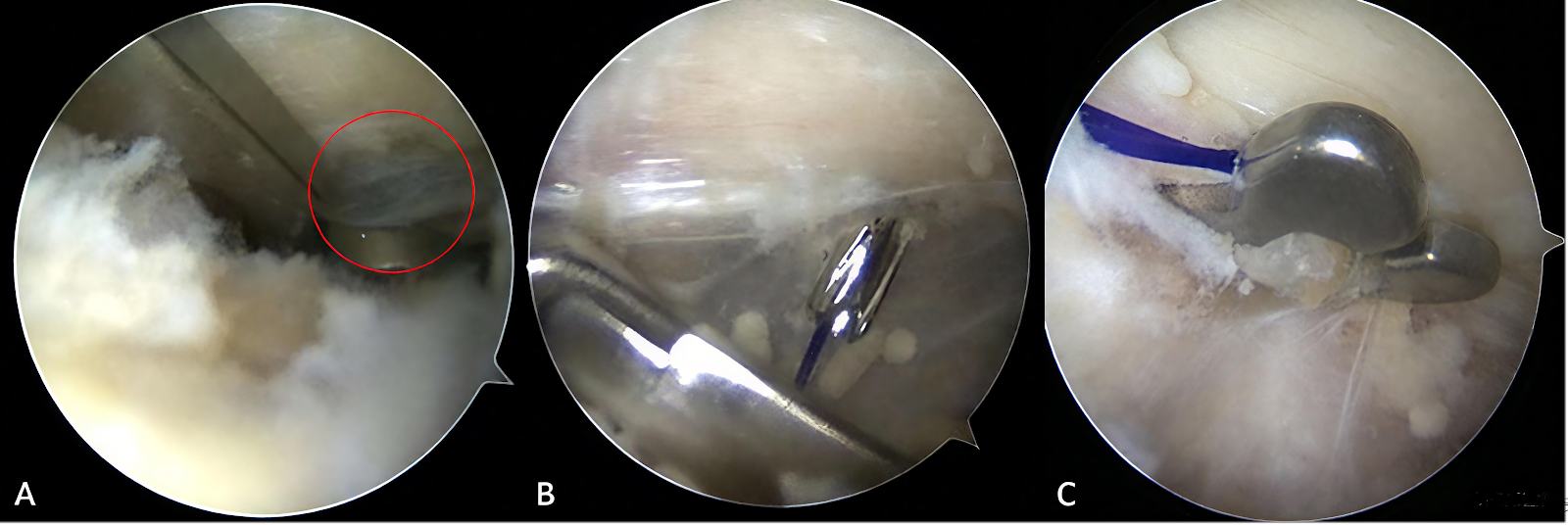
Diagnostic value of diffusion-weighted imaging with different diffusion sensitivity coefficients for spinal tuberculosis and Brucellar spondylitis
Graphical Abstract
Original ResearchVol 1899, Issue 27 • pp. 100-112DOI: 10.12307/2026.21424Jan 15, 2026

Dynamic evolution of evaluation standards for effectiveness and safety after anterior cruciate ligament reconstruction in the knee

Authors: Li Chengke, Gao Miaomiao, Lei Lei, Ma Rongxing, Zhang Jingyu, Hu Yongcheng

BACKGROUND: A combination of subjective and objective evaluation criteria is often required to more accurately and comprehensively assess knee function in patients undergoing anterior cruciate ligament reconstruction. OBJECTIVE: To review the evolving trends in effectiveness and safety evaluation criteria after anterior cruciate ligament reconstruction and analyze the dynamic shift in the use of subjective and objective assessment tools. METHODS: A systematic search of PubMed and Embase was conducted up to August 22, 2023 to identify studies assessing knee function after anterior cruciate ligament reconstruction. A total of 136 eligible studies meeting the inclusion criteria were included. The frequency of each evaluation standard was extracted and analyzed over time using Origin 2025 software. RESULTS AND CONCLUSION: (1) Between 1990 and 2005, objective measures were widely applied. Since 2005, subjective scoring systems, particularly patient-reported outcome measures, have increased sharply, surpassing objective standards in frequency from 2009 onward. (2) Early use was dominated by the Lysholm scale and Tegner activity score, while the International Knee Documentation Committee-Subjective Knee Form, Knee Injury and Osteoarthritis Outcome Score, and anterior cruciate ligament–return to sport after injury gradually emerged as the main tools in later years. (3) In contrast, objective assessments such as the KT1000/2000 arthrometer, Lachman test, and hop test remained relatively stable but showed an overall declining trend. (4) These findings indicate a paradigm shift from objective knee stability to patient-centered subjective experience in evaluating ACL reconstruction outcomes. (5) This study is the first to quantitatively reveal the dynamic evolution of mainstream evaluation tools, highlighting the current emphasis on combining subjective and objective criteria. The recommended combination is the International Knee Documentation Committee-Subjective Knee Form or Knee Injury and Osteoarthritis Outcome Score plus anterior cruciate ligament–return to sport after injury plus KT1000/2000 or hop test, to comprehensively reflect knee function recovery and patient perception, providing an evidence base for future comprehensive assessment approaches.

Dynamic evolution of evaluation standards for effectiveness and safety after anterior cruciate ligament reconstruction in the knee
Graphical Abstract
Original ResearchVol 1899, Issue 27 • pp. 100-112DOI: 10.12307/2026.21418Jan 15, 2026

Micromorphological characteristics of human scaphoid bone based on Micro CT imaging technology

Authors: ZHANG Dezhou, LYU Yuan, LI Kun, CHEN Jie, YANG Xiangrong, ZHAO Hailong, ZHANG Shaojie, MA Yuan, HAO Yunteng, YANG Yang, GAO Mingjie, WANG Zhiqiang, LI Zhijun, SHI Jun, WANG Xing

BACKGROUND: Clinically, due to the special anatomical characteristics and internal trabecular bone distribution of the scaphoid, the treatment effect of fractures is generally poor, often leading to nonunion and ischemic necrosis, which in turn causes wrist arthritis and loss of function. OBJECTIVE: To scan scaphoid specimens using Micro CT technology, analyze their internal microstructure characteristics, measure the trabecular bone microstructure parameters in each region, and discover regional differences in scaphoid trabecular bone, aiming to provide a scientific basis for the prevention, treatment, and fracture mechanism research of scaphoid fractures. METHODS: Bilateral scaphoid bones (10 cases) from 5 adult cadaver specimens were scanned by Micro CT. By selecting and reconstructing trabecular bone in three regions of interest (tubercle, waist, and body), the internal micromorphological characteristics of the scaphoid were observed in detail, and the differences in trabecular bone microstructure parameters among regions were measured and compared. RESULTS AND CONCLUSION: (1) Micro CT images showed that the cortical bone on the surface of the scaphoid was relatively thin, and the interior was filled with complex trabecular bone microstructure; the lamellar trabecular bone near the cortical bone was relatively dense, extending inward into rod-like trabecular bone. From sagittal, coronal, and transverse sections, the trabecular bone distribution in the waist was relatively sparse, while that in the body and tubercle was denser. (2) There were significant differences in bone volume fraction, bone surface area, bone surface area to tissue volume ratio, trabecular separation, trabecular number, trabecular connectivity, trabecular connection density, fractal dimension, bone mineral density, and bone mineral content of the scaphoid tubercle between left and right sides (P < 0.05). There were no significant differences in the trabecular bone microstructure parameters of the waist and body between left and right sides (P > 0.05). (3) There were significant differences in bone volume, bone volume fraction, bone surface area, bone surface area to tissue volume ratio, bone surface area to bone volume ratio, bone mineral density, and bone mineral content between the body and the tubercle/waist (P < 0.05). There was a significant difference in trabecular thickness between the body and the tubercle (P < 0.05). There were significant differences in trabecular separation and fractal dimension among the body, tubercle, and waist (P < 0.05). There were significant differences in trabecular number, trabecular connectivity, and trabecular connection density between the waist and the tubercle/body (P < 0.05). There were no significant differences in tissue volume and degree of anisotropy among the body, tubercle, and waist (P > 0.05). (4) The results showed that the trabecular bone microstructure parameters of the scaphoid had regional differences, among which the waist had lower bone density and strength, making it the most prone to fracture. This finding provides a theoretical basis for understanding the fracture mechanism of the scaphoid from the perspective of trabecular bone microstructure. At the same time, the trabecular bone structure characteristics of different parts of the scaphoid revealed in this study also provide a theoretical basis for designing targeted internal fixation instruments.

Micromorphological characteristics of human scaphoid bone based on Micro CT imaging technology
Graphical Abstract
Original ResearchVol 1899, Issue 27 • pp. 100-112DOI: 10.12307/2026.21427Jan 15, 2026

Bone grafting for repairing scaphoid nonunion

Authors: Guo Wen, Gao Binli, Wang Yang, Lin Sen

BACKGROUND: Due to the relatively special symptoms and anatomical structure of scaphoid fractures, untreated fractures or those with delayed treatment often lead to non-union of the fracture, carpal joint collapse, or loss of function, subsequently resulting in persistent or intermittent pain, swelling, and limited mobility in the carpal joint. Currently, although there are a wide variety of bone grafting techniques and the choice of treatment methods is complex, there is still no consensus on which bone grafting surgical method is superior. OBJECTIVE: To review the research status of scaphoid nonunion, summarize different bone grafting surgeries for the treatment of nonunion of fractures at home and abroad in recent years, explore the clinical efficacy, advantages and disadvantages of various bone grafting techniques, and provide guidance for clinical diagnosis and treatment. METHODS: A computer was used to search for relevant articles published in the PubMed, MEDLINE, EMBASE, CNKI, China Medical Library, VIP, and WanFang databases from 1980 to 2024. The Chinese and English search terms were “scaphoid nonunion, surgery, bone graft, bone flap, scaphoid proximal pole.” A total of 687 articles were retrieved, and 54 articles were selected for review through inclusion and exclusion criteria. RESULTS AND CONCLUSION: (1) Non-vascularized graft is still the mainstream treatment for scaphoid nonunion. Among them, simple cancellous bone transplantation has fast bone formation (healing rate 85%-100%) and is suitable for cases without deformity; cortical cancellous bone has strong support (healing rate 88%-93%), which is more conducive to restoring the morphology of the scaphoid. (2) There is no significant difference in the healing rate between iliac bone and distal radial donors, but the distal radial donor site has fewer complications (5% vs. 18%). (3) Vascularized grafts can significantly improve the efficacy of complex cases: radial styloid bone flap healing rate 81%-92%, pronator quadratus pedicled flap healing rate 93.3%, free medial femoral condyle flap has outstanding effect on joint surface reconstruction (healing rate 95%), free iliac bone flap success rate 91.7%-100%, but high technical requirements. (4) The treatment of scaphoid nonunion needs to follow the principle of individualization: non-vascularized grafts are suitable for simple, well-vascularized fractures, while vascularized grafts are aimed at ischemic necrosis or complex deformities. (5) In the future, standardized imaging evaluation (such as MRI grading), biological enhancement techniques (bone morphogenetic protein 2, 3D printed scaffolds), and the popularization of microsurgery are needed to improve efficacy, while combining patient age and occupational needs to formulate the optimal plan, balancing functional recovery and complication prevention.

Bone grafting for repairing scaphoid nonunion
Graphical Abstract
Original ResearchVol 1899, Issue 27 • pp. 100-112DOI: 10.12307/2026.21411Jan 15, 2026

Arthroscopy-assisted loop titanium plate versus clavicular hook plate for acute acromioclavicular joint dislocation: clinical efficacy and cost-effectiveness

Authors: Li Kanglin, Gao Shihua, Jiang Yongdong, Huang Ziqi, Wu Yufeng

BACKGROUND: Surgical management of acute Rockwood type III-V acromioclavicular joint dislocations remains controversial. While clavicular hook plate fixation is widely adopted, it carries risks of postoperative complications. The emerging arthroscopy-assisted loop titanium plate technique, despite its minimally invasive advantages, requires further validation regarding long-term efficacy and cost-effectiveness. OBJECTIVE: To compare the clinical efficacy and cost-effectiveness of modified arthroscopic loop titanium plate fixation versus clavicular hook plate fixation for the treatment of acute Rockwood type III-V acromioclavicular joint dislocations. METHODS: Eighty eligible patients with acute Rockwood type III-V acromioclavicular joint dislocations admitted at Zhongshan Hospital of Traditional Chinese Medicine Affiliated to Guangzhou University of Chinese Medicine between January 2021 and July 2024 were enrolled and randomly assigned to either the loop titanium plate group (n=40) or the hook plate group (n=40). Ultimately, 31 and 33 patients in each group completed follow-up, respectively. The loop titanium plate group underwent fixation using the FixButtonTM suspension system with 2.4 mm bone tunnels; the hook plate group received open reduction and internal fixation with a clavicular hook plate. Outcome measures included operative time, incision length, blood loss, hospitalization costs, visual analog scale (VAS) for pain, Constant-Murley score, coracoclavicular distance, acromioclavicular distance, and complication rates, with a follow-up of 6 months. RESULTS AND CONCLUSION: The loop titanium plate group had longer operative time, shorter incisions, less blood loss, but higher hospitalization costs (P < 0.05). At 6 months postoperatively, the excellent-to-good rate in the loop titanium plate group (94%) was significantly higher than that in the hook plate group (82%) (P < 0.05); VAS scores were lower and Constant-Murley scores were higher (P < 0.05); coracoclavicular and acromioclavicular distances were better restored (P < 0.05). The total complication rate in the loop titanium plate group (6%) was lower than that in the hook plate group (12%) (P < 0.05). These findings suggest that although the modified arthroscopic loop titanium plate technique requires longer operative time and higher costs, it significantly reduces trauma, alleviates pain, improves shoulder function, and lowers complication risk, offering superior short-term clinical efficacy. Despite higher upfront costs, the potential reduction in secondary surgeries may confer long-term economic benefits. This technique is more suitable for patients with high demands for cosmetic incisions and functional recovery. Future studies with larger sample sizes and longer follow-up are needed to further validate these conclusions.

Arthroscopy-assisted loop titanium plate versus clavicular hook plate for acute acromioclavicular joint dislocation: clinical efficacy and cost-effectiveness
Graphical Abstract
Original ResearchVol 1899, Issue 27 • pp. 100-112DOI: 10.12307/2026.21408Jan 15, 2026

Application of correction leverage technique in primary failure of distal locking screw during antegrade femoral intramedullary nailing

Authors: HE Zhiyu, WAN Zhihong, HUANG Yu, LIU Anming, WU Jiaqi, WANG Guoyou, ZHANG Lei, CHEN Xiaojun

BACKGROUND: Interlocking intramedullary nail fixation is the "gold standard" for the treatment of femoral shaft fractures, and the difficulty of distal locking nail implantation has always been a difficult problem to solve. OBJECTIVE: By comparing the clinical effects of correction leverage technique and free-hand locking nail technique, it is further explained whether the correction leverage technique can be fast and accurate. The distal locking screw of femoral intramedullary nail was placed without direct X-ray radiation exposure, thereby solving the problem of difficult distal locking screw placement. METHODS: A total of 52 patients with femoral shaft fractures who had difficulty in distal locking screw placement during interlocking intramedullary nail fixation were enrolled from the Department of Orthopedics and Traumatology, Affiliated Traditional Chinese Medicine Hospital of Southwest Medical University from July 2022 to September 2024. They were divided into two groups according to the placement protocol: correction leverage group (26 cases) used the correction leverage technique for distal locking screw placement, and free-hand group (26 cases) used the traditional free-hand technique. The distal locking screw placement time, number of X-ray exposures, and first-attempt accuracy were compared between the two groups. RESULTS AND CONCLUSION: (1) The distal locking screw placement time in the correction leverage group was significantly shorter than that in the free-hand group (t=-4.136, P < 0.001). (2) The number of X-ray exposures in the correction leverage group was less than that in the free-hand group (t=-19.696, P < 0.001). (3) The first-attempt accuracy in the correction leverage group (100%) was higher than that in the free-hand group (71%), with a significant difference (χ2=5.253, P < 0.05). (4) These results indicate that compared with simple free-hand screw placement, the correction leverage technique has the advantages of faster locking, higher accuracy, and lower X-ray radiation. This technique does not require auxiliary equipment and has strong operability, and is worthy of further clinical validation and promotion for interlocking intramedullary nail fixation of femoral shaft fractures.

Application of correction leverage technique in primary failure of distal locking screw during antegrade femoral intramedullary nailing
Graphical Abstract
Original ResearchVol 1899, Issue 27 • pp. 100-112DOI: 10.12307/2026.21402Jan 15, 2026

Relationship between gait parameter characteristics and joint function recovery after arthroscopic minimally invasive surgery in patients with knee osteoarthritis

Authors: Fang Chao, Deng Miao, Wang Yang, Gao Fei, Li Jiahang, Yu Fangfang

BACKGROUND: There are individual differences in the effectiveness of arthroscopic surgery in improving knee osteoarthritis, and subjective scoring scales may be biased in evaluating the clinical efficacy of arthroscopic treatment for knee osteoarthritis. OBJECTIVE: To explore the correlation between gait parameters and joint function recovery in patients with knee osteoarthritis after arthroscopic surgery. METHODS: A total of 98 patients with knee osteoarthritis admitted to Chongqing Armed Police Corps Hospital from October 2023 to October 2024 were selected as the research subjects. According to Lysholm knee function score after 6 months of follow-up, they were divided into the excellent group (n=63) and the fair group (n=35). Clinical data including gender, age, body mass index, disease duration, respiration, heart rate, Kellgren-Lawrence grade, smoking history, drinking history, hypertension history, location of onset, and postoperative complications were collected. The intraoperative and postoperative indicators, as well as the knee joint function scores and gait parameters at different times before and after surgery were compared between the two groups. Multivariate Logistic regression was used to analyze the independent influencing factors of knee joint function recovery. Stratified regression analysis was conducted to explore the impact of different clinical and pathological characteristics after treatment on gait parameters. Generalized estimating equations were used to analyze the differences in gait parameters among patients with different knee joint functions after treatment. Generalized additive models were used to analyze the impact of gait parameters on Lysholm score after treatment. Receiver operating characteristic curves were drawn to analyze the value of gait parameters in judging the recovery of knee joint function after treatment. RESULTS AND CONCLUSION: (1) There were significant differences in age, disease duration, Kellgren-Lawrence grade, and postoperative complications between the excellent and fair groups (P < 0.05). (2) The fair group had longer operation time, more intraoperative blood loss, longer postoperative swelling regression time, and longer rehabilitation time than the excellent group (P < 0.05). (3) After surgery, the fair group had higher Western Ontario and McMaster Universities Osteoarthritis Index and visual analog scale scores, and lower Lysholm score, step frequency, and step speed than the excellent group (P < 0.05). (4) Logistic regression analysis showed that age, Kellgren-Lawrence grade, postoperative complications, and postoperative swelling regression time were independent risk factors affecting knee joint function recovery (P < 0.05). (5) Stratified regression analysis showed that age, Kellgren-Lawrence grade, postoperative swelling regression time, and postoperative complications all had negative effects on step frequency and step speed (β < 0, P < 0.05). (6) Generalized estimating equation analysis showed that the degree of knee joint function recovery was associated with gait characteristics (β > 0, P < 0.05). (7) Generalized additive model analysis showed that the effects of step frequency and step speed on Lysholm score after treatment were linear. (8) Receiver operating characteristic curve analysis showed that the combined detection of step frequency and step speed had higher predictive efficacy (area under the curve > 0.85, P < 0.05). (9) These findings suggest that arthroscopic surgery can improve knee joint function in patients with knee osteoarthritis, and dynamic tracking of postoperative gait parameter changes combined with functional scoring scales reveals the association between step frequency, step speed, and knee joint function outcome, further clarifying its clinical predictive value and providing a new quantitative tool for clinical functional assessment to achieve more precise postoperative rehabilitation guidance.

Relationship between gait parameter characteristics and joint function recovery after arthroscopic minimally invasive surgery in patients with knee osteoarthritis
Graphical Abstract
Original ResearchVol 1899, Issue 27 • pp. 100-112DOI: 10.12307/2026.21409Jan 15, 2026

Application of a modified rectangular locking device in the treatment of femoral shaft fractures with intramedullary nailing

Authors: ZHANG Ping, ZHOU Da-fang, CHEN Hui, ZHANG Ying, LIU Qing, SHI Ce

BACKGROUND: Conventional rectangular targeting devices are divided into two modules, one left and one right, which hinders flexible intraoperative use and is prone to deformation and error. Even after connection, repeated fluoroscopic confirmation is still required for screw drilling and placement. OBJECTIVE: To investigate the key technology development and clinical application of a modified rectangular locking device for precise distal locking screw placement in intramedullary nailing of femoral shaft fractures. METHODS: Medical records of patients with femoral shaft fractures admitted to Affiliated Suqian Hospital of Xuzhou Medical University and Suyu District People's Hospital from 2021 to 2023 were collected. Patients aged 18-65 years, diagnosed with femoral shaft fractures (AO classification: A, B, or C) by radiographic examination, who underwent closed reduction and intramedullary nailing with distal locking screw placement using the modified rectangular locking device, and whose clinical data (including medical history, radiographic findings, surgical records, and follow-up records) were selected. Forty-one cases met the criteria, including 30 males and 11 females, aged 20-62 years, with an average age of (41.17±8.14) years. Intraoperative fluoroscopy times, locking success rate, time for successful distal locking screw placement, American Knee Society Score at 1 month postoperatively and after fracture healing, and fracture healing time were collected. RESULTS AND CONCLUSION: (1) All 41 patients underwent distal locking screw placement using the modified rectangular locking device. Intraoperative fluoroscopy times ranged from 0 to 2 times, with an average of (1.1±0.5) times; the locking success rate was 98%; the time for successful distal locking screw placement ranged from 6 to 10 minutes, with an average of (7.0±1.5) minutes; the American Knee Society Score at 1 month postoperatively ranged from 140 to 190, with an average of (150±15) points. (2) Thirty-eight patients were followed up completely for 12-24 months. Fracture healing time ranged from 9 to 14 months, with an average of (10.5±2.5) months. After fracture healing, the American Knee Society Score ranged from 150 to 190, with an average of (185±8) points, with 35 excellent and 3 good results. (3) The results indicate that compared with freehand locking, oblique fluoroscopic placement, arthroscopic-assisted placement, and electromagnetic navigation locking of distal locking screws, the modified rectangular locking device offers advantages including no dependence on arthroscopic or electromagnetic navigation equipment, no requirement for extensive surgical experience, simple steps, accurate locking, high repeatability, and reduced radiation exposure.

Application of a modified rectangular locking device in the treatment of femoral shaft fractures with intramedullary nailing
Graphical Abstract
Original ResearchVol 1899, Issue 27 • pp. 100-112DOI: 10.12307/2026.21404Jan 15, 2026

Morphometric analysis of principal stress trabeculae in the proximal femur

Authors: Cao Meng, Li Lun, Tian Simiao, Cao Fang, Song Liqun, Zhao Dewei

BACKGROUND: The principal compressive and tensile trabecular bones in the proximal femur are essential structures responsible for transmitting forces through the hip joint. Given their complex and heterogeneous composition, exploring their microstructural variations will help improve our understanding of principal stress trabeculae. OBJECTIVE: To extract trabecular microstructural information from various regions of femoral head specimens and study their morphometric patterns, elucidate the microstructural variations of principal stress trabeculae, and provide a theoretical basis for the design of future bionic hip prostheses. METHODS: Totally 11 fresh femoral heads were obtained from patients with femoral neck fractures. They were scanned by Micro-CT and reconstructed by image analysis software (InveonTM Acquisition Workplace). The principal compressive and tensile trabecular bones were segmented into five regions respectively. After selecting the volumes of interest for each region, morphometric analyses were subsequently performed on bone volume fraction, bone surface area fraction, trabecular thickness, trabecular number, trabecular spacing, trabecular pattern factor, and grayscale value. RESULTS AND CONCLUSION: (1) Among the principal compressive trabeculae, Region 5 exhibited the highest bone volume fraction, trabecular thickness, and grayscale value, followed by Region 4 and 1, with statistically significant differences between Region 5 and 1 (all P < 0.05). Moving from Region 3 to Region 2, 1, and Regions 4, 5, there was a decreasing trend in bone surface area fraction and trabecular number, accompanied by an increasing trend in trabecular thickness. (2) Among the principal tensile trabeculae, Region 3 displayed the highest values for bone volume fraction, trabecular thickness, trabecular number, and grayscale value, while Region 2 showed opposite trends, with statistically significant differences between the two regions, (excluding grayscale value; all P < 0.001). (3) This study proposes a reliable method for extracting the volume of interest of principal stress trabeculae, and for the first time proposes the theory that principal compressive trabeculae are a functionally graded porous structure, providing new insights into the microstructural anatomy of principal compressive trabeculae and potentially proving useful for the design of novel bionic hip prostheses.

Morphometric analysis of principal stress trabeculae in the proximal femur
Graphical Abstract
Original ResearchVol 1899, Issue 27 • pp. 100-112DOI: 10.12307/2026.21405Jan 15, 2026

Determination of vancomycin blood concentration and its relationship with safety in patients with orthopedic infection by ultra-performance liquid chromatography-tandem mass spectrometry

Authors: WANG Shi-li, LUO Yuan, QIAO Li, YANG Yong-hong, ZHANG Yu-jie, ZHANG Shun

BACKGROUND: Vancomycin is widely used as a first-line drug for treating methicillin-resistant Staphylococcus aureus infections in orthopedic perioperative infection prevention and treatment. However, its narrow therapeutic window and large individual differences make blood drug concentration monitoring crucial for ensuring efficacy and safety. OBJECTIVE: To establish the ultra-performance liquid chromatography-tandem mass spectrometry method to determine the concentration of vancomycin in plasma, and explore the correlation of blood drug concentration changes with clinical efficacy and acute renal impairment. METHODS: Totally 200 orthopedic patients with bone infection who were hospitalized in Beijing Jishuitan Hospital Guizhou Hospital from January 2020 to May 2022 were selected. Blood drug concentration was measured by ultra-performance liquid chromatography-tandem mass spectrometry. Renal function indexes (blood creatinine and urea nitrogen) were monitored, and the clinical efficacy and acute renal impairment were analyzed in patients with different vancomycin blood concentration levels. RESULTS AND CONCLUSION: (1) The established UPLC-MS/MS method had a linear range of 0.5-120.0 μg/mL (R²=0.997), intra-day relative standard deviation of 4.82%-6.57%, inter-day relative standard deviation of 10.2%-12.3%, and accuracy of 97.3%-106%. (2) The clinical effective rate in the 10-20 μg/mL group (82%) was significantly higher than that in the <10 μg/mL group (67%) (P < 0.05). (3) The incidence of renal impairment in the >20 μg/mL group (35.0%) was significantly higher than that in the 10-20 μg/mL group (16.0%) and <10 μg/mL group (6.7%) (P < 0.05). (4) The established UPLC-MS/MS method is sensitive and accurate, and can be used for clinical monitoring of vancomycin blood concentration. Maintaining vancomycin blood concentration within 10-20 μg/mL can achieve the best clinical efficacy and reduce the risk of nephrotoxicity. Blood concentration monitoring is of great significance for guiding individualized administration.

Determination of vancomycin blood concentration and its relationship with safety in patients with orthopedic infection by ultra-performance liquid chromatography-tandem mass spectrometry
Graphical Abstract
Original ResearchVol 1899, Issue 27 • pp. 100-112DOI: 10.12307/2026.21388Jan 15, 2026

Biomechanical evaluation of oblique pulling manipulation on C5/6 intervertebral discs with different levels of degeneration

Authors: Huang Xuecheng, Cai Qirui, Weng Rui, Chen Cairui, Yang Geng, Lin Dongxin

BACKGROUND: The oblique pulling manipulation has good therapeutic effects on cervical spondylosis, but its biomechanical mechanism of action on intervertebral discs with different degrees of degeneration is not clear. OBJECTIVE: To explore the biomechanical mechanism of the oblique pulling manipulation on the discs with different degrees of degeneration through three-dimensional finite element model. METHODS: The motion capture system was used to measure the kinematic parameters of the key steps during the oblique pulling manipulation, and a three-dimensional finite element model of the whole cervical spine of the C5/6 mildly, moderately, and severely degenerated intervertebral discs was established. The kinematic parameters were converted into moments and loaded onto the whole cervical spine of the mildly, moderately, and severely degenerated intervertebral discs in a step-by-step manner, so as to obtain the biomechanical parameters of the stress-strain of each structure during the simulated oblique pulling manipulation. RESULTS AND CONCLUSION: (1) In the simulation of the oblique pulling manipulation to the right side, with the increase of disc degeneration, the Von-Mise stress of the annulus fibrosus gradually increased, and the stress was concentrated on the right lateral side of the annulus fibrosus; the Von-Mise stress of the nucleus pulposus decreased, and the stress was concentrated at the junction of the nucleus pulposus and annulus fibrosus; the overall displacement of the intervertebral disc decreased, the stress of the left facet joint decreased, and the Von-Mise stress of the spinal cord increased, with the stress concentrated in the upper cervical spine. (2) In the simulation of the oblique pulling manipulation to the right side, with the increase of disc degeneration, the overall strain of the C5/6 intervertebral disc, the intradiscal pressure of the nucleus pulposus, and the stress of the left nerve root decreased, while the shear force of the intervertebral disc increased, the relative distance between the left nerve root and the intervertebral disc increased, and the relative distance between the right nerve root and the intervertebral disc did not change significantly. (3) The results indicate that the cervical oblique pulling manipulation improves the biomechanical imbalance of mildly and moderately degenerated intervertebral discs by adjusting the stress of the facet joints and increasing the distance between nerve roots, but for severely degenerated intervertebral discs, it should be used with caution due to the sharp increase of annulus fibrosus stress and the risk of spinal cord compression.

Biomechanical evaluation of oblique pulling manipulation on C5/6 intervertebral discs with different levels of degeneration
Graphical Abstract
Original ResearchVol 1900, Issue 28 • pp. 100-112DOI: 10.12307/2026.21371Jan 15, 2026

Screening biomarkers for premature ovarian insufficiency based on cellular senescence and endoplasmic reticulum stress with experimental validation

Authors: Yan Yuge, Wang Yanxi, Qi Xiang, Cao Shan, Zou Xiaoyan, Liu Yujuan

BACKGROUND: Ovarian granulosa cell senescence and endoplasmic reticulum stress are closely related to the development and progression of premature ovarian insufficiency; however, the underlying regulatory mechanisms remain unelucidated. OBJECTIVE: To identify potential biomarkers associated with cellular senescence and endoplasmic reticulum stress in granulosa cells in premature ovarian insufficiency using bioinformatic analysis and machine learning algorithms, with subsequent validation in animal experiments. METHODS: The premature ovarian insufficiency dataset GSE201276 was downloaded from the GEO database. Differentially expressed genes were screened, and weighted gene co-expression network analysis was performed to identify module genes. Gene sets related to cellular senescence and endoplasmic reticulum stress were obtained from the GeneCards database, and intersected with differentially expressed genes and module genes. Consensus clustering analysis was then performed to identify subtype-specific differentially expressed genes, followed by Gene Ontology and Kyoto Encyclopedia of Genes and Genomes enrichment analyses and immune infiltration analysis. Two machine learning algorithms were applied to screen key genes associated with cellular senescence and endoplasmic reticulum stress in granulosa cells, and a diagnostic model was constructed and validated. Finally, a premature ovarian insufficiency mouse model was established in C57BL/6J mice, and the model was verified by estrous cycle monitoring, hematoxylin-eosin staining, and serum ELISA. The expression of key genes was validated by real-time quantitative PCR and western blot. RESULTS AND CONCLUSION: Consensus clustering identified 911 subtype-specific differentially expressed genes associated with cellular senescence and endoplasmic reticulum stress. Gene Ontology enrichment analysis showed that these genes were mainly involved in biological processes such as negative regulation of cell cycle, meiosis, and female gonad development. Kyoto Encyclopedia of Genes and Genomes analysis revealed enrichment in pathways such as oocyte meiosis, progesterone-mediated oocyte maturation, and transforming growth factor beta signaling. Immune infiltration analysis showed significantly higher infiltration levels of M1 macrophages and resting dendritic cells in the premature ovarian insufficiency group (P < 0.05). Machine learning algorithms identified four key genes, and the diagnostic model and calibration curves showed that aurora kinase A and actin binding protein exhibited good predictive performance. Animal experiments showed that compared with the control group, the model group exhibited disrupted estrous cycles, reduced numbers of primary, secondary, and antral follicles, and increased numbers of atretic follicles (P < 0.01). Serum follicle-stimulating hormone levels were elevated, while anti-Müllerian hormone levels were decreased, with significant differences (P < 0.01). Compared with the control group, the mRNA and protein expression levels of aurora kinase A and actin binding protein in ovarian tissues of the model group were significantly decreased (P < 0.05). These results indicate that aurora kinase A and actin binding protein may participate in the development of premature ovarian insufficiency by regulating granulosa cell senescence and endoplasmic reticulum stress, and their specific regulatory roles and molecular mechanisms require further experimental validation.

Screening biomarkers for premature ovarian insufficiency based on cellular senescence and endoplasmic reticulum stress with experimental validation
Graphical Abstract
Original ResearchVol 1899, Issue 27 • pp. 100-112DOI: 10.12307/2026.21397Jan 15, 2026

Unicompartmental knee arthroplasty for severe medial compartment osteoarthritis with moderate lateral involvement: clinical outcomes

Authors: Cui Pengfei, Wen Zhangkun, Chen Fengjiang, Wang Zhen, Wang Changyao

BACKGROUND: In patients with severe medial compartment osteoarthritis of the knee combined with moderate lateral compartment osteoarthritis, it is controversial whether medial monondylar replacement alone can achieve satisfactory clinical benefit. OBJECTIVE: To compare and analyze the difference in clinical outcomes of unicompartmental knee arthroplasty and total knee arthroplasty in patients with heterogeneous degenerative changes of the knee (medial compartment Kellgren-Lawrence grade III-IV combined with lateral compartment Kellgren-Lawrence grade II). METHODS: Knee arthroplasty patients with severe medial ventricular osteoarthritis combined with mild and moderate lateral ventricular osteoarthritis shown by preoperative knee X-ray were selected and divided into unicompartmental knee arthroplasty group and total knee arthroplasty group according to different operation methods, and 50 patients were included in each group according to 1:1 pairing. Western Ontario and McMaster University Osteoarthritis Index (WOMAC), American Knee Association Score, joint amnesia score, postoperative gait parameters, and complications were collected at 3, 6 months, 1 and 2 years after surgery, and the relevant data were statistically analyzed to compare the differences in postoperative efficacy between the two groups. RESULTS AND CONCLUSION: (1) The American Knee Association Score and WOMAC scores of the unicompartmental knee arthroplasty group were better than those of the total knee arthroplasty group at 3, 6 months and 1 year after surgery (P < 0.05). Two years after surgery, American Knee Association Score and WOMAC score were not significantly different between two groups (P > 0.05). (2) There was no significant difference in joint amnesia score between the two groups at 3 months postoperatively (P > 0.05), but at 6 months, 1 year, and 2 years postoperatively, the unicompartmental knee arthroplasty group was higher than the total knee arthroplasty group (P < 0.05). (3) At 1 year postoperatively, the unicompartmental knee arthroplasty group had better gait speed and stride length than the total knee arthroplasty group (P < 0.05), while there was no significant difference in cadence between the two groups (P > 0.05). (4) No complications occurred in either group within 2 years postoperatively. (5) Follow-up confirmed that in patients with severe medial compartment osteoarthritis (Kellgren-Lawrence grade III-IV) combined with moderate lateral compartment degeneration (Kellgren-Lawrence grade II), unicompartmental knee arthroplasty had equivalent clinical efficacy to total knee arthroplasty at 2 years postoperatively, and the biomechanical advantages of unicompartmental knee arthroplasty were significant within 1 year postoperatively. During the follow-up period, no obvious progression of lateral compartment osteoarthritis (increase in Kellgren-Lawrence grade) was found, but long-term observation is needed. Before making a decision, clinicians should establish a multidimensional evaluation framework (including patient age, exercise load expectation, surgeon's unicompartmental knee arthroplasty volume, etc.), and fully inform patients of the risk of lateral compartment progression, and comprehensively evaluate whether to perform unicompartmental knee arthroplasty.

Unicompartmental knee arthroplasty for severe medial compartment osteoarthritis with moderate lateral involvement: clinical outcomes
Graphical Abstract
Original ResearchVol 1899, Issue 27 • pp. 100-112DOI: 10.12307/2026.21389Jan 15, 2026

Mechanical analysis of a bone cement-augmented cortical bone trajectory screw versus a new variable-diameter all-cortical bone screw

Authors: Xie Xuechen, Julaiti·Maitirouzi, Li Chunchao, Zhang Le, Wang Yixi, Paerhati·Rexiti

BACKGROUND: The scarcity of bone trabecular structures caused by osteoporosis is not enough to maintain screw holding power, which often leads to the failure of internal fixation surgery. The screw holding power is often increased by increasing the diameter and length of screws, changing the surface coating of screws, and reinforcing the nail track with bone cement. The cement reinforced cortical bone track nailing technique and the modified cortical bone track nailing technique using a new type of variable diameter screw have been proven to have good fixation effects, and now the related mechanical properties of the two need to be analyzed and compared. OBJECTIVE: Finite element analysis was used to compare the mechanical properties of bone cement-strengthened cortical bone trajectory nailing technology, cortical bone trajectory nailing technology, and modified cortical bone trajectory nailing technology using a new variable-diameter total cortical bone thread screw in lumbar spine internal fixation surgery. METHODS: Based on the CT scan data processing of three osteoporotic vertebrae, the L4 lumbar spine model was constructed, and the innovative variable-diameter all-cortical bone screw was applied in the modified cortical bone nailing technique, with the screw having a total length of 45 mm and a diameter varying from 5.5 to 4.3 mm. This was compared with the un-augmented cortical bone trajectory group (diameter 5.5 mm, length 40 mm) and the bone cement-augmented cortical bone trajectory group (diameter 5.5 mm, length 40 mm, central hole diameter 1 mm). The fixation strength of each group was compared by measuring axial pullout force, screw stability (load-displacement ratio in superior, inferior, left, and right directions), and vertebral range of motion. RESULTS AND CONCLUSION: (1) Axial pullout force: augmented cortical bone trajectory group > modified cortical bone trajectory screw group (P=0.0246), and both augmented and modified groups were greater than the un-augmented cortical bone trajectory group (P=0.0001 and P=0.00264, respectively). (2) Screw stability: when load was applied inferiorly, the load-displacement ratios were augmented cortical bone trajectory group > un-augmented and modified groups (all P < 0.05), and modified group > un-augmented group (P < 0.05). (3) Vertebral range of motion: under five loading conditions, the augmented cortical bone trajectory group showed less motion than the modified group, but the differences were not statistically significant (P > 0.05), and both augmented and modified groups showed less motion than the un-augmented group. (4) Compared with the modified group, the augmented group showed improved mechanical properties in screw load-displacement ratio and lumbar range of motion, but the differences were not statistically significant (P > 0.05). (5) These findings suggest that the bone cement-augmented cortical bone trajectory technique has better biomechanical properties than the new variable-diameter all-cortical bone screw, and may be a more suitable screw placement option for internal fixation in patients with osteoporosis.

Mechanical analysis of a bone cement-augmented cortical bone trajectory screw versus a new variable-diameter all-cortical bone screw
Graphical Abstract
Original ResearchVol 1899, Issue 27 • pp. 100-112DOI: 10.12307/2026.21391Jan 15, 2026

Finite element analysis of biomechanics of two internal fixation methods for Pauwels type III fractures based on fatigue life calculation

Authors: QU Aili, YU Junhui, SUN Jianbin, YE Peng, AN Weijun

BACKGROUND: In patients with Pauwels type III femoral neck fracture who are unable to undergo closed reduction, the traditional cannulated compression screw internal fixation method cannot effectively counteract the large shear force. To solve this problem, this study personalized an internally supported plate and used it in conjunction with the cannulated compression screw internal fixation protocol to improve treatment outcomes. OBJECTIVE: To compare the biomechanical performance of two internal fixation methods for Pauwels type III femoral neck fractures under gait loading: an "inverted triangle" arrangement of three screws and an "inverted triangle" arrangement of three screws combined with a medial support plate by finite element calculation. METHODS: Based on the CT scan data, inverse modeling was first performed using Mimics software to generate a point cloud model of the femur. Subsequently, the model was refined using Geomagic software to optimize its geometry and ensure the accuracy of the model. Finally, the processed model was imported into NX software to establish a femoral neck fracture model with a Pauwels angle of 70°. The mechanical and fatigue life results of the 3-nail [fully threaded (model 1), unthreaded (model 2)] model, 3-nail + personalized internal support plate [fully threaded (model 3), unthreaded (model 4)] model were computed based on the Ansys software for the gait loading. RESULTS AND CONCLUSION: (1) Under gait loading, the introduction of an internal support plate reduced the mean femoral stress compared with the 3-nail fixation approach, including a decrease in fracture and stump stresses of 6.6 MPa and 11.0 MPa, respectively; a decrease in displacement of 0.24 mm and 0.12 mm, respectively, and a reduction in relative displacement of the fracture surface. (2) The internal fixation method reduced the fatigue life of the bone system, and the addition of the internal support plate further reduced fatigue life. (3) The finite element analysis was sensitive to thread parameters, so the thread type and characteristics of screws should be considered in model construction. Compared with the 3-screw fixation alone, the addition of an internal support plate reduced stress and deformation levels, providing a more stable mechanical environment for bone healing. From the perspective of fatigue life, internal fixation reduced the life of the femoral system, and the more implants, the lower the life; the fixation scheme with the internal support plate had the lowest fatigue life. (4) This indicates that in clinical design of fixation schemes, the impact of implants on long-term healing outcomes should be fully considered, and the number of implants and fixation method should be comprehensively evaluated.

Finite element analysis of biomechanics of two internal fixation methods for Pauwels type III fractures based on fatigue life calculation
Graphical Abstract
Original ResearchVol 1899, Issue 27 • pp. 100-112DOI: 10.12307/2026.21401Jan 15, 2026

InterTAN versus proximal femoral nail anti-rotation for intertrochanteric fractures in the elderly: a comparison of joint function and stability

Authors: He Yun, Yiliyaer·Abudusimu, Xu Bin, Wang Guosheng

BACKGROUND: Elderly patients with intertrochanteric fractures often have multiple medical comorbidities and significant osteoporosis, posing considerable challenges for treatment. Proximal femoral nail anti-rotation (PFNA) and InterTAN are both commonly used intramedullary fixation systems. However, there remains insufficient evidence regarding the optimal selection of fixation systems for patients with different medical conditions and fracture types. OBJECTIVE: To compare the clinical outcomes and complication differences between InterTAN and PFNA in the treatment of elderly patients with intertrochanteric fractures. METHODS: A prospective randomized controlled trial was conducted. A total of 118 elderly patients with intertrochanteric fractures who met the inclusion criteria were enrolled. Patients were randomly divided into two groups: PFNA group (59 cases) underwent internal fixation with PFNA, and InterTAN group (59 cases) underwent internal fixation with InterTAN. Perioperative parameters, including operation time, intraoperative blood loss, fluoroscopy frequency, and hospital stay, were recorded. Follow-up assessments were conducted at 1, 3, 6, and 12 months postoperatively. The fracture healing rate, internal fixation stability, and complications were evaluated using Harris hip scores and imaging examinations. RESULTS AND CONCLUSION: (1) The PFNA group had significantly shorter operation time, less intraoperative blood loss, and fewer fluoroscopy exposures than the InterTAN group (P < 0.001), while hospital stay showed no significant difference (P > 0.05). (2) At 12 months postoperatively, the fracture healing rate was similar between the two groups (P > 0.05). However, the InterTAN group had a significantly lower rate of internal fixation instability (P < 0.05) and better Harris pain, function, and range of motion scores (P < 0.05). The overall complication rate did not differ significantly between groups (P > 0.05). (3) These findings suggest that PFNA offers advantages in perioperative trauma, while InterTAN shows potential benefits in fixation stability and functional recovery at 1-year follow-up. The choice of internal fixation system should be tailored to the patient's condition and fracture type.

InterTAN versus proximal femoral nail anti-rotation for intertrochanteric fractures in the elderly: a comparison of joint function and stability
Graphical Abstract
Original ResearchVol 1899, Issue 27 • pp. 100-112DOI: 10.12307/2026.21392Jan 15, 2026

Single-dose liposomal bupivacaine versus continuous ropivacaine infusion for adductor canal block analgesia after total knee arthroplasty

Authors: LIU Chenglong, WEI Shanwen, LI Di, ZOU Mingming, MA Yanxia

BACKGROUND: Effective analgesia after total knee arthroplasty is crucial for rehabilitation, and adductor canal block is a common method. Traditional single injection of local anesthetic has limited analgesic duration, while continuous catheter infusion is complex and costly. OBJECTIVE: To compare the efficacy and safety of single-dose liposomal bupivacaine versus continuous catheter infusion of ropivacaine for adductor canal block analgesia after total knee arthroplasty. METHODS: Eighty patients undergoing primary unilateral knee arthroplasty at Gaoyou People's Hospital from March 2024 to February 2025 were randomly divided into liposomal bupivacaine group and continuous catheter infusion group. The liposomal bupivacaine group received a single adductor canal block with 133 mg (10 mL) liposomal bupivacaine mixed with 5 mL of 0.75% ropivacaine. The continuous catheter infusion group received an adductor canal catheter with 0.25% ropivacaine via a pump (load 10 mL, infusion rate 6 mL/h). Pain scores at different time points, pain-free time, morphine rescue dose, and walking distance were compared to assess analgesic efficacy; complications were observed for safety. Operation time, cost, patient satisfaction, and hospital stay were also compared. RESULTS AND CONCLUSION: (1) At 6, 12, 24, 48, and 72 h postoperatively, there were no significant differences in resting and movement pain scores between groups (P > 0.05). Pain-free time was 19 h in the liposomal bupivacaine group and 22 h in the continuous infusion group (P > 0.05). (2) At 72 h, morphine rescue dose was 78.6 mg morphine equivalents in the liposomal bupivacaine group and 80.5 mg in the continuous infusion group (P > 0.05). (3) Operation time was significantly shorter in the liposomal bupivacaine group (6.1±1.4 min vs. 20.3±1.2 min, P < 0.05). (4) Patient satisfaction, hospital stay, and walking ability showed no significant differences (P > 0.05). (5) One case of transient femoral nerve palsy occurred in the continuous infusion group at 6 h postoperatively, resolving spontaneously. Each patient in the liposomal bupivacaine group saved 132 RMB in analgesic costs. (6) Within 72 h after total knee arthroplasty, single-dose liposomal bupivacaine and continuous ropivacaine infusion showed no differences in analgesic scores, pain-free time, morphine rescue dose, hospital stay, satisfaction, or walking ability, but single-dose liposomal bupivacaine was more convenient, saving time and cost.

Single-dose liposomal bupivacaine versus continuous ropivacaine infusion for adductor canal block analgesia after total knee arthroplasty
Graphical Abstract
Original ResearchVol 1899, Issue 27 • pp. 100-112DOI: 10.12307/2026.21393Jan 15, 2026

Finite element analysis of core decompression with ceramic rod implantation in osteonecrosis of the femoral head during the peri-collapse stage

Authors: Liang Yingjie, Yuan Lingli, Geng Chunhui, Zhang Zhongchuan, Zheng Wenming, Hu Tengfei, Tang Haoxu, Zhang Kunkun

BACKGROUND: The elastic modulus of β-tricalcium phosphate bioceramic rods is close to that of normal bone tissue, and it exhibits excellent biocompatibility and mechanical properties. It can be used as a supporting material inside the femoral head after core decompression. However, there are few biomechanical studies on osteonecrosis of the femoral head and the changes in stress and displacement of the femoral head after ceramic rod implantation. OBJECTIVE: To explore the biomechanical effects of core decompression with ceramic rod implantation in the treatment of osteonecrosis of the femoral head during the peri-collapse stage. METHODS: A total of 21 hips were selected from 19 patients with osteonecrosis of the femoral head implanted with ceramic rods at the peri-collapse stage. Preoperative and postoperative imaging data were obtained, and relevant CT images were loaded in Mimics 21.0 software to construct a three-dimensional model of the femoral head. A global model of the proximal femur that includes cortical and cancellous bone, as well as a model of the proximal cancellous bone of the femur were created. The preoperative MRI image data of the patients were imported, and the necrotic lesion model was made by using the graphic matching technology, which was saved in .stl format. They were transferred to Geomagic 2012 software for smoothing and precise surface processing. Subsequently, the ceramic rod was designed and modeled in SolidWorks 2021 software, and the relevant models were imported for assembly and Boolean operations. After ensuring no interference, ANSYS 2021 software was used to calculate and observe the stress and displacement of the weight-bearing area and necrotic area of the femoral head during single-leg stance and the push-off phase of walking. RESULTS AND CONCLUSION: (1) The area of maximum stress on the femoral head was located in the anterolateral superior part of the necrotic area. During single-leg stance, the stress values in the weight-bearing area and necrotic area were significantly lower postoperatively than preoperatively (P < 0.05), and the femoral head collapse value (displacement of the weight-bearing area) was lower than preoperatively (P < 0.05). (2) During the push-off phase of walking, with the increase in load, the stress values in the weight-bearing area and necrotic area and the femoral head collapse value (displacement of the weight-bearing area) increased, but they were still lower than preoperatively (P < 0.05). (3) It is suggested that core decompression combined with ceramic rod implantation helps to reduce the load on the weight-bearing area of the femoral head, effectively disperse the stress in the weight-bearing area, partially transfer the load to the femoral calcar, improve the local stress concentration, and effectively support the femoral head to prevent further collapse.

Finite element analysis of core decompression with ceramic rod implantation in osteonecrosis of the femoral head during the peri-collapse stage
Graphical Abstract
Original ResearchVol 1899, Issue 27 • pp. 100-112DOI: 10.12307/2026.21395Jan 15, 2026

Preoperative Planning Assisted Sleeve+ Extension Rod Combined with MBT Prosthesis in Revision for Non-infectious Knee Prosthesis Loosening

Authors: CAO Xun, ZHENG Shanbin, SUN Jiahao, CHEN Zhiyuan, ZHU Jiaqing, MA Bowen, XIA Tianwei, ZHANG Chao, SHEN Jirong

BACKGROUND: With the widespread application of total knee arthroplasty in China, non-infectious prosthesis loosening has become one of the main reasons for postoperative revision. For complex loosening cases, traditional revision techniques are relatively complex and difficult. The application of artificial intelligence-assisted preoperative planning combined with Sleeve extension rods and mobile bearing tray prostheses provides a new solution for precisely reconstructing joint stability and mechanical alignment, which is expected to improve the long-term outcomes of revision surgeries. OBJECTIVE: To explore the mid-and early-term clinical efficacy of revision surgery for non-infectious total knee prosthesis loosening using Sleeve extension rods combined with mobile bearing tray prostheses under the assistance of artificial intelligence-assisted preoperative planning. METHODS: A retrospective analysis was conducted on 17 patients with non-infectious prosthesis loosening after total knee arthroplasty in Department of Orthopedics and Traumatology, Jiangsu Provincial Hospital of Traditional Chinese Medicine from January 2021 to September 2024. There were 6 males and 11 females, aged 59-81 years (mean 72.06±6.10 years). The affected side was left in 8 cases and right in 9 cases. The duration of prosthesis use ranged from 2 to 22 years (mean 10.53±4.60 years). All cases were revisions after primary arthroplasty. Revision reasons included periprosthetic osteolysis with liner wear in 15 cases, femoral condyle old fracture causing loosening in 1 case, and tibial plateau prosthesis fracture in 1 case. According to AORI classification, there were 13 cases of type IIB and 4 cases of type IIA. The artificial intelligence-designed prosthesis sizes were recorded and compared with the actual intraoperative sizes. Visual analog scale (VAS) score, American Knee Society knee score, hip-knee-ankle angle, and knee range of motion were compared preoperatively, at 1 week, 6 months, and 12 months postoperatively to evaluate surgical efficacy. RESULTS AND CONCLUSION: (1) Except for one patient with poor incision healing at 1 month postoperatively, all other patients recovered well without adverse events such as deep vein thrombosis, infection, periprosthetic fracture, or prosthesis loosening. (2) The follow-up period ranged from 6 to 41 months (mean 23.63±12.50 months). At the last follow-up, 2 patients had slight soreness and discomfort after activity, and 1 patient had obvious pain during activity. (3) At the last follow-up after revision, resting pain, exercise pain VAS scores, affected side range of motion, hip-knee-ankle angle, and American Knee Society knee score were significantly improved compared with preoperative values (P < 0.01). (4) The matching rate of artificial intelligence preoperative design for femoral condyle and tibial plateau prostheses was 85%, and the matching rate for other components was 62%. (5) The use of Sleeve+ extension rod combined with MBT prosthesis for non-infectious knee revision can effectively correct joint alignment, fill bone defects, improve pain and knee range of motion, and enhance patients' quality of life, with good early and mid-term efficacy. Artificial intelligence preoperative planning generally helps improve surgical accuracy, reduce revision difficulty, minimize risks, and promote postoperative recovery.

Preoperative Planning Assisted Sleeve+ Extension Rod Combined with MBT Prosthesis in Revision for Non-infectious Knee Prosthesis Loosening
Graphical Abstract
Original ResearchVol 1899, Issue 27 • pp. 100-112DOI: 10.12307/2026.21396Jan 15, 2026

Establishment and validation of a high-fidelity finite element model of the wrist joint

Authors: XIONG Wantao, LIU Guangwei, WANG Yuding, SU Xingyu, CUI Guopeng, LI Yongyao

BACKGROUND: Current finite element models of the wrist joint predominantly focus on osseous and ligamentous structures, with insufficient incorporation of musculotendinous components, thereby limiting their fidelity and accuracy. OBJECTIVE: To establish a high-fidelity finite element model of the wrist joint, providing a reference for in-depth biomechanical investigations. METHODS: Upper limb CT and MRI data from a 33-year-old healthy male volunteer were imported into Mimics 20.0. Threshold-based selection, region growing, and image segmentation techniques were employed to reconstruct wrist-related bones and soft tissues (including muscles). The model underwent surface optimization, patch generation, and meshing in SolidWorks 2020 and HyperMesh 14.0. Material property assignment and ligament-cartilage contact interfaces were implemented in ABAQUS 6.13 to construct a three-dimensional finite element model of the wrist joint. Stress distribution across wrist structures under axial compression was analyzed. RESULTS AND CONCLUSION: (1) A three-dimensional finite element model encompassing the ulna, radius, distal humerus, carpal bones, metacarpals, pronator teres, pronator quadratus, supinator, lateral muscle group, volar muscle group, dorsal muscle group, interosseous membrane, major ligaments, and cartilage structures was successfully established, comprising 759 191 elements and 245 510 nodes. The stress distribution pattern at the radiocarpal joint under axial compression was obtained and compared with cadaveric studies from the literature, validating the model's authenticity and effectiveness. (2) In summary, based on human CT and MRI imaging data, a more complete wrist joint bone and soft tissue structure was reconstructed through computer software simulation, establishing the origin and insertion points of forearm muscles and their contact with bones during muscle course, resulting in a more realistic finite element model of the wrist joint.

Establishment and validation of a high-fidelity finite element model of the wrist joint
Graphical Abstract
Original ResearchVol 1900, Issue 28 • pp. 100-112DOI: 10.12307/2026.21373Jan 15, 2026

Pathogenesis and potential therapeutic targets of idiopathic pulmonary fibrosis: analysis of data from a large-scale genome-wide association study

Authors: Fan Zhiliang, Chai Yihui, Chen Guanglei, Li Qian, Gu Chunsong, Chen Yunzhi, Li Wen, Wu Damei, Pu Xiang

BACKGROUND: The gut–lung axis has emerged as a critical factor in the development of various pulmonary diseases. However, its role in idiopathic pulmonary fibrosis (IPF) remains insufficiently investigated, and the underlying causal relationships are yet to be clarified. This study integrates genome-wide association studies (GWAS), expression quantitative trait loci (eQTL) analysis, and colocalization-based molecular docking to comprehensively assess how gut microbiota may influence IPF through immune regulation, inflammatory mediators, and metabolic pathways. The research aims to provide mechanistic insights from genetic, transcriptional, and pharmacological perspectives. OBJECTIVE: To explore the causal relationship between gut microbiota and IPF, to elucidate the mediating effects of immune cells, inflammatory proteins, and circulating metabolites, to screen key microbial taxa and potential target genes, and to predict candidate therapeutic agents that may contribute to early diagnosis and drug development for IPF. METHODS: Using publicly available GWAS summary statistics for gut microbiota (473 species), immune cells (731 types), inflammatory proteins (91), metabolites (233), and IPF data from Finnish and eQTLGen databases, we performed univariate Mendelian randomization (MR) with inverse variance weighting and sensitivity analyses to explore causal links. Two-step mediation MR assessed whether immune cells, inflammatory proteins, and metabolites mediate the gut microbiota–IPF relationship. Additionally, MR and summary-data-based MR (SMR) were used to investigate causal relationships between gut microbiota and gene expression, followed by colocalization and druggability prediction, with molecular docking validation. RESULTS AND CONCLUSION: Bacteroides faecis, Megasphaera, and Pandoraea abundances showed causal relationships with IPF. B. faecis mediated IPF risk via 18:2 linoleic acid ratio and multiple CD4+ T cell subsets; Pandoraea risk was also influenced by different CD4+ T cell subsets; Megasphaera exerted protective effects mainly through interleukin-33, low-density lipoprotein-related metabolites, and CD4-CD8- T cell subsets. Further analysis identified GNF-Pf-2272, 5155877, and PpIX as potential drugs targeting KDM4C, CBR3, and YWHAG. Although the IPF GWAS data were predominantly from European populations, given the commonality of human genetic backgrounds across core pathways, these findings provide valuable reference for exploring gut microbiota modulation to reduce IPF risk in Chinese populations.

Pathogenesis and potential therapeutic targets of idiopathic pulmonary fibrosis: analysis of data from a large-scale genome-wide association study
Graphical Abstract
Original ResearchVol 1899, Issue 27 • pp. 100-112DOI: 10.12307/2026.21386Jan 15, 2026

Biomechanical analysis of titanium alloy porous spacer-enhanced high tibial osteotomy versus conventional T-shaped plate and bone grafting

Authors: Qing Mingsong, Sheng Xiaolei, Li Yuwan, Xu Zhi, Wang Lunhua, Liu Jinnan

BACKGROUND: High tibial osteotomy is an effective treatment for certain patients with knee osteoarthritis; however, traditional T-shaped plates have multiple limitations. OBJECTIVE: To compare the biomechanical performance of titanium alloy porous blocks with that of conventional T-shaped plates and bone grafting schemes in high tibial osteotomy using finite element analysis. METHODS: A computer simulation experiment was conducted, performing three-dimensional finite element analysis on a 55-year-old male patient who underwent high tibial osteotomy. Three different implant geometries were constructed: a conventional T-shaped plate high tibial osteotomy model (Model A), a bone graft–augmented high tibial osteotomy model (Model B), and a titanium alloy porous block–augmented high tibial osteotomy model (Model C). These models were used to evaluate the effects of each implant on total displacement and stress distribution under two loading conditions: standing and initial rising from a seated position. RESULTS AND CONCLUSION: (1) Validation results confirmed that the finite element models were effective. (2) In terms of stability, Model C (titanium alloy porous block–augmented high tibial osteotomy) demonstrated the best reduction in total displacement, with maximum displacements under both standing and rising conditions significantly lower than those of the other two models. (3) Stress analysis revealed that Model C had the lowest T-shaped plate stress levels, (40.9±36.5) MPa (standing) and (66.1±44.7) MPa (rising), reduced by 91.2% and 92.9% compared to Model A; additionally, the average stress at the lateral hinge site was significantly lower than Models A and B, indicating an advantage in reducing lateral hinge fracture risk. Stress distribution at the proximal osteotomy contact surface and hinge site showed that Model C had better stress stimulation effects, promoting bone healing while reducing hinge fracture risk. (4) These findings suggest that the titanium alloy porous block not only enhances initial stability of the surgical area but also optimizes stress transmission pathways and provides a favorable biocompatible environment, offering a new approach to address the limitations of existing plates in mechanical stability and biological fusion, with potential clinical application value.

Biomechanical analysis of titanium alloy porous spacer-enhanced high tibial osteotomy versus conventional T-shaped plate and bone grafting
Graphical Abstract
Original ResearchVol 1900, Issue 28 • pp. 100-112DOI: 10.12307/2026.21376Jan 15, 2026

A systematic review and network meta-analysis of neuromodulation techniques for promoting upper limb motor function after stroke

Authors: Fan Mengmeng, Ding Jiali, Wan Yujie, Huang Hailiang

OBJECTIVE: This study aimed to systematically compare the efficacy and safety of various neuromodulation techniques for upper limb motor function recovery after stroke, and to rank the relative advantages of different interventions through a network meta-analysis, thereby providing evidence-based guidance for clinical rehabilitation. METHODS: A comprehensive literature search was conducted in CNKI, WanFang, VIP, CBM, PubMed, EMbase, Web of Science, and Cochrane Library from inception to August 2025. Randomized controlled trials investigating different neuromodulation techniques for post-stroke upper limb motor impairment were included. Control group received sham stimulation or conventional rehabilitation, while trial group received additional neuromodulation therapies. The methodological quality of included studies was assessed using the Cochrane Risk of Bias Tool. Network meta-analyses were performed using Stata 16.0 and RevMan 5.4 software. RESULTS: A total of 51 randomized controlled trials were included, covering 12 neuromodulation stimulation modalities. Network meta-analysis results showed that compared with conventional treatment, high-frequency repetitive transcranial magnetic stimulation (MD=11.50, 95%CI: 6.83-16.16, P < 0.05) was most effective in improving basic upper limb motor function recovery; continuous theta burst stimulation (MD=12.10, 95%CI: 44.99-19.21, P < 0.05; MD=9.60, 95%CI: 1.32-17.88, P < 0.05) was most effective in improving the practicality and dexterity of upper limb function; and cathodal transcranial direct current stimulation (MD=15.40, 95%CI: 0.03-30.77, P < 0.05; MD=-0.83, 95%CI: -1.64 to -0.03, P < 0.05) was most effective in improving daily living activity limitations or with obvious spasticity. CONCLUSION: When the goal is to promote basic upper limb motor function recovery, high-frequency repetitive transcranial magnetic stimulation is most effective; for improving the practicality and dexterity of upper limb function, continuous theta burst stimulation is most effective; and for patients with impaired daily living ability accompanied by obvious spasticity, cathodal transcranial direct current stimulation is most effective.

A systematic review and network meta-analysis of neuromodulation techniques for promoting upper limb motor function after stroke
Graphical Abstract
Original ResearchVol 1900, Issue 28 • pp. 100-112DOI: 10.12307/2026.21375Jan 15, 2026

A network meta-analysis of effects of non-invasive neuromodulation techniques on language function in patients with aphasia after stroke

Authors: Li Sihui, Wang Qin, Cui Shenhong, Cheng Xiaofei, Feng Ziyun, Wang Dehua, Liang Chunting, Leng Jun

OBJECTIVE: Many studies have shown that non-invasive neuromodulation techniques can effectively improve the symptoms of non-fluent aphasia after stroke. However, the optimal stimulation protocols for these techniques still need to be further verified and explored. This article used a network meta-analysis method to systematically evaluate the effects of different non-invasive neuromodulation techniques on improving the language function of patients with non-fluent aphasia after stroke. METHODS: The CNKI, WanFang, VIP, CBM, PubMed, Cochrane Library, Embase, and Web of Science databases were searched for randomized controlled trials on the treatment of non-fluent aphasia after stroke with non-invasive neuromodulation techniques, with the search deadline of June 1, 2025. The control group received conventional treatment or sham stimulation, while the experimental group received non-invasive neuromodulation techniques in addition to the control treatment. Outcome measures included the Western Aphasia Battery, Chinese Aphasia Battery, and Communicative Abilities in Daily Living Scale. Stata 17.0 software was used for traditional meta-analysis and network meta-analysis, and GRADE was used to evaluate the evidence level of outcome measures. RESULTS: A total of 33 randomized controlled trials involving 10 non-invasive neuromodulation methods were included. (1) Traditional meta-analysis results showed that low-frequency repetitive transcranial magnetic stimulation (rTMS) over the right Broca's area, transcranial direct current stimulation (tDCS) over bilateral Broca's area, and tDCS over the left Broca's area improved Western Aphasia Battery scores (P < 0.001); low-frequency rTMS over the right Broca's area, low-frequency rTMS over the posterior superior temporal gyrus, and low-frequency rTMS over the right Broca's area combined with high-frequency rTMS over the left Broca's area improved Chinese Aphasia Battery scores (P < 0.05); low-frequency rTMS over the right Broca's area, tDCS over bilateral Broca's area, continuous theta burst stimulation over the right Broca's area combined with intermittent theta burst stimulation over the left Broca's area, high-frequency rTMS over the right Broca's area, and low-frequency rTMS over the right Broca's area combined with high-frequency stimulation over the left Broca's area improved Communicative Abilities in Daily Living Scale scores (P < 0.05). (2) Network meta-analysis results showed that low-frequency rTMS over the right Broca's area was more effective in improving Western Aphasia Battery scores [SMD=1.13, 95%CI(0.59, 1.67), P < 0.05] and Chinese Aphasia Battery scores [SMD=4.73, 95%CI(1.28, 8.18), P < 0.05], while tDCS over bilateral Broca's area was more effective in improving Communicative Abilities in Daily Living Scale scores [SMD=1.81, 95%CI(0.51, 2.12), P < 0.05]. (3) GRADE evidence level evaluation showed that the evidence levels for Western Aphasia Battery, Chinese Aphasia Battery, and Communicative Abilities in Daily Living Scale outcome measures were all low. CONCLUSION: Different non-invasive neuromodulation techniques can improve the language function of patients with non-fluent aphasia after stroke. Low-frequency rTMS over the right Broca's area has significant efficacy in improving multi-dimensional language function, especially in spontaneous speech coherence, auditory comprehension accuracy, and naming fluency; tDCS over bilateral Broca's area is more focused on improving patients' daily communication ability. However, the results are affected by the quantity and quality of included studies, and the evidence level is low, requiring more high-quality studies for further verification.

A network meta-analysis of effects of non-invasive neuromodulation techniques on language function in patients with aphasia after stroke
Graphical Abstract
Original ResearchVol 1900, Issue 28 • pp. 100-112DOI: 10.12307/2026.21374Jan 15, 2026

Meta-analysis of robot-assisted walking training on lower limb motor function improvement in Parkinson's disease patients

Authors: Ren Yi, Wang Qing, Yu Shaohong, Qiu Zhengang

OBJECTIVE: Studies have confirmed that robot-assisted walking training can effectively improve motor function in patients with neurological diseases such as stroke, spinal cord injury, and multiple sclerosis. Currently, different robot-assisted gait training devices differ in design and function, but their impact on Parkinson's disease patients remains unclear. Different robots can provide different motion parameters, motion frequencies, and training modes, but related research is scarce. Therefore, this article systematically evaluates the impact of robot-assisted walking training on lower limb motor function in Parkinson's disease patients. METHODS: Randomized controlled trials addressing the impact of robot-assisted walking training on lower limb motor function in Parkinson's disease patients were searched in English databases (PubMed, Web of Science, Embase, Cochrane Library) and Chinese databases (CNKI, VIP, Wanfang) from inception to April 20, 2025. Methodological quality was assessed using the Cochrane Risk of Bias tool, and meta-analysis was performed using RevMan 5.3 software. RESULTS: A total of 12 studies involving 526 patients were included. Meta-analysis showed that compared with the control group, robot-assisted walking training significantly improved Berg Balance Scale scores (MD=4.08, 95%CI 2.59 to 5.58, P<0.00001), Activities-specific Balance Confidence Scale scores (MD=4.31, 95%CI 2.97 to 5.83, P<0.00001), 6-minute walk test distance (MD=32.62, 95%CI 13.41 to 51.83, P=0.0009), Timed Up and Go test time (MD=-1.88, 95%CI -2.58 to -1.18, P<0.00001), cadence (MD=2.98, 95%CI 0.67 to 5.29, P=0.01), stride length (MD=9.11, 95%CI 7.06 to 11.15, P<0.00001), gait speed (MD=0.04, 95%CI 0.02 to 0.06, P=0.0001), Unified Parkinson's Disease Rating Scale part II score (MD=-2.05, 95%CI -2.55 to -1.55, P<0.00001), and part III score (MD=-3.73, 95%CI -4.17 to -3.29, P<0.00001). CONCLUSION: Robot-assisted walking training can effectively improve lower limb motor function in Parkinson's disease patients, specifically enhancing balance and walking ability, and improving gait parameters. Notably, intervention periods of 8 weeks or more showed greater improvements in walking endurance (6-minute walk test) and dynamic balance (Timed Up and Go test), with an average increase in gait speed of 0.04 m/s and stride length of 9.11 cm. However, large-sample, high-quality randomized controlled trials are still needed for further verification.

Meta-analysis of robot-assisted walking training on lower limb motor function improvement in Parkinson's disease patients
Graphical Abstract
Original ResearchVol 1899, Issue 27 • pp. 100-112DOI: 10.12307/2026.21381Jan 15, 2026

Comparison of biomechanical differences between cervical rotation and rotation-traction manipulations using finite element analysis

Authors: Weng Rui, Huang Xuecheng, Lin Dongxin, Xie Siyuan, Yu Yaoshuai, Chen Cairui, Tan Peng, Zhao Zilin, Xie Pusheng, Li Yikai

BACKGROUND: Currently, the biomechanical differences between cervical rotation manipulation and cervical rotation-traction manipulation for the treatment of cervical radiculopathy have not been systematically elucidated. OBJECTIVE: To compare the biomechanical differences between cervical rotation manipulation and cervical rotation-traction manipulation in the treatment of cervical spondylotic radiculopathy caused by cervical disc herniation, and to provide a basis for the rational selection of manipulation in clinical practice. METHODS: A 27-year-old Asian male patient with cervical spondylotic radiculopathy caused by left posterior cervical disc herniation compressing the nerve root was recruited. The CT scan data of the skull and cervical spine were extracted to construct a finite element model of the head and full cervical spine. After model validation, the key parameters of cervical rotation manipulation and rotation-traction manipulation were loaded into the model, and the effects of the two manipulations on the stress of intervertebral disc, facet joints, spinal cord and nerve roots, disc displacement, and intervertebral foramen volume were compared. RESULTS AND CONCLUSION: (1) In terms of Von-Mise stress, the maximum stresses of cervical rotation manipulation on the annulus fibrosus, nucleus pulposus, and facet joints were 0.903, 0.139, and 2.186 MPa, respectively, which were significantly increased by 18%, 13%, and 30% compared with rotation-traction manipulation (0.765, 0.123, 1.682 MPa); while the maximum stress on the spinal cord and nerve roots was 2.547 MPa, which was 7% lower than that of rotation-traction manipulation (2.738 MPa). (2) In terms of displacement, the maximum forward displacement of the herniated side of the intervertebral disc by cervical rotation manipulation was 1.067 mm, which was 11.1% more than that of rotation-traction manipulation (0.960 mm). (3) In terms of intervertebral foramen volume changes, both manipulations increased the volume after implementation compared with before, with rotation manipulation increasing by 15.5% and rotation-traction manipulation increasing by 19.8%, the latter being more effective in expanding the intervertebral foramen volume. (4) It is suggested that cervical rotation manipulation has advantages in promoting the forward displacement of the herniated disc, but it produces higher stress on the intervertebral disc and facet joints, which may easily cause disc damage; rotation-traction manipulation will cause slightly higher stress on the spinal cord and nerve roots, but it can more effectively expand the intervertebral foramen volume and reduce the risk of disc structural damage. In clinical treatment, the advantages and disadvantages of the two manipulations should be carefully weighed and selected based on the patient's specific condition.

Comparison of biomechanical differences between cervical rotation and rotation-traction manipulations using finite element analysis
Graphical Abstract
Original ResearchVol 1899, Issue 27 • pp. 100-112DOI: 10.12307/2026.21384Jan 15, 2026

Finite element analysis of the effect of morphological differences in endplate defects on biomechanics of lumbar intervertebral discs

Authors: YANG Yiting, LI Zheng, YANG Yong, FAN Chunsun, LU Yonggang

BACKGROUND: Endplate defects are one of the important causative factors of lumbar degeneration, and their morphological characteristics may significantly affect the local mechanical environment of the spine. However, the effects of their different morphologic defects on the biomechanical properties of the lumbar spine have not been fully elucidated. OBJECTIVE: To investigate the effects of focal marginal defects, focal central defects, and angular defects on the stress distribution of lumbar endplates, intervertebral discs, and small joints, and to reveal their underlying biomechanical mechanisms. METHODS: Lumbar CT images were obtained from a healthy 36-year-old male volunteer, and a complete endplate model of the L4-L5 segment was reconstructed. Three typical endplate defect models (focal marginal defect, focal central defect, and angular defect) were constructed based on the classification of vertebral endplate defects in clinical imaging studies. By applying dynamic loads and corresponding moments to simulate physiological spinal muscle loads and typical motion loads, such as stance, forward flexion, backward extension, lateral bending, and rotation, the biomechanical stress distribution characteristics and peak changes in the vertebral cartilage endplates, intervertebral disc annulus fibrosus, nucleus pulposus, and facet joints during physiological spinal movements were evaluated. The effects of different defect types on the biomechanical stability of the lumbar spine were explored. RESULTS AND CONCLUSION: (1) Different defect types significantly altered the stress transmission pathways of the endplate and adjacent structures; marginal defects mainly affected the lateral annulus fibrosus stress distribution, while central defects significantly changed load bearing during extension. (2) There was obvious stress gradient concentration at the defect edges, suggesting potential microdamage risk. (3) Angular endplate defects produced significant stress concentration under dynamic loads, possibly being one of the high-risk factors leading to segmental instability and accelerated degeneration. (4) The experimental results provide biomechanical evidence for the involvement of endplate defects in intervertebral disc degeneration and facet joint damage, and have important guiding value for early clinical identification of high-risk defect types and formulation of targeted prevention strategies.

Finite element analysis of the effect of morphological differences in endplate defects on biomechanics of lumbar intervertebral discs
Graphical Abstract
Original ResearchVol 1899, Issue 27 • pp. 100-112DOI: 10.12307/2026.21382Jan 15, 2026

Construction and validation of a deep learning prediction model for cervical instability

Authors: LU Guangqi, SUN Xinyue, HAN Xue, LIU Yakun, MA Mingming, MAO Hanze, ZHOU Shuaiqi, LIANG Long, LI Jing, HU Jiaming, ZHU Liguo, YU Jie, ZHUANG Minghui

BACKGROUND: Early prediction of cervical instability is crucial for the prevention and treatment of cervical spondylosis, and deep learning technology can provide robust support for intelligent prediction of cervical instability. OBJECTIVE: To develop a deep learning model of cervical instability based on cervical magnetic resonance imaging for early intelligent prediction of cervical instability. METHODS: This study recruited young and middle-aged participants (18-45 years), including both cervical instability patients and healthy controls, through the Spine Department Outpatient Clinic of Wangjing Hospital, China Academy of Chinese Medical Sciences, as well as community-based recruitment. All participants underwent cervical magnetic resonance imaging examinations. On the axial magnetic resonance imaging images, five key anatomical structures were manually annotated: intervertebral disc, facet, prevertebral muscle, deep muscle group in the back of the neck, and superficial muscle group in the back of the neck. A deep learning algorithm was then employed to develop a predictive model for cervical instability, utilizing both the original images and the delineated regions of interest. Finally, the model's predictive performance was systematically evaluated and validated. RESULTS AND CONCLUSION: (1) The study included a total of 308 young and middle-aged participants, comprising 196 individuals with cervical instability and 112 healthy controls. Based on enrollment time, the subjects' data were allocated to either the model training set or the test set. (2) The model demonstrated high predictive performance, with an area under the curve values of 0.97, an F1-score of 0.98, a precision of 0.98, and a recall of 0.97 in the training set. In the test set, these values were 0.97, 0.95, 1.00, and 0.90, respectively. (3) The results indicate that the deep learning model based on cervical magnetic resonance images can achieve early intelligent prediction of cervical instability with high predictive performance.

Construction and validation of a deep learning prediction model for cervical instability
Graphical Abstract
Original ResearchVol 1900, Issue 28 • pp. 100-112DOI: 10.12307/2026.21357Jan 15, 2026

Research hotspots and trends of optogenetics in behavioral neuroscience

Authors: Liu Yan, Zuo Qingchun, Li Weiying, Wu Xubo

BACKGROUND: Optogenetics has achieved considerable advances in emotion regulation, reward mechanisms, social behavior, and motor control, demonstrating broad prospects for investigating the pathological mechanisms underlying various neuropsychiatric disorders, including depression, autism spectrum disorder, Parkinson’s disease, and epilepsy. OBJECTIVE: To analyze the global scientific collaboration network of optogenetics in behavioral neuroscience, identify key research foci, and explore future research directions. METHODS: A bibliometric analysis was performed using CiteSpace and VOSviewer software to perform a visualized analysis of relevant literature indexed in the Web of Science Core Collection from January 2010 to December 2024. RESULTS AND CONCLUSION: (1) A total of 859 articles were included, involving 47 countries, 834 research institutions, and 5 525 authors, and 125 journals. Since 2020, the annual number of publications in optogenetics in behavioral neuroscience has increased significantly, indicating that this field has entered a stage of rapid development. The United States and European high-income countries are leading in research output and academic influence. The Chinese Academy of Sciences, Stanford University, Columbia University, University of California San Diego, and University of Washington are core institutions in the collaboration network. Deisseroth Karl, Stuber Garret D., and Duan Shumin are representative high-yield authors. (2) Optogenetics in behavioral neuroscience has initially formed a stable international collaboration network, with high-income countries playing a leading role in technological innovation and theoretical guidance. Research hotspots mainly focus on emotion regulation, reward and motivation mechanisms, and social behavior modeling. Future research hotspots are expected to focus on neural regulation of sleep comorbid emotional disorders and higher cognitive functions such as social cognition and decision-making, showing an overall trend from basic mechanisms to complex behavioral systems and from single-modal intervention to multimodal integration.

Research hotspots and trends of optogenetics in behavioral neuroscience
Graphical Abstract
Original ResearchVol 1900, Issue 28 • pp. 100-112DOI: 10.12307/2026.21372Jan 15, 2026

Research context and trend of TANK binding kinase 1 in autoimmunity and tumor prevention and treatment

Authors: Xu Canli, He Wenxing, Wang Yuping, Ba Yinying, Chi Li, Wang Wenjuan, Wang Jiajia

BACKGROUND: The research results on TANK binding kinase 1, a bi-directional tumor regulator, have been increasing yearly, but there is no bibliometric literature to analyze the information in the literature related to TANK binding kinase 1. OBJECTIVE: To explore the research status, hot spots, and trends of TANK binding kinase 1 based on bibliometric analysis. METHODS: We collected literature related to TANK binding kinase 1 in the last 10 years based on the SCIE database in the Web of Science Core Collection. The data were imported into CiteSpace 6.3.R1 and analyzed bibliometrically and visually with five options: country, author, institution, reference, and keyword. In addition, Origin 2021 was used to plot the relevant statistical graphs. RESULTS AND CONCLUSION: There was an upward trend in the number of publications and co-citations involved in TANK binding kinase 1 research. Dan-Dan Chen, Jian-Fang Gui, Qiwei Qin, and Shun Li were the four authors with the highest number of publications (n=11), while the Chinese Academy of Sciences, University of Chinese Academy of Sciences, Zhejiang University, Chinese Academy of Agricultural Sciences, and Wuhan University had a larger number of publications (> 50). The research hot spots of TANK binding kinase 1 in the last decade mainly focus on innate immunity, the cyclic gmp-amp synthase (cGAS)-stimulator of interferon genes (STING) pathway, NF-κB, inflammation, optineurin, expression, and cancer. The results indicate that scholars from various countries have conducted continuous and in-depth research in related fields in recent years, and TANK binding kinase 1 shows great scientific potential in autoimmune systems, signaling pathways, gene expression, and tumor prevention and treatment. However, academic cooperation among scholars and institutions is not close, and future scholars should strengthen cooperation and communication, grasp the research hotspots and trends of TANK binding kinase 1, expand the scope of research in disease fields, and provide more evidence for further elucidating the pharmacological mechanisms and pathological changes of diseases.

Research context and trend of TANK binding kinase 1 in autoimmunity and tumor prevention and treatment
Graphical Abstract
Original ResearchVol 1900, Issue 28 • pp. 100-112DOI: 10.12307/2026.21355Jan 15, 2026

Sarcopenia and cognitive impairment: a data analysis based on European population databases

Authors: Yin Xingxiao, Peng Hao, Song Yanping, Yao Na, Shen Zhen, Jiang Yang, Chen Hongbo, Huang Li, Song Yueyu, Li Yanqi, Chen Qigang

BACKGROUND: In recent years, multiple epidemiological studies have suggested a potential pathological link between sarcopenia and cognitive impairment. However, due to methodological limitations in traditional observational studies and difficulties in controlling confounding factors, their genetic-level causal relationship has not yet been fully elucidated. OBJECTIVE: To systematically analyze the causal relationship and underlying pathogenesis between sarcopenia and cognitive impairment in European populations using Mendelian randomization methods. METHODS: This study utilized genome-wide association study (GWAS) summary data for sarcopenia-related phenotypes (whole-body fat-free mass, hand grip strength, and walking speed) from the UK Biobank, and cognitive function GWAS summary data from the IEU database. After rigorous threshold filtering and linkage disequilibrium clumping, bidirectional Mendelian randomization analyses were performed. Forward analysis used sarcopenia-related traits as exposures and cognitive function as the outcome; reverse analysis swapped the direction. Inverse variance weighting was the primary analysis method, supplemented by weighted median, MR-Egger regression, and robust adjusted profile scoring. Heterogeneity and sensitivity analyses were conducted to ensure robustness. RESULTS AND CONCLUSION: Forward MR-IVW analysis showed that whole-body fat-free mass (OR=1.091, 95%CI: 1.001-1.188, P=0.045), left hand grip strength (OR=1.283, 95%CI: 1.077-1.527, P=0.005), right hand grip strength (OR=1.220, 95%CI: 1.022-1.456, P=0.027), and walking speed (OR=3.069, 95%CI: 1.997-4.717, P<0.001) were significantly positively associated with cognitive function. Reverse analysis showed that cognitive function had a significant positive causal effect only on walking speed (OR=1.023, 95%CI: 1.004-1.043, P=0.014), but not on fat-free mass or grip strength. Sensitivity analyses indicated some heterogeneity but no horizontal pleiotropy. The findings suggest a causal relationship between sarcopenia and cognitive impairment, indicating that sarcopenia may serve as a predictor for cognitive impairment, providing a theoretical basis for early clinical screening. This study, based on international public databases, offers new evidence for the association between sarcopenia and cognitive impairment in Chinese populations and has important reference value for early screening and prevention of both diseases.

Sarcopenia and cognitive impairment: a data analysis based on European population databases
Graphical Abstract
Original ResearchVol 1900, Issue 28 • pp. 100-112DOI: 10.12307/2026.21370Jan 15, 2026

Association between plasma metabolites and osteoarthritis

Authors: LI Yunpeng, LYU Yuqiang, ZHANG Jialin, TANG You, WANG Kai, ZHAO Wenzhi

BACKGROUND: In recent years, metabolic disorders have been confirmed to be closely related to the onset of osteoarthritis, but the causal relationship between plasma metabolites and osteoarthritis has not been systematically elucidated. OBJECTIVE: To explore the causal relationship between 1,400 plasma metabolites and 9 types of osteoarthritis using two-sample Mendelian randomization. METHODS: A genome-wide association study of 1,400 metabolites was used as the exposure. Nine types of arthritis, namely any-site osteoarthritis, early osteoarthritis, knee and/or hip osteoarthritis, knee osteoarthritis, hip osteoarthritis, spinal osteoarthritis, finger osteoarthritis, hand osteoarthritis, and thumb osteoarthritis, were set as the outcomes. Single nucleotide polymorphisms were used as instrumental variables, and sensitive single nucleotide polymorphisms were selected for Mendelian randomization analysis. The inverse variance weighted method was used as the main analysis approach. Meanwhile, four methods, namely MR-Egger, weighted median, simple mode, and weighted mode, were employed for cross-validation. MR-PRESSO, Cochran's Q test, and other methods were used for sensitivity and pleiotropy analyses. The false discovery rate method was used for further correction. RESULTS AND CONCLUSION: Mendelian randomization analysis showed that finger osteoarthritis, hand osteoarthritis, hip osteoarthritis, and spinal osteoarthritis had no results meeting FDR < 0.05. Any-site osteoarthritis, early osteoarthritis, knee and/or hip osteoarthritis, knee osteoarthritis, and thumb osteoarthritis were significantly causally associated with multiple metabolites. Metabolites such as glycine, serine, higenamine, and sulfate were closely related to multiple osteoarthritis types. Compared with some non-weight-bearing joint osteoarthritis (e.g., finger and hand osteoarthritis), plasma metabolites showed stronger sensitivity with weight-bearing joint osteoarthritis (e.g., knee and hip osteoarthritis). This study provides a theoretical basis for metabolic intervention strategies for osteoarthritis in the Chinese population and offers a methodological paradigm for mechanistic research on complex diseases in China.

Association between plasma metabolites and osteoarthritis
Graphical Abstract
Original ResearchVol 1900, Issue 28 • pp. 100-112DOI: 10.12307/2026.21352Jan 15, 2026

Metabolic dysfunction-related fatty liver disease: pathological mechanisms mediated by common and heterogeneous pathways

Authors: SUN Zhiyuan, XU Kai, TIAN Xuewen, SHANG Qinghui

BACKGROUND: In recent years, with the continuous maturity of the research system, metabolic dysfunction-related fatty liver disease has become independent from traditional non-alcoholic fatty liver disease. Its metabolic disorder background and heterogeneous disease progression patterns have updated the academic understanding of this type of disease. However, the relationship between the common occurrence mechanism of this type of disease and the pathological differences between individuals still needs to be further elucidated through systematic research. OBJECTIVE: To review the common pathways (such as insulin resistance and oxidative stress) commonly found in the pathological mechanism of metabolic dysfunction-related fatty liver disease, and to deeply explore its heterogeneous regulatory network (such as genetic variation and adipose tissue dysfunction), so as to analyze the interaction between the two. METHODS: A systematic search was conducted in Web of Science, PubMed, Embase, CNKI, Wanfang, and VIP databases for Chinese and English literature, with the search time limit from the establishment of each database to June 2025, focusing on the common metabolic disorder mechanisms, genetic/microenvironment heterogeneity pathways, and clinical phenotype classification of metabolic dysfunction-related fatty liver disease, sorting out relevant literature and integrating research evidence. RESULTS AND CONCLUSION: The pathological mechanism of metabolic dysfunction-related fatty liver disease revolves around the core of 'common pathways, heterogeneous regulation, dynamic interaction'. Among the common pathways, insulin resistance is the core link, which activates de novo lipogenesis in the liver, inhibits fatty acid oxidation, and jointly leads to abnormal lipid deposition in hepatocytes; activates the nuclear factor kappa B inflammatory pathway to aggravate hepatocyte injury, and upregulates the transforming growth factor beta pathway to promote liver fibrosis. The synergistic effects of oxidative stress and redox imbalance, and excessive fatty acid accumulation impair mitochondrial function, increase reactive oxygen species production, and destroy cell structure, while an imbalanced state (such as abnormal beta-hydroxybutyrate/acetoacetate ratio) further aggravates injury and promotes the progression of metabolic dysfunction-related fatty liver disease. In terms of heterogeneous regulation, PNPLA3 I148M inhibits triglyceride hydrolysis, TM6SF2 E167K reduces very low-density lipoprotein precursor secretion, independently driving the risk of metabolic dysfunction-related fatty liver disease, liver fibrosis, and cancer; adipose tissue dysfunction is key in lean metabolic dysfunction-related fatty liver disease, leading to ectopic fat deposition and decreased adiponectin levels; among the three metabolic subtypes, type A has lower cardiovascular risk, while types B/C progress rapidly in liver fibrosis. Therefore, the dynamic interaction between genetics, metabolism, and environment affects the disease trajectory, and differentiated intervention based on metabolic subtypes and genetic metabolic risk scores can provide theoretical support for precise risk stratification and personalized treatment of metabolic dysfunction-related fatty liver disease.

Metabolic dysfunction-related fatty liver disease: pathological mechanisms mediated by common and heterogeneous pathways
Graphical Abstract
Original ResearchVol 1900, Issue 28 • pp. 100-112DOI: 10.12307/2026.21369Jan 15, 2026

Oxidative stress and osteoporosis: a bibliometric analysis of literature from SCI core database

Authors: GUO Jun, LU Zheng, YU Jinling, HAO Yuanyuan, LIU Kaishun, LIU Xuexia, HUANG Yourong

BACKGROUND: Oxidative stress, representing an imbalance between oxidative and antioxidant systems in the body, plays a crucial role in the pathogenesis of osteoporosis. However, a systematic analysis of the current research status and trends in the field of oxidative stress and osteoporosis is lacking. OBJECTIVE: To analyze the current research status, hot topics, and trends in the field of oxidative stress and osteoporosis using bibliometric methods. METHODS: The Web of Science core database was searched using “oxidative stress” and “osteoporosis” as search terms, with the language limited to “English” and the document type limited to “article” and “review article.” The search period was from January 1, 1999 to December 31, 2024. After screening the literature according to the inclusion and exclusion criteria, CiteSpace (6.3.R1) and VOSviewer (1.6.20) software were used for data analysis and visualization of publication volume, country, institution, author, journal, and keywords. RESULTS AND CONCLUSION: (1) Publication volume analysis: A total of 2 558 articles were retrieved, with 2 416 articles included. From 1999 to 2024, the number of publications in the field of oxidative stress and osteoporosis showed a significant increasing trend, especially after 2011, reflecting the gradual deepening and rising popularity of research in this field. (2) Country analysis: China ranked first with 1 088 publications, but the average citations per article were relatively low. The United States ranked second with 353 publications, but the average citations per article were as high as 74.62, demonstrating international influence in research quality. (3) Institution analysis: Chinese institutions dominated in publication volume, but the level of international cooperation needs improvement. Among them, Shanghai Jiao Tong University and Soochow University had extensive cooperation and high citation counts. (4) Author and co-cited author analysis: A few core authors such as Almeida and Manolagas had significant influence in this field, with extremely high citation counts and diverse collaboration models, dominated by international authors. (5) Journal analysis: American journals such as the Journal of Bone and Mineral Research occupied a core position in the field of osteoporosis and oxidative stress, with significant academic authority and influence. (6) Keyword analysis: The research core focused on the interaction mechanism between “osteoporosis” and “oxidative stress.” High-frequency keywords included “reactive oxygen species,” “inflammation,” “osteoblasts,” and “osteoclasts.” Keyword clustering analysis showed that research hotspots concentrated on inflammation, oxidative stress and bone metabolic imbalance, population and clinical studies, and the development and application of antioxidant therapy. (7) The field of oxidative stress and osteoporosis is growing rapidly. Although China occupies a dominant position, its international influence needs improvement. Future research should deeply analyze the oxidative stress signaling network, explore cross-disease interactions, develop novel antioxidants and therapeutic methods, promote precision medicine and multi-omics technology applications, and strengthen international cooperation and exchange, in order to provide more scientific and effective solutions for the prevention and treatment of osteoporosis.

Oxidative stress and osteoporosis: a bibliometric analysis of literature from SCI core database
Graphical Abstract
Original ResearchVol 1900, Issue 28 • pp. 100-112DOI: 10.12307/2026.21368Jan 15, 2026

Bibliometric analysis of application of artificial intelligence in orthopedic imaging diagnosis

Authors: Yue Yuhang, Xie Liangyu, Shi Liupeng, Yin Zuozhen, Cao Shengnan, Shi Bin, Sun Guodong

BACKGROUND: In the process of applying artificial intelligence to orthopedic imaging, the technical system exhibits a clear hierarchical structure: machine learning is the primary pathway to achieving artificial intelligence, while convolutional neural networks, a branch of deep learning, have become the core model for image analysis. Clarifying this technical lineage helps to systematically review the research evolution and trends in this field through bibliometric methods. OBJECTIVE: To comprehensively analyze the research status and development trends of artificial intelligence in the field of orthopedic imaging based on bibliometric methods, providing ideas and methods for future research. METHODS: By searching the Web of Science Core Collection database, with keywords including artificial intelligence, deep learning, convolutional neural network, and orthopedic imaging, a total of 460 relevant English articles published between 2015 and 2025 were included. CiteSpace 6.4.R1, VOSviewer 1.6.20, and Bibliometrix software were used to conduct visual analysis from dimensions such as annual publication volume, country and institution distribution, author collaboration network, keyword co-occurrence, clustering, and burst word evolution. RESULTS AND CONCLUSION: (1) The number of publications in this field has steadily increased over the past 10 years. (2) China and the United States are the main publishing countries, with the United States showing outstanding performance in citation frequency and international collaboration influence; Sichuan University, the University of California, and Harvard University constitute a core collaborative institutional network. (3) Research hotspots mainly focus on bone age assessment, automated image segmentation, and the application of deep learning in fracture detection and osteoarthritis diagnosis. Related keywords such as bone age assessment, automated segmentation, and deep learning have continued to burst, indicating the evolutionary trajectory of research focus. (4) The research enthusiasm for artificial intelligence in orthopedic imaging continues to rise, with intelligent segmentation, disease grading, and multimodal data fusion being important future research directions. (5) This paper systematically reviews the field from a macro perspective, providing a reference for promoting the deep integration of artificial intelligence technology in orthopedic clinical practice; through bibliometric analysis, it constructs a knowledge map of the application of artificial intelligence in orthopedic imaging, systematically summarizes the research status and hotspots in this field, and aims to provide reference and guidance for future related research.

Bibliometric analysis of application of artificial intelligence in orthopedic imaging diagnosis
Graphical Abstract
Original ResearchVol 1900, Issue 28 • pp. 100-112DOI: 10.12307/2026.21367Jan 15, 2026

Extracorporeal shock wave therapy: current research status, hotspots, and trends

Authors: ZHANG Jingyi, ZHI Liang, YANG Zeyu, LI Yaning, HU Jia, WANG Jia, WANG Yulong, LONG Jianjun

BACKGROUND: Extracorporeal shock wave therapy, as a non-invasive and non-invasive treatment technique, is widely used in various fields. Currently, there is no systematic analysis of the latest research status, hot topics, and development trends in this field. OBJECTIVE: To analyze the research status, hotspots, and trends of extracorporeal shock wave therapy using bibliometric visualization software over the past 10 years. METHODS: Relevant literature in the field of extracorporeal shock wave therapy was retrieved from the Web of Science core database from January 1, 2015 to December 31, 2024. CiteSpace was used for analyzing publication volume, collaborations among countries/regions, institutions, and authors, citation analysis of journals and co-cited literature. Additionally, keyword co-occurrence, clustering, and burst analyses were conducted, and visualized knowledge maps were generated. RESULTS AND CONCLUSION: A total of 1 641 articles were included. The number of publications in the field of extracorporeal shock wave therapy is generally on the rise over the past 10 years. China, the United States, and Italy are the top three countries in terms of publication volume, while Chang Gung University, the University of California, and Harvard University are the top three research institutions. A total of 280 journals published articles related to extracorporeal shock wave therapy, among which Clinical Orthopaedics and Related Research was the most cited journal, and PLoS One had the highest centrality. The author with the highest publication volume was Wang, Ching-Jen from Chang Gung University, and there was little collaboration among high-yield authors and their research groups. The hot keywords in this field were mainly double-blind, pain, erectile dysfunction, plantar fasciitis, lateral epicondylitis, etc. Burst keywords included rabbit, ischemia, myocardial infarction, fasciopathy, muscle spasm, and erectile function, showing diversified research directions. Extracorporeal shock wave therapy is a non-invasive and safe treatment method. Pain management, musculoskeletal system diseases, and urological-related diseases are the research hotspots in the field of extracorporeal shock wave therapy in the past 10 years, and research on related mechanisms is also a focus of interest. Future research directions may focus on standard parameter research and long-term efficacy verification of extracorporeal shock wave therapy.

Extracorporeal shock wave therapy: current research status, hotspots, and trends
Graphical Abstract
Original ResearchVol 1900, Issue 28 • pp. 100-112DOI: 10.12307/2026.21366Jan 15, 2026

Post-stroke rehabilitation robotics: current research status and hot topics in and outside China

Authors: WANG Xueting, YANG Wei, WANG Pengqin

BACKGROUND: In recent years, the research on stroke rehabilitation robots has developed rapidly both domestically and internationally. It involves the intersection of multiple disciplines such as rehabilitation medicine, artificial intelligence, virtual reality, and sensor technology, and has become a research hotspot in the field of stroke rehabilitation. OBJECTIVE: To grasp the current status and hotspots of research in this field through a comparative analysis of domestic and international studies, and to predict future development trends. METHODS: The Web of Science Core Collection and CNKI databases were selected as data sources to collect relevant literature on stroke rehabilitation robots from 2005 to 2025. CiteSpace 6.2.R3 visualization software and bibliometric methods were used to compare the annual publication volume, countries, and keywords of included studies, analyze differences between domestic and international research, and summarize and prospect frontier technologies. RESULTS AND CONCLUSION: A total of 3,522 English and 717 Chinese articles were included. From 2005 to 2025, 81 countries participated in research, forming a cross-continental cooperation network centered on the United States, China, and Italy. Publication trends showed a yearly increase both domestically and internationally, with an average annual growth rate of 13.06% internationally and 20.17% domestically, the latter being about 1.5 times faster. Research trends indicated that international research has gone through stages of mechanism exploration, clinical translation, and intelligent integration, currently focusing on multidisciplinary intersection and technology integration such as robot perception systems and machine learning. Domestic research started with technology introduction and clinical validation, gradually developing into intelligent integration and precise rehabilitation, with significant progress in multimodal fusion such as brain-computer interfaces and virtual reality in recent years. Research hotspots: international research mainly focuses on design optimization of robot technology and multimodal technology integration, while domestic research emphasizes the impact on functional outcomes after stroke. Additionally, international research is more advanced in neurophysiological signal fusion, advanced algorithms, and model construction, whereas domestic research shows unique advantages in combining rehabilitation technology with traditional Chinese medicine therapies. The results indicate that the field of stroke rehabilitation robots is in a rapid development stage, with technology integration and clinical translation being the core trends for future development. Although domestic and international research have different emphases, both are committed to improving the intelligence, lightweight design, and clinical practicality of rehabilitation robots. Domestic research started later but is developing rapidly, gradually building a multimodal technology system characterized by intelligent integration and precise rehabilitation. In the future, domestic and international research can complement each other, with China contributing clinical big data and application scenarios, and foreign countries providing core technologies and innovative methods, jointly advancing breakthroughs and applications in stroke rehabilitation robot technology.

Post-stroke rehabilitation robotics: current research status and hot topics in and outside China
Graphical Abstract
Original ResearchVol 1900, Issue 28 • pp. 100-112DOI: 10.12307/2026.21323Jan 15, 2026

Molecular mechanisms of active compounds from Tripterygium wilfordii in prevention and treatment of rheumatoid arthritis

Authors: ZHANG Hongrui, WU Ruiqi, WANG Wenchi, PENG Qinglin, CUI Wei

BACKGROUND: Currently, traditional Chinese medicine has been proven to play a significant role in combating rheumatoid arthritis. The efficacy and mechanisms of active components of Tripterygium wilfordii against rheumatoid arthritis have gained increasing recognition among researchers. OBJECTIVE: To summarize the research progress on the anti-rheumatoid arthritis effects of active components from Tripterygium wilfordii in vitro and in vivo. METHODS: Relevant literature published from inception to March 2025 was retrieved from CNKI, WanFang, VIP, and PubMed databases. Search terms included “rheumatoid arthritis, synovial cells, bone erosion, osteoclast, Tripterygium wilfordii, signal path” in Chinese and English. Eighty-seven articles were ultimately selected for review. RESULTS AND CONCLUSION: (1) Triptolide effectively alleviates joint inflammation and inhibits the abnormal proliferation and migration of fibroblast-like synoviocytes. Triptolide inhibits the Janus kinase 2/signal transducer and activator of transcription 3 signaling pathway mediated by interleukin-6 and soluble interleukin-6 receptor, thereby suppressing downstream pro-inflammatory cytokines (e.g., interleukin-6, interleukin-17), and time-dependently inhibits the expression of circRNA0003353 in rheumatoid arthritis fibroblast-like synoviocytes, while increasing the level of anti-inflammatory cytokine interleukin-4, reducing cell viability and migration, demonstrating dual potential for anti-inflammatory and inhibition of pathological synovial hyperplasia. (2) Celastrol significantly reduces joint swelling, synovial hyperplasia, inflammatory cell infiltration, and bone erosion. Celastrol inhibits the reactive oxygen species/nuclear factor kappa B/NOD-like receptor pyrin domain-containing protein 3 signaling pathway, reducing secretion of pro-inflammatory cytokines interleukin-1β and interleukin-18 in serum and immune cells. In collagen-induced arthritis rat models, celastrol induces autophagy and inhibits the phosphatidylinositol 3 kinase/protein kinase B/mammalian target of rapamycin signaling pathway, significantly reducing levels of inflammatory cytokines such as tumor necrosis factor α and interleukin-1β, exerting cytoprotective and anti-inflammatory effects. (3) Wilforine can inhibit the inflammatory response of rheumatoid arthritis and potentially affect bone metabolism. In collagen-induced arthritis rat models, wilforine significantly downregulates levels of interleukin-6, interleukin-1β, and tumor necrosis factor α, and exerts therapeutic effects by inhibiting the abnormally activated Wnt/β-catenin signaling pathway. (4) The active components of Tripterygium wilfordii show good therapeutic effects in rheumatoid arthritis, but the mechanisms are complex, involving interactions of multiple genes, proteins, and signaling pathways. Current research has not fully elucidated the specific mechanisms, limiting their widespread clinical application. Future research should further explore the molecular mechanisms of active components and conduct large-scale clinical trials to verify efficacy and safety, while exploring combination strategies with other drugs to achieve better therapeutic outcomes.

Molecular mechanisms of active compounds from Tripterygium wilfordii in prevention and treatment of rheumatoid arthritis
Graphical Abstract
Original ResearchVol 1900, Issue 28 • pp. 100-112DOI: 10.12307/2026.21350Jan 15, 2026

Signaling pathways associated with dopaminergic neuronal axonal degeneration in Parkinson's disease

Authors: WANG Jiao, SONG Wenjun, XIN Rui, LIU Wei, YING Zhenhao

BACKGROUND: Clarifying the interactions between multiple signaling pathways and axonal pathological alterations, and elucidating the role and mechanisms of axonal degeneration in the onset and progression of Parkinson's disease will pave the way for research on the pathogenesis and pathological mechanisms of Parkinson's disease centered around axonal degenerative changes. OBJECTIVE: Through in-depth analysis of the roles and interactions of the signaling pathways mentioned in this review during the occurrence and development of Parkinson's disease, to uncover potential clinical early warning mechanisms and explore novel strategies for prevention and treatment, including targeted gene sites, drug therapy, and rehabilitation interventions. METHODS: A search of the PubMed database was conducted using the following keywords: "Parkinson, PD, axonal regeneration, aging, α-syn, pathological mechanism, autopsy, mitochondria, ER stress, inflammatory response, Nrf2/ Keap1, BDNF, NGF, NT3/TrkC, GDNF, RhoA, Rac/Cdc42, Wnt/β-catenin, SHH, Notch, Slit-Robo, Ephrin, Netrin, Semaphorin, integrin, ubiquitin-proteasome, autophagy-lysosome, apoptosis, exercise." Another search of CNKI database was conducted using the search terms of "Parkinson's, axonal degeneration, exercise, oxidative stress, brain-derived neurotrophic factor." Literature was screened based on inclusion and exclusion criteria, and 101 articles were finally included for review and analysis. RESULTS AND CONCLUSION: Studies have shown that Parkinson's disease lesions initially occur in the limbic system region of the brain or the olfactory bulb, and that early axonal degeneration usually precedes cytosolic degeneration. Abnormal protein folding and aggregation, mitochondrial dysfunction, endoplasmic reticulum stress, and inflammatory responses may directly lead to axonal damage; meanwhile, cellular stress responses, neurotrophic factors, cytoskeletal regulation, development and regeneration, axonal growth and guidance, and clearance of abnormal proteins contribute to the repair of damaged axons. Therefore, prevention and treatment strategies for Parkinson's disease should focus on promoting the activation and expression of repair pathways, such as the use of quinacrine and niclosamide or exercise-induced activation of brain-derived neurotrophic factor and other axonal repair pathways, which can effectively promote axonal repair; at the same time, inhibiting abnormal activation of damage pathways is also a key strategy, including knocking out α-synuclein, Parkin genes or using drugs such as empagliflozin to reduce oxidative stress and inflammatory responses, potentially delaying the progression of Parkinson's disease.

Signaling pathways associated with dopaminergic neuronal axonal degeneration in Parkinson's disease
Graphical Abstract
Original ResearchVol 1900, Issue 28 • pp. 100-112DOI: 10.12307/2026.21327Jan 15, 2026

Single hyperbaric oxygen for exercise-induced fatigue: an evaluation using conventional monitoring indicators

Authors: Xiang Yang, Hu Jiangping, Liu Qi, Fu Beilun, Li Miao, Zhu Huan, Qian Youling, Wang Kangfeng

BACKGROUND: Currently, the research on the fatigue elimination effect of hyperbaric oxygen therapy mainly involves two forms: single-session intervention and periodic multiple intervention, with the application research of single therapy being the main focus. However, the effectiveness of single-session hyperbaric oxygen therapy on exercise-induced fatigue remains controversial, affecting its application in sports training. OBJECTIVE: To summarize the intervention effect of a single hyperbaric oxygen therapy on exercise-induced fatigue from two aspects: the commonly used biochemical monitoring indicators and physiological monitoring indicators for exercise-induced fatigue, and proposes corresponding application strategies based on the current research status and training practice. METHODS: A literature search was conducted in Chinese databases (CNKI, Wanfang) and English databases (PubMed) using combinations of keywords such as 'hyperbaric oxygenation', 'micro-barometric oxygen', 'oxygen therapy', 'micro-hyperbaric oxygen' with 'exercise fatigue', 'high intensity exercise', 'heart rate', 'heart rate variability', 'rating of perceived exertion', 'blood urea', 'creatine kinase', 'testosterone', 'cortisol', 'white blood cell', 'hemoglobin'. The search period was from January 2001 to June 2025, and 62 articles were finally included for review. RESULTS AND CONCLUSION: (1) Single-session hyperbaric oxygen intervention can promote the elimination of exercise-induced fatigue, but its intervention effect on commonly used physiological monitoring indicators is better than that on biochemical indicators. The differences in fatigue type and fatigue degree (differences in fatigue induction protocols), insufficient dosage of hyperbaric oxygen, and metabolic characteristics of biochemical indicators in the body may be the main factors causing this issue. (2) In view of the current research status, it is recommended that future research should be conducted in the following directions: 'comprehensively comparing the advantages and disadvantages of different hyperbaric oxygen modes', 'deeply comparing the intervention effects of different hyperbaric oxygen intervention times on exercise-induced fatigue', 'clarifying the intervention effect of single-session hyperbaric oxygen therapy during non-acute exercise fatigue period', and 'establishing a comprehensive evaluation index system for the intervention effect of hyperbaric oxygen'.

Single hyperbaric oxygen for exercise-induced fatigue: an evaluation using conventional monitoring indicators
Graphical Abstract
Original ResearchVol 1900, Issue 28 • pp. 100-112DOI: 10.12307/2026.21321Jan 15, 2026

Evolution, development and molecular regulation of fish tooth

Authors: WANG Shuoran, HUANG Rui, DONG Lingyue, AN Wei, HUANG Xiaofeng

BACKGROUND: Fish tooth serves as a pivotal model for depicting evolution and development of vertebrate and human tooth. Recent advancements in molecular developmental biology have provided new insights in the developmental homology between the teeth and scales of fish, as well as the signaling pathways involved. However, comparative studies across species and integration of evolutionary mechanisms require further exploration. OBJECTIVE: To synthesize the evolutionary origins, morphological diversification, and molecular regulatory mechanisms of fish dentition, while critically comparing core propositions and limitations of existing hypotheses. METHODS: A systematic literature search was conducted using PubMed and China National Knowledge Infrastructure databases with search terms “fish teeth, teeth development, evolution of teeth, molecular regulation of teeth” in both English and Chinese. Articles published between 1970 and 2025 were screened. According to the inclusion criteria, 77 articles were ultimately included for comprehensive analysis. RESULTS AND CONCLUSION: Molecular evidence supports the revised “outside-in” hypothesis, confirming that ectodermal scales and endodermal mesenchyme synergistically evolve to form teeth. Fish dentin is classified into four types: orthodentin, osteodentin, pseudodentin, and vascular dentin. In cartilaginous fish, enameloid mineralization is initiated by tubular vesicles secreted by odontoblasts, whereas in teleosts, collagen fibers guide crystal growth, indicating an evolutionary transition from vesicle-mediated to collagen-templated enameloid mineralization. Sonic hedgehog signaling precisely regulates tooth replacement sites in cartilaginous fish, while zebrafish pharyngeal teeth depend on spatiotemporal activation of retinoic acid signaling, confirming functional conservation of core pathways (FGF, Shh, Wnt) but with regulatory mechanisms driven by natural selection, leading to species-specific adaptations.

Evolution, development and molecular regulation of fish tooth
Graphical Abstract
Original ResearchVol 1900, Issue 28 • pp. 100-112DOI: 10.12307/2026.21328Jan 15, 2026

Mechanisms and potential therapeutic strategies for skeletal muscle extracellular matrix aging

Authors: ZHOU Jing, SU Dongming, YANG Dan

BACKGROUND: Skeletal muscle aging has been primarily attributed to cellular dysfunction. Emerging evidence indicates that pathological remodeling of the extracellular matrix (ECM) is a core driver. However, a systematic discussion of ECM pathology and intervention strategies is lacking. OBJECTIVE: To systematically elucidate the pathological changes of the ECM in aged skeletal muscle and construct a vicious cycle model of 'component imbalance → physical stiffening → functional decline'. Based on this model, to review multiple potential intervention strategies targeting the ECM. METHODS: A systematic search of PubMed, Web of Science, Scopus, Embase, Cochrane Library, CNKI, Wanfang Data, and VIP was conducted from inception to September 1, 2025. Following predefined inclusion and exclusion criteria, 70 relevant studies were selected from 4,789 articles for comprehensive analysis and review of the pathological mechanisms and interventions for aged skeletal muscle ECM. RESULTS AND CONCLUSION: (1) Aging transforms the ECM from a functional matrix into an inhibitory fibrotic barrier, with core pathology involving three aspects: ① component imbalance: excessive collagen deposition; ② physical stiffening: accumulation of chemical cross-links (e.g., advanced glycation end products); ③ functional decline: impaired ECM signaling that inhibits muscle regeneration. (2) Targeting this pathological cycle, the authors propose a three-tier intervention framework: Tier 1, restoring dynamic balance and physical properties (e.g., exercise); Tier 2, targeting and eliminating upstream drivers (e.g., senescent cells); Tier 3, functional reconstruction using tissue engineering. (3) Pathological ECM remodeling is a key therapeutic target for muscle aging. The proposed 'pathological cycle-layered intervention' framework deepens the understanding of aging mechanisms and provides direction for future combination therapies and personalized precision medicine.

Mechanisms and potential therapeutic strategies for skeletal muscle extracellular matrix aging
Graphical Abstract
Original ResearchVol 1900, Issue 28 • pp. 100-112DOI: 10.12307/2026.21315Jan 15, 2026

Regulatory role of ADAMTS8 in proliferation and apoptosis of hypertrophic scar fibroblasts

Authors: ZHANG Jingyi, MA Fang, LIU Honglin, WANG Jianjun, XIA Tongtong, YANG Jiaqi, WANG Yajing, SHEN Jiangyong, JIANG Yideng

BACKGROUND: Studies have confirmed that A disintegrin and metalloproteinase with thrombospondin motifs 8 (ADAMTS8) plays a regulatory role in fibrosis, so it is of great clinical significance to explore the mechanism of ADAMTS8 in hypertrophic scars. OBJECTIVE: To investigate the regulatory effect of ADAMTS8 on hypertrophic scars. METHODS: (1) Immunohistochemical staining was used to detect the expression of type I collagen, type III collagen, alpha-smooth muscle actin and ADAMTS8 in normal human skin and hypertrophic scar tissues. Western blot was used to detect ADAMTS8 protein expression in normal skin and hypertrophic scar tissues. With hypertrophic scar as positive sample and normal skin as negative sample, receiver operating characteristic curve was drawn to analyze the ability of ADAMTS8 to predict and distinguish normal skin from hypertrophic scar. (2) STRING 12.0 platform was used to construct a protein-protein interaction network for ADAMTS8, and GO functional enrichment and KEGG pathway enrichment analyses were performed on the obtained targets. (3) Fibroblasts from human hypertrophic scar tissue were isolated and cultured. The 3rd to 6th generation fibroblasts were divided into three groups: control group (routine culture), Ad-NC group (transfected with empty adenovirus), and Ad-ADAMTS8 group (transfected with adenovirus overexpressing ADAMTS8). CCK-8 assay and EdU staining were used to detect cell proliferation activity, and flow cytometry and TUNEL staining were used to detect cell apoptosis. RESULTS AND CONCLUSION: (1) Immunohistochemical staining showed that the expression of type I collagen, type III collagen and alpha-smooth muscle actin in hypertrophic scars was higher than that in normal skin (P < 0.001), while ADAMTS8 expression was lower than that in normal skin (P < 0.001). Western blot showed that ADAMTS8 protein expression in hypertrophic scars was lower than that in normal skin (P < 0.001). Receiver operating characteristic curve showed that the area under the curve of ADAMTS8 predicting hypertrophic scar was 0.86, indicating that ADAMTS8 has good ability to distinguish hypertrophic scar from normal skin. (2) The top 41 genes were screened through STRING database. KEGG enrichment showed that ADAMTS8 was mainly involved in extracellular matrix receptor interaction, phosphatidylinositol-3-kinase-protein kinase B signaling pathway, efferocytosis and other biological processes and key mechanisms. GO enrichment showed that ADAMTS8 was involved in apoptosis-related pathway enrichment, including negative regulation of fibroblast growth factor receptor signaling pathway, fibroblast growth factor binding, negative regulation of apoptosis and apoptotic process. (3) CCK-8 assay and EdU staining showed that overexpression of ADAMTS8 inhibited the proliferation of hypertrophic scar fibroblasts; flow cytometry and TUNEL staining showed that overexpression of ADAMTS8 promoted apoptosis of hypertrophic scar fibroblasts. (4) These results indicate that ADAMTS8 expression is decreased in human hypertrophic scars, and overexpression of ADAMTS8 can inhibit proliferation and promote apoptosis of hypertrophic scar fibroblasts.

Regulatory role of ADAMTS8 in proliferation and apoptosis of hypertrophic scar fibroblasts
Graphical Abstract
Original ResearchVol 1900, Issue 28 • pp. 100-112DOI: 10.12307/2026.21326Jan 15, 2026

Exercise regulation of pyroptosis for the prevention and treatment of bone metabolic disorders

Authors: Zan Junhao, Hu Shujuan, Yuan Xinguo, Pu Rui

BACKGROUND: Pyroptosis is extensively involved in bone formation mediated by osteoblasts and bone resorption mediated by osteoclasts. Inflammatory factors released during pyroptosis contribute to bone metabolic imbalance. Exercise can inhibit pyroptosis and regulate the progression of bone metabolic disorders, which has become a research hotspot for preventing and treating such diseases. OBJECTIVE: To summarize the regulatory role of pyroptosis in bone metabolic disorders and to elucidate the molecular mechanisms by which exercise-mediated pyroptosis improves these conditions. METHODS: CNKI and PubMed were searched for relevant literature published from 1992 to 2025. The search terms were “exercise, pyroptosis, osteoporosis, osteoarthritis, rheumatoid arthritis, abnormal bone metabolism, osteoblasts, osteoclasts, bone marrow mesenchymal stem cells, osteocytes, chondrocytes” in Chinese and English, respectively. According to the inclusion and exclusion criteria, 80 articles were included for review. RESULTS AND CONCLUSION: Pyroptosis plays a critical role in the pathogenesis of bone metabolic disorders. Recent studies have indicated that exercise, as a safe and effective non-pharmacological intervention, can alleviate pyroptosis by inhibiting inflammasome activation, suppressing pyroptotic protein expression, modulating myokine secretion, and reducing oxidative stress, thereby decreasing bone resorption and increasing bone formation. However, current research on pyroptosis and bone metabolic disorders has limitations, and the specific pathways and regulatory mechanisms by which exercise-mediated pyroptosis participates in bone metabolic disorders require further investigation.

Exercise regulation of pyroptosis for the prevention and treatment of bone metabolic disorders
Graphical Abstract
Original ResearchVol 1900, Issue 28 • pp. 100-112DOI: 10.12307/2026.21322Jan 15, 2026

Synergistic imbalance in lumbar core muscles and novel targeted interventions for intervertebral disc degeneration

Authors: YOU Chenyang, JIANG Chao, CHE Yanjun

BACKGROUND: Research on the mechanical stability imbalance mechanism in intervertebral disc degeneration has long focused on the paraspinal muscles, with insufficient attention paid to the anterior/posterior abdominal wall and hip core muscle groups. There is a particular lack of systematic analysis of the synergistic actions of multiple muscle groups, and the link between molecular mechanisms and muscle function remains unclear. OBJECTIVE: To integrate evidence on the association between the anterior/posterior abdominal wall, paraspinal, and hip core muscle groups and intervertebral disc degeneration, to elucidate interaction of synergistic muscle imbalance with molecular pathways such as Piezo1–YAP, and to propose targeted prevention and treatment strategies. METHODS: A search was conducted in CNKI, WanFang, PubMed and Web of Science using a combination of MeSH terms (e.g., transversus abdominis[MeSH]) and free terms (e.g., TrA, IVDD) connected by Boolean operators (AND/OR) for muscle anatomy terms (transversus abdominis, gluteus maximus, etc.), disease terms (intervertebral disc degeneration, low back pain, etc.), and study types (RCT, cohort study, etc.). Finally, 61 articles were selected according to preset criteria for analysis. RESULTS AND CONCLUSION: There is a complex association between lumbar core muscles and intervertebral disc degeneration. The transversus abdominis maintains lumbar stability by regulating intra-abdominal pressure and thoracolumbar fascia tension; its decompensation (inhibition/atrophy) is an important pathological feature of intervertebral disc degeneration. Meanwhile, patients with intervertebral disc degeneration exhibit characteristic synergistic dysfunction of core muscles: (1) antagonistic compensation of abdominal wall muscles (overactivation of internal/external oblique to compensate for transversus abdominis dysfunction); (2) dual compensation in the quadratus lumborum region (intra-regional psoas-quadratus lumborum synergistic reorganization, inter-regional erector spinae-quadratus lumborum/psoas compensation); (3) gluteal muscle imbalance (gluteus maximus fatty infiltration/inhibition, gluteus medius protective compensation on the dominant side). These synergistic dysfunctions are core links in disrupting spinal stability and accelerating intervertebral disc degeneration. Multi-muscle synergistic imbalance (e.g., disruption of the gluteus-psoas-abdominal muscle kinetic chain) not only exacerbates local mechanical abnormalities but also affects overall spine-pelvic biomechanical balance through systemic compensation, and causes dysregulation of intra-abdominal pressure. Molecular mechanism studies indicate that abnormal mechanical loading activates the Piezo1-Ca²⁺-F-actin-YAP signaling axis, promoting extracellular matrix degradation and inflammatory responses; meanwhile, imbalance of the nuclear factor E2-related factor 2/nuclear factor κB pathway exacerbates oxidative stress and inflammatory microenvironment, forming a mechanical-biological vicious cycle. Intervention strategies targeting recovery of core muscle synergistic function (e.g., transversus abdominis targeted training, gluteal strengthening, correction of abnormal activation patterns) and their combination with molecular targeted drugs have important clinical potential. Future research should delve into the mechanisms of interaction among muscle groups and compensation patterns to optimize prevention and treatment strategies for intervertebral disc degeneration.

Synergistic imbalance in lumbar core muscles and novel targeted interventions for intervertebral disc degeneration
Graphical Abstract
Original ResearchVol 1900, Issue 28 • pp. 100-112DOI: 10.12307/2026.21320Jan 15, 2026

Xanthohumol combined with swimming ameliorates hepatic injury in rats with metabolic associated fatty liver disease

Authors: WANG Zheng, WU Weidong, ZHU Jingsheng

BACKGROUND: Xanthohumol is a natural polyphenol that exhibits biological activities such as antioxidant and anti-inflammatory properties. Recently, it has been found to potentially improve lipid metabolism disorders. As an aerobic exercise, swimming can effectively regulate body energy metabolism and reduce hepatic fat accumulation. However, the intervention effect and mechanism of their combined application on metabolic associated fatty liver disease remain unclear. OBJECTIVE: To investigate the effect of xanthohumol combined with swimming on the ferroptosis pathway mediated by nuclear factor erythroid 2-related factor 2 in rats with metabolic associated fatty liver disease. METHODS: Rats were randomly divided into seven groups: control group, model group, exercise group, low-, medium-, and high-dose xanthohumol, and combination groups, with 12 rats in each group. The rats in control group were fed with normal feed, while the rats in other groups were used to prepare metabolic associated fatty liver disease models. Rats in the exercise and combination groups received swimming training once a day, 6 days per week, for a total of 8 weeks. Rats in other groups were raised quietly. Rats in the low-, medium-, and high-dose xanthohumol groups were intragastrically administered 2 mL of 25, 50, and 100 mg/(kg·d) xanthohumol, respectively; rats in the combination group were intragastrically administered 2 mL of 100 mg/(kg·d) xanthohumol while undergoing swimming training; other groups were intragastrically administered 2 mL of 0.3% sodium carboxymethyl cellulose, for a total of 8 weeks. After treatment, serum alanine aminotransferase, aspartate aminotransferase, and free fatty acid levels were measured; hepatic lipid accumulation was observed by hematoxylin-eosin staining; hepatic malondialdehyde and reduced glutathione levels were detected according to kit instructions; hepatic ferrous ion content was measured by microassay; Western blot was used to detect the protein expression of nuclear factor erythroid 2-related factor 2, Kelch-like ECH-associated protein 1, and glutathione peroxidase 4 in liver tissue; RT-qPCR was used to detect the mRNA levels of ferroptosis-related genes (glutathione peroxidase 4, solute carrier family 7 member 11, ferritin heavy chain 1, ferroportin 1, and cationic transport regulator-like protein 1). RESULTS AND CONCLUSION: Compared with the exercise group and high-dose xanthohumol group, the combination group showed lower serum alanine aminotransferase, aspartate aminotransferase, and free fatty acid levels, improved liver morphology, decreased hepatic malondialdehyde level, increased reduced glutathione level, decreased hepatic ferrous ion content, increased protein expression of nuclear factor erythroid 2-related factor 2 (nuclear) and glutathione peroxidase 4, decreased protein expression of Kelch-like ECH-associated protein 1, increased mRNA levels of glutathione peroxidase 4, solute carrier family 7 member 11, ferritin heavy chain 1, and ferroportin 1, and decreased mRNA level of cationic transport regulator-like protein 1 (P < 0.05). These findings suggest that xanthohumol combined with swimming may improve hepatic injury in rats with metabolic associated fatty liver disease by regulating the ferroptosis pathway mediated by nuclear factor erythroid 2-related factor 2.

Xanthohumol combined with swimming ameliorates hepatic injury in rats with metabolic associated fatty liver disease
Graphical Abstract
Original ResearchVol 1900, Issue 28 • pp. 100-112DOI: 10.12307/2026.21324Jan 15, 2026

Hedgehog signaling pathway and diabetic osteoporosis: a potential target for specific drug therapy

Authors: Tian Tan, Bao Shanjun

BACKGROUND: The Hedgehog signaling pathway has been demonstrated to play a crucial role in osteogenesis, promoting osteoblast differentiation and maturation, maintaining bone metabolic homeostasis, enhancing glucose metabolism, and improving insulin resistance, thereby offering therapeutic potential for both osteoporosis and diabetes. Currently, the primary treatment strategy for diabetic osteoporosis involves a combination of hypoglycemic agents and calcium supplements. However, studies indicate that some antidiabetic drugs may disrupt calcium and phosphate balance, accelerating bone loss. Therefore, identifying effective therapeutic targets for diabetic osteoporosis is imperative. OBJECTIVE: To explore the relationship between Hedgehog signaling pathway activation and diabetic osteoporosis pathogenesis, providing a reference and theoretical basis for the subsequent development of targeted drugs for diabetic osteoporosis. METHODS: Literature on the link between Hedgehog signaling pathway transduction and the pathological mechanism of diabetic osteoporosis published from the inception of PubMed and CNKI databases up to July 2025 was retrieved. Chinese search terms included "diabetic osteoporosis, Hedgehog signaling pathway, osteogenic differentiation, glucose metabolism, Runx2, advanced glycation end products"; English search terms included "diabetic osteoporosis, hedgehog signaling pathway, osteogenic differentiation, glucose metabolism, Runx2, AGEs". A total of 81 articles were included after screening for relevance and avoiding duplication. RESULTS AND CONCLUSION: (1) The prevention and treatment of diabetic osteoporosis requires simultaneous regulation of bone metabolism and glucose metabolism. Activation of the Hedgehog signaling pathway promotes osteoblast differentiation and increases bone mass by initiating transcription of the target gene Runt-related transcription factor 2 and synergistically regulating with the Wnt signaling pathway. (2) Additionally, Hedgehog signaling pathway expression promotes the activation of phosphatidylinositol 3-kinase-protein kinase B and AMP-activated protein kinase signaling pathways, and reduces advanced glycation end products. Activation of these pathways enhances glucose transport and utilization, improving glucose metabolism, while reduced AGEs alleviate stress responses, inhibit pancreatic β-cell apoptosis, and maintain glucose homeostasis.

Hedgehog signaling pathway and diabetic osteoporosis: a potential target for specific drug therapy
Graphical Abstract
Original ResearchVol 1900, Issue 28 • pp. 100-112DOI: 10.12307/2026.21316Jan 15, 2026

Molecular mechanisms of Toddalia asiatica against rheumatoid arthritis: bioinformatics and molecular dynamics simulation

Authors: Deng Qian, Peng Zining, Meng Fanyu, Huang Yuanbo, Liu Nian, Yan Weitian, Li Zhaofu, Peng Jiangyun

BACKGROUND: The therapeutic potential of Toddalia asiatica in rheumatoid arthritis has garnered increasing attention, yet its mechanisms remain incompletely elucidated. OBJECTIVE: To investigate the underlying mechanisms of Toddalia asiatica in treating rheumatoid arthritis using bioinformatics combined with molecular dynamics simulation. METHODS: Active ingredients of Toddalia asiatica and their targets were retrieved. Drug targets were intersected with rheumatoid arthritis-related targets, followed by enrichment analysis of the overlapping genes. Molecular docking and molecular dynamics simulation were performed to validate the binding mechanisms of core active ingredients with key targets. RESULTS AND CONCLUSION: Through literature retrieval, 22 core active ingredients of Toddalia asiatica and their key targets against rheumatoid arthritis were identified. Enrichment analysis indicated that Toddalia asiatica may exert therapeutic effects by modulating disease-related signaling pathways (including cancer, infectious diseases, metabolic diseases, and cardiovascular diseases) as well as biological pathways related to metabolism, immunity, and inflammation. Meanwhile, the main components Dihydrochelerythrine and 8-Methoxychelerythrine specifically target phospholipase C gamma 2 (PLCG2) and mitogen-activated protein kinase 8 (MAPK8), respectively, suggesting that Toddalia asiatica may exert anti-rheumatoid arthritis effects through synergistic multi-pathway regulation.

Molecular mechanisms of Toddalia asiatica against rheumatoid arthritis: bioinformatics and molecular dynamics simulation
Graphical Abstract
Original ResearchVol 1901, Issue 29 • pp. 100-112DOI: 10.12307/2026.21365Jan 15, 2026

Exosomes and neuropathic pain: visualization analysis on literature

Authors: MA Jing, HAN Jing, SHI Linyu, WU Yuwei, YUAN Haiguang, LI Yongfeng

BACKGROUND: Neuropathic pain has a complex pathogenesis and limited clinical intervention outcomes. In recent years, exosomes have gradually emerged as a focal point in the study of neuropathic pain due to their unique intercellular communication functions and molecular delivery capabilities. OBJECTIVE: To systematically review research progress on exosomes in the field of neuropathic pain using bibliometric methods, summarize the knowledge framework and research hotspots, and provide theoretical foundations and translational strategies for advancing this field. METHODS: Based on the Web of Science Core Collection (WOSCC) database, literature on exosomes and neuropathic pain was retrieved from 2012-01-01 to 2025-03-31. VOSviewer and CiteSpace software were used for keyword co-occurrence, cluster analysis, burst detection, and collaboration network visualization. RESULTS AND CONCLUSION: A total of 313 articles were included. Among 42 countries or regions, the United States and China made significant contributions. Shanghai Jiao Tong University and Nantong University were the most prolific institutions. The most productive and co-cited journals were Neural Regeneration Research and International Journal of Molecular Sciences, respectively. A total of 382 authors were identified; Zhang Zhenggang had the most articles, and Zhang Yi had the most citations. Key high-frequency keywords included 'nerve regeneration', 'neuroinflammation', 'schwann cell', 'regenerative medicine', and 'spinal cord injury', which are key research areas for future development. Through bibliometric analysis, this study mapped the research trends of exosomes in neuropathic pain. Exosomes hold broad prospects in elucidating basic mechanisms and developing novel precision therapies for neuropathic pain. Future efforts should integrate multi-omics and engineered exosome platforms to advance this field.

Exosomes and neuropathic pain: visualization analysis on literature
Graphical Abstract
Original ResearchVol 1900, Issue 28 • pp. 100-112DOI: 10.12307/2026.21318Jan 15, 2026

Differential proteomic analysis of exercise-induced and pathological cardiac hypertrophy models in mice

Authors: QIN Di, QIN Xuelin, LI Zhu, YE Jiachi, CHEN Gan, LIN Yi, PENG Yong

BACKGROUND: Improving outcomes for patients with pathological cardiac hypertrophy by leveraging the mechanisms of exercise-induced cardiac hypertrophy is currently a significant focus in cardiovascular research. However, the molecular mechanisms underlying the differences between exercise-induced and pathological cardiac hypertrophy remain incompletely understood. OBJECTIVE: To identify potential therapeutic targets for pathological cardiac hypertrophy based on cardiac proteomics using mouse models of exercise-induced and pathological cardiac hypertrophy. METHODS: Twenty-one 6-8-week-old male C57BL/6J mice were randomly divided into control, exercise, and isoproterenol groups (n=7 per group). Exercise-induced cardiac hypertrophy was established by 8 weeks of continuous training, while pathological cardiac hypertrophy was induced by subcutaneous injection of isoproterenol for 7 days. After confirming successful modeling via heart mass index, heart-to-tibia ratio, hematoxylin-eosin staining, wheat germ agglutinin staining, and Sirius red staining, tandem mass tag technology was used to reveal differential protein expression and functional characteristics between the two hypertrophy models. RESULTS AND CONCLUSION: Compared with the control group, heart mass index and heart-to-tibia ratio were significantly increased in both exercise and isoproterenol groups (P < 0.001, P < 0.05). Isoproterenol group showed disordered cardiomyocyte arrangement, extensive inflammatory cell infiltration, and obvious cardiomyocyte damage. Cardiomyocyte cross-sectional area was significantly increased in both exercise and isoproterenol groups (P < 0.05, P < 0.01), and myocardial fibrosis area was significantly higher in the isoproterenol group than in the control group (P < 0.01). Compared with the control group, 46 differentially expressed proteins were identified in the exercise group, 302 in the isoproterenol group, and 340 between exercise and isoproterenol groups. Among these, two overlapping proteins were peroxisomal acyl-coenzyme A oxidase 1 (Acox1) and galectin-3 (Gal-3). Gene Ontology and Kyoto Encyclopedia of Genes and Genomes enrichment analyses suggested that exercise may induce physiological cardiac hypertrophy by upregulating Acox1 to promote fatty acid metabolism, while isoproterenol may induce pathological cardiac hypertrophy by downregulating Acox1 leading to peroxisome dysfunction and lipotoxicity. These findings suggest that Acox1 and Gal-3 may serve as potential intervention targets for pathological cardiac hypertrophy.

Differential proteomic analysis of exercise-induced and pathological cardiac hypertrophy models in mice
Graphical Abstract
Original ResearchVol 1900, Issue 28 • pp. 100-112DOI: 10.12307/2026.21319Jan 15, 2026

Transcriptomic analysis of potential targets of protocatechualdehyde in treatment of atherosclerosis

Authors: PENG Shijing, JIANG Tong, ZHAO Wenjie, WANG Hui, YANG Wenqing, KAN Dongfang

BACKGROUND: Protocatechualdehyde has the potential to delay the progression of atherosclerosis. Nevertheless, its specific mechanisms of action within multi-target regulatory networks remain unclear and require further investigation. OBJECTIVE: To investigate the potential targets of protocatechualdehyde in intervening atherosclerosis based on transcriptomics. METHODS: (1) Thirty ApoE-/- mice were randomly divided into a model group (n=10), a rosuvastatin group (n=10), and a protocatechualdehyde group (n=10). An atherosclerosis model was induced by feeding the mice with a high-fat diet for 12 weeks. Seven C57BL/6J mice were selected as a control group (without modeling). After successful modeling, the control group and model group were given physiological saline by gavage; the rosuvastatin group was given rosuvastatin by gavage, and the protocatechualdehyde group was given protocatechualdehyde by gavage, once a day for 12 consecutive weeks. After the last administration, samples were collected. Serum lipid levels were measured using an automatic biochemical analyzer. Aortic plaque pathology was assessed by gross oil red O staining, hematoxylin-eosin staining, and Masson staining of aortic root paraffin sections. (2) High-throughput sequencing was used to analyze the transcriptome expression profiles of aortic samples from the control, model, and protocatechualdehyde groups. Differential gene screening (FC > 2, q < 0.05), GO and KEGG enrichment analyses, weighted gene co-expression network analysis, and short time-series expression miner analysis were performed based on the Ouyi Cloud platform. A protein-protein interaction network was constructed using the STRING database, and core genes were screened using Cytoscape. (3) RT-PCR was used to detect the mRNA expression of Calm4 (calmodulin pseudogene 4), Kprp (keratinocyte proline-rich protein), Hrnr (filaggrin 2), and Lor (loricrin) in aortic samples from the control, model, and protocatechualdehyde groups to validate candidate targets. RESULTS AND CONCLUSION: (1) Protocatechualdehyde significantly reduced serum total cholesterol, triglycerides, high-density lipoprotein cholesterol, and low-density lipoprotein cholesterol levels in atherosclerotic mice. Gross oil red O staining and hematoxylin-eosin and Masson staining of aortic root paraffin sections showed that protocatechualdehyde reduced plaque formation, inhibited intimal thickening, increased collagen fiber content in plaques, and stabilized plaques. (2) Transcriptome analysis identified 191 differentially expressed genes, and Cytoscape analysis preliminarily identified Kprp, Calm4, Hrnr, and Lor as key candidate targets. (3) RT-PCR showed that the mRNA expression of Kprp, Calm4, Hrnr, and Lor in the model group was higher than that in the control group (P < 0.05), while the mRNA expression of Kprp, Calm4, and Lor in the protocatechualdehyde group was lower than that in the model group (P < 0.05). These results indicate that protocatechualdehyde intervention can significantly improve atherosclerotic plaques, and Kprp, Calm4, and Lor may be potential targets for protocatechualdehyde in the treatment of atherosclerosis.

Transcriptomic analysis of potential targets of protocatechualdehyde in treatment of atherosclerosis
Graphical Abstract
Original ResearchVol 1901, Issue 29 • pp. 100-112DOI: 10.12307/2026.21364Jan 15, 2026

Exosomes promote diabetic wound healing: a visual analysis of research hotspots and evolutionary trends

Authors: Jian Xichao, Shao Jingjie, Tang Shihan, Qi Fang, Deng Chengliang

BACKGROUND: Diabetes wound is one of the serious complications of diabetes patients, and its complex pathological mechanism and clinical treatment dilemma is still a major challenge. In recent years, exosomes have become a new focus in the field of diabetes wound research because they play a key role in intercellular communication, immune regulation, and tissue repair. OBJECTIVE: To investigate the research hotspots and evolutionary trends of exosomes in diabetic wound healing. METHODS: A systematic search was conducted in the Web of Science core collection to identify English literature focusing on exosomes in diabetic wound healing and published between the inception of the database and December 31, 2024. The annual publication volume was analyzed to track changes over time. Visual analyses using VOSviewer and CiteSpace software were performed on the retrieved literature to examine key aspects such as authors, countries, institutions, journals, and keywords, providing insights into the current research landscape and evolving hot topics in exosomes for diabetic wound healing. RESULTS AND CONCLUSION: From 2014 to 2024, a total of 424 publications on exosome-promoted diabetic wound healing were produced, contributed by 2,883 authors from 46 countries and featured in 199 journals. In the realm of exosome-promoted diabetic wound healing, China had the highest number of publications, followed by the United States. The journals 'Journal of Nanobiotechnology' and 'Advanced Healthcare Materials' published the most papers and had high influence. Author Chen Zhenbing and Huazhong University of Science and Technology were the most productive author and institution, respectively, but the researcher clusters have not yet reached a certain scale, and future collaboration needs to be strengthened. Global research focus mainly concentrated on 10 thematic clusters including adipose stem cells, diabetic wounds, diabetic wound healing, wound healing, endoplasmic reticulum stress, microvesicles, collagen, proteomics, and diabetic foot infection. The research hotspots in this field are undergoing a transition from molecular mechanisms to systematic interventions. Future research hotspots will focus on angiogenesis, macrophages, antibacterial, and hydrogels. On this basis, integrating multidisciplinary technologies to achieve more effective precision treatment and optimize management strategies for diabetic wounds.

Exosomes promote diabetic wound healing: a visual analysis of research hotspots and evolutionary trends
Graphical Abstract
Original ResearchVol 1900, Issue 28 • pp. 100-112DOI: 10.12307/2026.21312Jan 15, 2026

Function and molecular mechanism of physcion in regulating bone homeostasis

Authors: Qi Yuxin, Dang Yifan, Dai Liming, Zhang Xiaoling

BACKGROUND: Although physcion has been shown to have protective effects against osteoporosis, the exact mechanism is not fully understood. OBJECTIVE: Through multidimensional analysis of the regulatory effect of physcion on the AKT signaling pathway, the molecular mechanism of its regulation on osteoclast induced differentiation and osteogenic function induced differentiation is revealed. METHODS: (1) RAW264.7 cells and C3H10T1/2 cells were cultured in vitro and subsequently exposed to 0, 10, 20, 30, 40, 50, and 60 µmol/L physcion, respectively. The cytotoxicity of physcion was detected by cell counting kit-8 assay. (2) RAW264.7 cells and C3H10T1/2 cells were treated with different concentrations (0, 20, 40 µmol/L) of physcion during osteoclast and osteoblast differentiation, respectively. Differentiation ability was assessed by qPCR, Western Blot, and alkaline phosphatase staining. (3) Network pharmacology was used to analyze the regulation of physcion on osteoclast differentiation and related signaling pathways, and molecular docking was performed for target proteins. (4) Western Blot was used to verify the phosphorylation level of AKT in the downstream target signaling pathway AKT axis regulated by physcion. RESULTS AND CONCLUSION: (1) At concentrations of 0-60 µmol/L, cell viability in all groups was greater than 90%, indicating no significant cytotoxicity. (2) Physcion significantly inhibited the expression of osteoclast differentiation-related genes, with Acp5, CTSK, DC-STAMP, and Nfatc1 showing downregulation, but had no significant effect on osteoblast differentiation-related genes COL1A1, Runx2, OSX expression or alkaline phosphatase staining intensity. (3) Network pharmacology and molecular docking suggested that physcion affects osteoclast differentiation and regulates the PI3K-AKT pathway, with a binding energy of -10.72 kJ/mol to AKT1, indicating strong binding activity. (4) During osteoclast differentiation, the p-AKT/AKT ratio in RAW264.7 cells increased (n=3, P=0.0063), while physcion decreased this ratio. These findings indicate that physcion inhibits osteoclast differentiation by regulating the AKT signaling pathway, thereby modulating bone homeostasis.

Function and molecular mechanism of physcion in regulating bone homeostasis
Graphical Abstract
Original ResearchVol 1900, Issue 28 • pp. 100-112DOI: 10.12307/2026.21317Jan 15, 2026

Construction of an early knee osteoarthritis rat model: CatWalk-based gait analysis and evaluation

Authors: BAI Xue, TIAN Yukui, GUO Lei, SHI Mengni, CUI Xiaofeng, WANG Cheng, LI Jingxian, ZHU Qingguang, LIU Junchang

BACKGROUND: Existing animal models of knee osteoarthritis predominantly focus on mechanical injury factors but fail to simulate and observe the "cold-dampness obstruction" syndrome characteristics in traditional Chinese medicine. OBJECTIVE: To construct a traditional Chinese medicine-Western medicine integrated knee osteoarthritis model for cold-dampness obstruction syndrome and validate its efficacy via a multidimensional assessment. METHODS: Twenty-four male Sprague-Dawley rats (SPF-grade) were randomly divided into sham-operated, model, and cold-dampness obstruction groups. The latter two groups underwent anterior cruciate ligament transection of the right hind knee. The cold-dampness obstruction group received artificial cold-damp environment intervention (temperature 10.5 °C, humidity 90%, 4 h/day, for 4 weeks) starting 14 days post-surgery. The sham-operated group had skin incision and immediate closure. Before modeling and at 1, 2 weeks post-modeling, and 4 weeks after cold-damp intervention, traditional Chinese medicine syndrome scores and CatWalk gait analysis were performed. Right hind knee joint tissues were harvested for histopathological observation and Mankin scoring. RESULTS AND CONCLUSION: (1) Traditional Chinese medicine syndrome scores: The cold-dampness obstruction group showed significant mental fatigue, reduced activity, loose stools, dark purple tongue, dull fur, decreased food intake, and slower weight gain (P < 0.01). (2) CatWalk gait parameters: At 1 week post-modeling, compared with the sham-operated group, the model and cold-dampness obstruction groups showed decreased maximum contact intensity, print length, maximum intensity, average intensity of 15 maximum pixels, and increased swing phase of the right hind paw (all P < 0.01). The cold-dampness obstruction group also showed significantly decreased swing speed (P < 0.05). After 4 weeks in the artificial climate chamber, compared with the sham-operated group, the cold-dampness obstruction group showed significantly decreased maximum contact intensity, maximum intensity, average intensity of 15 maximum pixels (P < 0.01), increased swing phase (P < 0.01), and decreased swing speed (P < 0.05). (3) Histopathology: Mankin scores in the model and cold-dampness obstruction groups were significantly higher than in the sham-operated group (P < 0.01), and the cold-dampness obstruction group had significantly higher scores than the model group (P < 0.01). These results indicate that anterior cruciate ligament transection combined with cold-damp environment can successfully construct a cold-dampness obstruction type early knee osteoarthritis rat model. CatWalk gait parameters and traditional Chinese medicine syndrome scores provide an objective evaluation system for studying the mechanisms of traditional Chinese medicine in knee osteoarthritis.

Construction of an early knee osteoarthritis rat model: CatWalk-based gait analysis and evaluation
Graphical Abstract
Original ResearchVol 1900, Issue 28 • pp. 100-112DOI: 10.12307/2026.21314Jan 15, 2026

Shaoyang Shenggu Fang inhibits oxidative stress and delays cartilage aging in rats with knee osteoarthritis

Authors: Yong Qiao, Xin Sun, Guoyou Wang, Lei Zhang, Huarui Shen, Huan Liu, Taiyuan Guan

BACKGROUND: Preliminary studies have demonstrated that Shaoyang Shenggu Fang can alleviate joint cartilage degeneration and promote cartilage repair, but its specific mechanism for alleviating knee osteoarthritis symptoms remains unclear. The Wnt/β-catenin pathway and oxidative stress play crucial roles in maintaining articular cartilage homeostasis. OBJECTIVE: To investigate the molecular mechanisms by which Shaoyang Shenggu Fang regulates the Wnt/β-catenin pathway to inhibit oxidative stress in cartilage and thereby delay cartilage aging in a rat model of knee osteoarthritis. METHODS: Thirty-two Sprague-Dawley rats were randomly divided into four groups: a blank control group, a model group, a Western medicine group, and a Chinese medicine group. Animal models of knee osteoarthritis were established in all groups except for the blank control group by transecting the anterior cruciate ligament and resecting the anterior horn of the medial meniscus. After 28 days of modeling, the Chinese medicine group was administered concentrated Shaoyang Shenggu Fang at a dose of 16 g/(kg·d) by gavage, the Western medicine group received glucosamine hydrochloride solution at 4 mL/d, and the blank and model groups received the same volume of normal saline. After 4 weeks, hematoxylin-eosin staining and Safranin O-fast green staining were used to observe the degree of cartilage damage and degeneration. ELISA was used to detect serum levels of inflammatory factors and oxidative stress indicators. Western blot was used to detect the expression of p21Cip1, p16INK4a, and Wnt signaling pathway-related proteins in knee cartilage. RESULTS AND CONCLUSION: Compared with the model group, the Western medicine and Chinese medicine groups showed significant improvement in cartilage defects, thinning of the cartilage layer, and decreased density, with significantly lower Mankin scores (P < 0.05). Compared with the model group, serum levels of interleukin-1β, tumor necrosis factor-α, and interleukin-6 were significantly decreased in the Western medicine and Chinese medicine groups (P < 0.05), while superoxide dismutase and glutathione peroxidase levels were increased and malondialdehyde concentration was decreased (all P < 0.05). In the Chinese medicine group, the expression levels of p21Cip1, p16INK4a, and Wnt5a proteins were significantly decreased (P < 0.05 and P < 0.01), β-catenin and C-Myc protein expression levels were decreased (P < 0.05), and glycogen synthase kinase-3β protein expression was significantly increased (P < 0.05). These results suggest that Shaoyang Shenggu Fang can significantly reduce inflammation and alleviate cartilage aging in rats with knee osteoarthritis, and the potential mechanism may be through regulation of the Wnt/β-catenin pathway to inhibit cartilage oxidative stress.

Shaoyang Shenggu Fang inhibits oxidative stress and delays cartilage aging in rats with knee osteoarthritis
Graphical Abstract
Original ResearchVol 1900, Issue 28 • pp. 100-112DOI: 10.12307/2026.21313Jan 15, 2026

Effects of platelet-rich fibrin on osteogenic genes and bone microstructure in rats with peri-implant bone defect

Authors: YANG Li, WANG Chao, MA Xuliang, YAO Yao, WANG Ruicong, ZHANG Yixuan, MIAO Wei

BACKGROUND: Peri-implant bone defects may affect implant stability. Platelet-rich fibrin, a second-generation autologous platelet concentrate, contains abundant growth factors and fibrin scaffolds and can facilitate bone regeneration. Nevertheless, its mechanism of action in the context of peri-implant bone defects remains to be fully investigated. OBJECTIVE: To investigate the effects of platelet-rich fibrin on osteogenic genes, bone microstructure, and IκB kinase/inhibitor of nuclear factor-κB/nuclear factor-κB signaling pathway in rats with peri-implant bone defect using a rat tibia model to simulate peri-implant bone defects, combined with ligature-induced inflammation. METHODS: Thirty male Sprague-Dawley rats were selected, and 20 of them were selected to establish peri-implant bone defect model. After modeling, they were randomly divided into model group and platelet-rich fibrin group, with an average of 10 rats per group, and the remaining 10 rats were assigned to the control group. The control group and the model group were not treated with any intervention, and the platelet-rich fibrin group was treated with platelet-rich fibrin implantation at the bone defect site. After 8 weeks, Image-Pro-Plus software was used to detect implant-bone contact rate and new bone formation rate; Micro-CT was used to detect bone microstructure changes; hematoxylin-eosin staining was used to observe histopathological changes; western blot was used to detect the protein expression of nuclear factor-κB, inhibitor of nuclear factor-κB, and IκB kinase in tibial tissue; RT-PCR was used to detect the expression of osteogenic-related genes osteopontin, osteocalcin, and Runt-related transcription factor 2. RESULTS AND CONCLUSION: (1) At 4 and 8 weeks after surgery, the new bone formation rate and implant-bone contact rate in the model group and platelet-rich fibrin group were increased (P < 0.05); the new bone formation rate and implant-bone contact rate in the platelet-rich fibrin group were significantly higher than those in the model group (P < 0.05). (2) Compared with the control group, the model group showed decreased trabecular bone number, bone volume fraction, Lane-Sandhu histological score, and mRNA expression of Runt-related transcription factor 2, osteocalcin, and osteopontin (P < 0.05), while trabecular separation, and protein expression of IκB kinase, inhibitor of nuclear factor-κB, and nuclear factor-κB were increased (P < 0.05). Compared with the model group, the platelet-rich fibrin group showed increased trabecular bone number, bone volume fraction, Lane-Sandhu histological score, and mRNA expression of Runt-related transcription factor 2, osteocalcin, and osteopontin (P < 0.05), while trabecular separation, and protein expression of IκB kinase, inhibitor of nuclear factor-κB, and nuclear factor-κB were decreased (P < 0.05). (3) Micro-CT showed no new bone tissue formation in the model group, while a large amount of new bone formation and connection with bone ends were observed in the platelet-rich fibrin group. (4) Hematoxylin-eosin staining showed that the platelet-rich fibrin group had good bone repair status and a large number of new bone cells around the defect. These results suggest that platelet-rich fibrin can accelerate the process of bone cell repair, has a significant promoting effect on bone healing in rats with peri-implant bone defects, can increase the expression level of osteogenic-related genes, improve bone microstructure, and enhance the activity of the IκB kinase/inhibitor of nuclear factor-κB/nuclear factor-κB signaling pathway.

Effects of platelet-rich fibrin on osteogenic genes and bone microstructure in rats with peri-implant bone defect
Graphical Abstract
Original ResearchVol 1901, Issue 29 • pp. 100-112DOI: 10.12307/2026.21363Jan 15, 2026

Effects of human umbilical cord blood mesenchymal stem cells on pain and function in patients with knee osteoarthritis: a meta-analysis

Authors: Liu Yanzhe, Liu Hua, Yang Tubao, Liu Yupeng

OBJECTIVE: To conduct a meta-analysis concerning the effects of human umbilical cord blood mesenchymal stem cells on pain and function in patients with knee osteoarthritis. METHODS: Using the Chinese search terms “human umbilical cord blood, mesenchymal stem cells, knee joint-related diseases” and the English search terms “human cord blood, mesenchymal stem cell, MSC, knee osteoarthritis, knee joint disease, knee joint disorders, knee OA,” we conducted searches in the CNKI, WanFang, VIP, PubMed, Elsevier, and Web of Science databases. The search timeframe spanned from the establishment of each database until June 13, 2024. The quality of the included literature was assessed using the Cochrane Risk of Bias tool and the ROBINS-I tool. For meta-analysis, the Revman software was utilized, calculating mean differences for continuous variables and relative risks for dichotomous variables, along with 95% confidence intervals. RESULTS: Three randomized controlled trials and three case-control studies were included, totaling 248 subjects, with moderate quality. Meta-analysis showed: (1) The visual analog scale score in the experimental group was lower than that in the control group, with a significant difference (χ²=44.98, P < 0.001, I²=91%); (2) The Western Ontario and McMaster Universities Osteoarthritis Index in the experimental group was lower than that in the control group, with a significant difference (χ²=16.84, P < 0.001, I²=88%); (3) The Lysholm knee function score in the experimental group was higher than that in the control group, with a significant difference (χ²=0.12, P=0.73, I²=0%); (4) The incidence of adverse reactions in the experimental group was higher than that in the control group, with a significant difference (χ²=4.99, P < 0.001, I²=20%), with a combined risk difference of 0.21, translating to a number needed to treat of 5. CONCLUSION: Human umbilical cord blood mesenchymal stem cells can reduce pain and improve knee function in patients with knee osteoarthritis, achieving a good balance between safety and efficacy.

Effects of human umbilical cord blood mesenchymal stem cells on pain and function in patients with knee osteoarthritis: a meta-analysis
Graphical Abstract
Original ResearchVol 1901, Issue 29 • pp. 100-112DOI: 10.12307/2026.21362Jan 15, 2026

In vitro simulation of cellular exercise environments: advancements in methodology and signal simulation

Authors: CHEN Bingao, CHEN Hongbao, XIE Hao, DING Xinglei, YUAN Yu, ZHANG Jiahao, BAN Weikang, XU Shenghao, YUAN Yang

BACKGROUND: With an increasing understanding of the health benefits of exercise, research on the mechanisms of exercise intervention has become a focal point. Traditional studies rely on in vivo animal models or multi-omics techniques to indirectly infer exercise intervention mechanisms, but the research is not in-depth enough, and many disease models cannot achieve the prescribed exercise intensity. Therefore, in vitro cell-based exercise environment simulation techniques are of particular significance. Existing technologies primarily focus on the replication of single signals, failing to comprehensively simulate the interaction of multi-dimensional signals during exercise, which limits the understanding of exercise adaptation mechanisms. OBJECTIVE: To explore the technological advancements in in vitro cell-based exercise environment simulation, analyze the advantages of existing signal simulation techniques, and propose a new framework integrating multi-dimensional signals to promote the precise replication of exercise mechanisms and application research in related fields. METHODS: This study conducted a search in the PubMed and Web of Science databases using keywords such as Exercise, Physiology, Molecular Signals, Myokines, Exerkines, etc. After initial screening and removal of duplicates, 5,046 relevant articles were identified, and 99 were finally included after further screening. RESULTS AND CONCLUSION: Existing in vitro cell exercise simulation techniques have made some progress in simulating specific attributes of exercise (e.g., mechanical stretching, electrical signals), but they still fail to fully replicate the multi-dimensional signal interactions during exercise. By integrating multiple signals such as mechanical forces, electrophysiological stimuli, and biological factors, future simulation technologies are expected to more realistically reproduce the effects of exercise on cellular metabolism, gene expression, and phenotypic remodeling, providing a more precise experimental platform for studying exercise mechanisms. Furthermore, innovations and optimizations in in vitro exercise simulation technologies will provide important support for sports medicine, drug development, and regenerative medicine.

In vitro simulation of cellular exercise environments: advancements in methodology and signal simulation
Graphical Abstract
Original ResearchVol 1901, Issue 29 • pp. 100-112DOI: 10.12307/2026.21336Jan 15, 2026

Human umbilical cord mesenchymal stem cell transplantation protects against reproductive damage induced by high-altitude hypoxia exposure in male mice

Authors: Cui Shuo, Li Xiujuan, Wang Wenting, Yang Lihong, He Sheng, Lei Lijian, Xie Jun

BACKGROUND: High-altitude hypoxia has been reported to damage the male reproductive system, but whether stem cells can protect against male reproductive damage caused by high-altitude hypoxia has not been reported. OBJECTIVE: To investigate the preventive effect of human umbilical cord mesenchymal stem cell transplantation on reproductive damage in hypoxia-exposed male mice. METHODS: Human umbilical cord mesenchymal stem cells were isolated and cultured, and three-lineage differentiation and flow cytometry identification were performed. Twenty-one C57BL/6 male mice were randomly divided into control, hypoxia, and stem cell groups (n=7). The hypoxia and stem cell groups were exposed to a chronic intermittent hypoxia model simulating an altitude of 5,000 m (11.1% oxygen). In the stem cell group, 1×10^6 human umbilical cord mesenchymal stem cells were injected via the tail vein once a week for 6 weeks, while the other groups received PBS. Body mass, food intake, and water intake were monitored. After hypoxia exposure, testicular tissue was analyzed for morphology, ultrastructure, reactive oxygen species levels, and mitochondrial membrane potential; epididymal tissue was analyzed by hematoxylin-eosin staining and sperm motility; and the homing ability of stem cells was observed by DiL fluorescence tracing. RESULTS AND CONCLUSION: Human umbilical cord mesenchymal stem cell transplantation significantly improved water and food intake in hypoxic mice but had no significant effect on body mass. Morphological analysis showed that hypoxia caused edema of the testis and epididymis and shedding of spermatogenic cells, while stem cell transplantation alleviated these structural damages and reversed mitochondrial swelling and atrophy in germ cells. Additionally, stem cell transplantation significantly inhibited hypoxia-induced increase in reactive oxygen species, restored mitochondrial membrane potential, and improved sperm motility. Tracing experiments showed that after entering the mice, stem cells mainly accumulated in lung tissue, with low homing to the testis. In conclusion, human umbilical cord mesenchymal stem cell transplantation can protect the structure and function of germ cell mitochondria, reduce hypoxia-induced testicular and epididymal edema, and thereby restore spermatogenesis and sperm motility.

Human umbilical cord mesenchymal stem cell transplantation protects against reproductive damage induced by high-altitude hypoxia exposure in male mice
Graphical Abstract
Original ResearchVol 1901, Issue 29 • pp. 100-112DOI: 10.12307/2026.21358Jan 15, 2026

Mechanisms and clinical strategies of mesenchymal stem cell-derived extracellular vesicles intervening in cell regulatory networks to treat pulmonary fibrosis

Authors: Ding Yan, Nie Hongguang, Sun Yu

BACKGROUND: Pulmonary fibrosis is a chronic progressive lung disease characterized by abnormal deposition of extracellular matrix, with current therapeutic options remaining limited. Extracellular vesicles derived from mesenchymal stem cells, with their lipid membrane structure, can cross the internal barriers in the body and directly deliver various anti-fibrotic, immunomodulatory factors (such as growth factors, immunomodulatory cytokines, and chemokines), lipids and nucleic acids (mRNAs and miRNAs) and other bioactive substances to target cells in the lungs. OBJECTIVE: To systematically review the core mechanisms of mesenchymal stem cell extracellular vesicles in the treatment of pulmonary fibrosis, summarize and elaborate on how extracellular vesicles directly deliver the bioactive substances they carry to different target cells in the lungs, demonstrating their unique advantages in regulating the pulmonary fibrosis microenvironment, and provide a theoretical basis for future use of mesenchymal stem cell-derived extracellular vesicles in the treatment of pulmonary fibrosis. METHODS: A computer-based search was conducted in CNKI, PubMed, clinicaltrials.gov, and the Chinese Clinical Trial Registry. English search terms included "Mesenchymal stem cells, Extracellular vesicles, Pulmonary fibrosis, Alveolar epithelium, Microvascular endothelium, Macrophages, Neutrophils"; Chinese search terms included "间充质干细胞,细胞外囊泡,肺纤维化,上皮细胞,血管内皮细胞,巨噬细胞,中性粒细胞". A total of 56 articles were included for summary. RESULTS AND CONCLUSION: In alveolar epithelial cells, epithelial-mesenchymal transition is inhibited by regulating signaling pathways such as protein kinase B/glycogen synthase kinase 3β and transforming growth factor β/Smad, and specific miRNAs (e.g., miR-466f-3p, let-7) block pro-fibrotic pathway networks. In fibroblasts and endothelial cells, miR-21-5p and miR-218/miR-214-3p respectively interfere with fibroblast activation and endothelial-mesenchymal transition. Meanwhile, they reprogram monocytes, regulate macrophage polarization, inhibit dendritic cell maturation, and balance Th17/Treg responses, reshaping the immune microenvironment. Furthermore, engineered modifications (targeting peptide modification, drug co-loading) of mesenchymal stem cell-derived extracellular vesicles can enhance precise targeting of multiple cell subtypes in diseased areas, while vesicle heterogeneity, standardized production, and in vivo dynamic tracing remain key bottlenecks for clinical translation. Future research should combine single-cell sequencing and spatial multi-omics to deeply analyze the mechanisms of mesenchymal stem cell-derived extracellular vesicles in intervening in cell regulatory networks and develop novel cell-targeted clinical strategies for pulmonary fibrosis.

Mechanisms and clinical strategies of mesenchymal stem cell-derived extracellular vesicles intervening in cell regulatory networks to treat pulmonary fibrosis
Graphical Abstract
Original ResearchVol 1901, Issue 29 • pp. 100-112DOI: 10.12307/2026.21359Jan 15, 2026

Application and prospects of precision-medicine-driven breast cancer organoids in therapeutic drug discovery

Authors: Mou Jiancheng, Luo Jie, Liu Haotian, Yang Zhuotao, Mu Yuxiao, Qian Da, Meng Xuli

BACKGROUND: Breast cancer organoids, as a novel in vitro model, can not only simulate the biological characteristics of breast cancer but also to some extent reproduce the impact of the tumor microenvironment on the tumor, facilitating research on breast cancer and further promoting precision medicine. OBJECTIVE: To review the application status of breast cancer organoids in the field of therapeutic drugs, including chemotherapy, targeted therapy, and immunotherapy, over the past few years, and to discuss the existing limitations in order to further promote their application in breast cancer treatment. METHODS: The first author conducted a search in the China National Knowledge Infrastructure (CNKI) and PubMed databases in June 2025 for relevant literature published from January 2010 to June 2025. Chinese search terms included '类器官,乳腺类器官,乳腺癌类器官,乳腺癌模型实验验证,精准治疗,靶向治疗,化疗,免疫治疗,药物敏感性'; English search terms included 'organoid, breast organoid, breast cancer organoid, breast cancer experimental model, precision medicine, targeted therapy, chemotherapy, immunotherapy, drug sensitivity'. A total of 58 articles were included for review. RESULTS AND CONCLUSION: (1) Compared with traditional breast cancer cell experiments, which lack verification of tissue structure and cell-cell interactions as well as in vivo microenvironment, breast cancer organoids have diverse sources of primary tumor cells and continuously innovating culture systems. They can simulate cell-cell interactions and reproduce the biological characteristics of breast cancer and its tumor microenvironment, making breast cancer organoids one of the most promising tools in breast cancer research. This also provides greater potential for improving treatment resistance in clinical breast cancer patients through drug sensitivity screening. (2) The application of drug sensitivity test results from breast cancer organoids in clinical practice has yielded promising outcomes. By testing drug sensitivity in breast cancer organoids to common chemotherapeutic agents, targeted drugs, and immunotherapeutic drugs, the antitumor mechanisms and synergistic effects of multiple drugs can be verified, avoiding the use of drugs with primary resistance and high toxicity in patients, thereby enabling personalized treatment plans and evidence-based precision medicine strategies. (3) Breast cancer organoids still have limitations in practical applications such as drug screening and treatment, including low model construction success rates, difficulty in model growth, and lack of angiogenesis processes. To overcome these limitations, it is necessary to increase the source tissue volume, improve the culture system, and innovate culture techniques, which will facilitate comprehensive therapeutic drug selection through breast cancer organoids and aid in personalized precision medicine.

Application and prospects of precision-medicine-driven breast cancer organoids in therapeutic drug discovery
Graphical Abstract
Original ResearchVol 1901, Issue 29 • pp. 100-112DOI: 10.12307/2026.21361Jan 15, 2026

Mechanism of acupuncture regulating proliferation and differentiation of stem cells

Authors: LI Wenfang, DONG Mengwei, JIN Haizhu, YANG Wanpeng, BA Te, NAN Nan, LIU Yang, HAO Huiqin

BACKGROUND: Stem cells have the potential for self-renewal and multi-directional differentiation, which can enhance tissue repair through direct differentiation or paracrine and immunomodulatory microenvironments. Acupuncture can promote the proliferation and differentiation of stem cells through multi-pathway synergy, which expands the application range of acupuncture. OBJECTIVE: To review the types of stem cells and their differentiation potential, and to explore the role and mechanism of acupuncture in promoting stem cell proliferation and differentiation. METHODS: Articles published before March 2025 were searched in PubMed and CNKI databases using English search terms 'Electroacupuncture, Acupuncture, Neural stem cells, Bone marrow mesenchymal stem cells, Adipose mesenchymal stem cells' and Chinese search terms '电针, 针刺, 神经干细胞, 骨髓间充质干细胞, 脂肪间充质干细胞'. Literature related to acupuncture promoting stem cell proliferation and differentiation was included, while irrelevant content was excluded. Finally, 76 articles were selected for analysis. RESULTS AND CONCLUSION: (1) Stem cells belong to the category of kidney essence, and stem cells and kidney essence play a synergistic role, which can be used to guide the treatment of diseases with kidney essence deficiency, explaining modern medicine with traditional Chinese medicine theory. (2) Acupuncture can activate multiple signaling pathways and regulate growth factor expression to promote the proliferation and differentiation of endogenous or exogenous stem cells, providing new ideas for clinical treatment. (3) Although acupuncture plays an important role in stem cell proliferation and differentiation, it also faces many challenges in application; the establishment of stem cell engineering standards and the innovative development of acupuncture therapy are the primary prerequisites for in-depth exploration of the combined mechanism of acupuncture and stem cell therapy, ensuring the stability and efficiency of treatment effects.

Mechanism of acupuncture regulating proliferation and differentiation of stem cells
Graphical Abstract
Original ResearchVol 1901, Issue 29 • pp. 100-112DOI: 10.12307/2026.21360Jan 15, 2026

Innovative application of kidney organoids in acute kidney injury

Authors: Huang Zhengbo, Ou Min, Li Guoshun, Duan Fuhui, Liu Jianqi, Lou Juxiang, Zhao Yanxiu, Su Xiaoyan

BACKGROUND: In recent years, the application of kidney organoid technology in acute kidney injury has gradually become a research hotspot. Traditional animal models have species differences from humans, and physiological and pathological processes of their kidneys cannot fully represent the human situation. Kidney organoid technology forms 3D kidney models through stem cell culture, which can simulate the complex structure and function of human kidneys. It has shown great potential in disease modeling and mechanism exploration of acute kidney injury, prediction of drug nephrotoxicity, and exploration of regeneration and repair mechanisms. OBJECTIVE: To summarize the application progress of kidney organoids in acute kidney injury research, providing new technical means and research strategies for the prevention and treatment of acute kidney injury. METHODS: Literature related to organoids and acute kidney injury was searched in CNKI and PubMed databases. Chinese search terms included "acute kidney injury, organoid, pluripotent stem cells, 3D bioprinting, kidney-on-a-chip, regenerative medicine, kidney transplantation"; English search terms included "acute kidney injury, organoid, pluripotent stem cells, 3D bioprinting, kidney-on-a-chip, regenerative medicine, kidney transplantation". All retrieved literature were original research articles and relevant reviews, with the search time limit from database inception to April 2025. Finally, 99 articles were screened for analysis and summary. RESULTS AND CONCLUSION: (1) The cell sources for inducing kidney organoid formation reported in the literature mainly include pluripotent stem cells, embryonic stem cells, and urine-derived stem cells. These induced kidney organoids play important roles in in vitro drug screening, kidney development, and disease modeling. (2) 3D bioprinting and kidney-on-a-chip technology are emerging techniques for constructing kidney organoids. 3D bioprinting can precisely and specifically construct complex multicellular structures, while kidney-on-a-chip technology has characteristics such as high gas permeability, sensitivity, and low cost, which can extend organoid lifespan, increase biocompatibility, and are suitable for preclinical drug development and toxicity screening. (3) The combination of gene editing technology with kidney organoid models brings new perspectives and tools for kidney disease research, drug development, and regenerative medicine. It can construct kidney organoids with specific reporter genes or sensitive indicators, and amplify and classify specific kidney cell types in kidney organoids. (4) Kidney organoids show unique advantages in disease simulation, drug evaluation, and exploration of regenerative therapeutic strategies for acute kidney injury. They can serve as in vitro models to study the toxicity mechanisms of drugs such as cisplatin, doxorubicin, and red yeast rice supplements that cause acute kidney injury, screen high-throughput drugs and therapeutic targets, and also play an important role in the field of renal transplantation regenerative medicine.

Innovative application of kidney organoids in acute kidney injury
Graphical Abstract
Original ResearchVol 1901, Issue 29 • pp. 100-112DOI: 10.12307/2026.21354Jan 15, 2026

Stem cell-derived exosomes modulate the inflammatory microenvironment and enhance regenerative capacity of oligodendrocytes

Authors: ZHANG Xixian

BACKGROUND: The dynamic interplay between the inflammatory microenvironment and oligodendrocytes following neural injury constitutes a central pathological feature in neurodegenerative and demyelinating diseases. Stem cell-derived exosomes, leveraging their inherent low immunogenicity, efficient barrier-penetrating capacity, and targeted delivery of diverse pro-repair factors, play a pivotal role in modulating oligodendrocyte differentiation and the inflammatory microenvironment, thereby facilitating neural repair and regeneration. OBJECTIVE: To investigate the mechanisms by which stem cell-derived exosomes regulate the inflammatory microenvironment to enhance oligodendrocyte survival, differentiation, and myelin repair. It seeks to establish a novel "cell-free therapy" paradigm, utilizing exosome-mediated multi-component synergy (miRNAs, proteins, and metabolites) and microenvironmental adaptation for treating neurological disorders. METHODS: Literature searches were conducted in the China National Knowledge Infrastructure, PubMed, and WanFang databases, covering publications from 2010 to 2025. Chinese search terms included "exosomes, stem cells, engineered, diagnosis, inflammatory microenvironment, oligodendrocytes, signaling pathways," while English terms comprised "stem cell-derived exosomes, oligodendrocytes, inflammatory microenvironment, signaling pathway, regulatory mechanisms." Irrelevant studies were excluded, and 65 articles meeting inclusion criteria were systematically reviewed according to the inclusion and exclusion criteria. RESULTS AND CONCLUSION: (1) The biological characteristics of exosomes and their roles in the central nervous system were summarized, analyzing the impact of the inflammatory microenvironment on oligodendrocytes and the regulatory mechanisms of exosomes, including miRNA-mediated signaling pathway regulation, anti-inflammatory factor secretion, and immune cell function modulation. (2) The regulatory mechanisms of the inflammatory microenvironment on oligodendrocyte biological behavior and their roles in disease pathogenesis were elaborated. (3) An engineered exosome delivery system based on targeting peptide modification and functional molecule loading, combined with traditional Chinese medicine active ingredient regulation strategies, was proposed to construct a novel cell-free therapy paradigm. (4) The deep interaction between the exosome functional network and myelin homeostasis was explained at the molecular level, providing new therapeutic directions for the development of exosome-targeted delivery systems to intervene in central nervous system demyelinating diseases.

Stem cell-derived exosomes modulate the inflammatory microenvironment and enhance regenerative capacity of oligodendrocytes
Graphical Abstract
Original ResearchVol 1901, Issue 29 • pp. 100-112DOI: 10.12307/2026.21353Jan 15, 2026

Exosomal miRNA as an early diagnostic biomarker and potential therapeutic target for cerebral small vessel disease

Authors: LIU Yuxuan, GUAN Dongsheng, WANG Jing, REN Yihan

BACKGROUND: In recent years, microRNA (miRNA) has received extensive attention in the pathogenesis and diagnosis and treatment of cerebral small vessel disease, and is involved in the regulation of various pathological processes of cerebral small vessel disease. OBJECTIVE: To review the role of miRNA in the pathogenesis, diagnosis, and treatment of cerebral small vessel disease, and to provide effective therapeutic targets and new potential biomarkers for the early diagnosis of cerebral small vessel disease. METHODS: “microRNA, cerebral small vessel disease, blood-brain barrier, chronic cerebral hypoperfusion, inflammation, apoptosis, diagnosis, biomarkers” were used as English search terms for PubMed search. “Exosomal miRNA, cerebral small vessel disease” were used as Chinese search terms for CNKI search. The search time limit was from inception to January 2025. Through the preliminary screening of reading titles and abstracts, the literature with poor relevance and duplicate content was excluded, and finally 72 articles were included for inductive discussion. RESULTS AND CONCLUSION: (1) Through the excavation and discussion of the biological functions and characteristics of exosomal miRNAs, it was confirmed that exosomal miRNAs are important related components in the occurrence and progression of cerebral small vessel disease diseases. (2) Exosomal miRNA participates in the regulation of various pathological processes of cerebral small vessel disease, playing an important role in the pathological mechanism of cerebral small vessel disease by protecting the blood-brain barrier, improving chronic cerebral hypoperfusion, reducing inflammatory responses, and inhibiting apoptosis. (3) Exosomal miRNA can intervene at different stages of pathological development by targeting multiple signaling pathways, effectively targeting different pathological links of cerebral small vessel disease. (4) The combined use of multiple exosomal miRNAs can effectively improve the progression of cerebral small vessel disease. Different miRNAs play different roles at various stages of the pathological mechanism, and constructing a miRNA interaction network is of great significance for regulating the development of cerebral small vessel disease. (5) Exosomal miRNAs are widely and stably present in various body fluids, and their specific and significant expression in urine, serum, blood and other body fluids of patients with cerebral small vessel disease can serve as an effective basis for diagnosis. (6) Currently, the main clinical treatment is to inject miRNA mimics or antagonists to regulate downstream target gene expression. How to achieve the most effective therapeutic effect of miRNA still requires further research. (7) miRNA as exosomal content has great application prospects in the treatment of cerebral small vessel disease. Future research should further explore its mechanism of action to provide effective ideas and further optimize clinical treatment plans for cerebral small vessel disease.

Exosomal miRNA as an early diagnostic biomarker and potential therapeutic target for cerebral small vessel disease
Graphical Abstract
Original ResearchVol 1901, Issue 29 • pp. 100-112DOI: 10.12307/2026.21356Jan 15, 2026

Advantages and potential of cell-derived exosomes in oral tissue regeneration

Authors: Li Jiapeng, Zuleina·Abula, Jia Qianqian, Nigare·Yunusijiang, Sun Jiaqi, Zhao Jin, Wu Zeyu

BACKGROUND: Stem cells show a great potential in oral tissue regeneration but face challenges such as immune rejection and tumor formation. Exosomes are nanoscale extracellular vesicles secreted by cells, reducing immunogenicity and tumor risks while maintaining stem cell functions, such as promoting angiogenesis and tissue repair. OBJECTIVE: To summarize the mechanisms and roles of exosomes in oral tissue regeneration, explore exosome engineering strategies and the challenges and future directions in the application of exosomes in oral regenerative medicine. METHODS: The relevant literature published from the WanFang and PubMed databases from their inception to 2025 was searched using Chinese search terms “stem cells, exosomes, dental pulp regeneration, periodontal regeneration” and English search terms “exosomes, stem cells, dentistry, regenerate.” Finally, 94 articles were included for review and analysis. RESULTS AND CONCLUSION: (1) Exosomes have lower immunogenicity and no tumorigenic risk compared with stem cells. They are more stable and easier to store and transport. Additionally, exosomes can penetrate dense tissues for targeted delivery, avoiding ethical and immune rejection issues associated with stem cell therapy, making them a safer and more effective treatment option. (2) Exosomes have shown significant efficacy in regenerating dental pulp, periodontal tissues, craniofacial bone, salivary glands, nerves, and skin, promoting tissue repair and regeneration through multiple mechanisms, demonstrating broad application prospects. (3) Engineering strategies such as preconditioning, isolation and purification, and targeted modification can enhance exosome function, improving therapeutic potential and clinical feasibility. However, current technologies still have limitations, and further optimization is needed to promote widespread application of exosomes.

Advantages and potential of cell-derived exosomes in oral tissue regeneration
Graphical Abstract
Original ResearchVol 1901, Issue 29 • pp. 100-112DOI: 10.12307/2026.21345Jan 15, 2026

Single-cell sequencing data identifies differentially expressed genes and immune cell subtypes in periodontitis patients

Authors: Qiu Xuedi, Guo Chao, He Jiayue, Zhou Zheng

BACKGROUND: Periodontitis is a chronic inflammatory disease. Previous research has predominantly focused on specific immune cells or cytokines. Therefore, systematically elucidating its immune mechanisms and discovering novel therapeutic targets hold significant implications. OBJECTIVE: To analyze the expression profiles of periodontitis-associated immune cell subpopulations and identify key differentially expressed genes with a causal relationship to the disease, thereby exploring potential molecular mechanisms and key genes involved in periodontitis and immune cell dynamics. METHODS: Single-cell RNA sequencing data from the GEO database were used to analyze immune cell subset heterogeneity and identify differentially expressed genes. Mendelian randomization analysis was performed using expression quantitative trait loci data to infer causal relationships between immune cell gene expression and periodontitis risk. Pathway enrichment and immune infiltration analyses were performed on the identified causal genes to reveal the associations between differentially expressed genes and immune cells with the development and progression of periodontitis. CellChat trajectory analysis was used to explore intercellular communication. To validate key findings, gingival tissue samples were collected from 20 patients with periodontitis diagnosed by the Department of Stomatology at The First Affiliated Hospital of Shihezi University (periodontitis group) and 20 healthy gingival tissue samples from patients undergoing orthodontic or impacted tooth extraction (control group). RT-qPCR and immunohistochemistry were used to detect the expression of key genes. RESULTS AND CONCLUSION: Comprehensive analysis identified 23 immune cell clusters in periodontitis and three key genes with significant causal relationships to periodontitis risk: annexin A1 (ANXA1), solute carrier family 11 member 1 (SLC11A1), and vimentin (VIM). Pathway enrichment analysis revealed their involvement in key immune regulatory mechanisms. Further analyses of immune subtype receptor-ligand interactions and key cell subtype trajectories characterized the distinct roles of ANXA1, SLC11A1, and VIM in disease progression. Compared with healthy controls, the mRNA expression levels of ANXA1, SLC11A1, and VIM were upregulated in periodontitis tissues (P < 0.05). This study reveals the key roles of immune cell subpopulations in periodontitis and validates causal genes (ANXA1, SLC11A1, VIM) associated with the disease.

Single-cell sequencing data identifies differentially expressed genes and immune cell subtypes in periodontitis patients
Graphical Abstract
Original ResearchVol 1901, Issue 29 • pp. 100-112DOI: 10.12307/2026.21351Jan 15, 2026

Action mechanism of mesenchymal stem cells and their derivatives in the treatment of liver fibrosis

Authors: Fan Longyu, Yuan Xiao, Xie Yanan, Yin Xiaoxuan

BACKGROUND: Multiple chronic liver diseases that fail to heal will progress to the stage of liver fibrosis. If not treated in a timely manner, they will eventually develop into liver cancer, severely threatening the safety of patients' lives. However, there is currently no specific drug for the treatment of liver fibrosis. Recent studies have demonstrated that mesenchymal stem cell therapy has significant advantages over traditional treatment protocols, providing a new direction for the treatment of liver fibrosis. OBJECTIVE: To review the mechanisms of action of mesenchymal stem cells and their derivatives in the treatment of liver fibrosis. METHODS: The Chinese and English keywords "mesenchymal stem cells, mesenchymal stromal cells, MSCs, liver fibrosis, hepatic fibrosis, hepatocyte death, liver cell death, hepatocyte-like cells, immunomodulation, macrophage, hepatic stellate cells, clinical trials, clinical studies" were used and searched in CNKI and PubMed databases, a total of 81 eligible articles were selected for this review. RESULTS AND CONCLUSION: Through summarizing existing studies, the mechanisms by which mesenchymal stem cells and their derivatives exert anti-fibrotic effects and delay disease progression have been identified. These specific mechanisms include reducing hepatocyte death, differentiating into hepatocyte-like cells, regulating immune responses, and inhibiting hepatic stellate cell activation, confirming that mesenchymal stem cells and their derivatives can serve as a new direction for the treatment of liver fibrosis-related diseases.

Action mechanism of mesenchymal stem cells and their derivatives in the treatment of liver fibrosis
Graphical Abstract
Original ResearchVol 1901, Issue 29 • pp. 100-112DOI: 10.12307/2026.21341Jan 15, 2026

Overexpression of collagen triple helix repeat-containing protein 1 promotes proliferation and osteogenic differentiation of human periodontal ligament stem cells

Authors: ZHOU Rui, ZHANG Xuesong, YANG Donghong, JIN Yinan, YANG Yuqi, YE Zhihui

BACKGROUND: Collagen triple helix repeat-containing protein 1 (CTHRC1) is a positive regulator of bone formation. However, its role and underlying mechanisms in periodontal ligament stem cells (PDLSCs) remain unclear. OBJECTIVE: To investigate the effects of CTHRC1 on the proliferation and osteogenic differentiation of PDLSCs and its mechanism of action. METHODS: Human PDLSCs were isolated and cultured in vitro, and cells were transfected with a lentiviral vector overexpressing CTHRC1. The effect of CTHRC1 overexpression on the proliferation activity of PDLSCs was determined by CCK-8 assay and flow cytometry. The effect of CTHRC1 overexpression on the osteogenic differentiation of PDLSCs was determined by alkaline phosphatase (ALP) activity and Alizarin Red staining. Western blot was used to detect the expression of extracellular signal-regulated kinase 1/2 (ERK1/2) and phosphorylated ERK1/2 (p-ERK1/2) after CTHRC1 overexpression. After blocking the ERK1/2 signaling pathway, the expression of osteogenic differentiation-related factors Runt-related transcription factor 2 (Runx2), osteocalcin (OCN), and Osterix was detected by Western blot and qRT-PCR. RESULTS AND CONCLUSION: CCK-8 and flow cytometry results showed that CTHRC1 overexpression promoted the proliferation of PDLSCs. ALP activity and Alizarin Red staining showed that CTHRC1 overexpression promoted the osteogenic differentiation of PDLSCs. Western blot results showed that CTHRC1 overexpression activated the ERK1/2 signaling pathway. Western blot and qRT-PCR results showed that CTHRC1 overexpression promoted the expression of Runx2, OCN, and Osterix at both protein and mRNA levels. When the ERK signaling pathway was inhibited by the specific inhibitor PD98059, the upregulation of osteogenic-related factors was partially suppressed. These results suggest that overexpression of CTHRC1 can promote the proliferation and osteogenic differentiation of PDLSCs, and its osteogenic differentiation effect may be related to the activation of the ERK1/2 signaling pathway.

Overexpression of collagen triple helix repeat-containing protein 1 promotes proliferation and osteogenic differentiation of human periodontal ligament stem cells
Graphical Abstract
Original ResearchVol 1901, Issue 29 • pp. 100-112DOI: 10.12307/2026.21347Jan 15, 2026

Rutin promotes osteogenic differentiation of MC3T3-E1 cells: regulating the formation of neutrophil extracellular traps

Authors: LI Jie, LIU Yang, WANG Dayu, WAN Qiang, ZHU Jiayi, FENG Wenjun, CHEN Jinlun, JIE Ke, HUANG Yiwei, XIN Pengfei, ZENG Jianchun, ZENG Yirong, ZHANG Haitao

BACKGROUND: Rutin can effectively prevent osteoporosis, but its mechanism of action remains unclear. OBJECTIVE: To investigate the effect of rutin on osteogenesis of MC3T3-E1 cells under the action of neutrophil extracellular traps. METHODS: (1) Human myeloid leukemia dHL60 cells were stimulated with phorbol 12-myristate 13-acetate to induce neutrophil extracellular trap formation. dHL60 cells were divided into 4 groups: control group received Hank's balanced salt solution; the other three groups received 50 nmol/L phorbol 12-myristate 13-acetate; the latter two groups additionally received 250 μmol/L rutin or 250 μmol/L rutin plus 5 U/mL DNase I. Apoptosis of dHL60 cells was detected by flow cytometry; mRNA and protein expression of neutrophil extracellular trap marker genes were detected by RT-qPCR and western blot. (2) MC3T3-E1 cells were divided into 6 groups: control group received Hank's balanced salt solution; the other five groups received 50 nmol/L phorbol 12-myristate 13-acetate; dHL60 cells and 50 nmol/L phorbol 12-myristate 13-acetate; 100 μmol/L rutin; dHL60 cells, 50 nmol/L phorbol 12-myristate 13-acetate, and 250 μmol/L rutin; dHL60 cells, 50 nmol/L phorbol 12-myristate 13-acetate, 250 μmol/L rutin, and 5 U/mL DNase I. Apoptosis of MC3T3-E1 cells under neutrophil extracellular traps was detected by flow cytometry; alkaline phosphatase staining and alizarin red staining were used to determine osteogenic and mineralization abilities; RT-qPCR and western blot were used to detect osteogenic-related gene and protein expression. RESULTS AND CONCLUSION: (1) Compared with the blank control group, rutin significantly inhibited the mRNA and protein expression of protein arginine deiminase 4, myeloperoxidase, and neutrophil elastase in dHL60 cells (P < 0.000 1); compared with the rutin group, the combination of rutin and DNase I had a more significant downregulation effect (P < 0.05), indicating that rutin can significantly inhibit neutrophil extracellular trap formation. (2) After inducing neutrophil extracellular traps from dHL60 and co-culturing with MC3T3-E1, the mRNA and protein expression of Runt-related transcription factor 2, β-catenin, and bone morphogenetic protein 2 in MC3T3-E1 cells were significantly downregulated (P < 0.000 1), and the apoptosis rate significantly increased (P < 0.000 1), indicating that neutrophil extracellular traps can significantly inhibit osteogenic ability and promote apoptosis of MC3T3-E1 cells in vitro. After intervention with rutin alone or rutin combined with DNase I, the apoptosis and osteogenic ability of MC3T3-E1 cells under neutrophil extracellular traps were significantly improved, and the effect of rutin combined with DNase I was more significant than rutin alone, indicating that rutin may inhibit neutrophil extracellular trap formation, thereby improving the osteogenic ability of MC3T3-E1 cells. (3) Molecular docking and molecular dynamics simulations showed that rutin binds well to protein arginine deiminase 4, myeloperoxidase, and neutrophil elastase target proteins, indicating that rutin can target and inhibit neutrophil extracellular trap formation.

Rutin promotes osteogenic differentiation of MC3T3-E1 cells: regulating the formation of neutrophil extracellular traps
Graphical Abstract
Original ResearchVol 1901, Issue 29 • pp. 100-112DOI: 10.12307/2026.21346Jan 15, 2026

Cerebrospinal fluid-contacting neurons differentiating into motor neurons promote functional recovery in spinal cord-injured mice

Authors: Tang Min, Shangguan Zeyu, Li Qizhe, Tan Wei, Li Qing

BACKGROUND: Cell transplantation is one of the effective approaches for repairing spinal cord injury. Our research team previously found that transplanted cerebrospinal fluid-contacting neurons can survive and promote motor function recovery in mice with spinal cord injury. However, whether these transplanted cerebrospinal fluid-contacting neurons differentiate into functional neurons and thereby facilitate motor function recovery remains unclear. OBJECTIVE: To investigate whether transplanted cerebrospinal fluid-contacting neurons differentiate into functional neurons in vivo and contribute to motor function recovery after spinal cord injury. METHODS: Primary cells containing cerebrospinal fluid-contacting neurons were isolated from the cervical spinal cord of C57BL/6 neonatal mice within 24 hours of birth and cultured adherently. Cells were transduced with a lentivirus carrying a multimodal imaging gene, and cerebrospinal fluid-contacting neurons were selected and purified using puromycin. Differentiation was induced with serum-containing differentiation medium, and expression of neuronal marker NeuN and motor neuron marker ChAT was detected by immunofluorescence. Thirty C57BL/6 mice were randomly divided into three groups: transplantation group and PBS group underwent T10 spinal cord injury by clip compression, while sham group only had laminectomy. One week after injury, cerebrospinal fluid-contacting neurons were transplanted in situ in the transplantation group, and an equal volume of PBS was injected in the PBS group. At 1, 4, and 8 weeks after transplantation, immunofluorescence was used to detect expression of motor neuron marker ChAT in spinal cord tissue. At 8 weeks, immunofluorescence was used to detect synaptic marker SYN, inhibitory transmitter marker GAD65/67, and excitatory transmitter marker vGLUT1; hematoxylin-eosin staining was used to observe spinal cord morphology; BMS motor function score and footprint analysis were used to assess motor function recovery. RESULTS AND CONCLUSION: (1) Cerebrospinal fluid-contacting neurons expressed neural stem cell characteristics in vitro and could differentiate into motor neurons. (2) Transplanted cerebrospinal fluid-contacting neurons could survive long-term in vivo and differentiate into motor neurons. (3) The proportion of cerebrospinal fluid-contacting neurons differentiating into motor neurons was highest at 8 weeks (P < 0.0001). (4) At 8 weeks after transplantation, cerebrospinal fluid-contacting neurons co-expressed SYN, GAD65/67, and vGLUT1, indicating synaptic connections with host neurons. (5) BMS scores of PBS group were consistently lower than those of transplantation group (P < 0.001); footprint analysis showed more coordinated gait in transplantation group with only toe dragging, while PBS group showed obvious hindlimb dragging. (6) Hematoxylin-eosin staining showed large cavities in the injured area of PBS group, while cavities were reduced in transplantation group. These results indicate that transplanted cerebrospinal fluid-contacting neurons can differentiate into motor neurons both in vitro and in vivo, form synaptic connections, and thereby improve motor function in spinal cord-injured mice.

Cerebrospinal fluid-contacting neurons differentiating into motor neurons promote functional recovery in spinal cord-injured mice
Graphical Abstract
Original ResearchVol 1901, Issue 29 • pp. 100-112DOI: 10.12307/2026.21348Jan 15, 2026

Protective effect of optimization of the whole blood separation process to prepare therapeutic-grade platelet lysate on cardiomyocytes from hypoxic injury

Authors: Jiang Lihong, Lin Fuwen, Chen Ying, Huang Yuchen, Peng Shaojing, Chen Jierun, Su Changshan, Zhong Zhoulin

BACKGROUND: Platelets are important blood resources, yet in routine blood bank processes they are often filtered out along with white blood cells as medical waste. Optimizing whole blood separation processes to prepare platelet lysate products and exploring their applications in tissue engineering and regenerative medicine is of great value. OBJECTIVE: To optimize whole blood separation to prepare therapeutic-grade platelet lysate and to investigate the protective effect of platelet lysate on hypoxic injury of cardiomyocytes. METHODS: Platelets were isolated from 21 qualified whole blood units under closed blood bag and tubing conditions, and 21 platelet lysates were prepared by freeze-thawing. The mass concentration ranges of platelet-derived growth factor AA, platelet-derived growth factor BB, platelet-derived growth factor AB, vascular endothelial growth factor, epidermal growth factor, insulin-like growth factor 1, fibroblast growth factor, and transforming growth factor beta 1 in platelet lysates were measured using enzyme-linked immunosorbent assay kits. Bacterial contamination was assessed by colony culture method and mycoplasma contamination by PCR detection kit. A cardiomyocyte hypoxia model was established to evaluate the protective effect of platelet lysate on hypoxic injury. RESULTS AND CONCLUSION: (1) The mass concentration ranges of major growth factors and cytokines in platelet lysates were: platelet-derived growth factor AA 12.86-24.17 μg/L, platelet-derived growth factor BB 0.25-0.32 μg/L, platelet-derived growth factor AB 85.09-114.91 μg/L, vascular endothelial growth factor 10.57-58.37 μg/L, epidermal growth factor 0.43-0.69 μg/L, insulin-like growth factor 1 106-204.9 μg/L, fibroblast growth factor 0.03-0.06 μg/L, and transforming growth factor beta 1 124.17-192.38 μg/L. (2) Colony culture and mycoplasma detection results were negative. (3) Low volume fraction (1%) platelet lysate yielded the highest proliferation efficiency of cardiomyocytes; low volume fraction (1%) platelet lysate stimulated cardiomyocytes to produce high levels of superoxide dismutase and glutathione peroxidase to protect cardiomyocytes. This study established a method for preparing therapeutic-grade platelet lysate by optimizing the whole blood separation process, which can improve the utilization rate of blood resources. Platelet lysate has high levels of major growth factors and can significantly promote the repair of hypoxic injured cardiomyocytes.

Protective effect of optimization of the whole blood separation process to prepare therapeutic-grade platelet lysate on cardiomyocytes from hypoxic injury
Graphical Abstract
Original ResearchVol 1901, Issue 29 • pp. 100-112DOI: 10.12307/2026.21344Jan 15, 2026

miR-9 regulates the differentiation of neural stem cells in mouse cerebral cortex

Authors: LIU Yingzhao, MA Yanxia, LIN Yaofa, ZHANG Guoqiao, MIAO Weiliang, JIA Yanli, CHI Chenshen, SONG Wangsheng, LI Di, LIU Chenglong, ZHANG Haonan

BACKGROUND: Neural stem cells located in the ventricular zone and subventricular zone are crucial for cortical neurodevelopment and the treatment of neurodegenerative diseases. However, their precise regulatory mechanisms remain incompletely understood. miRNA-9 is one of the most abundantly expressed miRNAs in the vertebrate embryonic and adult brain, playing diverse roles during development. Nevertheless, the role of miR-9 in neural stem cell differentiation remains unclear. OBJECTIVE: To investigate the role of miR-9 in regulating the differentiation of neural stem cells in the ventricular zone and subventricular zone. METHODS: Neural stem cells were isolated from the ventricular zone and subventricular zone of embryonic day 14.5 ICR mice and cultured in proliferation medium for 3-4 days to form neurospheres. Stemness was identified by Pax6/Nestin immunofluorescence double staining. The expression profile of miR-9 was detected by qRT-PCR in telencephalon tissues at embryonic days 12.5, 14.5, 16.5, 18.5 and postnatal days 0, 7, as well as in embryonic day 14.5 neural stem cells cultured in vitro. Neural stem cells were transfected with miR-9 inhibitor or mimic using transfection reagents. After 24 hours, cells were differentiated for 3-4 days (neurons) and 6-8 days (glial cells). The differentiation of each lineage was quantified by immunofluorescence staining for Tuj1 (neuronal marker), myelin basic protein (oligodendrocyte marker), and glial fibrillary acidic protein (astrocyte marker). RESULTS AND CONCLUSION: qRT-PCR results showed that miR-9 was highly expressed in early embryonic telencephalon (E12.5-E14.5) and gradually decreased with development (E16.5 to P7). In E14.5 neural stem cells, miR-9 expression level was close to 90% of the internal reference RNU6B. Functional experiments showed that compared with the control group, the miR-9 inhibition group had decreased proportions of Tuj1-positive neurons and myelin basic protein-positive oligodendrocytes, while the proportion of glial fibrillary acidic protein-positive astrocytes increased. Conversely, the miR-9 overexpression group had increased proportions of Tuj1-positive neurons and myelin basic protein-positive oligodendrocytes, and decreased proportion of glial fibrillary acidic protein-positive astrocytes, with significant differences (P < 0.001). These results indicate that miR-9 plays a bidirectional regulatory role in neural stem cell differentiation: (1) It participates in the temporal regulation of neurogenesis through developmental stage-specific expression patterns (high early, downregulated later); (2) It maintains the balance of trilineage differentiation by promoting neuronal and oligodendrocyte differentiation while inhibiting astrocyte generation.

miR-9 regulates the differentiation of neural stem cells in mouse cerebral cortex
Graphical Abstract
Original ResearchVol 1901, Issue 29 • pp. 100-112DOI: 10.12307/2026.21343Jan 15, 2026

Role and mechanism of emodin in slowing down the senescence of HT-22 cells induced by high glucose

Authors: Rao Binchan, Xu Yongjie, Xu Mengling, Chen Di, Zhu Liying, Yang Siyuan, Li Xing, Wang Zhengrong, Pan Wei

BACKGROUND: The occurrence of diabetic encephalopathy may be closely related to neuronal aging, but its underlying molecular mechanism is not fully understood. Therefore, exploring the role of neuronal senescence in diabetic encephalopathy is of great significance for further revealing the pathogenesis of diabetic encephalopathy. OBJECTIVE: To investigate the effect and mechanism of emodin on senescence of HT-22 cells under high glucose conditions. METHODS: HT-22 cells were divided into control group (glucose concentration 25 mmol/L), high glucose group (glucose concentration 55 mmol/L), and high glucose + emodin group (glucose concentration 55 mmol/L, emodin concentration 100 µmol/L) and cultured for 48 h. The growth state of cells in each group was observed under microscope; CCK-8 assay was used to detect cell viability; ELISA was used to detect telomerase reverse transcriptase activity; RT-qPCR and western blot were used to detect the expression of senescence-related proteins P53, P21, and P16; immunofluorescence, RT-qPCR and western blot were used to detect the expression of lamin A/C. RESULTS AND CONCLUSION: Compared with the control group, the high glucose group showed obvious growth inhibition under microscope, characterized by decreased cell number, increased cell volume, and flattened morphology; compared with the high glucose group, the high glucose + emodin group showed significantly increased cell number and more regular morphology. Compared with the control group, cell viability was significantly decreased in the high glucose group (P < 0.0001); compared with the high glucose group, cell viability was significantly increased in the high glucose + emodin group (P < 0.0001). Compared with the control group, telomerase reverse transcriptase activity was significantly decreased in the high glucose group (P < 0.001). Compared with the control group, the expression levels of P53, P21, and P16 were significantly increased in the high glucose group (P < 0.05); compared with the high glucose group, the expression levels of P53, P21, and P16 were significantly decreased in the high glucose + emodin group (P < 0.05). Compared with the control group, the expression level of lamin A/C was significantly decreased in the high glucose group (P < 0.0001); compared with the high glucose group, the expression level of lamin A/C was significantly increased in the high glucose + emodin group (P < 0.05). The results indicate that emodin may slow down the senescence of HT-22 cells induced by high glucose by upregulating the expression of lamin A/C.

Role and mechanism of emodin in slowing down the senescence of HT-22 cells induced by high glucose
Graphical Abstract
Original ResearchVol 1901, Issue 29 • pp. 100-112DOI: 10.12307/2026.21342Jan 15, 2026

Non-coding RNA-activated by DNA damage promotes proliferation and inhibits apoptosis of induced pluripotent stem cell-derived cardiomyocytes

Authors: Huan Kanghui, Jiang Yujian, Bian Weihua

BACKGROUND: Although cell transplantation offers a promising approach for the treatment of myocardial infarction, the low transplantation rate limits its application. Therefore, promoting the proliferation of transplanted cells and reducing apoptosis are the key issues to be solved urgently to improve the therapeutic effect. OBJECTIVE: To investigate the effects of non-coding RNA-activated by DNA damage (NORAD) on the proliferation of human induced pluripotent stem cell-derived cardiomyocytes and their apoptosis induced by oxygen-glucose deprivation/reoxygenation, as well as the effects of transplanting NORAD-overexpressing human induced pluripotent stem cell-derived cardiomyocytes (hiPSC-NORADOECMs) on cardiac function in a murine model of myocardial infarction. METHODS: The expression of NORAD in the hearts of mice at different ages (3 days old and 8 weeks old) was measured by real-time fluorescence quantitative polymerase chain reaction (RT-qPCR). A cellular model of hiPSC-NORADOECMs was established by infecting human induced pluripotent stem cells with a lentiviral vector designed to specifically upregulate NORAD, followed by directed differentiation into cardiomyocytes. The overexpression efficiency was confirmed by RT-qPCR. Proliferation was assessed by immunofluorescence staining for Ki67. Apoptosis was induced by oxygen-glucose deprivation/reoxygenation (OGD/R). Intracellular reactive oxygen species (ROS) levels were measured by flow cytometry, and the expression of apoptosis-related proteins (Bax, Bcl-2, Cleaved Caspase-3) was detected by western blot. In vivo, hiPSC-NORADOECMs were transplanted into the infarcted myocardium of mice, and cardiac function was evaluated by echocardiography after 4 weeks. RESULTS AND CONCLUSION: NORAD expression was significantly higher in 3-day-old neonatal mouse hearts compared with 8-week-old adult hearts. hiPSC-NORADOECMs were successfully generated, showing increased Ki67 expression compared with control cells. Overexpression of NORAD inhibited OGD/R-induced ROS production, decreased Bax and Cleaved Caspase-3 protein levels, and increased Bcl-2 levels. Transplantation of hiPSC-NORADOECMs significantly improved cardiac function in myocardial infarction mice. These findings indicate that NORAD overexpression promotes hiPSC-CM proliferation and inhibits apoptosis by reducing ROS production, thereby enhancing the reparative capacity of hiPSC-CMs in myocardial infarction.

Non-coding RNA-activated by DNA damage promotes proliferation and inhibits apoptosis of induced pluripotent stem cell-derived cardiomyocytes
Graphical Abstract
Original ResearchVol 1901, Issue 29 • pp. 100-112DOI: 10.12307/2026.21340Jan 15, 2026

Retrospective analysis of central nervous system diseases related to non-primary infiltration after allogeneic hematopoietic stem cell transplantation

Authors: Chen Shiyu, Zhang Xiaohan, Li Xiaoqing, Du Xin

BACKGROUND: Allogeneic hematopoietic stem cell transplantation may be complicated by central nervous system diseases not related to primary disease infiltration. There is no clear conclusion on the clinical symptoms, possible causes, and prognosis. OBJECTIVE: To explore the clinical characteristics, risk factors, and prognosis of non-primary infiltration-related central nervous system diseases after allogeneic hematopoietic stem cell transplantation, in order to provide evidence-based basis for early clinical diagnosis, etiological intervention, and prognosis improvement. METHODS: A retrospective analysis was conducted on clinical data, laboratory characteristics, and treatment processes of 298 patients with hematological diseases who developed non-primary infiltration-related central nervous system diseases after allogeneic hematopoietic stem cell transplantation from January 2015 to June 2024. They were divided into a non-primary infiltration-related central nervous system disease group (n=19) and a control group (without such diseases, n=279). Risk factors were analyzed statistically, and clinical symptoms, possible causes, and prognosis were evaluated. RESULTS AND CONCLUSION: (1) Among 298 patients, 19 developed non-primary infiltration-related central nervous system diseases, with an incidence of 6.4%. (2) The median onset time was 16 days (2-45 days) after transplantation. The main initial symptom was convulsions, accompanied by elevated blood pressure, headache, visual decline, consciousness disorders, and psychiatric behavioral abnormalities. (3) Univariate analysis showed significant associations between the occurrence of these diseases and granulocyte engraftment time, platelet engraftment time, history of central nervous system leukemia before transplantation, and grade III-IV graft-versus-host disease. Etiological analysis revealed: calcineurin inhibitor-related encephalopathy (2 cases), central nervous system injury (4 cases), central nervous system infection (4 cases), transplantation-associated thrombotic microangiopathy (4 cases), central nervous system graft-versus-host disease (1 case), intracranial hemorrhage (2 cases), endocrine metabolic encephalopathy (1 case), and unknown cause (1 case). (4) As of the follow-up date, the cumulative mortality in the disease group was 47% (9/19), significantly higher than 28.6% (80/279) in the control group. Further analysis showed that the estimated overall survival rates at 1 and 2 years after transplantation were significantly lower in the disease group than in the control group. In conclusion, non-primary infiltration-related central nervous system diseases after allogeneic hematopoietic stem cell transplantation are caused by multiple transplantation-related factors. Timely identification of pathogenic factors and precise diagnosis and treatment are crucial for improving the prognosis of patients with these complications.

Retrospective analysis of central nervous system diseases related to non-primary infiltration after allogeneic hematopoietic stem cell transplantation
Graphical Abstract
Original ResearchVol 1901, Issue 29 • pp. 100-112DOI: 10.12307/2026.21333Jan 15, 2026

Culture and identification of adipose-derived stem cells from periprostatic adipose tissue

Authors: Zhou Shukui, Liu Jinpeng, Gao Wenlong, Yang Shengke, Liao Hong, Wu Yi, Li Zeng

BACKGROUND: Periprostatic adipose tissue is the white visceral adipose tissue that is closest to the prostate, which is part of the prostate cancer tumor microenvironment and plays a key role in the occurrence and progression of prostate cancer. OBJECTIVE: To investigate the ability of adipose-derived stem cells derived from periprostatic adipose tissue to form three-dimensional cell sheets. METHODS: Periprostatic adipose tissue was harvested from patients undergoing radical prostatectomy. Adipose-derived stem cell suspensions were prepared using a combination of enzymatic digestion and mechanical dissection. Adipose-derived stem cell proliferation was assessed using a CCK-8 assay. Expression of stem cell-associated antigens CD34/CD44/CD45/CD90/CD105 was determined by flow cytometry. Multidirectional differentiation potential of the stem cells was assessed using osteogenic/adipogenic/chondrogenic differentiation assays. Adipose-derived stem cells were cultured for three weeks in low-glucose DMEM containing 100 μg/mL vitamin C and 10% fetal bovine serum to construct cell sheets, followed by histological analysis and scanning electron microscopy. RESULTS AND CONCLUSION: Adipose-derived stem cells from periprostatic adipose tissue exhibited a long spindle or fusiform shape, aligned growth, and consistent morphology. Primary culture reached 95% confluence at 9-10 days with good cell viability, and no obvious senescence was observed up to passage 15. Flow cytometry showed expression rates of CD44, CD90, and CD105 at 98.24%, 84.99%, and 89.14%, respectively, while CD34 and CD45 were expressed at 0.64% and 1.02%. After 3 weeks of osteogenic, adipogenic, and chondrogenic induction, the cells could differentiate into osteoblasts, adipocytes, and chondrocytes. After continuous culture for 3 weeks, the cells formed a three-dimensional cell sheet with a smooth surface and uniform texture, rich in extracellular matrix components such as fibronectin and type I collagen. Scanning electron microscopy revealed a flat surface with aligned long spindle-shaped cells and abundant extracellular matrix deposition between cells. This study successfully isolated adipose-derived stem cells from periprostatic adipose tissue of prostate cancer patients and constructed a three-dimensional cell sheet by stimulating extracellular matrix secretion with vitamin C over 3 weeks of continuous culture.

Culture and identification of adipose-derived stem cells from periprostatic adipose tissue
Graphical Abstract
Original ResearchVol 1901, Issue 29 • pp. 100-112DOI: 10.12307/2026.21338Jan 15, 2026

Exosomes derived from human umbilical cord mesenchymal stem cells in treatment of diabetic foot ulcers

Authors: LI Tianbo, YU Zeyang, QIN Xinyuan, WANG Jiangning, GAO Lei

BACKGROUND: Exosomes derived from mesenchymal stem cells play an important role in regulating apoptosis, promoting cell regeneration, and improving the wound microenvironment, making them a hot research topic in the treatment of diabetic foot ulcers. OBJECTIVE: To explore the clinical application value of exosomes derived from human umbilical cord mesenchymal stem cells in the repair of diabetic foot ulcers. METHODS: A retrospective analysis was conducted on the data from 72 patients with diabetic foot ulcers treated between May 2022 and April 2025. Thirty-six patients received treatment with exosomes derived from human umbilical cord mesenchymal stem cells (observation group), and 36 patients were managed with vacuum-assisted closure therapy (control group). Ulcer healing rate, incidence of adverse events, serum inflammatory markers, growth factor levels, total wound healing time, and ulcer recurrence rate during follow-up were compared between the two groups after 2 weeks of treatment. RESULTS AND CONCLUSION: (1) After 2 weeks of treatment, the ulcer healing rate in the observation group (48.03±6.12)% was significantly higher than that in the control group (30.13±6.38)%, with a significant difference (P < 0.05). (2) Ulcers healed in both groups, with healing time in the observation group (30.42±2.30) d significantly shorter than that in the control group (43.94±3.46) d (P < 0.05). (3) The incidence of adverse events during treatment was 13.89% in the control group and 19.44% in the observation group, with no significant difference (P > 0.05). (4) Serum levels of interleukin-6, C-reactive protein, and procalcitonin decreased significantly in both groups, with a greater decrease in the observation group (P < 0.01); serum levels of vascular endothelial growth factor, basic fibroblast growth factor, and platelet-derived growth factor increased significantly in both groups, with a greater increase in the observation group (P < 0.01). (5) After ulcer healing, the observation group was followed for an average of 9.4 months, with 5 cases of ulcer recurrence; the control group was followed for an average of 9.8 months, with 10 cases of recurrence. The recurrence rate in the observation group was significantly lower than that in the control group (13.9% vs. 27.8%, P < 0.05). These results indicate that compared with vacuum-assisted closure therapy, application of exosomes derived from human umbilical cord mesenchymal stem cells for repairing diabetic foot ulcers can effectively inhibit inflammation, promote ulcer healing, and reduce recurrence rate without increasing the risk of related adverse events, demonstrating both efficacy and safety.

Exosomes derived from human umbilical cord mesenchymal stem cells in treatment of diabetic foot ulcers
Graphical Abstract
Original ResearchVol 1901, Issue 29 • pp. 100-112DOI: 10.12307/2026.21331Jan 15, 2026

Quercetin promotes osteogenic differentiation of senescent jaw bone marrow mesenchymal stem cells

Authors: WANG Hengxin, LI Hongkun, XU Nuo, LI Anping, WANG Xinjing, ZHANG Tong

BACKGROUND: Age-related degeneration is closely associated with bone metabolic imbalance. In the jaw, this manifests as alveolar bone resorption, tooth loosening, and even loss. Impaired osteogenic differentiation potential of senescent jaw bone marrow mesenchymal stem cells is a critical factor hindering jaw bone regeneration. Quercetin, a natural flavonoid compound, exhibits antioxidant, anti-inflammatory, and cell differentiation-regulating properties, yet effect and mechanism of quercetin in osteogenic differentiation of senescent jaw bone marrow mesenchymal stem cells remain unclear. OBJECTIVE: To investigate the effects of quercetin on the proliferation, migration, osteogenic differentiation, and senescence of aged jaw bone marrow mesenchymal stem cells. METHODS: Jaw bone marrow mesenchymal stem cells were isolated from the mandibles of 10 8-week-old SD rats and cultured using a combination of bone marrow flushing and bone slice digestion. Jaw bone marrow mesenchymal stem cells were subcultured to the third and seventh passages, serving as the young and senescent groups, respectively. The quercetin group was treated with quercetin based on the senescent group. CCK-8 assay was used to detect the effects of 0.01, 0.1, 1, 10, 100 μmol/L quercetin on proliferation of senescent jaw bone marrow mesenchymal stem cells, and the optimal concentration was selected. Cell scratch assay was used to observe cell migration ability. RT-qPCR and western blot were used to detect the expression of senescence markers. β-galactosidase staining was used to observe the proportion of positive cells. After 7 days of osteogenic induction, RT-qPCR and western blot were used to detect the expression of osteogenic markers. After 14 days of osteogenic induction, alkaline phosphatase staining was performed. After 21 days of osteogenic induction, alizarin red staining was performed. Western blot was used to detect the expression of phosphorylated protein kinase B, protein kinase B, phosphorylated mammalian target of rapamycin, and mammalian target of rapamycin. RESULTS AND CONCLUSION: Compared with the young group, the proliferation ability of the senescent group decreased. Compared with the senescent group, 1 μmol/L quercetin significantly promoted the proliferation of senescent jaw bone marrow mesenchymal stem cells (P < 0.01). Compared with the senescent group, the migration ability of senescent jaw bone marrow mesenchymal stem cells in the quercetin group was improved, the proportion of β-galactosidase positive cells was significantly reduced, and the mRNA and protein expression of senescence-related P16, P53, and P21 were decreased (P < 0.05). After osteogenic induction, compared with the senescent group, the quercetin group showed increased calcium nodule formation, alkaline phosphatase staining area, and mRNA and protein expression of alkaline phosphatase, osteopontin, and Runt-related transcription factor 2 (P < 0.05). Compared with the senescent group, the phosphorylation levels of protein kinase B and mammalian target of rapamycin in the quercetin group were significantly reduced (P < 0.05). These results indicate that quercetin can inhibit senescence of jaw bone marrow mesenchymal stem cells caused by multiple passages and promote osteogenic differentiation by regulating the protein kinase B/mammalian target of rapamycin signaling pathway.

Quercetin promotes osteogenic differentiation of senescent jaw bone marrow mesenchymal stem cells
Graphical Abstract
Original ResearchVol 1901, Issue 29 • pp. 100-112DOI: 10.12307/2026.21339Jan 15, 2026

Effect of lactylated mixed lineage kinase domain-like protein on stemness expression of breast tumor stem cells

Authors: WU Fang, TAO Xiang, HE Wenying, XIE Jixin, LIAO Hailei, WANG Libin, WANG Lijuan

BACKGROUND: Mixed lineage kinase domain-like protein is one of the key executor proteins in the necroptosis pathway and plays an important role in various diseases. However, the mechanism by which its lactylation affects the formation and differentiation of breast tumor stem cells remains unclear. OBJECTIVE: To investigate the effect of mixed lineage kinase domain-like protein K230 site lactylation on the stemness expression of breast tumor stem cells. METHODS: The differences in protein lactylation between breast tumor MCF-7 adherent cells and spheroidal stem cells were analyzed by mass spectrometry. The mixed lineage kinase domain-like protein and its lactylation sites related to tumor stem cells were screened. A mixed lineage kinase domain-like protein K230R mutant plasmid vector was constructed and transfected into MCF-7 breast tumor cells. The proliferation and migration abilities of the cells were detected by CCK-8 and scratch assays. The effect of mixed lineage kinase domain-like protein K230R mutation on the formation of breast tumor stem cells was verified by suspension spheroid formation assay. Western blot was used to detect the expression of stemness and epithelial-mesenchymal transition-related proteins. RESULTS AND CONCLUSION: The lactylation level of mixed lineage kinase domain-like protein at K230 was higher in breast tumor stem cells. Compared with the wild-type group, the K230R mutant group showed significantly reduced scratch healing rate and spheroid formation rate, significantly increased expression of epithelial-related proteins, and significantly decreased expression of mesenchymal-related and stemness-related proteins. The study indicates that lactylation of mixed lineage kinase domain-like protein at K230 promotes epithelial-mesenchymal transition and enhances stemness expression of breast tumor stem cells, thereby promoting the occurrence and development of breast tumors.

Effect of lactylated mixed lineage kinase domain-like protein on stemness expression of breast tumor stem cells
Graphical Abstract
Original ResearchVol 1901, Issue 29 • pp. 100-112DOI: 10.12307/2026.21337Jan 15, 2026

Transplantation of human umbilical cord mesenchymal stem cells to repair myelination disorders in neonatal rats with white matter injury

Authors: ZHANG Shujuan, XU Qianqian, WANG Chao, LI Yunhui, ZHU Yanping

BACKGROUND: Myelination deficits are a core feature of white matter injury in preterm infants. In recent years, human umbilical cord mesenchymal stem cells have been applied in various animal models of brain injury, demonstrating the capacity to promote myelin repair. Elucidating the regulatory mechanisms by which human umbilical cord mesenchymal stem cells enhance neural myelination will contribute to optimizing therapeutic strategies and facilitating clinical translation. OBJECTIVE: To clarify the reparative effect of human umbilical cord mesenchymal stem cells on myelination disorders caused by maturation arrest of the oligodendrocyte lineage in neonatal rats with white matter injury. METHODS: Seventy-two 2-day-old Sprague-Dawley rats were randomly divided into sham operation group, white matter injury group, and human umbilical cord mesenchymal stem cell transplantation group (n=24 per group). A neonatal rat model of white matter injury was established by combining low-dose lipopolysaccharide with hypoxia-ischemia. On day 14 after modeling, pathological changes in white matter were observed by hematoxylin-eosin staining; the positive expression, protein and mRNA levels of oligodendrocyte lineage transcription factor 2, neural/glial antigen 2, and myelin basic protein were detected by immunohistochemistry, western blot, and real-time quantitative PCR. On day 28 after modeling, myelin formation was observed by Luxol fast blue staining, and spatial learning and memory ability were tested by Morris water maze. RESULTS AND CONCLUSION: On day 14 after modeling, hematoxylin-eosin staining showed that in the white matter injury group, a large number of cells degenerated and necrosed, and nerve fibers were arranged disorderly; in the human umbilical cord mesenchymal stem cell transplantation group, cell morphology was close to normal and nerve fibers were arranged relatively neatly. On day 14 after modeling, there was no statistically significant difference in the positive expression, protein and mRNA levels of oligodendrocyte lineage transcription factor 2 among groups (P > 0.05). Compared with the sham operation group, the positive expression, protein and mRNA levels of neural/glial antigen 2 were upregulated (P < 0.05), while those of myelin basic protein were downregulated (P < 0.05) in the white matter injury group. Compared with the white matter injury group, the positive expression, protein and mRNA levels of neural/glial antigen 2 were downregulated (P < 0.05), while those of myelin basic protein were upregulated (P < 0.05) in the human umbilical cord mesenchymal stem cell transplantation group. On day 28 after modeling, Luxol fast blue staining showed that compared with the sham operation group, myelin expression was decreased in the white matter injury group (P < 0.05); compared with the white matter injury group, myelin expression was increased in the human umbilical cord mesenchymal stem cell transplantation group (P < 0.05). On day 28 after modeling, Morris water maze results showed that compared with the sham operation group, the escape latency was prolonged and the number of platform crossings was decreased in the white matter injury group (P < 0.05); compared with the white matter injury group, the escape latency was shortened and the number of platform crossings was increased in the human umbilical cord mesenchymal stem cell transplantation group (P < 0.05); there was no statistically significant difference in average swimming distance among groups (P > 0.05). These findings indicate that human umbilical cord mesenchymal stem cells can promote the maturation of the oligodendrocyte lineage, repair myelination disorders, and improve cognitive function in neonatal rats with white matter injury.

Transplantation of human umbilical cord mesenchymal stem cells to repair myelination disorders in neonatal rats with white matter injury
Graphical Abstract
Original ResearchVol 1901, Issue 29 • pp. 100-112DOI: 10.12307/2026.21332Jan 15, 2026

Human adipose multilineage-differentiating stress-enduring cells on treatment of ischemic stroke in rats

Authors: GAO Hongmei, ZHANG Kun, XIAO Dongjie, LIU Hua

BACKGROUND: Mesenchymal stem cells have shown good therapeutic effects in ischemic stroke, while the role of multilineage-differentiating stress-enduring (Muse) cells isolated from adipose-derived mesenchymal stem cells in ischemic stroke needs further study. OBJECTIVE: To explore the neurorestorative effect of intravenous administration of adipose Muse cells on ischemic stroke in rats. METHODS: The Muse cells expressing stage-specific embryonic antigen 3 were sorted by magnetic beads after long-term (4 hours) trypsin incubation of human adipose-derived mesenchymal stem cells. The middle cerebral artery occlusion model was established in rats. After successful modeling, the adipose-derived mesenchymal stem cell group and adipose-derived Muse group were injected with 200 μL adipose-derived mesenchymal stem cell suspension or adipose Muse cell suspension (containing 2×10^5 cells) via the tail vein, while the saline group received 200 μL saline. Behavioral scores were assessed at days 3 and 7 after transplantation. Hematoxylin-eosin staining was used to observe brain tissue damage at day 3. Immunofluorescence was used to detect microtubule-associated protein 2 and Ki67 expression in the damaged area. TUNEL staining was used to observe apoptosis. Western blot was used to detect growth-associated protein 43 and Bcl-2/Bax protein expression. RESULTS AND CONCLUSION: After magnetic bead sorting, flow cytometry showed that the expression rate of stage-specific embryonic antigen 3 in positively sorted cells was as high as 80%. At day 7 after transplantation, compared with the saline group, the neurological deficit score was reduced in both the adipose-derived mesenchymal stem cell group and the adipose-derived Muse group (P < 0.05). At day 3, compared with the adipose-derived mesenchymal stem cell group, the adipose-derived Muse group showed a lower neurological deficit score (P < 0.05). Hematoxylin-eosin staining showed reduced inflammatory response and vacuolation in the cerebral cortex of rats in both cell-treated groups, and immunofluorescence for microtubule-associated protein 2 showed that the adipose-derived Muse group had more obvious inhibition of neuronal loss. TUNEL and Ki67 staining showed reduced apoptosis and increased proliferation in the damaged area in both cell-treated groups. Western blot results showed that the Bcl-2/Bax ratio and growth-associated protein 43 expression were increased in both cell-treated groups, and the adipose-derived Muse group was superior to the adipose-derived mesenchymal stem cell group in inhibiting apoptosis. These results indicate that both adipose-derived mesenchymal stem cells and adipose-derived Muse cells promote neural repair in rats, with adipose-derived Muse cells playing a better role in inhibiting apoptosis.

Human adipose multilineage-differentiating stress-enduring cells on treatment of ischemic stroke in rats
Graphical Abstract
Original ResearchVol 1901, Issue 29 • pp. 100-112DOI: 10.12307/2026.21335Jan 15, 2026

Overexpression of programmed death ligand 1 enhances immunosuppressive capacity of human umbilical cord mesenchymal stromal cells against T cells

Authors: Liang Zihan, Wang Rui, Sun Lei, Jin Ranran, Lyu Pengju, Li Yalong, Cheng Chaofei, Yue Han, Shen Sining

BACKGROUND: The T cell immunosuppressive activity of mesenchymal stromal cells offers new hope for the treatment of autoimmune diseases. Amimatoside injection, a drug of human umbilical cord mesenchymal stromal cells, has been approved for the treatment of acute graft-versus-host disease (GVHD) primarily affecting the digestive tract, after steroid therapy failure, in patients aged 14 years and older. Therefore, further exploration of the T cell immunosuppressive potential of mesenchymal stromal cells can lay a foundation for the treatment of autoimmune diseases. OBJECTIVE: To investigate the effect of programmed death ligand 1 (PD-L1) gene overexpression on the inhibition of CD4+ T cell proliferation by human umbilical cord mesenchymal stromal cells (hUC-MSCs). METHODS: (1) hUC-MSCs were cultured in vitro to passages 0, 1, 2, and 3, and the percentage of PD-L1-positive cells was detected by flow cytometry. (2) hUC-MSCs were divided into experimental group and negative control group. The experimental group was transduced with lentivirus-mediated PD-L1 gene, while the negative control group was transduced with lentivirus carrying empty plasmid vector. Transfection efficiency was assessed by flow cytometry, real-time quantitative PCR, and western blot. (3) CD4+ T cells were enriched from healthy human peripheral blood using magnetic beads, labeled with carboxyfluorescein diacetate succinimidyl ester (CFSE), and co-cultured with hUC-MSCs from the experimental and negative control groups at a ratio of 5:1. The proportion of CFSE-dim CD4+ T cells was detected by flow cytometry. (4) RNA sequencing was performed on hUC-MSCs from both groups, and single-cell RNA sequencing was performed on the experimental group. Subgroups were identified based on function, and bioinformatics analysis was used to depict heatmaps of marker genes, enriched signaling pathways, and gene regulatory networks for each subgroup. RESULTS AND CONCLUSION: (1) The percentage of PD-L1-positive cells in hUC-MSCs gradually decreased with increasing passage number. (2) hUC-MSCs stably overexpressing PD-L1 were successfully constructed, with significantly increased PD-L1 expression in the experimental group. (3) Transcriptome sequencing data suggested that PD-L1-overexpressing hUC-MSCs promoted the upregulation of genes related to immune effector regulatory pathways. (4) Co-culture of CD4+ T cells with hUC-MSCs showed a significant decrease in the proportion of CD4+ T cells in the experimental group. (5) Based on single-cell RNA sequencing results, PD-L1-overexpressing hUC-MSCs could be divided into three functional subgroups with heterogeneity; PD-L1 gene expression was higher in subgroup 1, and the significantly high expression of histone methyltransferase SETDB1 might be closely related to enhanced T cell immunosuppressive function. These findings indicate that PD-L1 gene overexpression can significantly enhance the T cell immunosuppressive capacity of hUC-MSCs. Single-cell RNA sequencing can effectively classify PD-L1 gene-modified hUC-MSCs based on their functional characteristics, providing theoretical support for improving the clinical efficacy of hUC-MSCs.

Overexpression of programmed death ligand 1 enhances immunosuppressive capacity of human umbilical cord mesenchymal stromal cells against T cells
Graphical Abstract
Original ResearchVol 1901, Issue 29 • pp. 100-112DOI: 10.12307/2026.21334Jan 15, 2026

Inhibitory effect of complement 1q/tumor necrosis factor-related protein 4 on 3T3-L1 preadipocyte differentiation

Authors: Maireyanmu·Rozi, Wang Hongping, Zhang Cuiping, Xia Juan, Shen Tian, Lei Tao, Lu Jun, Gao Jie

BACKGROUND: Adipocyte differentiation is a complex biological process involving the transformation of preadipocytes into mature adipocytes. This process plays a key role in the development and progression of obesity and related metabolic diseases. In recent years, the complement C1q/tumor necrosis factor-related protein family, as a new member of the adipokine family, has become a research hotspot in the field of metabolic regulation. OBJECTIVE: To investigate the effect of complement 1q/tumor necrosis factor-related protein 4 on the differentiation of 3T3-L1 preadipocytes and its underlying mechanism. METHODS: The CCK-8 assay was used to determine the effects of different concentrations of complement 1q/tumor necrosis factor-related protein 4 on the viability of 3T3-L1 preadipocytes, and a safe concentration was selected for intervention. 3T3-L1 preadipocytes were divided into a control group, an inducer group, and a complement 1q/tumor necrosis factor-related protein 4 group. 3T3-L1 preadipocytes were first contact-inhibited for 2 days, followed by adipogenic inducer and complement 1q/tumor necrosis factor-related protein 4 treatment. On day 10 of differentiation, oil red O staining was used to observe lipid droplet formation; RT-qPCR and western blot were used to detect the mRNA and protein expression levels of CCAAT/enhancer binding protein alpha, peroxisome proliferator-activated receptor gamma, and fatty acid binding protein 4; and triglyceride and total cholesterol assay kits were used to measure intracellular triglyceride and total cholesterol levels. RESULTS AND CONCLUSION: The highest safe concentration of complement 1q/tumor necrosis factor-related protein 4 with no toxic effect on 3T3-L1 preadipocytes was 1,000 ng/mL. Compared with the control group, the inducer group showed increased lipid droplet formation, elevated triglyceride and total cholesterol levels (P < 0.05), and significantly upregulated mRNA and protein expression of CCAAT/enhancer binding protein alpha, peroxisome proliferator-activated receptor gamma, and fatty acid binding protein 4 (P < 0.05). Compared with the inducer group, the complement 1q/tumor necrosis factor-related protein 4 group showed reduced lipid droplet formation, decreased triglyceride and total cholesterol levels (P < 0.05), and significantly downregulated mRNA and protein expression of CCAAT/enhancer binding protein alpha, peroxisome proliferator-activated receptor gamma, and fatty acid binding protein 4 (P < 0.05). These results indicate that complement 1q/tumor necrosis factor-related protein 4 can inhibit the differentiation of preadipocytes into mature adipocytes, and the mechanism may be related to the downregulation of CCAAT/enhancer binding protein alpha, peroxisome proliferator-activated receptor gamma, and fatty acid binding protein 4 expression.

Inhibitory effect of complement 1q/tumor necrosis factor-related protein 4 on 3T3-L1 preadipocyte differentiation
Graphical Abstract
Original ResearchVol 1902, Issue 30 • pp. 100-112DOI: 10.12307/2026.21438Jan 15, 2026

Osteoarthritis characteristic genes and prediction of targeted food-medicine homology traditional Chinese medicine: bioinformatics analysis and kinetic simulation

Authors: LI Zhengpeng, SHAO Weigang, ZENG Hao, XIANG Kelin, ZHANG Botao, ZOU Shunyi, CHEN Sheng, QI Wen

BACKGROUND: Early diagnosis and treatment of osteoarthritis remain a significant challenge due to the lack of highly specific biomarkers. OBJECTIVE: To screen characteristic genes of osteoarthritis, predict potential food-medicine homology traditional Chinese medicine and their core components, and validate their therapeutic potential through molecular docking and molecular dynamics simulations. METHODS: This study is based on three datasets (GSE55235, GSE169077, and GSE55457) from the GEO database, including a total of 25 normal samples and 26 osteoarthritis samples. It combines genes extracted from the eQTL database as exposure factors, and osteoarthritis data from the IEU openGWAS database (407,746 samples) as outcome factors. Core biomarkers were identified using least absolute shrinkage and selection operator regression, random forest, and support vector machine algorithms. CIBERSORT was used to evaluate immune infiltration characteristics and single-gene gene set enrichment analysis was performed. Potential traditional Chinese medicines were predicted using Coremine Medical and HERB databases, and food-medicine homology traditional Chinese medicines and their core components were screened, followed by molecular docking and molecular dynamics simulations. RESULTS AND CONCLUSION: ① Two genes, glucose transporter 3 (GLUT3) and atypical chemokine receptor 1 (ACKR1), were identified as characteristic genes of osteoarthritis, showing good diagnostic efficacy (AUC > 0.8) and involvement in metabolic regulation, cell signal transduction, and inflammatory responses, closely related to glucose metabolism, immune regulation, and inflammatory signaling pathways. ② Seven food-medicine homology traditional Chinese medicines were screened, including Cornus officinalis, Perilla frutescens, Ganoderma lucidum, Gastrodia elata, bitter almond, clove, and Rehmannia glutinosa, with core components β-sitosterol and stigmasterol. Molecular docking and dynamics simulations showed that stigmasterol had the best affinity with GLUT3 and the complex exhibited high stability. ③ This study systematically reveals the key roles of GLUT3 and ACKR1 in the pathogenesis of osteoarthritis, preliminarily validates the possibility of food-medicine homology traditional Chinese medicines intervening in the pathological process of osteoarthritis through multi-target and multi-pathway mechanisms, providing new molecular evidence for early diagnosis and targeted therapy, as well as theoretical support for prevention and treatment strategies. This research, from the perspective of traditional Chinese medicine and modern molecular biology, can provide a reference for the clinical application of traditional Chinese medicine in osteoarthritis.

Osteoarthritis characteristic genes and prediction of targeted food-medicine homology traditional Chinese medicine: bioinformatics analysis and kinetic simulation
Graphical Abstract
Original ResearchVol 1902, Issue 30 • pp. 100-112DOI: 10.12307/2026.21434Jan 15, 2026

Potential targets and drug prediction for gout: identification of druggable genes

Authors: Tian Xuanhe, Tong Siyu, Teng Fei, Zhong Shuai, Zhao Xiaohu, Zhang Yuya, Liu Yuan, Jiang Ping

BACKGROUND: Existing pharmacological treatments for gout are frequently limited by substantial side effects, underscoring the urgent need to discover novel therapeutic targets and develop more targeted drugs. OBJECTIVE: To identify genetic targets for gout, and to predict promising therapeutic compounds as well as traditional Chinese medicines by integrating druggable gene datasets with Mendelian randomization and colocalization analysis approaches. This work will lay a foundation for in-depth exploration of the pathogenesis of gout in the Chinese population, and provide insights for the clinical management and development of new targeted drugs. METHODS: Gout-related datasets were obtained from the Finnish database FinnGen R11. Blood expression quantitative trait loci data were obtained from the GWAS catalog website developed by the MRC Integrative Epidemiology Unit at the University of Bristol. Mendelian randomization analysis was performed to identify potential targets; colocalization analysis was used to identify key susceptibility genes for gout. Gene Ontology and Kyoto Encyclopedia of Genes and Genomes enrichment analyses were used to explore gene functions, and protein-protein interaction networks were used to screen closely interacting targets. The Drug-Gene Interaction Database developed by Washington University School of Medicine in St. Louis was used to predict compounds with potential therapeutic effects. Molecular docking was used to predict the binding degree of compounds to core targets. The Coremine Medical database founded by PubGene was used to predict traditional Chinese medicines related to core genes. All databases used are public resources. A gout cell model was established using monosodium urate crystal-induced RAW264.7 cells to preliminarily verify the expression of key genes and the intervention effect of compounds. CCK-8 assay and cell invasion assay were used to screen safe doses and optimal administration concentrations. ELISA was used to measure inflammatory factor levels, and real-time fluorescence quantitative reverse transcription PCR was used to detect mRNA expression of key targets and pathways. RESULTS AND CONCLUSION: (1) Mendelian randomization analysis identified 40 potential gene targets significantly associated with gout; colocalization analysis identified Jun proto-oncogene as a key susceptibility gene for gout; protein-protein interaction network showed that Jun proto-oncogene, mitogen-activated protein kinase 3, and 3-hydroxy-3-methylglutaryl-CoA reductase had close interactions. (2) Gene Ontology and Kyoto Encyclopedia of Genes and Genomes enrichment results showed that potential targets mainly regulate mitogen-activated protein kinase, tumor necrosis factor, ErbB, interleukin-17, hypoxia-inducible factor 1, Toll-like receptor and other signaling pathways, and intervene in positive regulation of extracellular signal-regulated kinase 1/2 cascade, glutathione metabolism, ubiquitin protein regulation and other processes. (3) Based on potential targets, 372 compounds with potential intervention effects were predicted, including capsaicin, 5,6-benzoflavone, L-glutamic acid, quercetin, honokiol, kaempferol, cinnamaldehyde, and andrographolide. (4) Molecular docking showed that capsaicin and 5,6-benzoflavone had high binding affinity with core targets such as Jun proto-oncogene. (5) 79 potential targeted traditional Chinese medicines were predicted, including Atractylodes, Magnolia officinalis, Smilax glabra, Alisma orientale, and Salvia miltiorrhiza, with efficacy mainly concentrated in clearing heat and detoxifying, promoting blood circulation and removing blood stasis, and resolving phlegm and dampness. (6) In cell experiments, CCK-8 and cell invasion assay results showed that the optimal safe dose of capsaicin was 50 μmol/L. In the model group, the expression of key gene Jun proto-oncogene was significantly upregulated, and capsaicin could significantly downregulate the mRNA expression of Jun proto-oncogene and mitogen-activated protein kinase pathway-related genes such as c-Jun N-terminal kinase, extracellular signal-regulated kinase 1/2, and p38, and reduce the levels of interleukin-6, interleukin-1β, and tumor necrosis factor α in cell supernatant. (7) Data mining results suggest that compounds such as capsaicin and 5,6-benzoflavone and traditional Chinese medicines such as Atractylodes and Smilax glabra may exert therapeutic effects on gout by intervening in targets such as Jun proto-oncogene and mitogen-activated protein kinase 3, regulating tumor necrosis factor, Th-17, hypoxia-inducible factor 1 and other pathways, and mitogen-activated protein kinase cascade, protein ubiquitination, and glutathione metabolism. Among them, the key susceptibility gene JUN can serve as a potential diagnostic marker for gout. Treatment methods mainly focusing on clearing heat and detoxifying combined with promoting blood circulation and removing blood stasis can be key to gout treatment. (8) Cell experiments preliminarily verified the expression of JUN gene and mitogen-activated protein kinase pathway in gout cell model and the intervention effect of capsaicin, providing a basis and foundation for the next step of gout diagnosis and treatment targets and new drug development.

Potential targets and drug prediction for gout: identification of druggable genes
Graphical Abstract
Original ResearchVol 1902, Issue 30 • pp. 100-112DOI: 10.12307/2026.21441Jan 15, 2026

Bibliometric analysis of exercise therapy interventions for adolescent idiopathic scoliosis

Authors: Sun Kui, Huang Hailun, Wei Yatao, Liu Yongai, Gao Heng

BACKGROUND: Adolescent idiopathic scoliosis is one of the most common spinal deformities in adolescents. As an important non-surgical treatment, exercise therapy for adolescent idiopathic scoliosis is widely used in clinical practice, yet there is a lack of systematic analysis of research trends and developments in this field. OBJECTIVE: To analyze the research hotspots, developmental dynamics, and international collaboration patterns in exercise therapy for improving adolescent idiopathic scoliosis from 1999 to 2024 using bibliometric methods, and to provide references for future research directions. METHODS: Based on the Web of Science Core Collection database, relevant literature published between January 1999 and September 2024 was retrieved, and 188 studies were ultimately included. Visualization analysis of countries/regions, institutions, authors, journals, keywords, and co-cited references was conducted using VOSviewer and CiteSpace software. RESULTS AND CONCLUSION: The number of publications in the field of exercise therapy for adolescent idiopathic scoliosis has undergone three stages, with sustained growth in recent years. China and the United States lead in publication output, while the United States has the highest average citations per paper, and Italy serves as a central hub for international collaboration. Schroth training, individualized treatment plans, and quality of life assessment are current research hotspots. Keyword evolution indicates a shift towards precise assessment and personalized intervention. Through visualization, this study identifies core author groups, high-impact journals, and key references in the field, revealing a clear path from general conservative treatment to specific, technology-integrated approaches, providing a systematic reference for scholars to quickly grasp the field's landscape and plan research directions.

Bibliometric analysis of exercise therapy interventions for adolescent idiopathic scoliosis
Graphical Abstract
Original ResearchVol 1901, Issue 29 • pp. 100-112DOI: 10.12307/2026.21329Jan 15, 2026

Acanthopanax exosome-like nanovesicles promote osteogenic differentiation of human bone marrow mesenchymal stem cells

Authors: Zhong Zhuolan, Peng Zhina, Tian Xiaohong, Han Cuifei, Zhang Zihan, Chu Jiaqi

BACKGROUND: Acanthopanax and its extracts exhibit osteogenic effects, but the osteogenic potential and mechanisms of acanthopanax exosome-like nanovesicles remain unclear. OBJECTIVE: To investigate the molecular mechanism by which acanthopanax exosome-like nanovesicles promote osteogenic differentiation of human bone marrow mesenchymal stem cells and their preventive role in osteoporosis. METHODS: (1) Human bone marrow mesenchymal stem cells were extracted by gradient density centrifugation. Exosome-like nanovesicles derived from acanthopanax were isolated by differential centrifugation and sucrose gradient density centrifugation. (2) Human bone marrow mesenchymal stem cells were treated with 0, 2.5, and 5 μg/mL acanthopanax exosome-like nanovesicles. Osteogenic differentiation was assessed by alkaline phosphatase staining, Alizarin red staining, qRT-PCR, and western blot assay. (3) Key pathways were identified by transcriptome sequencing and verified using a transforming growth factor β1 receptor inhibitor. (4) An ovariectomized rat model of osteoporosis was established, and acanthopanax exosome-like nanovesicles were administered intraperitoneally for 12 weeks. Bone microarchitecture was analyzed by Micro-CT, and osteogenic protein expression was detected by histological staining. RESULTS AND CONCLUSION: (1) Acanthopanax exosome-like nanovesicles exhibited a typical cup-shaped or disc-shaped morphology. (2) In vitro experiments confirmed that acanthopanax exosome-like nanovesicles dose-dependently promoted osteogenic differentiation of bone marrow mesenchymal stem cells, as evidenced by increased alkaline phosphatase activity, enhanced mineralization nodule formation, and upregulated osteogenic gene expression. (3) Transcriptome analysis revealed that acanthopanax exosome-like nanovesicles activated the transforming growth factor β1/Smad2/3 signaling pathway, upregulating transforming growth factor β1 and phosphorylated Smad2/3 protein expression, and the transforming growth factor β1 receptor inhibitor partially suppressed their osteogenic effect. (4) Animal experiments showed that after intervention with 5 mg/kg acanthopanax exosome-like nanovesicles, bone mineral density, bone volume fraction, and trabecular thickness were significantly increased in ovariectomized rats (P < 0.05), collagen fiber formation was evident, and the expression of Runt-related transcription factor 2, osteocalcin, and transforming growth factor β1 proteins in bone tissue was upregulated. No obvious toxicity was observed in major organs by histological examination. These results indicate that acanthopanax exosome-like nanovesicles promote osteogenic differentiation of human bone marrow mesenchymal stem cells by activating the transforming growth factor β1/Smad2/3 pathway and effectively improve osteoporosis.

Acanthopanax exosome-like nanovesicles promote osteogenic differentiation of human bone marrow mesenchymal stem cells
Graphical Abstract
Original ResearchVol 1901, Issue 29 • pp. 100-112DOI: 10.12307/2026.21330Jan 15, 2026

Senescent bone marrow mesenchymal stem cells promote multiple myeloma cell proliferation through galectin-3

Authors: FAN Tingting, XIANG Miaomiao, YUAN Xiaoshuang, YANG Xu, YANG Bo, TIAN Ting, CHEN Xiaoxu, TANG Dongxin, WANG Feiqing, LIU Yang, LI Yanju

BACKGROUND: Studies showed that multiple myeloma microenvironment has the function of inducing mesenchymal stem cells to become senescent phenotype, while the effect of senescent bone marrow mesenchymal stem cells on multiple myeloma cells is rarely reported. OBJECTIVE: To investigate the effect of senescent bone marrow mesenchymal stem cells on the proliferation of multiple myeloma cells through paracrine galectin-3. METHODS: Bone marrow blood was collected from healthy donors, and bone marrow mesenchymal stem cells were extracted by Ficoll density gradient centrifugation and adherent purification. The third-generation bone marrow mesenchymal stem cells were taken and induced with 200 µmol/L hydrogen peroxide solution for 2 h, then cultured with L-DMEM complete medium for 24 h to construct a senescent bone marrow mesenchymal stem cell model. The model was identified by β-galactosidase staining and senescence gene P21 expression. RT-qPCR was used to detect the expression of galectin-3 in senescent bone marrow mesenchymal stem cells. The supernatant of senescent bone marrow mesenchymal stem cells was collected and concentrated by centrifugation to prepare conditioned medium, which was used to culture multiple myeloma cell line U266 for 24 h. CCK-8 was used to detect U266 cell proliferation, flow cytometry was used to detect U266 cell apoptosis, and RT-qPCR and western blot were used to detect BCL-2 protein and mRNA expression in U266 cells. Bone marrow from multiple myeloma patients and healthy individuals was collected, and galectin-3 levels were detected by ELISA. RESULTS AND CONCLUSION: After hydrogen peroxide induction, the number of β-galactosidase positive cells significantly increased, and the mRNA expression of P21 and galectin-3 was upregulated (P < 0.01). Compared with the control group, after culturing U266 cells with senescent bone marrow mesenchymal stem cell conditioned medium for 24 h, cell proliferation increased (P < 0.05), apoptosis rate decreased (P < 0.05), and BCL-2 protein and mRNA expression levels increased (P < 0.05). The level of galectin-3 in bone marrow of multiple myeloma patients was significantly higher than that of healthy individuals (P < 0.05). The results indicate that senescent bone marrow mesenchymal stem cells may promote the proliferation of multiple myeloma cells by upregulating BCL-2 expression through paracrine galectin-3.

Senescent bone marrow mesenchymal stem cells promote multiple myeloma cell proliferation through galectin-3
Graphical Abstract
Original ResearchVol 1902, Issue 30 • pp. 100-112DOI: 10.12307/2026.21437Jan 15, 2026

Causal relationship between plasma metabolites and osteonecrosis: a large sample analysis based on genome-wide association study database and FinnGen database

Authors: Wei Qiuyu, Yu Shaoyong, Zhou Zheyi, Wu Gang

BACKGROUND: Osteonecrosis is a disabling and refractory disease with a high prevalence rate in China, necessitating the exploration of potential biomarkers for early prevention, diagnosis, and treatment. Metabolomic studies have demonstrated correlations between human metabolites and osteonecrosis; however, the causal relationship between plasma metabolites and osteonecrosis remains unclear. OBJECTIVE: To investigate the causal association between 1,400 plasma metabolites and osteonecrosis using Mendelian randomization and provide supporting evidence. METHODS: Public data on 1,400 plasma metabolites (exposure factors) and osteonecrosis (outcome factor) were collected. The plasma metabolite data were derived from a genome-wide association study (GWAS) on blood metabolites published in Nature Genetics in January 2023, which included 1,091 blood metabolites and 309 metabolite ratios from 8,299 individuals in the Canadian Longitudinal Study on Aging (CLSA) cohort. The single-nucleotide polymorphism data for osteonecrosis were obtained from the FinnGen public database R12 dataset, comprising 475,307 samples, including 2,043 osteonecrosis cases and 473,264 controls, all of European ancestry. Mendelian randomization analyses (inverse variance weighting, MR-Egger, weighted median, simple mode, and weighted mode) were performed using Rstudio software, followed by heterogeneity tests, pleiotropy tests, and Steiger directionality tests to ensure robustness and reliability. RESULTS AND CONCLUSION: Three plasma metabolites showed significant causal associations with osteonecrosis (P < 0.05): adenosine monophosphate to valine ratio (OR=1.303, 95%CI=1.110-1.531, P=0.001, PFDR=0.07), oxidized cysteinylglycine level (OR=0.888, 95%CI=0.791-0.998, P=0.046, PFDR=0.05), and 3β,17β-androstenediol disulfate level (OR=1.121, 95%CI=1.020-1.231, P=0.018, PFDR=0.06). The adenosine monophosphate to valine ratio and 3β,17β-androstenediol disulfate level were risk factors for osteonecrosis, while oxidized cysteinylglycine level was a protective factor. These findings suggest causal relationships between three plasma metabolites and osteonecrosis, potentially serving as biomarkers for early diagnosis and targets for intervention. Although based on European population data, this study provides valuable reference for osteonecrosis research in China, and future domestic researchers may achieve early diagnosis and precise treatment by detecting and regulating metabolite levels.

Causal relationship between plasma metabolites and osteonecrosis: a large sample analysis based on genome-wide association study database and FinnGen database
Graphical Abstract
Original ResearchVol 1902, Issue 30 • pp. 100-112DOI: 10.12307/2026.21442Jan 15, 2026

Bibliometric analysis of research hotspots on mitochondria and spinal cord injury treatment

Authors: Wang Lei, Hu Baoyang, Fang Fang

BACKGROUND: Mitochondria are vital for axonal regeneration and apoptosis following spinal cord injury, making them an essential area of therapeutic research for this condition. OBJECTIVE: To summarize literature on mitochondria and spinal cord injury treatment, using bibliometric methods to elucidate the current research landscape, global trends, and comprehensive academic understanding in this field. METHODS: We retrieved relevant literature published between 1990 and 2024 from the Web of Science Core Collection database on topics related to mitochondria and the treatment of spinal cord injury. Visual analysis was conducted using CiteSpace 5.8 and VOS Viewer 1.6.18 software to draw knowledge maps and examine metrics such as publications, centrality, and H-index. Keyword analysis, co-cited journal analysis, co-cited document analysis, highly co-cited document analysis, and publication burst analysis were conducted to identify research hotspots and directions within the field. RESULTS AND CONCLUSION: A total of 558 publications were included in this bibliometric analysis, involving 48 countries, 3,036 authors, 713 institutions, and 252 journals. The number of publications in the field of mitochondria and spinal cord injury treatment has shown an increasing trend year by year. The United States and China have performed prominently in this field, ranking among the top globally in terms of publication output, citation frequency, core authors, and research institutions. Currently, research hotspots in this field focus on apoptosis and oxidative stress. Future research directions may center on molecular mechanisms, inflammatory responses, and neural repair. The field of mitochondria and spinal cord injury treatment holds great promise, with steady improvement in overall research level in recent years, but still faces challenges such as limited high-quality literature and significant regional disparities in research progress, which require further efforts by researchers.

Bibliometric analysis of research hotspots on mitochondria and spinal cord injury treatment
Graphical Abstract
Original ResearchVol 1902, Issue 30 • pp. 100-112DOI: 10.12307/2026.21435Jan 15, 2026

Zinc finger DHHC-type containing 2 emerges as a novel therapeutic target in osteoarthritis pathogenesis: genome-wide data analysis in European populations

Authors: Wei Bingqi, Zhang Xinyue, Ren Xingyue, Sun Jiahui, Chen Liu, Li Yijing, Qi Yifan, Wang Shangzeng

BACKGROUND: Studies have suggested that palmitoylation-mediated regulation offers distinct advantages in osteoarthritis. Therefore, it is essential to utilize whole-genome data to explore novel key drug-targetable tissue-constructing hubs of palmitoylation regulation in osteoarthritis from a genetic perspective. OBJECTIVE: To explore novel key drug targets involved in palmitoylation-mediated regulation of osteoarthritis pathogenesis through Mendelian randomization analysis, thereby providing valuable insights for developing targeted therapeutic strategies against osteoarthritis. METHODS: We identified 31 palmitylation-related genes from three independent studies and cross-referenced them with 15 695 druggable genes from the eQTLGen Consortium database (which is publicly available and aims to better understand diseases at the plasma proteome gene level, containing multiple druggable gene targets). This yielded 22 potential palmitoylation drug targets. Using drug-target Mendelian randomization, sensitivity analysis, and colocalization analysis, we identified novel drug targets for palmitoylation-regulated osteoarthritis (from GWAS Catalog database, established by the National Human Genome Research Institute, summarizing data from published genome-wide association studies). GeneMANIA and STRING interaction network analyses were performed to explore potential interacting proteins of the novel drug target. After further validation with osteoarthritis validation genes (also from GWAS Catalog), we determined the interacting proteins of the novel drug target. RESULTS AND CONCLUSION: (1) After matching 31 palmitoylation genes, 22 potential palmitoylation drug targets were obtained. Through Mendelian randomization, sensitivity analysis, and colocalization analysis, zinc finger DHHC-type containing 2 (ZDHHC2) was identified as a novel drug target for palmitoylation-regulated osteoarthritis. GeneMANIA and STRING analyses revealed 7 potential interacting proteins, and validation with osteoarthritis genes suggested a strong interaction between ZDHHC3 and ZDHHC2. These results indicate that ZDHHC2 is a novel key drug target in palmitoylation-regulated osteoarthritis pathogenesis, and ZDHHC3, as an interacting protein, may exert synergistic effects, facilitating future construction of efficient and safe drug prevention and treatment chains for osteoarthritis patients through palmitoylation regulation. (2) The use of international databases and European populations provides important reference for Chinese biomedical and clinical research, offering clues for osteoarthritis research in the Chinese population from a genetic perspective. This approach can also be used to screen drug gene targets in Chinese populations, target palmitoylation regulation, and promote personalized and precise medication for osteoarthritis prevention and treatment.

Zinc finger DHHC-type containing 2 emerges as a novel therapeutic target in osteoarthritis pathogenesis: genome-wide data analysis in European populations
Graphical Abstract
Original ResearchVol 1902, Issue 30 • pp. 100-112DOI: 10.12307/2026.21436Jan 15, 2026

Bibliometric and visualization analysis of the mechanism of osteogenic factors and neurotransmitters in the bone-brain axis

Authors: WANG Degang, MEI Junhua, WANG Junli, ZHENG Li, CHEN Guohua

BACKGROUND: In recent years, numerous studies have confirmed a close relationship between the skeletal system and the central nervous system, making the bone-brain axis a research hotspot in interdisciplinary fields; however, no studies have yet conducted a bibliometric and visualization analysis of this field. OBJECTIVE: To comprehensively analyze the research trends, hotspots, and future development directions in the bone-brain axis field utilizing bibliometric methods, providing data support and reference for subsequent studies. METHODS: A systematic literature search was conducted in the Web of Science Core Collection database to collect studies related to bone-brain axis published between 2015 and 2024. Visualization tools such as VOSviewer and CiteSpace were employed to analyze publication trends, collaboration networks, institutional contributions, and keyword co-occurrence patterns. RESULTS AND CONCLUSION: ①A total of 7,461 publications were included, showing a significant upward trend in publication volume over the past decade (2015-2024), indicating that bone-brain axis research has become an academic hotspot with increasing attention. ②The United States and China dominated the field, with the USA publishing 2,397 papers (32.1%) and China 2,307 papers (30.9%). Harvard Medical School and Zhejiang University were the most productive and central institutions. ③Professor Wang Wei was the most prolific author, focusing on the interaction between bone marrow and neuroinflammation. ④The journal Bone published the most papers (over 800), while PLOS ONE had the highest average citations per paper (45), indicating its influence. ⑤Core keywords included 'Bone Marrow', 'Stem Cells', 'Osteoporosis', and 'Neuroinflammation', reflecting fundamental research directions. Emerging frontiers included 'Extracellular Vesicles', 'Alzheimer's Disease', 'Inflammation', and 'Oxidative Stress', highlighting the importance of inflammation and neurodegenerative diseases. ⑥Future research directions include exploring the specific mechanisms of osteogenic factors and neurotransmitters in the bone-brain axis, elucidating the molecular mechanisms of inflammation, oxidative stress, and extracellular vesicles in neurodegenerative diseases and bone metabolic disorders, and promoting the translation of basic research to clinical applications.

Bibliometric and visualization analysis of the mechanism of osteogenic factors and neurotransmitters in the bone-brain axis
Graphical Abstract
Original ResearchVol 1902, Issue 30 • pp. 100-112DOI: 10.12307/2026.21440Jan 15, 2026

Mendelian randomization analysis identifies potential drug targets for spinal osteoarthritis

Authors: Zhao Ruikai, Wang Yu, Guo Xiaohui, Sun Zehua, Wang Xu

BACKGROUND: Spinal osteoarthritis is a common degenerative spinal disease that severely affects quality of life of patients, but its exact molecular mechanism remains unclear. OBJECTIVE: To identify plasma proteins related to spinal osteoarthritis through Mendelian randomization analysis and provide a reference for finding new potential therapeutic targets in this disease field. METHODS: Protein data were obtained from the deCODE Genetics database (A total of 35 559 Icelandic individuals were included, and genetic association information of 4 907 plasma proteins was detected, https://www.decode.com/summarydata/). Spinal osteoarthritis data were obtained from the Osteoarthritis Genetics Consortium (A total of 826 690 samples are available for free download via https://msk.hugeamp.org/downloads.html). All data are open source and comply with ethical requirements. Wald ratio or inverse variance weighting was used to assess the causal relationship between 4 907 plasma proteins and spinal osteoarthritis, with Bonferroni correction applied to the P-values. In addition, Steiger directional test was performed to exclude reverse causality; colocalization analysis to exclude linkage disequilibrium; phenotype scanning to exclude horizontal pleiotropy; and external validation to exclude chance findings. Finally, the online analysis tool Enrichr was used to screen small-molecule compounds targeting causal proteins, and molecular docking was performed for the top-ranked compounds to predict their binding modes and energies, thereby identifying the most stable and possible binding modes. RESULTS AND CONCLUSION: Among 4 907 proteins, 1 878 significant protein quantitative trait loci for 1 553 proteins were screened. After Mendelian randomization analysis, four proteins were identified to have strong causal relationships with spinal osteoarthritis: monocyte CD14, interleukin-12 subunit beta (IL12B), hepatocyte growth factor-like protein (MST1), and Semaphorin-4A (SEMA4A). IL12B was negatively correlated with spinal osteoarthritis, while the others were positively correlated. Drug prediction results showed that tesmilifene targeting CD14, montelukast targeting IL12B, naphthylamine targeting MST1, and colchicine targeting SEMA4A all exhibited good binding abilities in molecular docking, with the lowest binding energies all below -6.0 kJ/mol. Comprehensive analysis indicates that these four plasma proteins may serve as potential biomarkers or drug targets for clinical screening, prevention, and intervention of spinal osteoarthritis, and also provide theoretical basis and reference value for related research in the Chinese population.

Mendelian randomization analysis identifies potential drug targets for spinal osteoarthritis
Graphical Abstract
Original ResearchVol 1902, Issue 30 • pp. 100-112DOI: 10.12307/2026.21423Jan 15, 2026

Signaling pathways related to active ingredients of ginseng in the treatment of musculoskeletal degenerative diseases

Authors: Xia Tiange, Zhou Yi, Li Shaoshuo, Wang Jianwei, Shao Yang

BACKGROUND: Current therapies for musculoskeletal degenerative diseases merely alleviate symptoms with significant adverse effects. As a traditional Chinese medicine, active ingredients of ginseng exhibit protective effects on bone and muscle through multi-target regulation of signaling pathways, and breakthroughs have been made in mechanism research in recent years. OBJECTIVE: To provide a systematical review of the latest molecular mechanisms of active ingredients of ginseng (ginsenosides, polysaccharides, and peptides) in preventing and treating musculoskeletal degenerative diseases via key signaling pathways, providing a basis for targeted drug development. METHODS: A systematic search was performed in multiple databases, including PubMed, Web of Science, Embase, CNKI, Wanfang, and VIP, with the search period from inception to April 2025. Chinese search terms included "musculoskeletal diseases, osteoporosis, osteoarthritis, intervertebral disc herniation, sarcopenia, ginsenosides, ginseng polysaccharides, ginseng peptides, signaling pathways"; English search terms included "musculoskeletal degenerative diseases, osteoporosis, osteoarthritis, intervertebral disc degeneration, sarcopenia, ginsenosides, ginseng polysaccharides, ginseng peptides, signaling pathways". Finally, 75 eligible articles were included. RESULTS AND CONCLUSION: (1) Ginsenosides (e.g., Rg3, Rh4, Rc): target the karyopherin α2-nuclear factor-κB axis to inhibit osteoclast differentiation and reduce bone resorption; activate the sirtuin 1 pathway to enhance mitochondrial biogenesis and delay sarcopenia; regulate Yes-associated protein 1/transcriptional coactivator and p38 mitogen-activated protein kinase to alleviate intervertebral disc degeneration. (2) Polysaccharides: processing techniques affect immunomodulatory activity, inhibiting inflammation via the mitogen-activated protein kinase/nuclear factor-κB pathway. (3) Peptides: activate the NAD+/sirtuin 1/peroxisome proliferator-activated receptor γ coactivator 1α axis to improve mitochondrial function. These findings suggest that ginseng exerts synergistic regulation of bone metabolism balance, inhibition of cartilage degradation, and delay of muscle aging through a "multi-component, multi-target" mechanism, but challenges remain regarding low bioavailability and lack of large-scale clinical trials for translation.

Signaling pathways related to active ingredients of ginseng in the treatment of musculoskeletal degenerative diseases
Graphical Abstract
Original ResearchVol 1902, Issue 30 • pp. 100-112DOI: 10.12307/2026.21426Jan 15, 2026

Metabolic dysregulation in osteoarthritis: mechanisms and targeted therapeutic strategies

Authors: GUO Shanshan, MA Ding, DONG Bingchen

BACKGROUND: Osteoarthritis has traditionally been considered as a degenerative joint disorder, with the central role of metabolic dysregulation in its pathogenesis long overlooked. Recent studies reveal a strong association between metabolic syndromes, such as obesity and diabetes, and osteoarthritis progression, yet the molecular mechanisms by which metabolic dysregulation results in joint degeneration remain poorly elucidated. OBJECTIVE: To systematically review advances in the pathological mechanisms of metabolic dysregulation in osteoarthritis, integrating multi-omics evidence to decode the regulatory network of the "metabolism-joint axis," and to provide novel perspectives for the clinical prevention and treatment of metabolic dysregulation in osteoarthritis. METHODS: An online search of the China National Knowledge Infrastructure and Wanfang databases was conducted using the terms "osteoarthritis, metabolic dysregulation, adipokine, glycolysis, mitochondrial dysfunction, metabolomics, MRI" in Chinese to retrieve relevant literature. Additionally, PubMed and Web of Science were searched using the English terms "osteoarthritis, metabolic dysregulation, adipokine, glycolysis, mitochondrial dysfunction, metabolomics, MRI." After screening titles and abstracts, as well as evaluating full texts, 108 articles were finally included for review. RESULTS AND CONCLUSION: Epidemiological studies confirm that metabolic syndrome (obesity, diabetes, dyslipidemia) is positively correlated with osteoarthritis risk; each 1 kg/m2 increase in body mass index raises the risk of knee osteoarthritis by 15%, and involvement of non-weight-bearing joints supports the independent pathogenic role of metabolic factors. Imbalance of the adipokine network drives osteoarthritis progression: obesity leads to abnormal leptin/adiponectin ratio (leptin increased 3.2-fold, adiponectin decreased 40%), which accelerates cartilage degradation by activating matrix metalloproteinase 13 (increased 2.1-fold) and inhibiting tissue inhibitor of metalloproteinase 2. Intracellular metabolic reprogramming is prominent: osteoarthritic chondrocytes exhibit a triad of enhanced glycolysis, mitochondrial dysfunction, and lipid droplet deposition (free fatty acids increased 1.8-fold). A metabolic-inflammatory positive feedback loop: adipose tissue releases interleukin-1β/tumor necrosis factor-α to activate the nuclear factor-κB pathway in joints, which in turn inhibits insulin receptor signaling and exacerbates metabolic disturbance. Clinical translation: serum adiponectin combined with synovial fluid lactate can predict osteoarthritis progression; early use of AMP-activated protein kinase agonists combined with muscle training and nutritional intervention is recommended for patients with metabolic syndrome.

Metabolic dysregulation in osteoarthritis: mechanisms and targeted therapeutic strategies
Graphical Abstract
Original ResearchVol 1902, Issue 30 • pp. 100-112DOI: 10.12307/2026.21431Jan 15, 2026

Meta-analysis of blood flow restriction training to improve knee function and muscle strength in patients after anterior cruciate ligament reconstruction

Authors: Chen Ziang, Li Qinlong, Wu Xue, Zhou Yue

OBJECTIVE: Blood flow restriction training is a low-load training method designed to enhance muscle strength and promote muscle hypertrophy. Its characteristics align well with the rehabilitation needs following anterior cruciate ligament reconstruction. However, its actual efficacy remains unclear. Therefore, this study systematically evaluated the effectiveness of blood flow restriction training on knee joint rehabilitation in patients after anterior cruciate ligament reconstruction and compared the differences in knee rehabilitation outcomes between blood flow restriction training and traditional resistance training. METHODS: By searching EBSCO, Embase, PubMed, The Cochrane Library, Web of Science English databases, CBM, CNKI, VIP, WanFang Data Chinese databases and clinical trial center database (ClinicalTrials.gov), the randomized controlled trials of knee rehabilitation after anterior cruciate ligament reconstruction were collected. The search time was from the establishment of the database to October 31, 2024. Two reviewers independently screened literature, extracted data, and assessed the risk of bias of included studies. The meta-analysis was performed using RevMan 5.4 software and Stata 14.0 software. RESULTS: A total of 11 studies involving 317 patients were included. Meta-analysis results showed that blood flow restriction training was superior to traditional resistance training in improving knee muscle strength [SMD=0.75, 95%CI (0.46, 1.04), P < 0.000 01], knee muscle mass [SMD=0.48, 95%CI (0.30, 0.66), P < 0.000 01], and knee function [SMD=2.69, 95%CI (1.32, 4.07), P=0.000 1]. Subgroup analysis showed that when training frequency was ≥3 times per week, knee muscle mass [SMD=0.43, 95%CI (0.23, 0.63), P < 0.000 1] was more improved than traditional resistance training. CONCLUSION: Blood flow restriction training can better improve knee muscle strength, knee muscle mass, and knee function in patients after anterior cruciate ligament reconstruction than traditional resistance training. Moreover, during the rehabilitation period, blood flow restriction training at least three times per week can better increase knee muscle mass.

Meta-analysis of blood flow restriction training to improve knee function and muscle strength in patients after anterior cruciate ligament reconstruction
Graphical Abstract
Original ResearchVol 1902, Issue 30 • pp. 100-112DOI: 10.12307/2026.21419Jan 15, 2026

A new strategy for preventing and treating orthopedic diseases by regulating ferroptosis through signaling pathways

Authors: HE Long, GAO Shuang, CHEN Chao, QIN Guozhong, RAN Qingsen, WANG Zhengchun, YANG Yafeng, REN Hang, QIU Yunkai, YANG Yang, LI Wei

BACKGROUND: Experiments have confirmed that ferroptosis is closely associated with a variety of orthopedic diseases. However, the specific mechanisms by which the regulation of ferroptosis leads to orthopedic diseases remain unclear. Current evidence suggests that signaling pathways may be an important approach for regulating the occurrence of ferroptosis. OBJECTIVE: To summarize the relevant signaling pathways involved in the regulation of ferroptosis in orthopedic diseases (osteoarthritis, spinal cord injury, osteoporosis, intervertebral disc degeneration, rheumatoid arthritis, osteosarcoma, steroid-induced osteonecrosis of the femoral head), to describe the key regulators of the ferroptosis pathway in orthopedic diseases through the modulation of the conduction of signaling pathways, and to conduct an in-depth study on the regulatory mechanisms of ferroptosis in orthopedic diseases and provide a theoretical basis for the prevention and treatment of such diseases. METHODS: Databases including PubMed, Elsevier, Web of Science, and CNKI were searched for relevant literatures on ferroptosis and related orthopedic diseases from the establishment of these databases up to February 2025. The search terms were "ferroptosis, osteoarthritis, osteoporosis, spinal cord injury, intervertebral disc degeneration, osteosarcomas, rheumatoid arthritis, steroid-induced osteonecrosis of the femoral head". A total of 138 articles were included for review. RESULTS AND CONCLUSION: (1) Under the regulation of multiple signaling pathways, the accumulation of intracellular iron ions, reactive oxygen species, and other substances can be induced, causing ferroptosis in osteoblasts, chondrocytes, osteosarcoma cells, etc., leading to changes in the microenvironment, thereby promoting or inhibiting the occurrence of related orthopedic diseases. (2) Studies have confirmed that signaling pathway-regulated ferroptosis is of great significance in the pathogenesis of orthopedic diseases. (3) However, the interaction mechanisms among signaling pathways, ferroptosis, and orthopedic diseases are still in the preliminary stage, and further research is needed to provide more strategies for the treatment of orthopedic diseases.

A new strategy for preventing and treating orthopedic diseases by regulating ferroptosis through signaling pathways
Graphical Abstract
Original ResearchVol 1902, Issue 30 • pp. 100-112DOI: 10.12307/2026.21430Jan 15, 2026

A new perspective on exercise for the prevention and treatment of type 2 diabetes mellitus: pyroptosis

Authors: Zhao Yanan, Cao Liquan, Tan Sijie

BACKGROUND: Pyroptosis, a pro-inflammatory form of programmed cell death, plays a key role in innate immunity. However, its excessive activation can induce the body's inflammatory response and the development of disease, e.g., pancreatic β-cell pyroptosis is closely associated with the pathogenesis of type 2 diabetes mellitus. Recent studies suggest that exercise may suppress β-cell pyroptosis, thereby offering potential benefits for the prevention and management of type 2 diabetes mellitus and its complications. Nevertheless, the precise mechanisms underlying the interplay among β-cell pyroptosis, type 2 diabetes mellitus, and exercise remain unclear. OBJECTIVE: To review a series of pyroptosis pathways and the molecular mechanisms of pancreatic β-cell pyroptosis and nucleotide-binding oligomerization domain-like receptor protein 3 (NLRP3) inflammasome in the pathophysiological process of type 2 diabetes mellitus, and to summarize the effects and potential mechanisms of different exercise modalities in regulating the pyroptosis of pancreatic β-cells and preventing and controlling type 2 diabetes mellitus, thereby providing theoretical and practical guidance for the prevention and control of type 2 diabetes mellitus and for the intervention of exercise. METHODS: A systematic literature search was conducted in CNKI, VIP, WanFang, Google Scholar, Web of Science, and PubMed for relevant literature published from January 2000 to April 2025. High-quality studies on exercise regulation of pyroptosis in the prevention and treatment of type 2 diabetes mellitus were screened. Based on explicit inclusion and exclusion criteria, the quality of the literature was assessed and integrated, and finally 57 articles were analyzed and summarized. RESULTS AND CONCLUSION: ① NLRP3 inflammasome-mediated pyroptosis plays an important role in the development and progression of type 2 diabetes mellitus; ② Various exercise interventions, especially aerobic exercise, can effectively improve pancreatic β-cell damage and enhance insulin sensitivity by inhibiting NLRP3-related pathways and reducing inflammatory responses; ③ Tai Chi, Yijinjing, resistance exercise, and hypoxic exercise also show certain regulatory potential. In summary, exercise intervenes in pyroptosis through multiple pathways, providing theoretical support and practical directions for new targets in the prevention and treatment of type 2 diabetes mellitus.

A new perspective on exercise for the prevention and treatment of type 2 diabetes mellitus: pyroptosis
Graphical Abstract
Original ResearchVol 1902, Issue 30 • pp. 100-112DOI: 10.12307/2026.21433Jan 15, 2026

Outcome measures in randomized controlled trials of vestibular peripheral vertigo: a systematic evaluation of traditional Chinese medicine treatment

Authors: HOU Mingkai, GENG Siyu, QIN Jinghan, SHEN Liya, HUANG Haoxuan, ZHAO Qianao, ZHANG Daopei, ZHANG Huailiang

Background: Vestibular peripheral vertigo is characterized by a wide range of diseases, profound impact, and difficulty in prevention and treatment, posing a significant public health issue globally. Currently, randomized controlled trials (RCTs) of traditional Chinese medicine (TCM) for vestibular peripheral vertigo are complex and diverse, lacking standardized and systematic categorization, which constrains the quality of evidence-based evidence and clinical translation value. Objective: To systematically review RCTs of TCM for vestibular peripheral vertigo, analyze clinical research characteristics and outcome measures, to optimize the development of clinical guideline indicator sets and provide reference for future clinical trial design. Methods: PubMed, Web of Science, The Cochrane Library, EMbase, CNKI, VIP, Wanfang, and China Biology Medicine disc were searched from inception to April 18, 2025, to collect RCTs of TCM for vestibular peripheral vertigo. Two researchers independently conducted literature screening, data extraction, and quality assessment. Qualitative analysis was used to summarize clinical outcome measures and related trial design elements. Results and Conclusion: A total of 166 RCTs involving 14,718 patients were included, with 119 types of outcome measures. Outcome measures were categorized into 7 domains based on functional attributes: symptoms/signs, TCM syndromes, physical and chemical examinations, safety, long-term prognosis, quality of life, and others. Currently, the design of RCTs of TCM for vestibular peripheral vertigo has not formed a unified standard, with lack of standardization in TCM syndrome types and measurement tools, and numerous methodological quality issues. Clinical outcome measures exhibit prominent heterogeneity, imbalance between endpoint and surrogate indicators, unreasonable selection, neglect of economic indicators, and incomplete safety event and long-term prognosis evaluation systems. It is recommended that future researchers improve methodological quality, rationally design outcome measures, and align with the characteristics of TCM clinical trial efficacy evaluation, to promote the standardization of clinical guidelines and core indicator sets for vestibular peripheral vertigo, and provide more scientific and effective evidence-based medicine evidence for precise prevention and treatment.

Outcome measures in randomized controlled trials of vestibular peripheral vertigo: a systematic evaluation of traditional Chinese medicine treatment
Graphical Abstract
Original ResearchVol 1902, Issue 30 • pp. 100-112DOI: 10.12307/2026.21429Jan 15, 2026

Role of non-coding RNAs in Alzheimer’s disease and treatment with traditional Chinese medicine

Authors: WANG Shaona, LI Ruiyang, AN Lanhua, ZHANG Jinsheng

BACKGROUND: The etiology of Alzheimer’s disease is diverse and its pathogenesis remains complex and incompletely understood. In recent years, non-coding RNAs have been demonstrated to play a key role in the regulation of amyloid-β abnormal deposition, tau hyperphosphorylation, neuroinflammation activation, mitochondrial dysfunction, and synaptic damage, offering new perspectives for elucidating mechanisms underlying diseases and development of drugs. Additionally, by regulating non-coding RNA networks, traditional Chinese medicine exhibits the advantages of multi-pathway intervention. OBJECTIVE: To review recent studies on the role of regulatory non-coding RNAs and transfer RNAs in the pathological mechanisms of Alzheimer’s disease, summarize the current status of traditional Chinese medicine monomers, compound formulas, and acupuncture in regulating different non-coding RNAs to exert anti-Alzheimer’s disease effects, and provide theoretical basis and direction reference for future optimization of clinical treatment strategies and development of novel drugs. METHODS: Using Chinese and English search terms including 'non-coding RNA, microRNA, long non-coding RNA, circular RNA, transfer RNA, Alzheimer’s disease, traditional Chinese medicine', relevant literature published from January 2015 to July 2025 was retrieved from CNKI and PubMed databases. According to inclusion and exclusion criteria, 101 articles were finally included for review. RESULTS AND CONCLUSION: (1) The occurrence and development of Alzheimer’s disease originate from a multifactorial interrelated pathological network, mainly including amyloid-β deposition, tau hyperphosphorylation, neuroinflammation activation, mitochondrial dysfunction, oxidative stress, synaptic structural and functional abnormalities, and calcium homeostasis imbalance, which can also interweave and synergistically promote disease progression. (2) Various regulatory non-coding RNAs such as microRNAs, long non-coding RNAs, circular RNAs, and transfer RNAs among housekeeping non-coding RNAs can regulate the above pathological processes at different levels, thereby affecting disease progression. (3) Many active components of traditional Chinese medicine monomers such as berberine, catalpol, Panax notoginseng saponins, ginsenoside Rg1, β-asarone, triptolide, and tanshinone IIA; traditional Chinese medicine compound formulas such as Anshen Dingzhi Formula, Tiaoxin Formula, Bushen Tiansui Formula; and acupuncture and moxibustion can upregulate or downregulate specific non-coding RNAs to intervene in multiple pathological links of Alzheimer’s disease, exert neuroprotective effects, and delay the occurrence and development of the disease.

Role of non-coding RNAs in Alzheimer’s disease and treatment with traditional Chinese medicine
Graphical Abstract
Original ResearchVol 1902, Issue 30 • pp. 100-112DOI: 10.12307/2026.21432Jan 15, 2026

Optimal exercise prescription for chronic low back pain in adults: a network meta-analysis

Authors: Zhao Ke, Zhang Ping, Xi Yongping, Zeng Fanzhi, Zhang Liru

OBJECTIVE: Traditional meta-analyses have confirmed that exercise intervention can effectively improve chronic low back pain, but the evidence for which specific exercise method is more effective is still insufficient. Therefore, this article used a network meta-analysis to explore the differences in the effects of different exercise elements on improving chronic low back pain in adults. METHODS: Randomized controlled trials of exercise intervention for nonspecific chronic low back pain were searched in PubMed, Web of Science, Embase, Cochrane, CNKI, WanFang, and VIP databases. The trial group received any type of exercise intervention, while the control group received non-exercise interventions such as drug therapy and physical therapy. The search period was from the database inception to March 1, 2025. Boolean operators (AND/OR) were used to combine keywords for the search. Two reviewers independently completed literature screening, data extraction, and risk of bias assessment. Stata 17.0 software was used for network meta-analysis, and the surface under the cumulative ranking curve (SUCRA) was used to rank the effects of exercise dose variables. RESULTS: A total of 40 randomized controlled trials were included. Network meta-analysis showed that core stability training, mat exercise, traditional Chinese exercises, combined exercise, and other exercises were superior to the control group in improving chronic low back pain [SMD=-0.76, 95%CI(-1.39, -0.13), P < 0.05; SMD=-1.67, 95%CI(-2.48, -0.86), P < 0.05; SMD=-2.09, 95%CI(-3.37, -0.80), P < 0.05; SMD=-1.60, 95%CI(-2.71, -0.49), P < 0.05; SMD=-1.40, 95%CI(-2.40, -0.40), P < 0.05]. Suspension training was less effective than traditional Chinese exercises and mat exercise [SMD=1.50, 95%CI(0.05, 2.95), P < 0.05; SMD=1.09, 95%CI(0.11, 2.06), P < 0.05]. Traditional Chinese exercises were superior to core stability training [SMD=-1.32, 95%CI(-2.64, -0.01), P < 0.05]. Regarding exercise duration, sessions of 15-20 min and 30-40 min were superior to control [SMD=-1.96, 95%CI(-3.55, -0.36), P < 0.05; SMD=-1.44, 95%CI(-2.12, -0.76), P < 0.05]. Regarding frequency, 3 times/week and 6-7 times/week were superior to control [SMD=-1.03, 95%CI(-1.69, -0.37), P < 0.05; SMD=-1.83, 95%CI(-2.75, -0.91), P < 0.05], and 6-7 times/week was superior to 1-2 times/week [SMD=-1.30, 95%CI(-2.61, -0.06), P < 0.05]. Regarding duration of program, 4 weeks, 12-13 weeks, and ≥16 weeks were significantly superior to control [SMD=-0.81, 95%CI(-1.50, -0.12), P < 0.05; SMD=-1.63, 95%CI(-2.82, -0.43), P < 0.05; SMD=-2.14, 95%CI(-3.36, -0.92), P < 0.05], and ≥16 weeks was superior to 6 weeks [SMD=-1.55, 95%CI(-3.03, -0.07), P < 0.05]. SUCRA results showed that traditional Chinese exercises, 15-20 min per session, 6-7 times per week, and ≥16 weeks ranked highest in their respective categories. CONCLUSION: Traditional Chinese exercises (Tai Chi, Qigong, Wuqinxi, Baduanjin), 15-20 min per session, 6-7 times per week, and ≥16 weeks may be the most effective for relieving chronic low back pain in adults. However, due to the limited number of included studies, further research is needed to provide stronger evidence.

Optimal exercise prescription for chronic low back pain in adults: a network meta-analysis
Graphical Abstract
Original ResearchVol 1902, Issue 30 • pp. 100-112DOI: 10.12307/2026.21422Jan 15, 2026

Blood cells and the occurrence and progression of osteoporosis: biomarkers and emerging therapeutic strategies

Authors: Hu Yingnan, Shi Wanwan, Wang Datao

BACKGROUND: Osteoporosis is a systemic bone metabolic disease characterized by deterioration of bone microstructure and increased bone fragility. Blood is not only a medium of bone metabolism, but also a major factor in the regulation of bone metabolism. The traditional theory of "osteogenesis-osteoclast" is difficult to fully explain its complex pathogenesis, but bone immunology reveals the core role of the interaction between the immune system and bone, and the effects of blood cells in osteoporosis have become a research hotspot. OBJECTIVE: To summarize the mechanism of action of blood cells in osteoporosis and evaluate its potential for related biomarkers and emerging therapeutic approaches. METHODS: A systematic literature search was conducted in CNKI, Wanfang, PubMed, and Web of Science databases using Chinese and English search terms including "blood cells, osteoporosis, biomarkers, bone metabolism, neutrophils, macrophages, bone marrow mesenchymal stem cells". A total of 65 articles were included for review. RESULTS AND CONCLUSION: Blood cells influence the occurrence and development of osteoporosis through mechanisms such as inflammation, immune response, and metabolic regulation. Blood cell-based biomarkers can serve as indicators for early screening of osteoporosis and provide effective targets for diagnosis and treatment. With advances in CRISPR-Cas9 gene editing, single-cell technology, and novel monoclonal antibody development, blood cells are expected to play an important role in the treatment of osteoporosis.

Blood cells and the occurrence and progression of osteoporosis: biomarkers and emerging therapeutic strategies
Graphical Abstract
Original ResearchVol 1902, Issue 30 • pp. 100-112DOI: 10.12307/2026.21425Jan 15, 2026

Systematic review of the effect of 3D-printed exoskeleton on hand function rehabilitation in stroke patients

Authors: Tang Ya, Li Long, Huang Du, Huang Zhaolu

OBJECTIVE: To systematically evaluate the clinical efficacy of different types of 3D-printed exoskeletons in hand function rehabilitation for stroke patients based on the International Classification of Functioning, Disability and Health framework. METHODS: A comprehensive search was conducted in PubMed, Cochrane Library, Web of Science, Embase, CNKI, and CBM databases for literature published between January 2015 and December 2024 on 3D-printed exoskeleton interventions for post-stroke hand function rehabilitation. Two researchers independently screened studies, extracted data, and assessed methodological quality following the PRISMA guidelines for systematic reviews. RESULTS: A total of 13 studies involving 62 stroke patients from the United States, China, the Netherlands, Italy, Singapore, and Turkey were finally included. Based on the Brunnstrom stages, exoskeletons were categorized into four types: assistive (Stage I), corrective (Stages II–III), training-oriented (Stages IV–VI), and compensatory (Stages IV–VI). Interventions were delivered via institution-based, home-based, or combined institution-home rehabilitation, with session durations ranging from 15 to 90 minutes, frequencies of 3 to 7 sessions per week, and durations of 5 to 8 weeks. Results showed that assistive exoskeletons improved fine hand use and self-care; corrective exoskeletons improved joint mobility, muscle tone, grip strength, and fine hand use; training-oriented exoskeletons improved joint mobility, upper limb muscle strength, and self-care; compensatory exoskeletons enhanced fine hand use and hand and arm use. CONCLUSION: Based on the ICF framework, evidence indicates that 3D-printed exoskeletons improve hand function, activity, and participation in stroke patients. It is recommended to select matching exoskeleton types according to Brunnstrom stages and develop individualized intervention plans to achieve optimal rehabilitation outcomes.

Systematic review of the effect of 3D-printed exoskeleton on hand function rehabilitation in stroke patients
Graphical Abstract