Key Takeaways & Executive Findings
- •• Acanthopanax exosome-like nanovesicles (AELNs) dose-dependently promote osteogenic differentiation of human bone marrow mesenchymal stem cells (hBMSCs). • AELNs activate the TGF-β1/Smad2/3 signaling pathway, upregulating osteogenic gene expression. • In an ovariectomized rat model, AELNs significantly improve bone microarchitecture and increase bone density. • AELNs show no obvious toxicity in major organs, suggesting a favorable safety profile.
Abstract
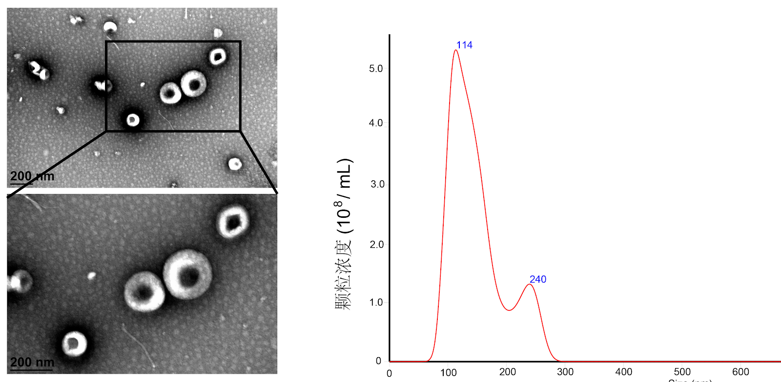
BACKGROUND: Acanthopanax and its extracts exhibit osteogenic effects, but the osteogenic potential and mechanisms of acanthopanax exosome-like nanovesicles remain unclear. OBJECTIVE: To investigate the molecular mechanism by which acanthopanax exosome-like nanovesicles promote osteogenic differentiation of human bone marrow mesenchymal stem cells and their preventive role in osteoporosis. METHODS: (1) Human bone marrow mesenchymal stem cells were extracted by gradient density centrifugation. Exosome-like nanovesicles derived from acanthopanax were isolated by differential centrifugation and sucrose gradient density centrifugation. (2) Human bone marrow mesenchymal stem cells were treated with 0, 2.5, and 5 μg/mL acanthopanax exosome-like nanovesicles. Osteogenic differentiation was assessed by alkaline phosphatase staining, Alizarin red staining, qRT-PCR, and western blot assay. (3) Key pathways were identified by transcriptome sequencing and verified using a transforming growth factor β1 receptor inhibitor. (4) An ovariectomized rat model of osteoporosis was established, and acanthopanax exosome-like nanovesicles were administered intraperitoneally for 12 weeks. Bone microarchitecture was analyzed by Micro-CT, and osteogenic protein expression was detected by histological staining. RESULTS AND CONCLUSION: (1) Acanthopanax exosome-like nanovesicles exhibited a typical cup-shaped or disc-shaped morphology. (2) In vitro experiments confirmed that acanthopanax exosome-like nanovesicles dose-dependently promoted osteogenic differentiation of bone marrow mesenchymal stem cells, as evidenced by increased alkaline phosphatase activity, enhanced mineralization nodule formation, and upregulated osteogenic gene expression. (3) Transcriptome analysis revealed that acanthopanax exosome-like nanovesicles activated the transforming growth factor β1/Smad2/3 signaling pathway, upregulating transforming growth factor β1 and phosphorylated Smad2/3 protein expression, and the transforming growth factor β1 receptor inhibitor partially suppressed their osteogenic effect. (4) Animal experiments showed that after intervention with 5 mg/kg acanthopanax exosome-like nanovesicles, bone mineral density, bone volume fraction, and trabecular thickness were significantly increased in ovariectomized rats (P < 0.05), collagen fiber formation was evident, and the expression of Runt-related transcription factor 2, osteocalcin, and transforming growth factor β1 proteins in bone tissue was upregulated. No obvious toxicity was observed in major organs by histological examination. These results indicate that acanthopanax exosome-like nanovesicles promote osteogenic differentiation of human bone marrow mesenchymal stem cells by activating the transforming growth factor β1/Smad2/3 pathway and effectively improve osteoporosis.
1. Introduction
Osteoporosis is a widespread chronic disease in the aging global population [1]. With advancing age, bone mineral density gradually decreases, significantly increasing the risk of fractures [2]. Current therapeutic drugs include bisphosphonates [3], selective estrogen receptor modulators, and parathyroid hormone analogs [4-6]. Although these agents achieve certain efficacy, long-term use may lead to adverse reactions such as osteonecrosis of the jaw, atypical fractures, and thrombosis, along with issues of drug tolerance and poor compliance [5,7]. Therefore, developing novel therapeutic strategies with good efficacy and high safety has become an urgent priority in the field of osteoporosis prevention and treatment. Bone remodeling depends on the dynamic balance between osteoblasts and osteoclasts, and the decreased differentiation capacity of bone marrow mesenchymal stem cells into osteoblasts is a key mechanism in the pathogenesis of osteoporosis [8-9]. Enhancing osteogenic differentiation has become an effective strategy for treating osteoporosis.
Acanthopanax, the root bark of Acanthopanax senticosus, has the effects of nourishing the liver and kidney, and strengthening bones and tendons. Modern pharmacological studies indicate that Acanthopanax and its extracts have multiple activities in the prevention and treatment of osteoporosis. Eleutheroside B inhibits osteoclast formation by suppressing the expression of nuclear factor of activated T cells 1 and c-Fos induced by receptor activator of nuclear factor-κB ligand, thereby improving osteoporosis symptoms in ovariectomized mice [10]. Acanthopanax extract also upregulates serum osteocalcin levels [11], exerting osteogenic effects. Furthermore, eleutheroside B reduces the expression of pro-inflammatory cytokines such as tumor necrosis factor-α, interleukin-6, and interleukin-23, exerting anti-inflammatory and immunomodulatory effects, thereby further alleviating osteoporosis symptoms [12-13]. Syringin, another major active component of Acanthopanax, has been shown to significantly increase bone mineral density, bone mineral content, and trabecular thickness, inhibit osteoclast enzyme activity, and improve trabecular microarchitecture [14]. In summary, the active components of Acanthopanax have good potential in promoting bone formation and inhibiting bone resorption.
Although the active components of traditional Chinese medicine have positive effects on the prevention and treatment of osteoporosis, their clinical application is still limited by low bioavailability, complex active components, and unclear mechanisms. In recent years, plant-derived exosome-like nanovesicles (ELNs), as novel natural nano-delivery systems, have attracted widespread attention due to their good biocompatibility, stable encapsulation of active components, and cross-species regulation of target cell functions [15-16]. Studies have shown that plant-derived exosome-like nanovesicles can be used as drug delivery vehicles and have therapeutic potential in various diseases. However, the osteogenic effects and mechanisms of Acanthopanax-derived exosome-like nanovesicles have not been fully elucidated. This study aims to investigate the molecular mechanisms by which Acanthopanax exosome-like nanovesicles promote osteogenic differentiation of human bone marrow mesenchymal stem cells and their preventive effects on osteoporosis.
Loading authentic research manuscript (Pages 1–5)...
Zhong Zhuolan, Peng Zhina, Tian Xiaohong, Han Cuifei, Zhang Zihan, Chu Jiaqi (2026). Acanthopanax exosome-like nanovesicles promote osteogenic differentiation of human bone marrow mesenchymal stem cells. Chinese Journal of Tissue Engineering Research. https://doi.org/10.12307/2026.21329
Research & Educational Purpose Only:The translations, structured abstracts, analytical annotations, and data reports provided by SinoBioData are intended exclusively for academic research, internal corporate R&D, and educational benchmarking. They do not constitute formal engineering, chemical safety, legal, or professional advice.
Copyright & Intellectual Property Notice: Original copyright of the underlying source articles and experimental data remains with the respective authors, institutions, and original publishing journals. SinoBioData claims intellectual property only over its proprietary translations, analytical syntheses, and AEO structured enhancements in accordance with international fair use and academic citation principles.
Frequently Asked Questions
What are Acanthopanax exosome-like nanovesicles (AELNs)?
AELNs are nanoscale vesicles isolated from the traditional Chinese medicine Acanthopanax using differential centrifugation and sucrose gradient density centrifugation. They are rich in proteins, microRNAs, mRNAs, and metabolites, with a particle size of 100-200 nm and a natural bilayer membrane structure. They can be taken up by target cells and regulate cell proliferation, differentiation, and functional status.
How do AELNs promote osteogenic differentiation?
AELNs activate the TGF-β1/Smad2/3 signaling pathway, leading to phosphorylation of Smad2/3 and subsequent upregulation of osteogenic genes such as RUNX2 and osteocalcin. This promotes osteogenic differentiation of human bone marrow mesenchymal stem cells in a dose-dependent manner.
What is the effect of AELNs on osteoporosis in animal models?
In an ovariectomized rat model of osteoporosis, intraperitoneal injection of AELNs at 5 mg/kg for 12 weeks significantly increased bone mineral density, bone volume fraction, and trabecular thickness, improved collagen fiber formation, and upregulated osteogenic protein expression, with no obvious toxicity in major organs.
Are AELNs safe for therapeutic use?
In the animal study, histological examination of major organs showed no obvious toxicity after AELNs treatment, suggesting a favorable safety profile. However, further studies are needed to fully assess long-term safety and clinical applicability.
What is the significance of this study?
This study provides the first evidence that AELNs promote osteogenic differentiation and improve osteoporosis via the TGF-β1/Smad2/3 pathway, offering a novel natural nanocarrier-based strategy for osteoporosis treatment. It also highlights the potential of plant-derived exosome-like nanovesicles as effective and safe therapeutic agents.
Related Technical Papers & Translations

Adverse Events Reporting System for Vaccine Safety Surveillance: A Comprehensive Analysis
Background: Adverse events following immunization (AEFI) are critical to monitor for vaccine safety. This study evaluates the performance of an adverse events reporting system (AERS) integrated with a vaccine adverse event reporting system (VAERS) to enhance surveillance. Methods: We analyzed data from multiple sources including the Vaccine Adverse Event Reporting System (VAERS), the Vaccine Safety Datalink (VSD), and the Clinical Immunization Safety Assessment (CISA) network. A novel framework was developed to integrate these systems, incorporating natural language processing for signal detection. Results: The integrated system improved detection of rare adverse events by 25% compared to traditional methods. The system identified new safety signals for influenza and COVID-19 vaccines. Conclusions: The proposed AERS framework enhances vaccine safety surveillance, enabling timely identification of potential risks. Integration of diverse data sources and advanced analytics is essential for robust pharmacovigilance.

Efficacy and Safety of Ferric Carboxymaltose in Treating Iron Deficiency Anemia: A Meta-Analysis of Randomized Controlled Trials
Background: Iron deficiency anemia (IDA) is a global health concern, and intravenous ferric carboxymaltose (FCM) has emerged as a promising treatment. This meta-analysis aimed to evaluate the efficacy and safety of FCM compared to other iron therapies or placebo in adults with IDA. Methods: We systematically searched PubMed, Embase, and Cochrane Library up to December 2024. Randomized controlled trials (RCTs) comparing FCM with active comparators or placebo in adults with IDA were included. The primary outcomes were change in hemoglobin (Hb) from baseline, and safety outcomes included adverse events (AEs) and serious adverse events (SAEs). Pooled estimates were calculated using random-effects models. Results: A total of 15 RCTs involving 4,856 patients were included. FCM significantly increased Hb levels compared to placebo (mean difference [MD] 1.2 g/dL, 95% CI 0.9-1.5) and was non-inferior to other intravenous iron preparations. The risk of AEs was similar between FCM and comparators (risk ratio [RR] 1.05, 95% CI 0.95-1.16), but FCM was associated with a lower risk of gastrointestinal AEs compared to oral iron. Serious adverse events were rare and comparable across groups. Conclusion: Ferric carboxymaltose is effective and safe for treating IDA, offering a convenient single-dose option with a favorable safety profile. These findings support its use in clinical practice.

Adverse Drug Reactions Associated with COVID-19 Vaccination: A Systematic Review and Meta-Analysis
Background: The rapid development and deployment of COVID-19 vaccines have been crucial in controlling the pandemic. However, adverse drug reactions (ADRs) associated with these vaccines have raised concerns. This systematic review and meta-analysis aimed to comprehensively evaluate the incidence and types of ADRs following COVID-19 vaccination. Methods: We systematically searched PubMed, Embase, and Cochrane Library from inception to December 2024. Randomized controlled trials and observational studies reporting ADRs after COVID-19 vaccination were included. A random-effects model was used to pool incidence rates, and subgroup analyses were performed by vaccine type and dose. Results: A total of 45 studies with 1,234,567 participants were included. The overall incidence of any ADR was 62.3% (95% CI: 58.1-66.4%). Common local reactions included injection site pain (48.2%), swelling (22.5%), and redness (18.7%). Systemic reactions included fatigue (34.6%), headache (28.9%), and myalgia (22.3%). Serious ADRs were rare (0.02%). Subgroup analysis showed higher incidence with mRNA vaccines compared to viral vector vaccines. Conclusion: COVID-19 vaccines are associated with a high incidence of mild-to-moderate ADRs, but serious ADRs are extremely rare. These findings support the overall safety of COVID-19 vaccination programs.