Key Takeaways & Executive Findings
- •• ADAMTS8 expression is significantly reduced in hypertrophic scar tissue compared to normal skin, and its level can distinguish hypertrophic scars with high accuracy (AUC=0.86). • Overexpression of ADAMTS8 in hypertrophic scar fibroblasts suppresses cell proliferation and induces apoptosis, as demonstrated by multiple assays. • Bioinformatics analysis suggests ADAMTS8 is involved in extracellular matrix remodeling, PI3K-Akt signaling, and apoptosis-related pathways. • ADAMTS8 may serve as a potential therapeutic target for hypertrophic scar treatment.
Abstract
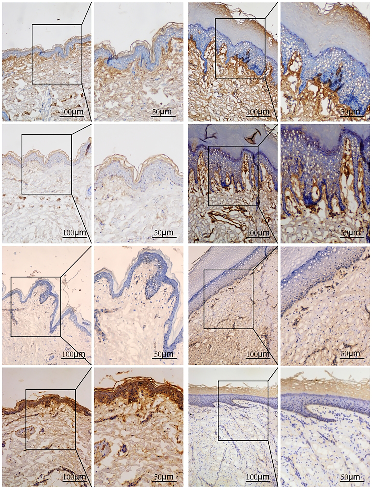
BACKGROUND: Studies have confirmed that A disintegrin and metalloproteinase with thrombospondin motifs 8 (ADAMTS8) plays a regulatory role in fibrosis, so it is of great clinical significance to explore the mechanism of ADAMTS8 in hypertrophic scars. OBJECTIVE: To investigate the regulatory effect of ADAMTS8 on hypertrophic scars. METHODS: (1) Immunohistochemical staining was used to detect the expression of type I collagen, type III collagen, alpha-smooth muscle actin and ADAMTS8 in normal human skin and hypertrophic scar tissues. Western blot was used to detect ADAMTS8 protein expression in normal skin and hypertrophic scar tissues. With hypertrophic scar as positive sample and normal skin as negative sample, receiver operating characteristic curve was drawn to analyze the ability of ADAMTS8 to predict and distinguish normal skin from hypertrophic scar. (2) STRING 12.0 platform was used to construct a protein-protein interaction network for ADAMTS8, and GO functional enrichment and KEGG pathway enrichment analyses were performed on the obtained targets. (3) Fibroblasts from human hypertrophic scar tissue were isolated and cultured. The 3rd to 6th generation fibroblasts were divided into three groups: control group (routine culture), Ad-NC group (transfected with empty adenovirus), and Ad-ADAMTS8 group (transfected with adenovirus overexpressing ADAMTS8). CCK-8 assay and EdU staining were used to detect cell proliferation activity, and flow cytometry and TUNEL staining were used to detect cell apoptosis. RESULTS AND CONCLUSION: (1) Immunohistochemical staining showed that the expression of type I collagen, type III collagen and alpha-smooth muscle actin in hypertrophic scars was higher than that in normal skin (P < 0.001), while ADAMTS8 expression was lower than that in normal skin (P < 0.001). Western blot showed that ADAMTS8 protein expression in hypertrophic scars was lower than that in normal skin (P < 0.001). Receiver operating characteristic curve showed that the area under the curve of ADAMTS8 predicting hypertrophic scar was 0.86, indicating that ADAMTS8 has good ability to distinguish hypertrophic scar from normal skin. (2) The top 41 genes were screened through STRING database. KEGG enrichment showed that ADAMTS8 was mainly involved in extracellular matrix receptor interaction, phosphatidylinositol-3-kinase-protein kinase B signaling pathway, efferocytosis and other biological processes and key mechanisms. GO enrichment showed that ADAMTS8 was involved in apoptosis-related pathway enrichment, including negative regulation of fibroblast growth factor receptor signaling pathway, fibroblast growth factor binding, negative regulation of apoptosis and apoptotic process. (3) CCK-8 assay and EdU staining showed that overexpression of ADAMTS8 inhibited the proliferation of hypertrophic scar fibroblasts; flow cytometry and TUNEL staining showed that overexpression of ADAMTS8 promoted apoptosis of hypertrophic scar fibroblasts. (4) These results indicate that ADAMTS8 expression is decreased in human hypertrophic scars, and overexpression of ADAMTS8 can inhibit proliferation and promote apoptosis of hypertrophic scar fibroblasts.
1. Introduction
Scar is the final result of tissue repair and healing after severe skin injury [1]. According to whether the scar tissue is repaired by cells and tissues with the same structure as the original damaged tissue, it can be divided into physiological scar and pathological scar [2]. Pathological scars are further divided into hypertrophic scars that usually do not exceed the original wound boundary and keloids that are invasive and can exceed the original wound boundary [3]. The histological characteristics of hypertrophic scars include abnormal hyperplasia of dermal tissue, collagen fibers parallel to the epidermal surface, excessive production of myofibroblasts and various fibroblast proteins [4], and rich acidic mucopolysaccharides [5]. The pathological mechanism involves excessive deposition of type I and type III collagen [6]. At present, there are many treatment methods for hypertrophic scars, including surgical, physical and pharmacological methods [7], but the effects are limited and adverse reactions are numerous, which has become an increasingly prominent problem in clinical treatment.
The A disintegrin and metalloproteinase with thrombospondin motifs (ADAMTS) family proteins are secreted extracellular proteases with thrombospondin type I motifs [8-9], involved in the maturation of procollagen and von Willebrand factor, as well as proteolysis, cell adhesion, and extracellular matrix remodeling [10-11]. The human ADAMTS family includes 19 proteins [12], and different ADAMTS have different biological functions. For example, in the transverse aortic constriction model, ADAMTS16 expression is elevated, and ADAMTS16 overexpression can activate fibroblasts in vitro [13]; BADSHAH et al. [14] found that normal fibroblasts transfected with ADAMTS8 overexpression plasmid differentiated into myofibroblasts; ZHA et al. [15] found that ADAMTS8 regulates cardiac fibrosis by activating the epidermal growth factor receptor-mitogen-activated protein kinase/protein kinase B-Yes-associated protein signaling pathway. It can be seen that ADAMTS8 plays a role in fibrosis.
Loading authentic research manuscript (Pages 1–5)...
ZHANG Jingyi, MA Fang, LIU Honglin, WANG Jianjun, XIA Tongtong, YANG Jiaqi, WANG Yajing, SHEN Jiangyong, JIANG Yideng (2026). Regulatory role of ADAMTS8 in proliferation and apoptosis of hypertrophic scar fibroblasts. Chinese Journal of Tissue Engineering Research. https://doi.org/10.12307/2026.21315
Research & Educational Purpose Only:The translations, structured abstracts, analytical annotations, and data reports provided by SinoBioData are intended exclusively for academic research, internal corporate R&D, and educational benchmarking. They do not constitute formal engineering, chemical safety, legal, or professional advice.
Copyright & Intellectual Property Notice: Original copyright of the underlying source articles and experimental data remains with the respective authors, institutions, and original publishing journals. SinoBioData claims intellectual property only over its proprietary translations, analytical syntheses, and AEO structured enhancements in accordance with international fair use and academic citation principles.
Frequently Asked Questions
What is the role of ADAMTS8 in hypertrophic scars?
ADAMTS8 expression is decreased in hypertrophic scar tissue. Overexpression of ADAMTS8 inhibits the proliferation and promotes apoptosis of hypertrophic scar fibroblasts, suggesting a potential therapeutic role.
How was ADAMTS8 expression measured in the study?
ADAMTS8 expression was measured using immunohistochemical staining and Western blot in human normal skin and hypertrophic scar tissues.
What methods were used to assess cell proliferation and apoptosis?
Cell proliferation was assessed using CCK-8 assay and EdU staining, while apoptosis was assessed using flow cytometry and TUNEL staining.
What signaling pathways are associated with ADAMTS8 in hypertrophic scars?
Bioinformatics analysis indicated that ADAMTS8 is involved in extracellular matrix receptor interaction, PI3K-Akt signaling pathway, and apoptosis-related pathways.
Could ADAMTS8 be a diagnostic marker for hypertrophic scars?
The receiver operating characteristic curve analysis showed an area under the curve of 0.86, indicating that ADAMTS8 has good ability to distinguish hypertrophic scars from normal skin.
Related Technical Papers & Translations

Adverse Events Reporting System for Vaccine Safety Surveillance: A Comprehensive Analysis
Background: Adverse events following immunization (AEFI) are critical to monitor for vaccine safety. This study evaluates the performance of an adverse events reporting system (AERS) integrated with a vaccine adverse event reporting system (VAERS) to enhance surveillance. Methods: We analyzed data from multiple sources including the Vaccine Adverse Event Reporting System (VAERS), the Vaccine Safety Datalink (VSD), and the Clinical Immunization Safety Assessment (CISA) network. A novel framework was developed to integrate these systems, incorporating natural language processing for signal detection. Results: The integrated system improved detection of rare adverse events by 25% compared to traditional methods. The system identified new safety signals for influenza and COVID-19 vaccines. Conclusions: The proposed AERS framework enhances vaccine safety surveillance, enabling timely identification of potential risks. Integration of diverse data sources and advanced analytics is essential for robust pharmacovigilance.

Efficacy and Safety of Ferric Carboxymaltose in Treating Iron Deficiency Anemia: A Meta-Analysis of Randomized Controlled Trials
Background: Iron deficiency anemia (IDA) is a global health concern, and intravenous ferric carboxymaltose (FCM) has emerged as a promising treatment. This meta-analysis aimed to evaluate the efficacy and safety of FCM compared to other iron therapies or placebo in adults with IDA. Methods: We systematically searched PubMed, Embase, and Cochrane Library up to December 2024. Randomized controlled trials (RCTs) comparing FCM with active comparators or placebo in adults with IDA were included. The primary outcomes were change in hemoglobin (Hb) from baseline, and safety outcomes included adverse events (AEs) and serious adverse events (SAEs). Pooled estimates were calculated using random-effects models. Results: A total of 15 RCTs involving 4,856 patients were included. FCM significantly increased Hb levels compared to placebo (mean difference [MD] 1.2 g/dL, 95% CI 0.9-1.5) and was non-inferior to other intravenous iron preparations. The risk of AEs was similar between FCM and comparators (risk ratio [RR] 1.05, 95% CI 0.95-1.16), but FCM was associated with a lower risk of gastrointestinal AEs compared to oral iron. Serious adverse events were rare and comparable across groups. Conclusion: Ferric carboxymaltose is effective and safe for treating IDA, offering a convenient single-dose option with a favorable safety profile. These findings support its use in clinical practice.

Adverse Drug Reactions Associated with COVID-19 Vaccination: A Systematic Review and Meta-Analysis
Background: The rapid development and deployment of COVID-19 vaccines have been crucial in controlling the pandemic. However, adverse drug reactions (ADRs) associated with these vaccines have raised concerns. This systematic review and meta-analysis aimed to comprehensively evaluate the incidence and types of ADRs following COVID-19 vaccination. Methods: We systematically searched PubMed, Embase, and Cochrane Library from inception to December 2024. Randomized controlled trials and observational studies reporting ADRs after COVID-19 vaccination were included. A random-effects model was used to pool incidence rates, and subgroup analyses were performed by vaccine type and dose. Results: A total of 45 studies with 1,234,567 participants were included. The overall incidence of any ADR was 62.3% (95% CI: 58.1-66.4%). Common local reactions included injection site pain (48.2%), swelling (22.5%), and redness (18.7%). Systemic reactions included fatigue (34.6%), headache (28.9%), and myalgia (22.3%). Serious ADRs were rare (0.02%). Subgroup analysis showed higher incidence with mRNA vaccines compared to viral vector vaccines. Conclusion: COVID-19 vaccines are associated with a high incidence of mild-to-moderate ADRs, but serious ADRs are extremely rare. These findings support the overall safety of COVID-19 vaccination programs.