Key Takeaways & Executive Findings
- •• Anterior transpedicular screw fixation of the subaxial cervical spine is anatomically feasible, with entry points and trajectories varying by vertebral level. • Entry point locations and angles showed no significant differences between genders or sides, supporting standardized approaches. • For most patients, screws of at least 30 mm length and 4.0 mm diameter (males) and 28 mm length and 3.5 mm diameter (females) are safe and reliable. • Individualized preoperative planning using 3D CT reconstruction is essential to ensure accurate screw placement and avoid complications.
Abstract
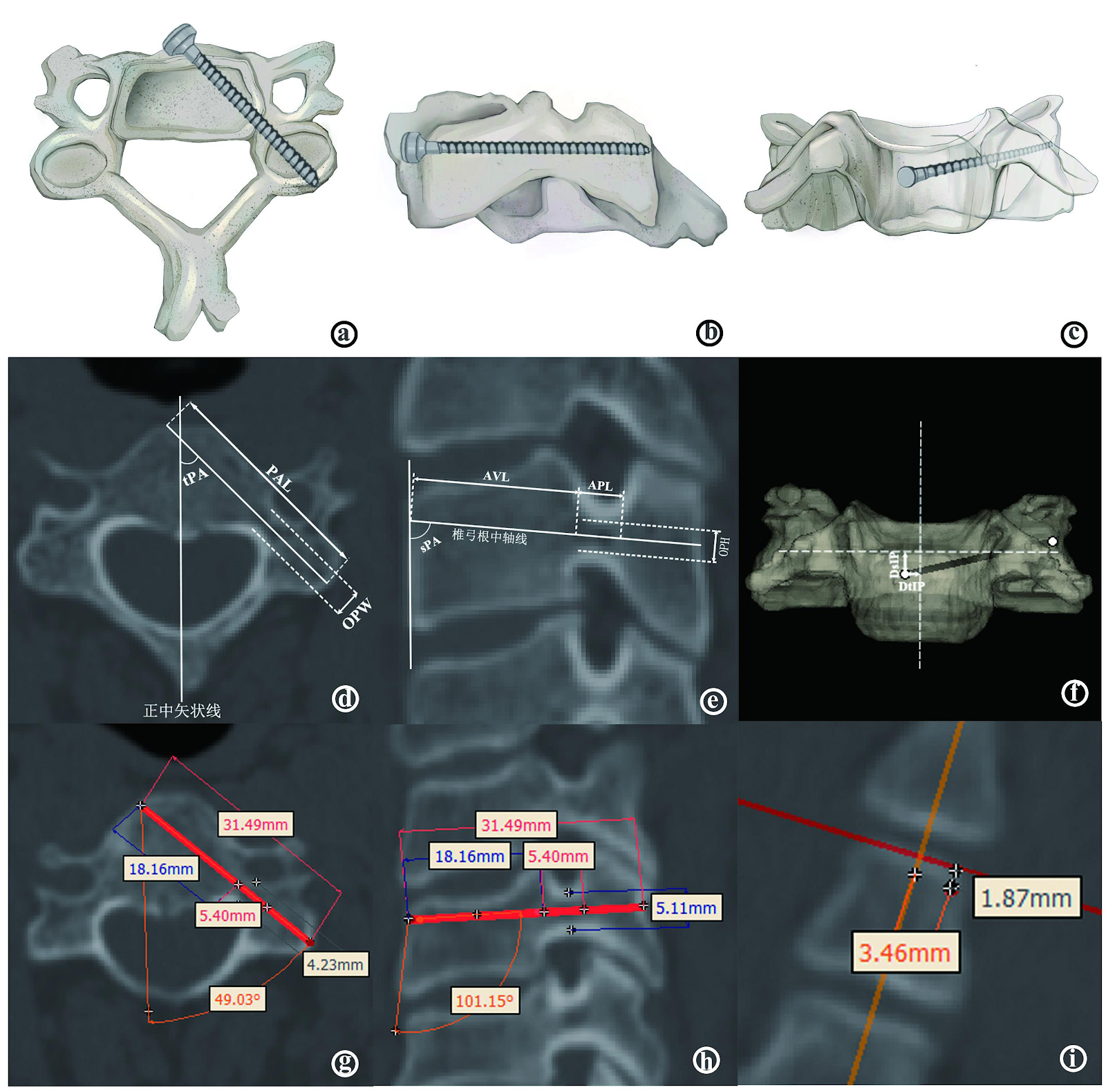
BACKGROUND: The anterior transpedicular screw fixation system can achieve adequate decompression and strong fixation in a single anterior surgery. However, due to its unique anatomical structure, the risk of screw placement is relatively high. Previous domestic and international scholars have confirmed the feasibility of this approach through anatomical measurements, but there are some drawbacks such as relatively small sample sizes, difficulties in locating the pedicle axis, and limitations in measurement methods. OBJECTIVE: To perform morphological measurements of the adult cervical spine based on imaging to provide anatomical guidance for anterior transpedicular pedicle screw fixation. METHODS: 3D CT scan data of 50 adult cervical vertebrae were imported into the Mimics system for 3D reconstruction. Morphological data were measured, including pedicle axis distance, pedicle width, pedicle height, pedicle horizontal axial angle, pedicle sagittal angle, distance from the entry point in the transverse plane, distance to the entry point in the sagittal plane, axial vertebral length and axial pedicle length. RESULTS AND CONCLUSION: (1) Positioning of nail entry points: C3 and C4 were located on the opposite side of the median sagittal plane of the vertebral body, with distances from the median sagittal line of approximately 2.060 mm and 2.310 mm. C5 could be located on the same side of the median sagittal line as or opposite to the median sagittal line, with an average value of approximately 1.224 mm. C6-C7 were located on the same side of the body, with distances of 1.132 mm and 2.538 mm from the midline; the distance from the upper endplate increased gradually from C3 to C7, with average values ranging from 2.362 to 7.350 mm. (2) Direction of nail entry: the transverse angle increased gradually from C3 to C4 (46.32°-47.36°) and decreased from C5 to C7 (44.03° to 37.80°); the sagittal angle required caudal deviation for C3-C4 (95.75° and 100.93°) and cephalad deviation for C5-C7 (104.38°, 110.34°, and 104.86°). (3) There were no significant differences in entry point location and direction between genders or sides (P > 0.05). For screw selection, except for individual patients with cervical developmental abnormalities, for most subaxial cervical pedicle screws, it is safe and reliable to choose screws at least 30 mm long and 4.0 mm in diameter for males, and at least 28 mm long and 3.5 mm in diameter for females. (4) Morphological measurements confirmed that anterior transpedicular screw fixation of the subaxial cervical spine is feasible, but individualized principles should be followed to formulate personalized fixation plans.
1. Introduction
Anterior cervical decompression and fusion is a common treatment for cervical spine diseases, offering advantages such as shorter operative time and reduced surgical trauma and complications [1-3]. However, the biomechanical stability of traditional anterior cervical plate and screw fixation is not satisfactory, with issues such as insufficient pullout resistance and poor fatigue resistance [4-5]. To achieve both thorough anterior decompression and strong posterior fixation in a single procedure, KOLLER et al. [6-7] first proposed the concept of cervical anterior transpedicular screw (ATPS) fixation in 2008. Due to the unique anatomical structure of the pedicle, with the spinal cord medially, the vertebral artery laterally, and nerve roots passing above and below [8-10], the risk of screw placement is relatively high.
In response, many renowned scholars have systematically described the anterior transpedicular screw fixation technique and obtained anatomical data through measurements, confirming the feasibility and clinical application prospects of this approach [7,11-13]. However, current domestic anatomical studies on anterior cervical pedicle screw placement have limitations such as relatively small sample sizes and restricted measurement methods. To more precisely define the morphological parameters for anterior transpedicular screw fixation, this study utilized the interactive medical image control system Mimics software to perform individualized and quantitative analysis of screw placement based on 3D CT imaging of 50 patients. This approach enables more accurate morphological measurements and explores the morphological characteristics, thereby providing anatomical references for clinical practice.
Loading authentic research manuscript (Pages 1–5)...
HU Yin, XING Le, HAN Kangen, LI Junchao, QIAN Mengran, GU Hongwen, YU Hailong, WANG Hongwei (2026). Morphological measurement of anterior cervical pedicle screw placement assisted by Mimics three-dimensional CT reconstruction. Chinese Journal of Tissue Engineering Research. https://doi.org/10.12307/2026.21417
Research & Educational Purpose Only:The translations, structured abstracts, analytical annotations, and data reports provided by SinoBioData are intended exclusively for academic research, internal corporate R&D, and educational benchmarking. They do not constitute formal engineering, chemical safety, legal, or professional advice.
Copyright & Intellectual Property Notice: Original copyright of the underlying source articles and experimental data remains with the respective authors, institutions, and original publishing journals. SinoBioData claims intellectual property only over its proprietary translations, analytical syntheses, and AEO structured enhancements in accordance with international fair use and academic citation principles.
Frequently Asked Questions
What is the clinical significance of anterior transpedicular screw fixation?
Anterior transpedicular screw fixation allows for adequate decompression and strong fixation in a single anterior surgery, providing better biomechanical stability than traditional anterior plate fixation. However, it requires precise anatomical knowledge due to the proximity of vital structures.
How does Mimics software aid in cervical pedicle screw placement?
Mimics is an interactive medical image control system that enables segmentation, measurement, simulation, and 3D modeling from CT data. It allows for accurate reconstruction of cervical vertebrae and precise measurement of pedicle morphology, facilitating individualized screw placement planning.
What are the key anatomical parameters measured in this study?
The study measured pedicle axis distance, pedicle width, pedicle height, pedicle horizontal axial angle, pedicle sagittal angle, entry point distances in transverse and sagittal planes, axial vertebral length, and axial pedicle length.
Are there significant differences in pedicle morphology between genders or sides?
The study found no significant differences in entry point location and direction between genders or sides (P > 0.05), suggesting that standardized approaches may be applicable, but individualized planning is still recommended.
What screw sizes are recommended for anterior cervical pedicle fixation?
For most patients, screws of at least 30 mm length and 4.0 mm diameter are recommended for males, and at least 28 mm length and 3.5 mm diameter for females, to ensure safety and reliability.
Related Technical Papers & Translations

Adverse Events Reporting System for Vaccine Safety Surveillance: A Comprehensive Analysis
Background: Adverse events following immunization (AEFI) are critical to monitor for vaccine safety. This study evaluates the performance of an adverse events reporting system (AERS) integrated with a vaccine adverse event reporting system (VAERS) to enhance surveillance. Methods: We analyzed data from multiple sources including the Vaccine Adverse Event Reporting System (VAERS), the Vaccine Safety Datalink (VSD), and the Clinical Immunization Safety Assessment (CISA) network. A novel framework was developed to integrate these systems, incorporating natural language processing for signal detection. Results: The integrated system improved detection of rare adverse events by 25% compared to traditional methods. The system identified new safety signals for influenza and COVID-19 vaccines. Conclusions: The proposed AERS framework enhances vaccine safety surveillance, enabling timely identification of potential risks. Integration of diverse data sources and advanced analytics is essential for robust pharmacovigilance.

Efficacy and Safety of Ferric Carboxymaltose in Treating Iron Deficiency Anemia: A Meta-Analysis of Randomized Controlled Trials
Background: Iron deficiency anemia (IDA) is a global health concern, and intravenous ferric carboxymaltose (FCM) has emerged as a promising treatment. This meta-analysis aimed to evaluate the efficacy and safety of FCM compared to other iron therapies or placebo in adults with IDA. Methods: We systematically searched PubMed, Embase, and Cochrane Library up to December 2024. Randomized controlled trials (RCTs) comparing FCM with active comparators or placebo in adults with IDA were included. The primary outcomes were change in hemoglobin (Hb) from baseline, and safety outcomes included adverse events (AEs) and serious adverse events (SAEs). Pooled estimates were calculated using random-effects models. Results: A total of 15 RCTs involving 4,856 patients were included. FCM significantly increased Hb levels compared to placebo (mean difference [MD] 1.2 g/dL, 95% CI 0.9-1.5) and was non-inferior to other intravenous iron preparations. The risk of AEs was similar between FCM and comparators (risk ratio [RR] 1.05, 95% CI 0.95-1.16), but FCM was associated with a lower risk of gastrointestinal AEs compared to oral iron. Serious adverse events were rare and comparable across groups. Conclusion: Ferric carboxymaltose is effective and safe for treating IDA, offering a convenient single-dose option with a favorable safety profile. These findings support its use in clinical practice.

Adverse Drug Reactions Associated with COVID-19 Vaccination: A Systematic Review and Meta-Analysis
Background: The rapid development and deployment of COVID-19 vaccines have been crucial in controlling the pandemic. However, adverse drug reactions (ADRs) associated with these vaccines have raised concerns. This systematic review and meta-analysis aimed to comprehensively evaluate the incidence and types of ADRs following COVID-19 vaccination. Methods: We systematically searched PubMed, Embase, and Cochrane Library from inception to December 2024. Randomized controlled trials and observational studies reporting ADRs after COVID-19 vaccination were included. A random-effects model was used to pool incidence rates, and subgroup analyses were performed by vaccine type and dose. Results: A total of 45 studies with 1,234,567 participants were included. The overall incidence of any ADR was 62.3% (95% CI: 58.1-66.4%). Common local reactions included injection site pain (48.2%), swelling (22.5%), and redness (18.7%). Systemic reactions included fatigue (34.6%), headache (28.9%), and myalgia (22.3%). Serious ADRs were rare (0.02%). Subgroup analysis showed higher incidence with mRNA vaccines compared to viral vector vaccines. Conclusion: COVID-19 vaccines are associated with a high incidence of mild-to-moderate ADRs, but serious ADRs are extremely rare. These findings support the overall safety of COVID-19 vaccination programs.