🧬 SinoBioData Academic Portal
🏛️ Indexed Academic JournalOriginal: 生物化学与生物物理学报

Acta Biochimica et Biophysica Sinica

Premier Chinese Biomedical Journal indexed in SinoBioData: Acta Biochimica et Biophysica Sinica (生物化学与生物物理学报).

Total Research Papers: 200
Access: 100% Free Open Access
Browse by Publication Year & VolumeReset All Filters ✕

Published Research PapersFiltered: Year 2026 • 58

Showing 83 of 200 peer-reviewed papers with full Graphical Abstracts.

Original ResearchVol. 58, Issue 8 • pp. 1736-1747DOI: 10.3724/abbs.2026076

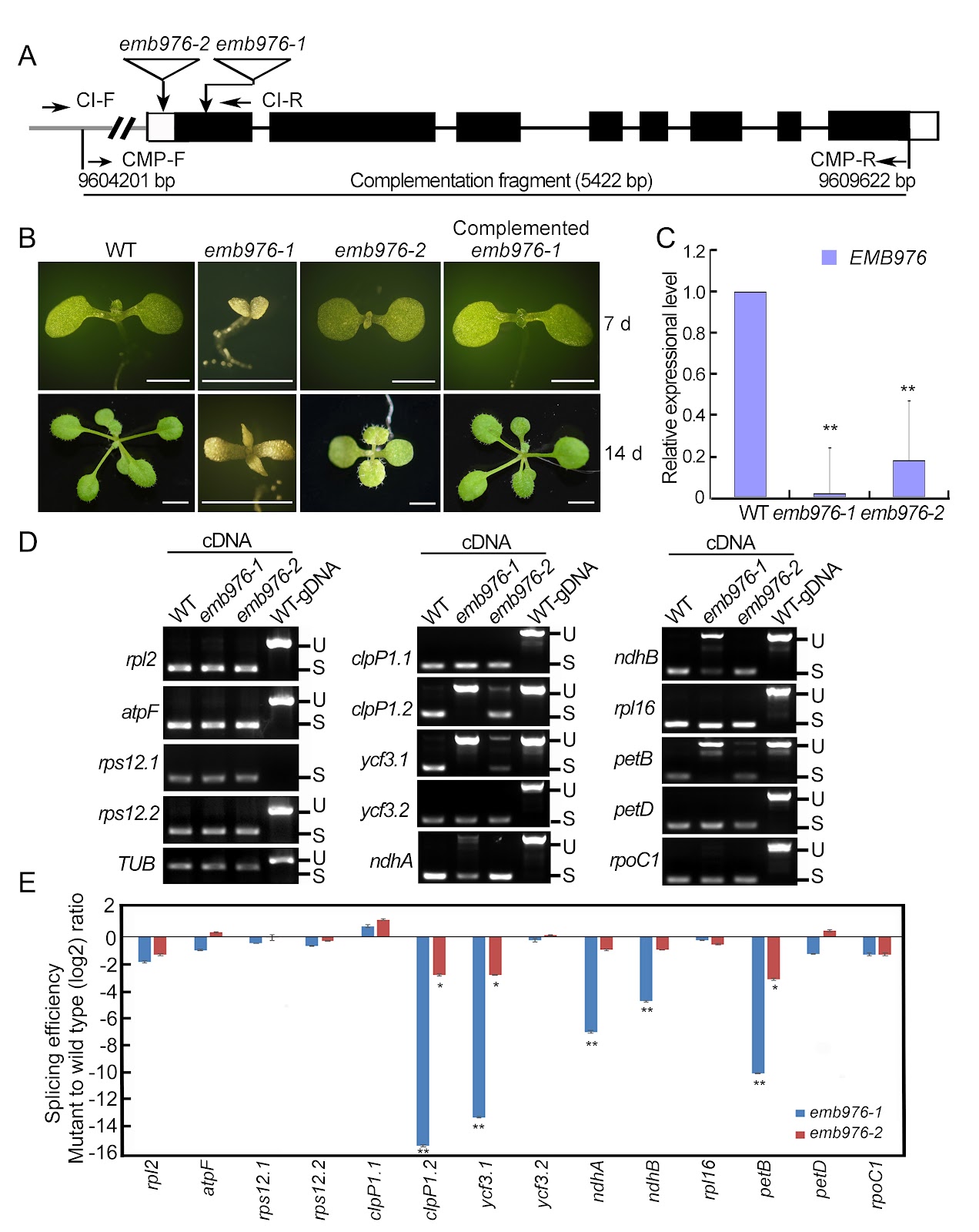
A protein-RNA complex orchestrated by EMB1006, EMB1270, EMB976, and CFM2 facilitates clpP1 intron 2 splicing in Arabidopsis chloroplasts

Authors: Liqun Zhang, Fangsheng Liao, Ying Wei, Keyi Yang, Yawen Zhu, Li Zhang, Wenyujie Shi, Shuya Zhou, Jirong Huang, Yong-Lan Cui, Weihua Huang

In Arabidopsis, the PPR proteins EMB1006, EMB1270, and EMB976 are all essential for the splicing of plastid clpP1 intron 2 (clpP1.2), although each also targets other distinct RNAs. The precise mechanism underlying their co-ordinated action in clpP1.2 splicing remains unclear. In this study, RNA electrophoretic mobility shift assays, guided by PPR code prediction, confirm that EMB1006 specifically binds to a sequence near the 3′ end of clpP1 exon 2. Additionally, immunoprecipitation coupled with mass spectrometry reveals that EMB1006 forms a complex with EMB1270, EMB976, and CFM2. Direct interactions between EMB1006 and EMB1270 or CFM2 are further supported by yeast two-hybrid (Y2H) and semi-in vivo pull-down assays. However, no direct interactions between EMB976 and EMB1006, CFM2 or EMB1270 are detected by Y2H. Based on these findings and previous evidence that EMB1270 binds to clpP1 intron 2 and interacts with CFM2, we propose a model in which EMB1006 and EMB1270 bind to distinct sites on clpP1 pre-mRNA. Together with CFM2 and possible indirect association with EMB976, they assemble into a protein-RNA complex that facilitates the splicing of clpP1.2 in chloroplasts.

A protein-RNA complex orchestrated by EMB1006, EMB1270, EMB976, and CFM2 facilitates clpP1 intron 2 splicing in Arabidopsis chloroplasts
Graphical Abstract
Original ResearchVol. 58, Issue 7 • pp. 1579-1596DOI: 10.3724/abbs.2026054

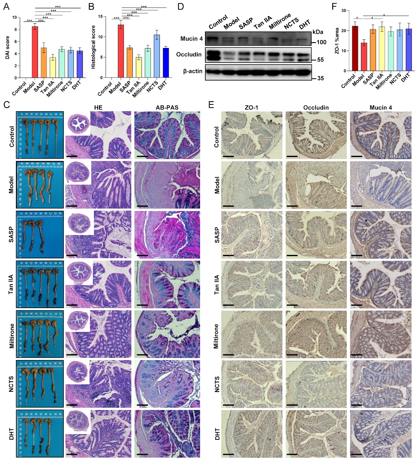
Tanshinones from Salvia miltiorrhiza alleviate ulcerative colitis via reprogramming the gut microbiota-metabolite axis

Authors: Zhe Liu, Chan Hui, Guochao Zhang, Haicheng Yang, Yi Wang, Yaqian Shi, Chao Wang, Yanfei Liu, Xia Gao, Yuting Wen

The anti-inflammatory properties of the traditional herb Salvia miltiorrhiza Bunge are well-established, yet its precise mechanism of action in ulcerative colitis (UC) remains unclear. Herein, we evaluate the therapeutic potential of four major tanshinones–tanshinone IIA (Tan IIA), miltirone, neocryptotanshinone, and dihydrotanshinone I–in a murine dextran sulfate sodium (DSS)-induced colitis model. Our results show that tanshinones effectively alleviate disease severity, suppress systemic and local inflammation, and restore intestinal barrier integrity. Integrated multi-omics analysis reveals that the therapeutic efficacy originates from a comprehensive reprogramming of the gut microbiota-metabolite axis. Specifically, tanshinones reverse colitis-associated dysbiosis and rectify metabolic disturbances in linoleic acid metabolism, bile acid biosynthesis, and amino acid utilization. Correlation network analysis identifies key functional modules linking beneficial microbes (e.g., Akkermansia) to anti-inflammatory lipid mediators and associating pathobionts (e.g., Desulfovibrio) with disrupted bile acid metabolism. Notably, supplementation with Akkermansia muciniphila synergizes with Tan IIA to amplify barrier restoration and metabolic normalization. Our findings establish that tanshinones ameliorate UC through microbiota-driven metabolic reprogramming, wherein the restructured microbial community actively shapes a therapeutic metabolic output. This work elucidates a metabolite-mediated mechanism of action and positions tanshinones as promising microbiome-targeting therapeutics for inflammatory bowel disease.

Tanshinones from Salvia miltiorrhiza alleviate ulcerative colitis via reprogramming the gut microbiota-metabolite axis
Graphical Abstract
Original ResearchVol. 58, Issue 8 • pp. 1761-1771DOI: 10.3724/abbs.2026010

Distinct miR319a identified from Persicaria chinensis mediates cross-kingdom suppression of cervical cancer by targeting ITGA3

Authors: Yueyue Yan, Dan Bai, Lei Li, Leimei Xu, Hua Yang, Yuhui Wang, Han Feng, Lan Zhu

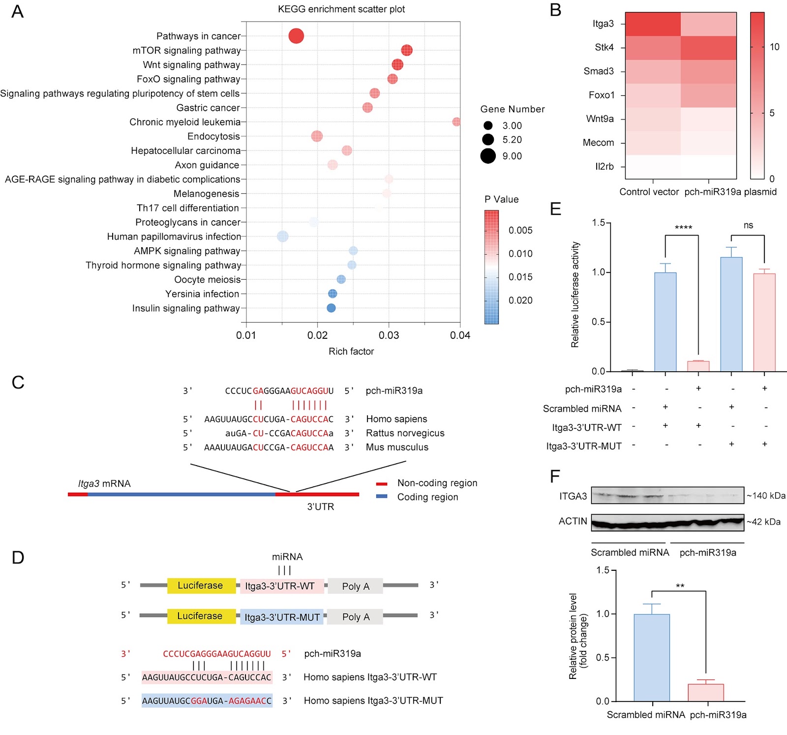
Persicaria chinensis, a well-known traditional Chinese medicinal herb that is both edible and medicinal, has been widely acknowledged for its therapeutic effects, such as anti-inflammatory, antioxidant, and antitumor activities. However, the role of miRNAs from this plant in the cross-kingdom regulation of human diseases has not been investigated. In this study, we analyze the miRNA expression profile of P. chinensis using high-throughput sequencing and identify a total of 673 miRNAs, including 422 novel miRNAs that are unique to this plant and 251 conserved miRNAs. Among the conserved miRNAs, pch-miR319a is found to be the most abundant. Moreover, food-oriented pch-miR319a accumulates in the uterus and tumors and exhibits a rich repertoire of target genes within cancer-related pathways, demonstrating significant cross-kingdom regulatory potential. Utilizing the dual-luciferase reporter gene assay, we demonstrate that pch-miR319a from P. chinensis targets the Itga3 gene, which is associated with cervical cancer progression. Overexpression of pch-miR319a significantly decreases the viability, migration, and induces apoptosis of HeLa cervical cancer cells in vitro. Moreover, in a syngeneic mouse tumor model of cervical cancer, treatment with pch-miR319a effectively inhibits tumor growth and downregulates the expressions of ITGA3 and the proliferation marker Ki-67. Our study highlights the potential of pch-miR319a from P. chinensis as a novel therapeutic agent for cervical cancer by targeting ITGA3 and provides new insights into the cross-kingdom regulatory mechanisms of plant miRNAs in human diseases.

Distinct miR319a identified from Persicaria chinensis mediates cross-kingdom suppression of cervical cancer by targeting ITGA3
Graphical Abstract
Original ResearchVol. 58, Issue 8 • pp. 1896-1900DOI: 10.3724/abbs.2026030

Development of an in vitro TurboID labeling assay for the detection of protoRAG-interacting proteins in the amphioxus

Authors: Qingyi Lu, Jie Xu, Junye Hong, Enfan Xiao, Qiuzhu Wei, Yuhe Sun, Zihan Zhao, Yuhang Zhang, Guangrui Huang

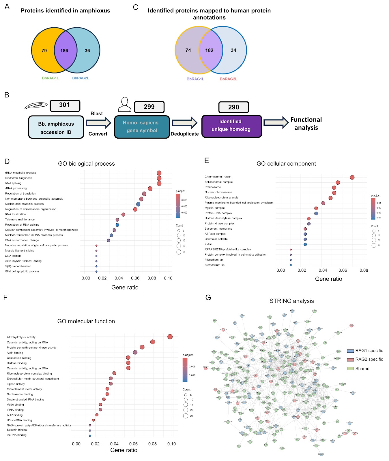
The recombination-activating gene (RAG)-mediated V(D)J rearrangement mechanism is essential for generating the diversity of the adaptive immune system in jawed vertebrates. The RAG1 and RAG2 proteins form a tetrameric complex that recognizes and cleaves pairs of recombination signal sequences (RSSs). In jawed vertebrates, the N-terminal region of RAG1 and the C-terminal region of RAG2 are critical for the regulation of physiological function and enhancement of recombination accuracy. Moreover, the intracellular activity of RAG1/2 is rigorously regulated and is thought to represent the aspect shaped by host coevolution. However, these regulatory mechanisms remain incompletely understood, impeding our deeper understanding of the physiological functions of RAG. Previous studies have shown that jawed vertebrate RAG evolved from invertebrate RAG-like (RAGL) transposases and underwent key structural changes during evolution. RAGL genes have been identified in multiple invertebrate species, but from Branchiostoma belcheri (Bb), the RAG-like protein in Bb amphioxus (BbRAG1L) and the RAG2-like protein in Bb amphioxus (BbRAG2L) are among the most extensively characterized to date. Lancelets (amphioxus) represent the most basal extant chordates (cephalochordates) that diverged from the other two chordate lineages (urochordates and vertebrates). Consequently, amphioxus has been considered a “living fossil” for studying the origins and evolution of adaptive immunity. Homologs of the core structural domain of RAG1 were discovered in the amphioxus genome in 2005, and the homologs were subsequently shown to exhibit recombinase activity in cooperation with murine-derived RAG2. A large number of transposon genes were then identified in the amphioxus genome, followed by the identification of target site duplication (TSD) and terminal inverted repeat (TIR) sequences in the amphioxus genome. Notably, BbRAG2L lacks the C-terminal region present in murine RAG2, including the acidic hinge and PHD domain. BbRAG1L coupled with BbRAG2L displays significant transposition activity in vitro and in human cell lines. In contrast, vertebrate RAG exhibits limited transposon activity in vitro and extremely low transposon activity in vivo. This leads to the question of how RAG lost its transposase activity during evolution. Structural comparison and functional experiments of BbRAGL (protoRAG) and vertebrate RAG have revealed that vertebrates undergo essential adaptive changes to eliminate the primitive transposase activity of protoRAG. Research on BbRAGL has therefore provided strong support for the hypothesis that RAG originated from a domesticated transposon. Given the similarity in both structure and function between BbRAG1L and vertebrate RAG and the fact that BbRAGL proteins are the only RAGL proteins that can currently be efficiently expressed in vertebrate cells, characterizing the binding partners of BbRAGL involved in amphioxus is of great significance for understanding the regulation of BbRAGL and its adaptation and evolution. Parallel comparison of BbRAGL and RAG interaction networks may further clarify how BbRAGL transitioned from a transposase to a recombinase during host coevolution and reveal general principles of vertebrate intracellular RAG regulation. In recent years, proximity labeling techniques have greatly facilitated approaches to identifying RAG cofactors. For example, RAG1 fused to biotin ligase RAG1 fused to a biotin ligase found in Escherichia coli (BirM) has been used to define dynamic interaction networks of RAG-associated proteins. Inspired by these advances, we sought to apply a similar approach to identify the BbRAGL cofactors in amphioxus and build a regulatory network of BbRAGL that could shed light on its relationship with host evolution. However, endogenous BbRAGL expression is undetectable in the adult amphioxus (data not shown), and the generation of transgenic amphioxus lines expressing proximity-based labeling proteins remains technically challenging. These constraints make it difficult to analyze native cofactors of endogenous BbRAGL in vivo. Because BbRAGL can be expressed in vertebrate cell lines, we instead developed an in vitro proximity-labeling strategy. We fused TurboID (TbID), an engineered biotin ligase, to the N-terminus of BbRAG1L or BbRAG2L (Figure 1A), purified the fusion proteins, and incubated them with protein lysates prepared from the hepatic cecum and colons of adult B. belcheri amphioxus in vitro to label the interacting proteins. The proteins were finally analyzed by liquid chromatography-tandem mass spectrometry (LC-MS/MS) (Figure 1B). A pTT5 vector was used to express plasmids encoding maltose-binding protein (MBP) and TurboID with or without full-length BbRAG1L or BbRAG2L, yielding pTT5-MBP-TurboID, pTT5-MBP-TurboID-BbRAG1L, and pTT5-MBP-TurboID-BbRAG2L. Plasmids expressing MBP-BbRAG1L and MBP-BbRAG2L were also generated.

Development of an in vitro TurboID labeling assay for the detection of protoRAG-interacting proteins in the amphioxus
Graphical Abstract
Original ResearchVol. 58, Issue 8 • pp. 1905-1909DOI: 10.3724/abbs.2026037

Identification and experimental validation of core genes associated with breast cancer brain metastasis via machine learning

Authors: Zhaoda Duan, Chunjiao Yu, Wenjie Yang, Qiaoling Ruan, Rui Zhang, Yongfang Zhao, Shan Yan

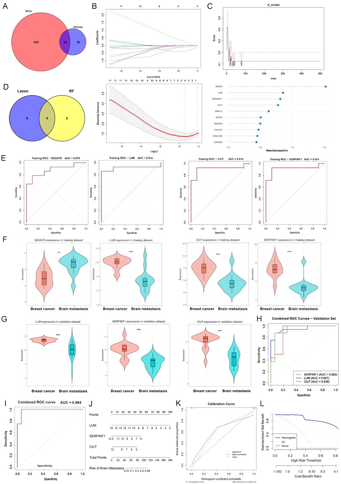
Breast cancer (BC) is the most common malignancy among women, with approximately 2.3 million new cases diagnosed annually, accounting for approximately 11.6% of all cancer cases worldwide. Distant metastasis is the primary cause of mortality in BC patients, with nearly 50% of patients ultimately developing metastatic disease. The predominant metastatic sites of BC include the lung, liver, brain, and bone, each exhibiting distinct biological characteristics that drive the organ-specific tropism of cancer cells. Among these, brain metastasis represents a significant cause of mortality in BC patients and is particularly prevalent in those with human epidermal growth factor receptor 2 (HER2)-positive or triple-negative breast cancer (TNBC) subtypes. Breast cancer brain metastasis (BCBM) can manifest in three forms: choroid plexus metastasis (rare), leptomeningeal metastasis (approximately 8%), and parenchymal metastasis, the most common presentation, with multiple lesions in 78% of cases and solitary lesions in 14%. Distinct anatomical regions of the brain provide different micro-environments, which in turn shape epidemiological patterns, biological behaviors, and therapeutic vulnerabilities of metastatic cancer. With the continuous advancement of systemic therapies and imaging surveillance, brain metastases from BC have become increasingly prevalent, accounting for approximately 10%–30% of all metastatic breast cancer (MBC) cases. The continuous progression of BCBM often compromises patients’ cognitive and sensory functions, leading to neurological impairment and severely limiting quality of life (QOL). Notably, the mortality rate within one year after diagnosis remains at 80%. Current therapeutic strategies for BCBM primarily include surgery, whole-brain radiotherapy (WBRT), stereotactic radiosurgery (SRS), chemotherapy, or combinations thereof. Although these approaches provide some clinical benefit, the efficacy remains limited due to the blood-brain barrier (BBB), which restricts drug penetration and contributes to chemoresistance. Therefore, elucidating the molecular mechanisms underlying BCBM is imperative to identify novel diagnostic biomarkers and therapeutic targets, with the ultimate goal of improving treatment efficacy and patient prognosis. Bioinformatics provides a powerful platform and data foundation for exploring the mechanisms of tumor initiation and progression. High-throughput platforms for gene expression analysis have gained significant popularity, with next-generation sequencing (NGS) and microarray analysis now widely applied as essential tools in medical oncology. These techniques have diverse clinical applications, including molecular cancer classification, prediction of therapeutic response, prognostic assessment, molecular diagnostics, and the discovery of novel drugs and therapeutic targets. Weighted gene coexpression network analysis (WGCNA) has been widely applied in studies of gene regulatory networks, biomarker discovery, and elucidation of the molecular mechanisms underlying complex phenotypes. In this study, we utilized the BCBM microarray dataset GSE43837. We performed differential expression analysis and WGCNA clustering using the R packages limma and WGCNA to identify potential gene modules and candidate targets. GSE43837 consists of 19 nonmetastatic primary breast tumor samples and 19 breast cancer brain metastasis samples. Differential expression analysis, with thresholds set at |logFC| > 1 and P < 0.05, identified 245 upregulated and 188 downregulated genes (Supplementary Table S1 and Supplementary Figure S1A). WGCNA further confirmed that the constructed network satisfied the scale-free topology criterion, with the optimal soft-threshold power determined to be 14 based on model fit and mean connectivity (Supplementary Figure S1B). Using the dynamic tree cut method, we clustered genes into multiple modules, each representing a group of coexpressed genes with varying degrees of correlation among modules (Supplementary Figure S1C,D). Notably, the midnightblue and black modules showed stronger correlations, and a significant positive relationship was observed between gene significance (GS) and module membership (MM) within these modules (Supplementary Figure S1E). This finding suggests that the core genes in these modules are highly representative and stable within the coexpression network. A total of 89 BCBM-related candidate genes were extracted from these key modules (Supplementary Table S2). To further identify key feature genes associated with BCBM, we applied two machine learning methods, LASSO regression and random forest (RF), to the 29 overlapping genes obtained from the intersection of DEGs and hub module genes (Figure 1A and Supplementary Table S3). In the LASSO regression analysis, the optimal penalty parameter λ was determined by cross-validation, yielding a set of candidate genes with nonzero regression coefficients (Figure 1B). Concurrently, in the RF model, 500 decision trees were constructed, and the classification ...

Identification and experimental validation of core genes associated with breast cancer brain metastasis via machine learning
Graphical Abstract
Original ResearchVol. 58, Issue 8 • pp. 1887-1895DOI: 10.3724/abbs.2026014

Super-resolution imaging reveals higher-order structures within common fragile sites in human mitotic chromosomes

Authors: Jiabin Wang, Daniel M. Czajkowsky, Zhifeng Shao

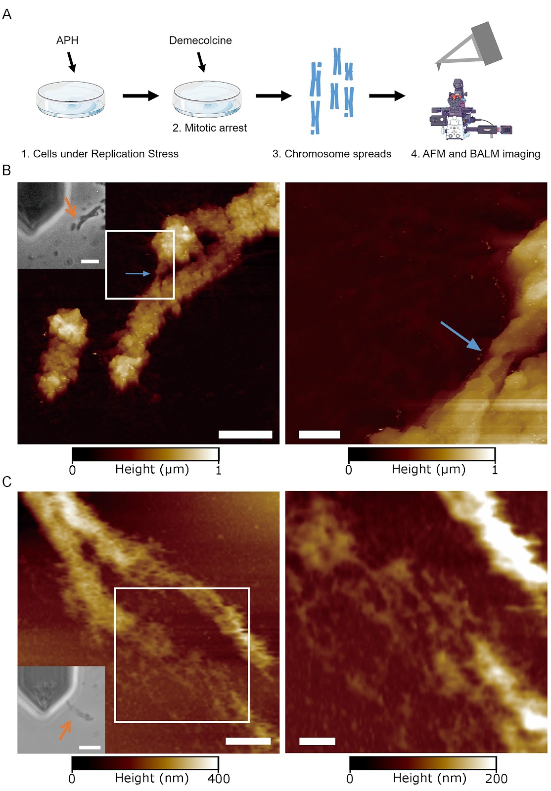
Common fragile sites (CFSs) are large genomic loci that are frequently deleted under replication stress and are thought to play a role in carcinogenesis as well as developmental disorders during early embryogenesis. They often appear as gaps or breaks in mitotic chromosomes by conventional optical microscopy and are also loci that are replicated in mitosis in a process called mitotic DNA synthesis (MiDAS). However, we still have a very poor understanding of the potential mechanisms underlying their genomic instability. We recently found that in normal mitotic chromosomes, there is a two-level hierarchy in the higher-order chromatin structure, with mechanically resistant ~90 nm Mitotic NanoDomains (MNDs) assembling into ~125 nm compact granules (CGs). In this work, we combine multiple super-resolution imaging techniques, including binding-activated localization microscopy (BALM), atomic force microscopy (AFM), and stochastic optical reconstruction microscopy (STORM), to characterize CFSs. Our super-resolution data indicate that sites that appear as gaps by conventional microscopy can be filled with chromatin of the size and shape of MNDs but not CGs. Moreover, we find that MiDAS loci only form an array of MNDs with no CGs. Taken together, our work suggests that under replication stress, CFS loci can not only be replicated but also assembled into higher-order chromatin. However, this organization is limited to the level of MNDs, which suggests that a failure to progress beyond MNDs to form CGs constitutes a key structural deficiency at these loci.

Super-resolution imaging reveals higher-order structures within common fragile sites in human mitotic chromosomes
Graphical Abstract
Original ResearchVol. 58, Issue 8 • pp. 1719-1735DOI: 10.3724/abbs.2026058

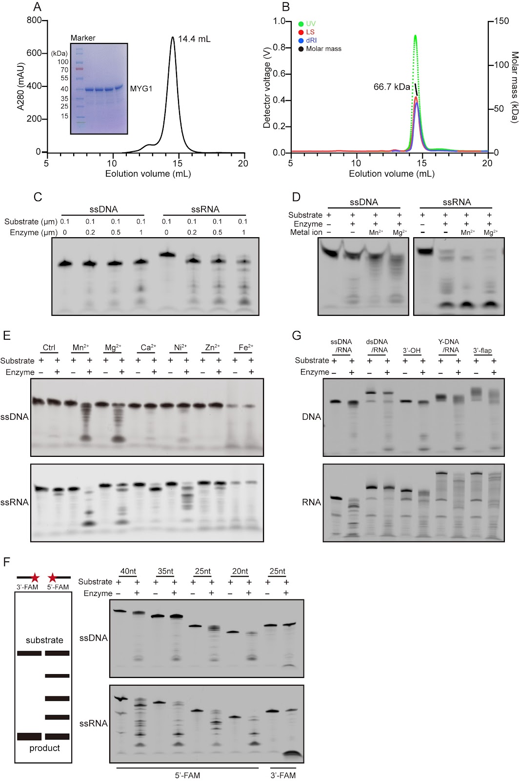
Biochemical and structural studies reveal the substrate specificity and catalytic mechanism of MYG1 as a two-metal ion-dependent 3′→5′ exonuclease

Authors: Chao Lan, Ziyue Chen, Guanchao Wang, Jianping Ding

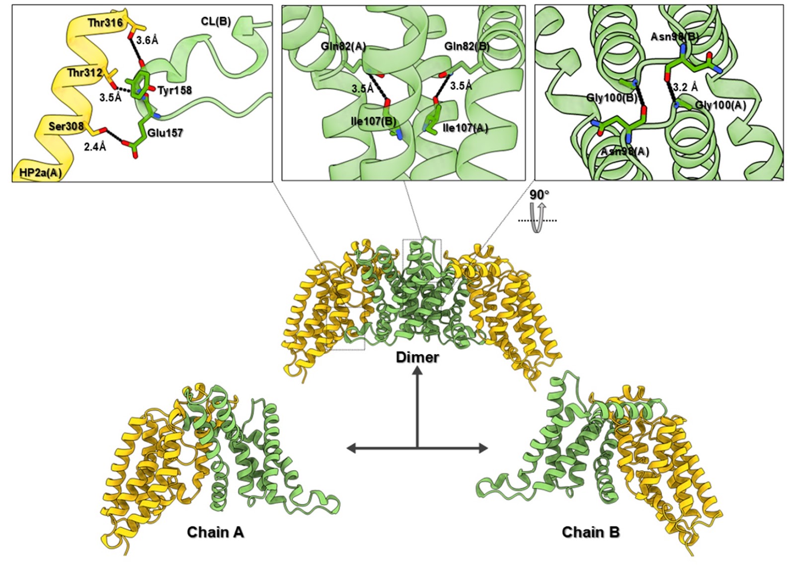
Nucleases are a class of enzymes that specifically cleave nucleic acids in all living organisms. They play crucial roles in essential biological processes, including the regulation of gene expression, DNA damage repair, and RNA processing and degradation. MYG1 (melanocyte proliferating gene 1) is a highly conserved eukaryotic protein that exhibits 3′→5′ exonuclease activity. This study systematically characterizes the enzymatic properties of MYG1 and determines its structures in complexes with metal ions and various mono- and poly-(deoxy)nucleotides. The functional roles of key residues involved in metal ion binding and substrate binding in the catalytic reaction are examined through site-directed mutagenesis, enzymatic activity assay, and structure determination. Our biochemical and structural data together demonstrate that MYG1 is a Mn2+- or Mg2+-dependent 3′→5′ exonuclease capable of cleaving a variety of nucleic acids with different structures. It exhibits the highest activity for single-stranded RNA and a nucleotide preference for U in single-stranded RNA and dT in single-stranded DNA. Mechanistically, MYG1 functions as a dimer, with the active site formed by the catalytic domain of monomer 1 and the substrate-binding domain of monomer 2, and cleaves nucleic acids through a two-metal ion-mediated catalytic mechanism. These findings establish a molecular basis for further investigations into the biological functions and molecular mechanisms of MYG1 within cells and its potential roles in human diseases.

Biochemical and structural studies reveal the substrate specificity and catalytic mechanism of MYG1 as a two-metal ion-dependent 3′→5′ exonuclease
Graphical Abstract
Original ResearchVol. 58, Issue 7 • pp. 1597-1610DOI: 10.3724/abbs.2025191

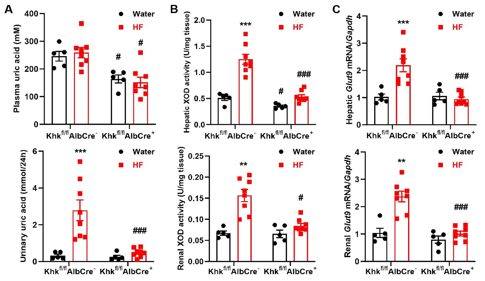
The effect of liver-specific ketohexokinase deletion on the intestinal-liver-kidney axis in high-fructose-induced metabolic syndrome mice

Authors: Huiru Yang, Xiaoli Yi, Shanshan Song, Mulan Wang, Wenting Tan, Ying Zhu, Jun Yu, Chuanming Xu

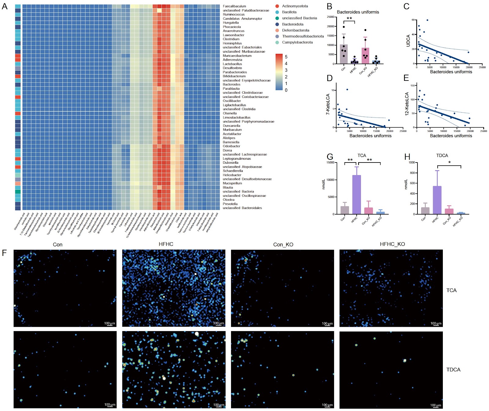
The liver is a crucial site for fructose uptake and metabolism, a function intricately linked to fructose-associated pathologies. This study examines the role of hepatic ketohexokinase (KHK) in metabolic syndrome induced solely by high-fructose intake. Liver-specific Khk-deficient mice are generated and fed with a 20% fructose solution for 3 months, after which the features of metabolic syndrome are examined. Compared with fructose-fed floxed controls, fructose-fed liver-specific Khk-deficient mice present alleviated liver injury and hepatic steatosis, along with lower triglyceride levels in the plasma and liver, plasma aspartate transaminase and alanine transaminase levels, and mRNA levels of genes related to triglyceride and fatty acid synthesis. Liver-specific Khk deficiency also leads to lower uric acid levels in the plasma and urine, as well as xanthine oxidase activity and Glut9 mRNA levels in the liver and kidneys of fructose-fed mice. Although intestinal villus length and epithelial barrier integrity remain unaffected, the deletion of liver Khk significantly reduces fructose-stimulated KHK, Glut2, Glut5, and aldolase B expression in the intestine and kidneys, suggesting inhibited fructose absorption and metabolism in these tissues. In the adipose tissue, fructose-induced increases in adipocyte size and tumor necrosis factor-α and interleukin-6 mRNA levels are blocked by liver-specific Khk deficiency, indicating improved remodeling of adipose tissue and reduced inflammation in adipocytes. Overall, liver-specific Khk deletion is sufficient to protect against metabolic syndrome induced by excessive fructose intake. Our findings underscore the critical role of liver KHK-mediated fructose metabolism in driving the physiological and pathological consequences associated with fructose consumption along the intestinal-liver-kidney axis.

The effect of liver-specific ketohexokinase deletion on the intestinal-liver-kidney axis in high-fructose-induced metabolic syndrome mice
Graphical Abstract
Original ResearchVol. 58, Issue 7 • pp. 1557-1567DOI: 10.3724/abbs.2025257

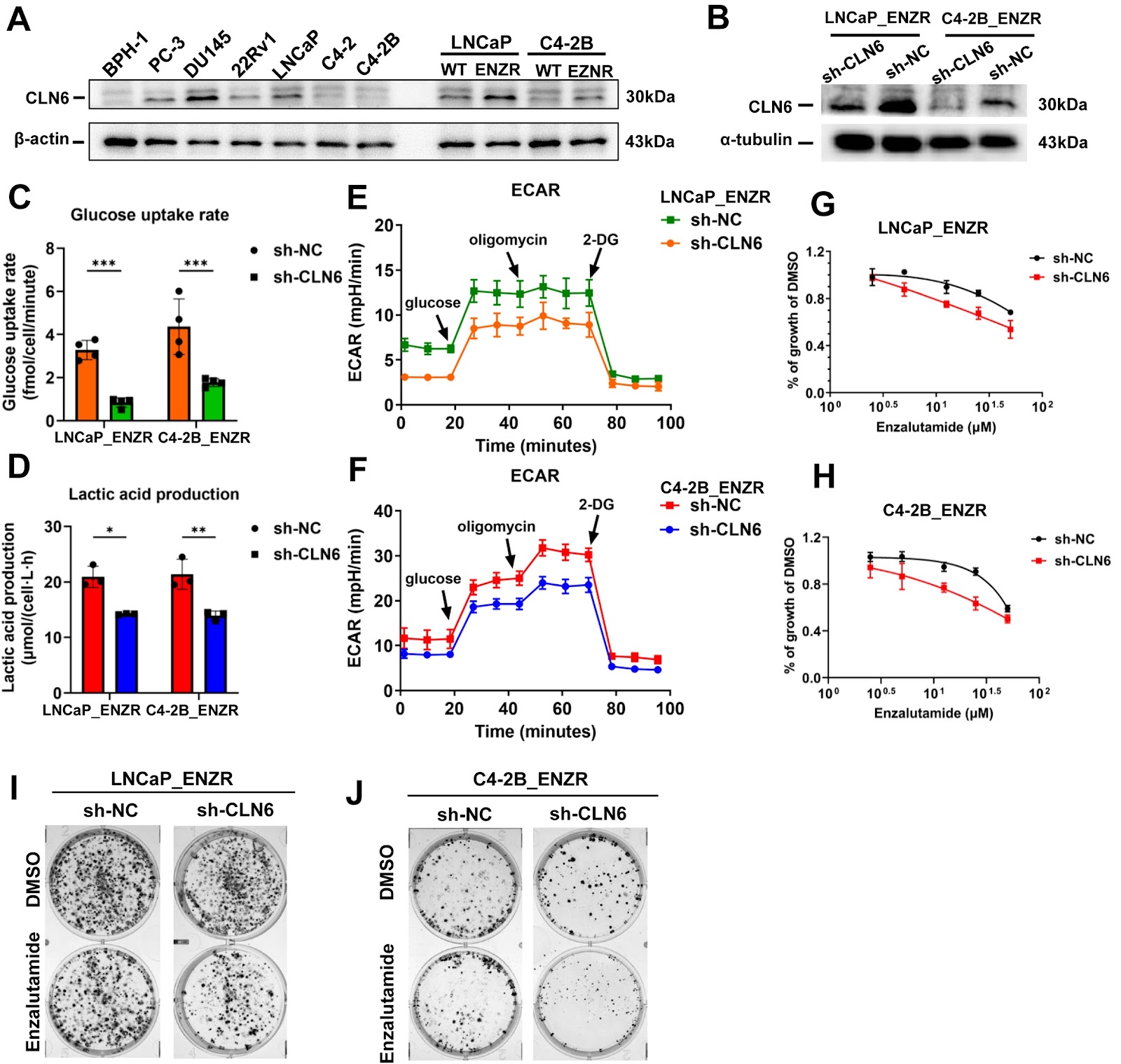
Glycolysis reprogramming predicts poor prognosis and drives therapy resistance via CLN6 in lethal prostate cancer

Authors: Zhouda Cai, Jianming Lu, Shanshan Mo, Jipu Liu, Chuanfan Zhong, Yongding Wu, Fen Zou, Jianheng Ye, Zhaodong Han, Yuxiang Liang, Le Zhang, Fengping Liu, Weide Zhong

Lethal prostate cancer is marked by tumor heterogeneity and resistance to androgen receptor signaling inhibitors (ARSIs). In this study we identify glycolysis as a driver of disease progression and therapy resistance. Using single-sample gene set enrichment analysis (ssGSEA) on the SU2C cohort, we demonstrate that elevated glycolysis activity is associated with poor progression-free and overall survival. The glycolysis-based prognostic score (GLY score) is derived from the HALLMARK_GLYCOLYSIS gene set which includes CLN6, SDHC, B4GALT2, RPE, NANP, and KIF20A, via LASSO-Cox regression. The GLY score effectively stratifies risk in the SU2C and WDCT cohorts, with higher scores predicting worse outcomes and increased SYNE1 mutation frequency. Pan-cancer analysis across TCGA datasets confirm its prognostic value. In vitro, enzalutamide-resistant prostate cancer cell lines exhibit heightened glycolysis, and 2-DG inhibition reverses this effect, restoring drug sensitivity. CLN6 knockdown reduces glycolytic activity and cell proliferation. The GLY score offers robust prognostic value, and CLN6 represents a promising therapeutic target for precision medicine in lethal prostate cancer.

Glycolysis reprogramming predicts poor prognosis and drives therapy resistance via CLN6 in lethal prostate cancer
Graphical Abstract
Original ResearchVol. 58, Issue 7 • pp. 1637-1652DOI: 10.3724/abbs.2025206

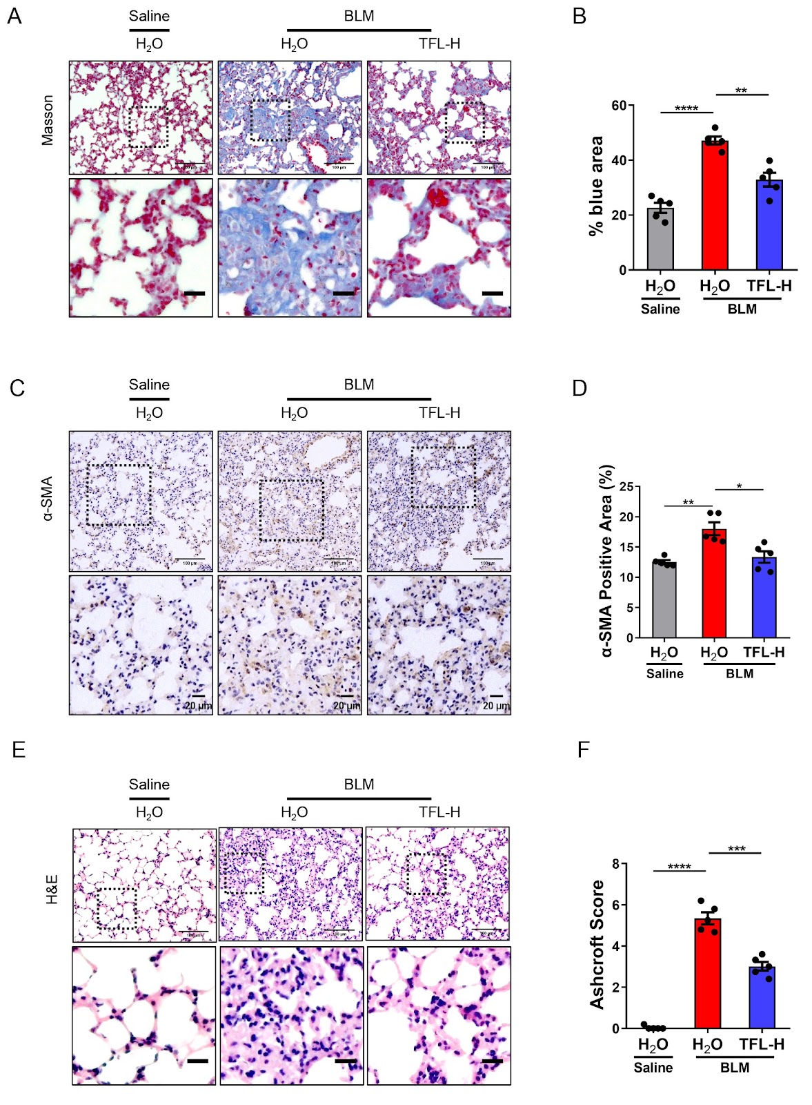
Total flavonoids of litchi seed attenuates cellular senescence by inhibiting the production of SASP through p65 suppression and ameliorates pulmonary fibrosis

Authors: Xiaocui Li, Yao Wei, Qiuchen Cheng, Suyu Xiao, Sisi Yao, Daikang Yang, Jilong Wang, Liping Chen, Qing Li, Tingzheng Zhan

Anti-aging foods not only benefit elderly individuals but also drive the development of safe and effective natural drugs. Here, we report that the addition of total flavonoids of litchi seed (TFL) delays replicative senescence and stress-induced senescence. TFL alleviates the senescence-associated secretory phenotype (SASP) and reduces the degree of DNA damage caused by bleomycin (BLM). TFL also counteracts stress-induced pulmonary senescence and fibrosis. TFL reduces the protein level of p21 in mouse lung and alleviates pulmonary fibrosis. Transcriptome profiling further reveals that TFL plays a key role in its anti-aging mechanism by inhibiting the SASP. Mechanistically, TFL suppresses p65 protein expression, thereby inhibiting IL-1α and IL-1β and delaying cellular senescence. Gut microbiome analysis reveals that the abundance and functions of the mouse gut microbiome change after BLM exposure and that TFL treatment reverses these changes. Overall, we provide a theoretical basis for the future application of TFL as a potential anti-aging product.

Total flavonoids of litchi seed attenuates cellular senescence by inhibiting the production of SASP through p65 suppression and ameliorates pulmonary fibrosis
Graphical Abstract
Original ResearchVol. 58, Issue 7 • pp. 1671-1675DOI: 10.3724/abbs.2026039

Small chemical molecule CPP promotes angiogenesis in surgically created severe lower limb ischemia and diabetes-induced limb vascular reduction models

Authors: Xinyu Dong, Yangyang Zhang, Congyao Zhao, Xiaomeng Yan, Xiaohui Chi, Xinyu Xie, Baoxiang Zhao, Jian Zhang, Li Wang, Junying Miao, Zhaomin Lin

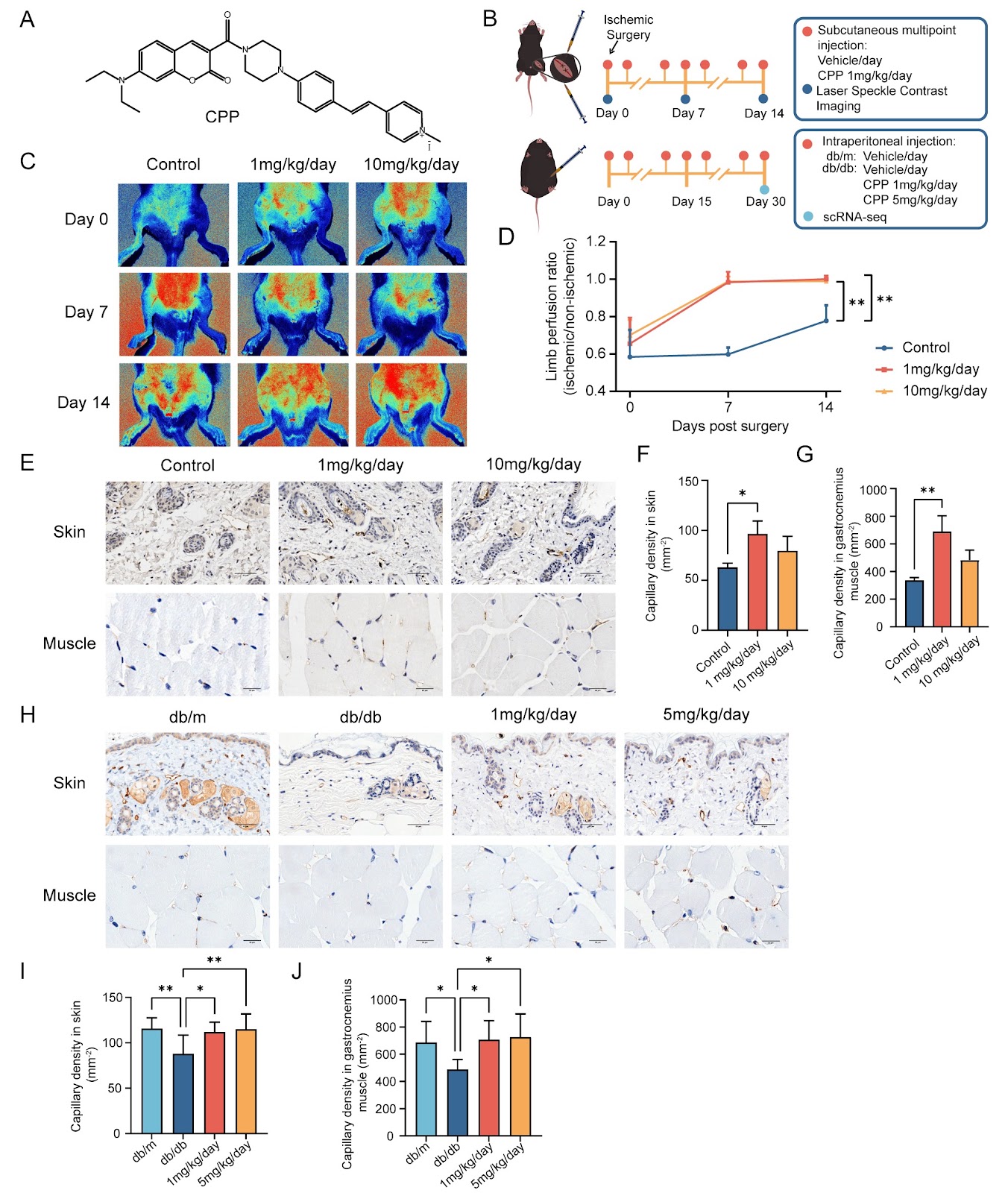
Patients with peripheral artery disease (PAD) commonly experience chronic limb-threatening ischemia (CLTI) in the end stage, leading to severe functional impairment of the limbs, amputation, and even death. Among the various disease factors contributing to PAD, diabetes is significantly associated with PAD risk, leading to more severe symptoms and a poorer prognosis. Promoting angiogenesis at ischemic sites and improving blood flow are key to the recovery of limbs from ischemic injury. Angiogenic therapies based on cells and small-molecule drugs are considered important and promising strategies. Cell-based therapies can be classified based on cell source into allogeneic therapies and autologous therapies. Autologous cells offer the advantage of not requiring immunosuppression but may be limited by factors such as cell unavailability and scarcity due to autologous factors. Allogeneic cells, while offering a more abundant cell source, present drawbacks, including immune incompatibility and ethical concerns, which restrict their application. Small chemical molecules offer advantages such as convenience, low cost, ease of synthesis and storage, and rapid action without the need for introducing exogenous genes. They circumvent the high costs associated with cell therapies and potential side effects from allogeneic cell transplantation, holding significant promise and potential in both biomedical research and clinical applications. Fibroblasts are a cell type with functional and transcriptional heterogeneity and cellular fate plasticity, making them a promising source of seed cells for clinical tissue and organ repair and regeneration. Small molecules have become recognized as crucial tools for the induction of cellular reprogramming. In our laboratory’s previous research, a novel small chemical molecule probe, CPP ((E)-4-(4-(4-(7-(diethylamino)-2-oxo-2H-chromene-3-carbonyl)piperazin-1-yl)styryl)-1-methylpyridin-1-ium iodide), was identified as an inhibitor of prolyl-4-hydroxylase 2 (PHD2). CPP induces the differentiation of cultured human dermal fibroblasts into vascular endothelial cells (VECs) via the PHD2/hypoxia-inducible factor-1α/hairy-related transcription factor 1 (PHD2/HIF1α/HEY1) signaling pathway. The differentiated VECs exhibited therapeutic efficacy in treating lower limb ischemia in mice, indicating that CPP holds promise as a therapeutic candidate for limb ischemia. In this study, we aimed to investigate the capability of CPP to directly induce angiogenesis in vivo under conditions of vascular injury. Critical limb ischemia (CLI) occurs at the end stage of PAD. To evaluate the in situ therapeutic effects of CPP on CLI, a CLI model was established using C57BL/6 mice (Hubei Research Center of Laboratory Animals, Wuhan, China). CPP was synthesized and provided by the laboratory of Professor Baoxiang Zhao at Shandong University (Jinan, China). Following surgery, continuous subcutaneous multipoint injections of CPP were administered for 14 d to mimic localized drug treatment (Figure 1A). Laser speckle blood flow imaging was used to assess lower limb perfusion on days 0, 7, and 14 (Figure 1B, upper panel). The results showed that by day 7 post-surgery, both CPP concentrations significantly enhanced perfusion in the ischemic limb compared to the control group. From days 7 to 14, perfusion changes in both CPP-treated groups plateaued. Meanwhile, the control group showed increased perfusion. Throughout the treatment period, no significant differences were observed between the 1 and 10 mg/kg/day CPP treatment groups (Figure 1C,D). On day 14, the capillary density in the skin and muscle was significantly higher in the 1 mg/kg/day CPP group than in the control group. Although the 10 mg/kg/day group had a slightly higher density than the control, the difference was not statistically significant (Figure 1E–G). In addition, the organ toxicity

Small chemical molecule CPP promotes angiogenesis in surgically created severe lower limb ischemia and diabetes-induced limb vascular reduction models
Graphical Abstract
Original ResearchVol. 58, Issue 7 • pp. 1509-1518DOI: 10.3724/abbs.2025223

Fibroblast growth factor 13 deficiency attenuates doxorubicin-induced cardiotoxicity by regulating Parkin-mediated myocardial injury

Authors: Jiabing Han, Xuyan Li, Yiming Dong, Yidan Wang, Simeng Lv, Yiyi Zhang, Ran Zhao, Yingke Yan, Yanxue Han, Yu Wang, Jing Yang, Cong Wang, Chuan Wang

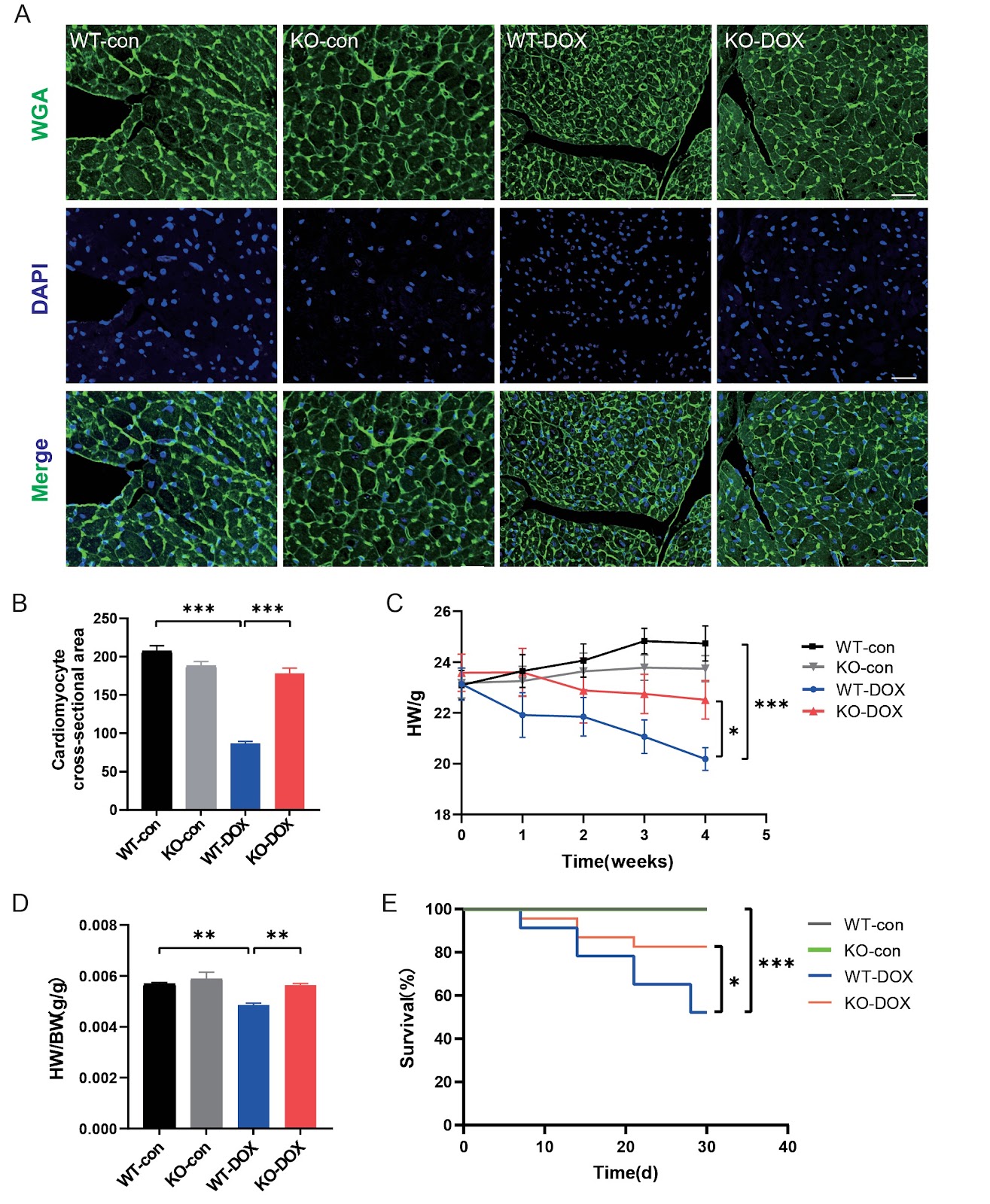
The clinical use of doxorubicin (DOX) as a chemotherapeutic agent is limited by its cardiotoxic effects. Fibroblast growth factor (FGF) isoform 13, a distinct type of FGF, has been increasingly recognized as an important regulator of cardiovascular disease. However, its role in doxorubicin-induced cardiotoxicity remains unknown. Therefore, the objective of this study is to investigate the role and mechanism of FGF13 in doxorubicin-induced cardiac injury. C57BL/6 mice are used to establish Dox-induced cardiotoxicity models. The results reveal that mouse weight, cardiomyocyte cross-sectional area, ejection fraction and fractional shortening are decreased in the DOX group. In contrast, Fgf13 deficiency mitigates doxorubicin-mediated cardiotoxicity, as indicated by increased mouse weight, cardiomyocyte cross-sectional area, ejection fraction and fractional shortening. Mechanistically, the protein expressions of bax and cleaved caspase 3 are elevated in the DOX-treated group, along with decreased JC-1 fluorescence intensity and bcl-2 expression, whereas Fgf13 knockout prevents these alterations. In addition, Parkin, but not p53, interacts with FGF13 and is upregulated in response to Fgf13 deficiency in a mouse model of doxorubicin-induced cardiotoxicity. Overall, Fgf13 knockout attenuates doxorubicin-induced cardiomyocyte apoptosis and mitochondrial damage through the modulation of Parkin, indicating that FGF13 may serve as a promising therapeutic target for DOX-induced cardiotoxicity.

Fibroblast growth factor 13 deficiency attenuates doxorubicin-induced cardiotoxicity by regulating Parkin-mediated myocardial injury
Graphical Abstract
Original ResearchVol. 58, Issue 7 • pp. 1544-1556DOI: 10.3724/abbs.2025253

A novel biomarker SNHG11 promotes tumor progression and oxidative phosphorylation in clear cell renal cell carcinoma

Authors: Zhuoyuan Lin, Chaojiang Chen, Jianxin Li, Jun Zhao, Jia Xu, Le Zhang, Chuanfan Zhong, Shanshan Mo, Jianming Lu, Yu Zheng

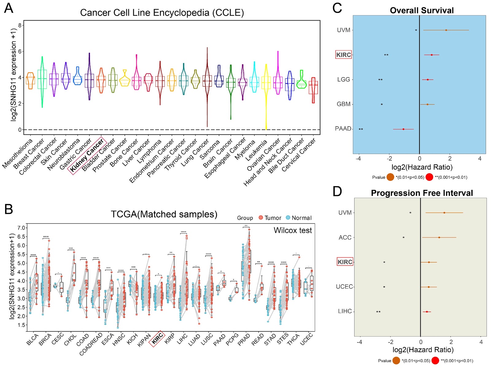
Kidney renal clear cell carcinoma (KIRC) is the most common and aggressive subtype of renal cell carcinoma and is characterized by poor prognosis and high molecular heterogeneity. Long noncoding RNAs (lncRNAs) have emerged as crucial regulators in cancer, yet the functional role of SNHG11 in KIRC remains unclear. In this study, we perform integrated multiomics analysis using data from the TCGA and ICGC cohorts and reveal that SNHG11, a methylation-associated lncRNA, is significantly correlated with poor clinical outcomes. In vitro and in vivo assays demonstrate that SNHG11 promotes tumor proliferation and progression. Mechanistically, SNHG11 enhances oxidative phosphorylation, as evidenced by increased ATP production, disrupted mitochondrial membrane potential, and altered NAD+/NADH ratios. Furthermore, SNHG11 expression is associated with somatic mutation patterns, particularly those involving BAP1 and PBRM1, indicating potential crosstalk between epigenetic regulation and genetic alterations. These findings reveal that SNHG11 is a novel biomarker in KIRC and a potential therapeutic target.

A novel biomarker SNHG11 promotes tumor progression and oxidative phosphorylation in clear cell renal cell carcinoma
Graphical Abstract
Original ResearchVol. 58, Issue 7 • pp. 1437-1440DOI: 10.3724/abbs.2026110

Metabolic reprogramming—the nexus of cellular adaptations, organ crosstalk, and therapeutic innovations in human diseases

Authors: Jun Ren, Hartmut Schlüter, Marcel Kwiatkowski, Ling Lin

For decades, cellular metabolism was viewed as a static network of biochemical pathways designed solely to produce ATP and fulfill basic energy needs. Classical pathophysiology frequently attributes systemic disorders, including cardiovascular disease, oncogenesis and tissue fibrosis, to structural defects, hormonal dysregulations or localized inflammation. However, advanced multi-omics frameworks have driven a paradigm shift, with metabolic reprogramming being recognized not merely as a passive consequence of disease but also as a primary driver of initiation, progression, and therapeutic resistance across human pathologies. This Special Issue, titled “Metabolic Reprogramming”, brings together 17 comprehensive studies alongside 3 New Phenomena exploring these transformations. The contributions span four interrelated thematic areas: (1) systemic cardiometabolic and gestational syndromes; (2) acute and chronic organ injury; (3) oncological metabolic dependencies; and (4) systemic organ-axis interdependencies. By integrating transcriptomics, metabolomics, single-cell deconvolution, and targeted biochemical analyses, these studies elucidate the precise molecular mechanisms governing metabolic remodeling.

Metabolic reprogramming—the nexus of cellular adaptations, organ crosstalk, and therapeutic innovations in human diseases
Graphical Abstract
Original ResearchVol. 58, Issue 6 • pp. 1402-1412DOI: 10.3724/abbs.2025224

Bioinformatics classification of the MgtE Mg2+ channel and de novo protein design for the stabilization of its novel subclass

Authors: Zhixuan Zhao, Kimiho Omae, Wataru Iwasaki, Ziyi Zhang, Fazhi Pan, Eun-Jin Lee, Koichi Ito, Motoyuki Hattori

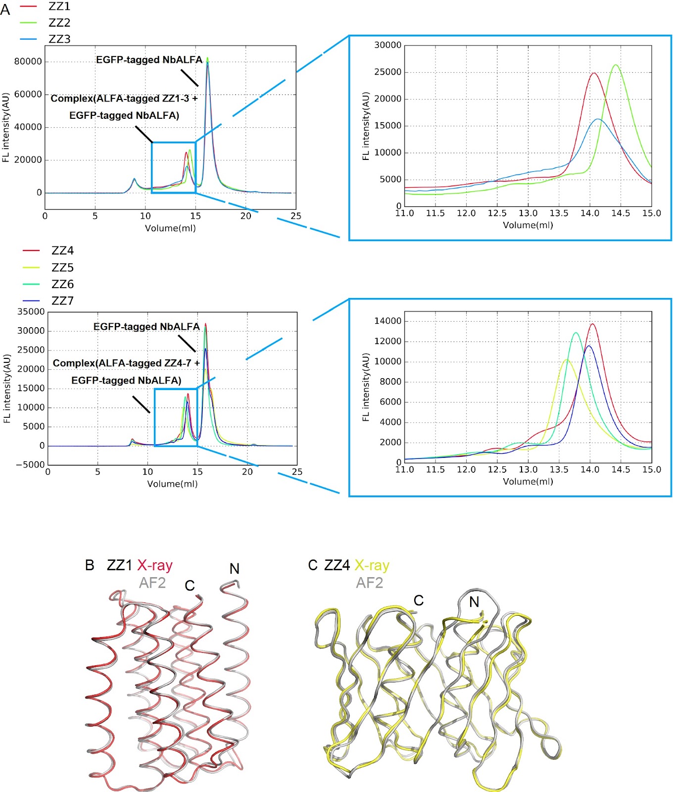
MgtE channels play crucial roles in Mg2+ homeostasis and are implicated in bacterial survival under antibiotic exposure. Previous structural and biophysical studies have focused predominantly on Thermus thermophilus MgtE, leaving the structural and mechanistic diversity of MgtE family proteins largely unexplored. In this study, via a genome mining approach, we identify diverse MgtE homologs, including a novel subclass termed the “mini-N type”, which lacks the canonical cytoplasmic N and CBS domains but possesses a unique small N-like domain. Despite extensive expression screening, mini-N-type homologs cannot be stably purified. To address this issue, we design a series of de novo proteins and determine their crystal structures. A selected de novo protein is fused to a mini-N-type MgtE, enabling successful purification and preliminary cryo-EM imaging. Our findings demonstrate that de novo-designed protein fusions serve as powerful tools for stabilizing and purifying otherwise unstable membrane proteins, opening new avenues for structural and functional studies of otherwise inaccessible membrane proteins.

Bioinformatics classification of the MgtE Mg2+ channel and de novo protein design for the stabilization of its novel subclass
Graphical Abstract
Original ResearchVol. 58, Issue 5 • pp. 1155-1169DOI: 10.3724/abbs.2026055

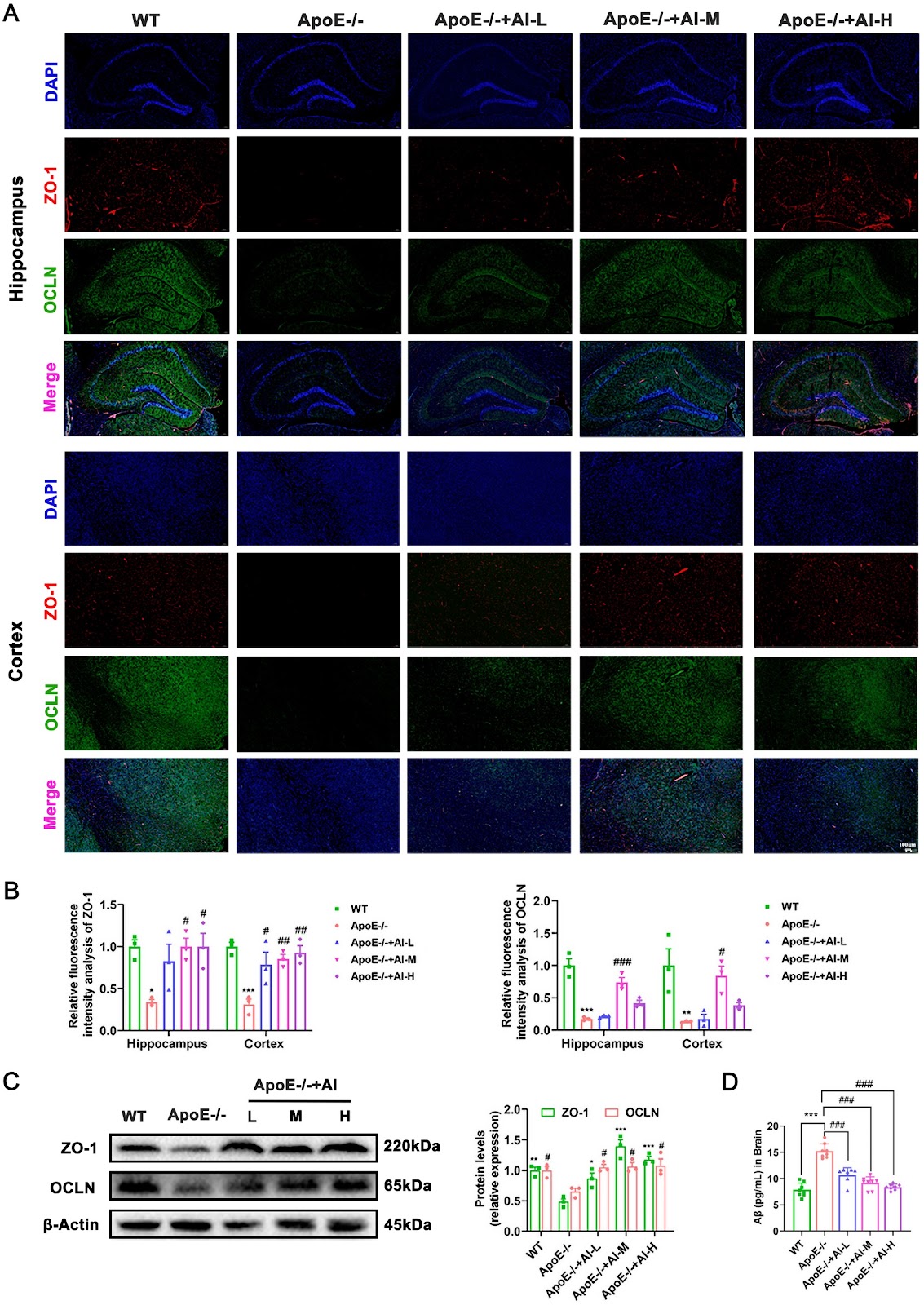
Atractylenolide I mitigates Alzheimer’s disease pathology in ApoE–/– mice via ARG1/nNOS axis and lipid homeostasis regulation

Authors: Xun Zhou, Rui Wang, Jingsi Yan, Xiaolang Wu, Dongsheng Yuan, Qi Wang, Huilin Li, Wei Zhao

Apolipoprotein E (ApoE) serves as a critical molecular nexus between Alzheimer’s disease (AD) and atherosclerosis, two age-associated inflammatory disorders that share vascular pathology, amyloid-beta (Aβ) deposition, and lipid dysregulation. Atractylenolide I (AI), a promising therapeutic candidate derived from Atractylodes macrocephala Koidz., exhibits multimodal bioactivities with demonstrated anti-inflammatory and neuroprotective properties. To explore its therapeutic potential against AD pathology, we use high-fat diet (HFD)-fed ApoE knockout (ApoE–/–) mice treated with or without AI for 12 weeks. Integrated bioinformatics analyses and experimental validation reveal that AI treatment markedly attenuates systemic lipid dyshomeostasis, particularly cerebral lipid deposition, suppresses neuroinflammation via downregulation of M1 macrophage polarization markers, and restores cognitive function through neuronal preservation in hippocampal regions. Mechanistically, AI orchestrates cholesterol efflux by up-regulating ATP-binding cassette transporter A1 (ABCA1) and liver X receptor (LXR) expression, while concurrently modulating the abundance of arginine biosynthesis metabolites (urea, malic acid, and creatinine) to rebalance neurovascular homeostasis. Notably, western blot and RT-qPCR analyses reveal that AI differentially regulates key enzymes including arginase 1 (ARG1) and simultaneously upregulates the expression of neuronal nitric oxide synthase (nNOS). Further molecular docking and surface plasmon resonance (SPR) analyses confirm the direct binding of AI to ARG1, indicating a novel neuroprotective mechanism involving the modulation of arginine metabolism. These findings delineate the pleiotropic effects of AI against AD pathology and establish a preclinical foundation for the development of AI-based therapeutics targeting neurodegenerative-cardiovascular comorbidities.

Atractylenolide I mitigates Alzheimer’s disease pathology in ApoE–/– mice via ARG1/nNOS axis and lipid homeostasis regulation
Graphical Abstract
Original ResearchVol. 58, Issue 6 • pp. 1281-1290DOI: 10.3724/abbs.2026042

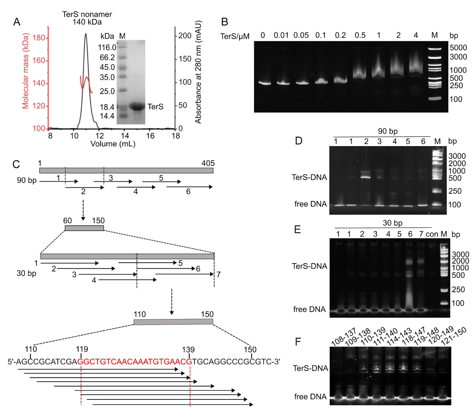
Structural and functional insights into the distinct DNA recognition mechanisms of the terminase small subunit TerS from cyanophages

Authors: De-Qin Dong, Feng Yang, Kang Du, Kang Xu, Wen-Bin Cheng, Yuxing Chen, Cong-Zhao Zhou, Yong-Liang Jiang

Efficient genome packaging is a critical step in the phage life cycle, directly influencing the viral maturation and infectivity. In tailed phages, this process is driven by a packaging motor composed of a portal protein and a terminase complex. The terminase complex usually consists of a large subunit (TerL) and a small subunit (TerS), which cooperate to recognize, cleave, and translocate genomic DNA into the capsid. However, due to the remarkable diversity and complexity of phage packaging systems, the molecular mechanisms governing TerS-mediated DNA recognition remain poorly understood. Here, we report the 3.51 Å cryo-electron microscopy structure of the TerS from the short-tailed cyanophage Pam5, which infects the host Pseudanabaena mucicola Chao 1806. Pam5 TerS assembles into a nonameric ring with a radially symmetric spiral architecture. Biochemical assays show that Pam5 TerS recognizes the genomic DNA via a specific interaction between the N-terminal helix-turn-helix (HTH) domain of TerS and a 21-bp DNA sequence within the terS gene. In contrast, the TerS from another short-tailed cyanophage, Pam1, which infects the same host, binds to DNA in a sequence-independent manner. These findings reveal that cyanophages, even infecting the same host, could adopt two distinct DNA recognition strategies: HTH-mediated sequence-dependent or sequence-independent modes. This work provides structural and mechanistic insights into the diverse DNA-recognition strategies of TerS and advances our understanding of the evolutionary plasticity of viral genome packaging mechanisms.

Structural and functional insights into the distinct DNA recognition mechanisms of the terminase small subunit TerS from cyanophages
Graphical Abstract
Original ResearchVol. 58, Issue 6 • pp. 1431-1432DOI: 10.3724/abbs.2026006

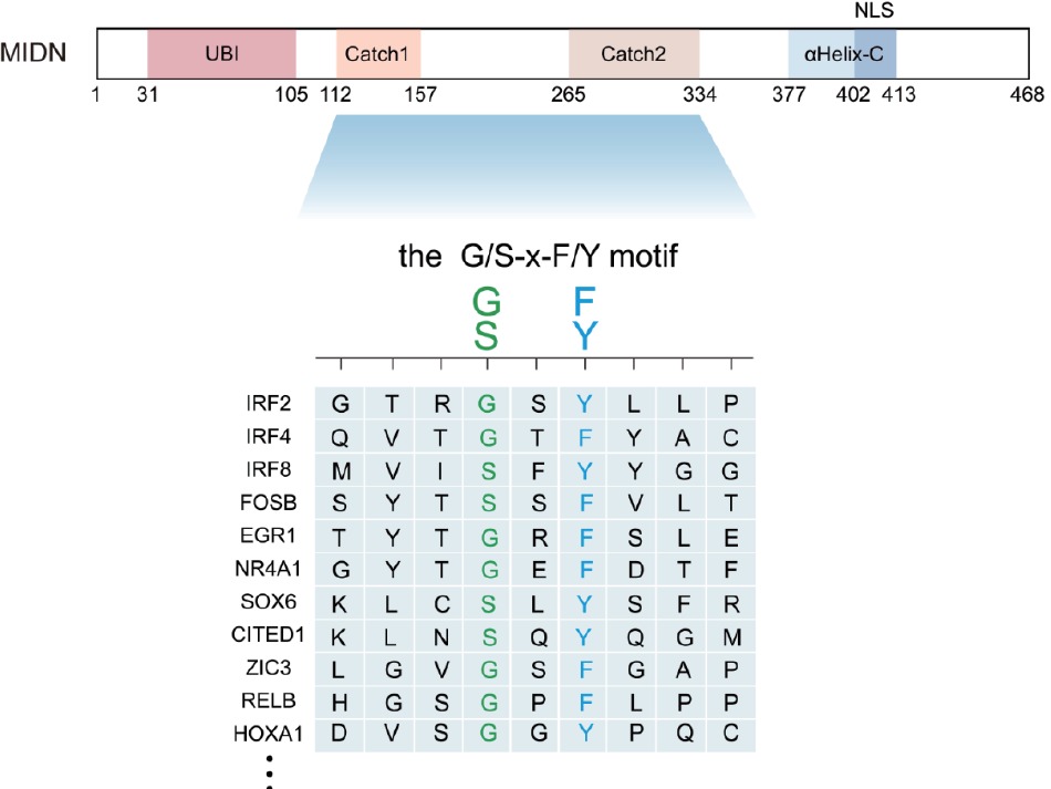
Caught the ‘Catch’ of midnolin: structural basis for broad substrate specificity in ubiquitin-independent proteasomal degradation

Authors: Chuanyin Li, Ronggui Hu

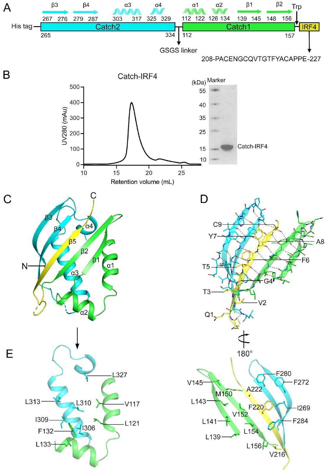
Protein homeostasis serves as the foundation for every cellular decision—division, differentiation, stress adaptation, or death—by precisely balancing the proteome across abundance, quality, spatial distribution, and temporal dynamics; its dysregulation drives numerous human pathologies, including cancers and neurological disorders. In the traditional ubiquitin-dependent degradation cascade, target proteins are marked by covalent attachment of polyubiquitin chains, a process requiring E1 activating enzymes, E2 conjugating enzymes, and E3 ligases that confer substrate specificity. This ubiquitin signal is then recognized by the 19S regulatory particle of the proteasome, which unfolds and translocates the tagged protein into the 20S core for proteolytic destruction. The ubiquitin-independent proteasomal degradation pathway mediated by midnolin (MIDN) has recently emerged as a distinct and biologically important mechanism for regulating nuclear protein turnover. While earlier genetic, biochemical, and cryo-electron microscopy studies established MIDN as a proteasome-associated adaptor for immediate-early gene (IEG) products, the molecular logic underlying its broad yet selective substrate recognition remains unresolved. In the current study, Zhong et al. provide a comprehensive structural and biochemical analysis of the MIDN Catch domain bound to IRF4-derived peptides, substantially advancing our understanding of MIDN substrate recognition. By solving multiple crystal structures of the Catch-IRF4 complex, including wild-type and systematically engineered mutants, the authors demonstrate that MIDN recognizes substrates through a conserved β-strand insertion mechanism at the interface of the Catch1 and Catch2 subdomains. This investigation generalizes and expands prior structural observations of MIDN-IRF4 to diverse substrates, demonstrating that β-strand complementation constitutes a universal recognition mechanism utilized by MIDN. A major conceptual advance of this study is the identification of a minimal and generalizable recognition principle centered on two highly conserved positions within the substrate motif. The authors show that a reciprocal phenylalanine-glycine interaction between the substrate and Catch2—forming an “F-G zipper”—constitutes the dominant energetic determinant for binding. Disruption of this zipper severely compromises protein stability and binding, explaining prior functional observations that single-point mutations in IRF4 or EGR1 abolish MIDN-mediated degradation. In contrast, flanking residues within the binding motif display remarkable tolerance to substitution. Through combined mutagenesis, thermostability analysis, AlphaFold3 modeling, and structural determination, the study demonstrates that these positions occupy large and plastic hydrophobic pockets in the Catch domain. This architectural flexibility provides a direct molecular explanation for how MIDN can accommodate a wide spectrum of substrates while preserving selectivity. From these data, the authors derive a concise consensus recognition motif (G/S-x-F/Y) embedded within an unstructured or loop region, offering a predictive framework for identifying additional MIDN targets. Importantly, this work bridges a critical gap between previous cryo-EM studies describing MIDN-proteasome engagement and earlier substrate-specific structural analyses. Whereas prior studies clarified how MIDN delivers captured substrates to the proteasome, the present study elucidates how MIDN initially selects and binds those substrates. Together, these findings unify MIDN biology across structural, biochemical and functional dimensions. Overall, this study represents a significant advance in the field of proteostasis by revealing how a ubiquitin-independent adaptor achieves broad substrate specificity through a simple yet robust structural principle. Beyond MIDN, the work provides a paradigm for short-linear-motif-based proteasomal targeting and has important implications for immune regulation, neurodegeneration and cancer biology.

Caught the ‘Catch’ of midnolin: structural basis for broad substrate specificity in ubiquitin-independent proteasomal degradation
Graphical Abstract
Original ResearchVol. 58, Issue 3 • pp. 638-648DOI: 10.3724/abbs.2025148

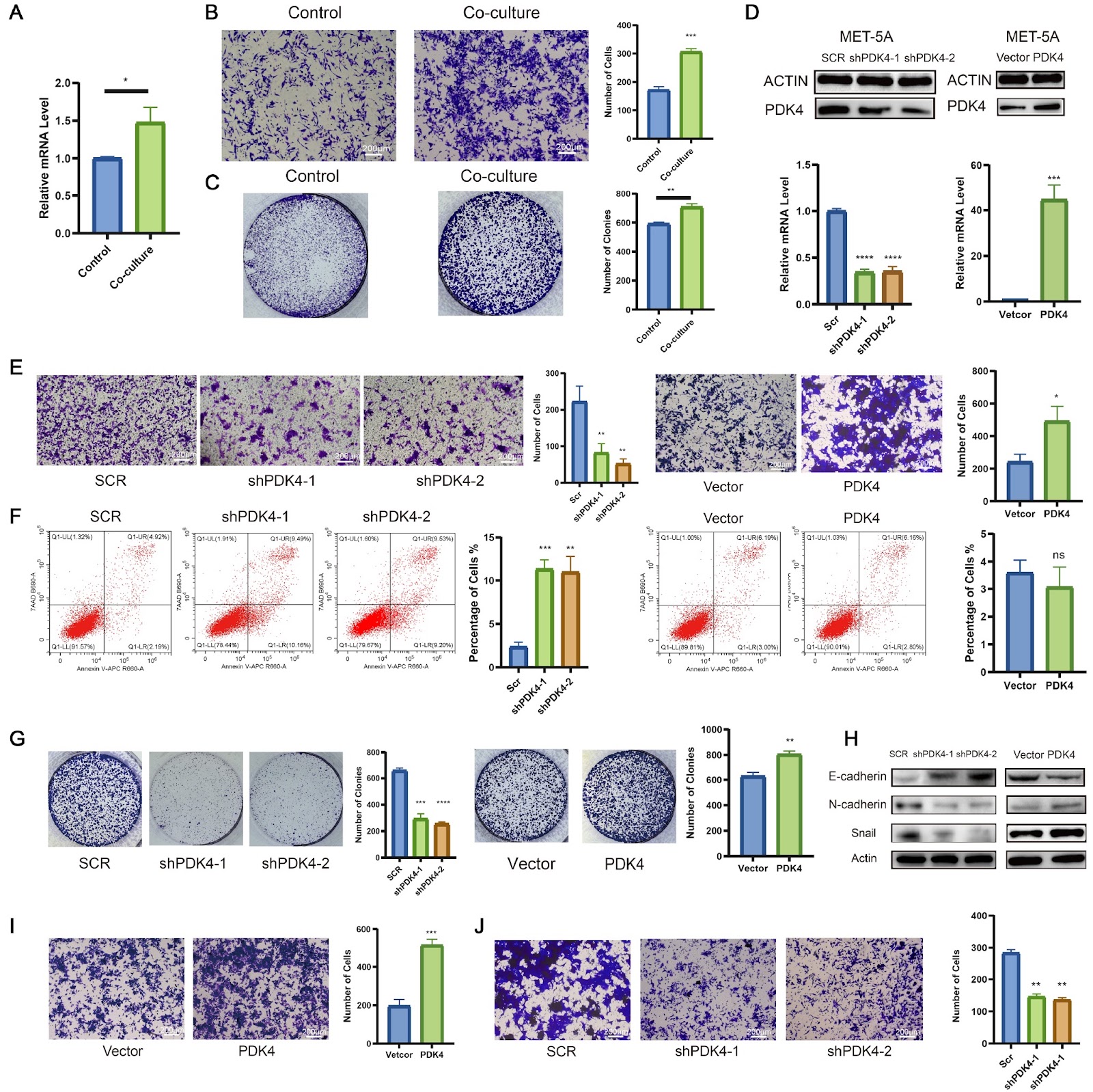
PDK4-driven metabolic reprogramming enhances mesothelial cell invasion in colorectal cancer peritoneal metastasis

Authors: Lingyu Han, Qinyuan Zhang, Yuchen Wu, Wenqin Luo, Shaobo Mo, Hongsheng Fang, Qingguo Li, Renjie Wang, Guoxiang Cai, Weixing Dai

Mesothelial cells play an important role in colorectal cancer peritoneal metastasis (CRC-PM), where they support tumor growth and invasion. In this study, we investigate the molecular mechanisms by which mesothelial cells contribute to CRC metastasis. Using single-cell RNA sequencing (scRNA-seq) on tissue samples from 12 CRC patients with peritoneal metastasis, we identify PDK4 as a key gene in mesothelial cells during metastasis. The expression of PDK4 is significantly greater in mesothelial cells undergoing mesothelial-to-mesenchymal transition (MMT) compared to normal peritoneal cells, suggesting its involvement in mesothelial cell reprogramming during peritoneal metastasis. In vitro experiments show that coculturing mesothelial cells with CRC cells leads to increased PDK4 expression, which in turn enhances mesothelial cell migration and invasion. Knockdown of PDK4 reduces mesothelial cell invasion, while overexpression of PDK4 increases invasive ability, highlighting its critical role in mesothelial cell invasion. Additionally, PDK4 promotes metabolic changes, specifically increasing fatty acid oxidation (FAO), which is necessary for mesothelial cell invasion. Blocking FAO reduces the invasive ability of PDK4-overexpressing mesothelial cells, while restoring FAO in PDK4-knockdown cells rescues their invasion potential. Further analysis shows that PDK4 enhances the acetylation of β-catenin, a protein involved in cell movement, and that this modification is crucial for mesothelial cell invasion. Our results suggest that PDK4 regulates mesothelial cell invasion through β-catenin acetylation following metabolic reprogramming, offering a potential target for therapies aimed at inhibiting CRC-PM.

PDK4-driven metabolic reprogramming enhances mesothelial cell invasion in colorectal cancer peritoneal metastasis
Graphical Abstract
Original ResearchVol. 58, Issue 6 • pp. 1235-1249DOI: 10.3724/abbs.2026002

Biochemical and structural studies of the midnolin Catch domain bound with both wild-type and mutant IRF4 peptides reveal the molecular basis for its broad substrate specificity

Authors: Yanling Zhong, Ziyue Chen, Guanchao Wang, Jianping Ding

The newly discovered midnolin-proteasome pathway is a unique ubiquitin-independent mechanism for degrading nuclear proteins, which is crucial for maintaining cellular protein homeostasis. The Catch domain of midnolin is essential for substrate recognition and binding, yet the underlying mechanism for its broad substrate specificity remains elusive. Transcription factor IRF4, essential for the functions of B and T cells, is a substrate of midnolin. This study presents comprehensive biochemical and structural analyses of the human midnolin Catch domain in complexes with both wild-type and mutant IRF4 peptides. The crystal structure of the Catch-IRF4 complex reveals that the Catch domain creates a substrate-binding groove at the interface of the Catch1 and Catch2 subdomains, recognizing and binding to the 215-QVTGTFYAC-223 sequence motif of IRF4. The binding motif of IRF4 forms a β-strand that is embedded into the substrate-binding groove, resulting in an antiparallel five-stranded β-sheet. The interactions between the IRF4 peptide and the Catch domain are predominantly hydrophobic and exhibit high spatial complementarity. Additionally, the biochemical, modeling and structural data indicate that the V2 and A8 positions of the IRF4 sequence motif can be substituted with other hydrophobic or small polar residues (G/A/V/L/I/M/P/F/Y/C/S/T), but not with large polar and charged residues (D/N/E/Q/H/K/R). The G4 position can be replaced by Ser, while the F6 position can be substituted with Tyr. These results suggest that the Catch domain can recognize and bind to a variety of substrates containing the sequence motif x[G/A/V/L/I/M/P/F/Y/C/S/T]x[G/S]x[F/Y]x[G/A/V/L/I/M/P/F/Y/C/S/T]x or briefly the G/SxF/Y motif (where x represents polar residues) located in an unstructured or loop region on the protein surface, and the hydrophobic interactions and spatial complementarity between the binding motifs of substrates and the Catch domain govern the substrate specificity. Collectively, these findings elucidate the molecular basis for midnolin’s broad substrate specificity.

Biochemical and structural studies of the midnolin Catch domain bound with both wild-type and mutant IRF4 peptides reveal the molecular basis for its broad substrate specificity
Graphical Abstract
Original ResearchVol. 58, Issue 6 • pp. 1423-1426DOI: 10.3724/abbs.2025222

Mixed fungal polysaccharides enhance intestinal health, antioxidant capacity, and microbiota diversity in broiler chickens

Authors: Bingyu Zhu, Enze Zhang, Min Yang, Ye Zhang, Can Liu, Runxin Jiao, Mengling Peng, Jie Zhou, Jianbo Cheng, Juhua Wang

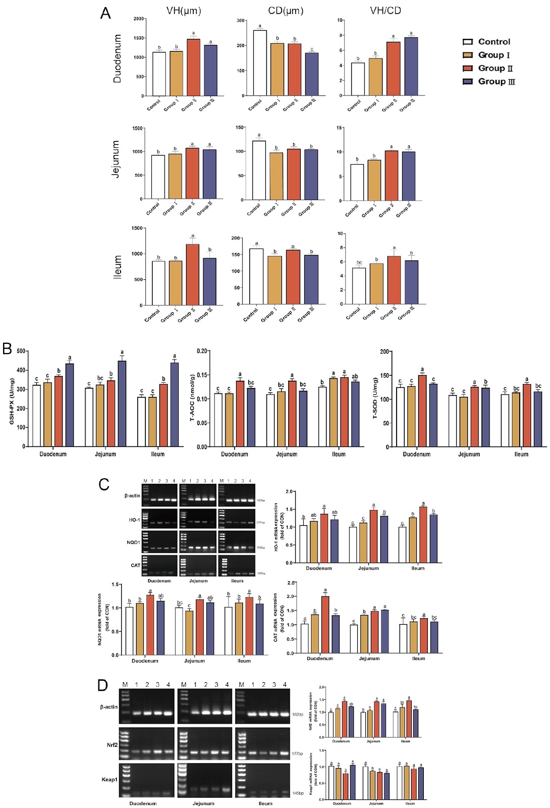
Poultry production faces escalating challenges from intensive farming practices, where stressors, including high stocking density, pathogen exposure, and dietary fluctuations, disrupt intestinal integrity, microbiota balance, and antioxidant defenses. These disruptions impair nutrient absorption, growth performance, and immune function, leading to significant economic losses. Although antibiotics have historically mitigated such issues, growing restrictions due to antimicrobial resistance necessitate natural alternatives. Fungal polysaccharides (FP)—notably lentinan (LNT) from Lentinula edodes and polysaccharide from Ganoderma lucidum (GLP), are promising candidates owing to their immunomodulatory, antioxidant, and prebiotic properties. However, existing research focuses predominantly on individual FP, neglecting potential synergies in blended formulations. Structurally, LNT (β-(1→3)-D-glucan backbone) enhances rumen volatile fatty acid production and fiber degradation, whereas GLP (heterogeneous α/β-glycans) potently activates the Nrf2/HO-1 antioxidant pathway and modulates Th1/Th2 immunity. These divergent mechanisms imply complementary effects when combined. In our previous experiments on broiler feeding, we reported that a combination of GLP (68.32% polysaccharide content, composed of mannose, glucose, arabinose, rhamnose, and galactose at a molar ratio of 1.00:16.37:18.82:1.42:17.42) and LNT (76.52% polysaccharide content, composed of mannose, galacturonic acid, arabinose, galactose, glucose, and rhamnose at a molar ratio of 1.00:15.22:8.23:2.05:1.78:4.26) at a 1:1 ratio maximally promoted broiler growth (unpublished data), but their impacts on intestinal morphology, antioxidant signaling, and the microbiota remain uncharacterized. We therefore hypothesize that mixed FP synergistically may enhance intestinal health by simultaneously improving nutrient absorption, activating antioxidant pathways, and stabilizing microbial ecosystems. To investigate the effects of mixed FP on intestinal development, 240 one-day-old Arbor Acres male broilers were randomly assigned to the 0 mg/kg FP (Control), 200 mg/kg FP (Group I), 400 mg/kg FP (Group II), and 600 mg/kg FP (Group III) groups. Broilers were housed in three-tier battery cages (0.7 m × 0.7 m × 0.4 m; 12 broilers/cage), with five replicate cages per experimental group maintained under identical conditions. The experiments were approved by the College of Animal Science and Technology in Anhui Agricultural University (approval number: SYXK 2016-007). All the cages were subjected to a 16 h light: 8 h dark cycle with ad libitum access to water and twice-daily feeding (09:00/16:00) of basal diets (Supplementary Table S1). On day 42, the duodenum, jejunum, and ileum segments were collected, fixed in 4% paraformaldehyde, sectioned at 5 μm, and stained with hematoxylin-eosin. Villus height (VH), crypt depth (CD), and VH/CD ratios were measured via Case Viewer software. The results revealed that Group II significantly increased VH and VH/CD across all the intestinal segments while reducing CD (Figure 1A; P < 0.05 vs the control); these findings suggest enhanced nutrient absorption capacity and intestinal health. To evaluate antioxidant capacity and signaling pathway activation, intestinal tissues were homogenized in PBS (1:9, w/v). The total antioxidant capacity (T-AOC), total superoxide dismutase (T-SOD), and glutathione peroxidase (GSH-Px) activities were determined via commercial kits (Nanjing Jiancheng Bioengineering Institute, Nanjing, China)). For gene expression analysis, total RNA was extracted and reverse-transcribed. The qPCR was performed via specific primers for HO-1, NQO1, CAT, Nrf2, and Keap1, with β-actin used as the reference gene (primer sequences and product sizes are listed in Supplementary Table S2). The results demonstrated that Group II significantly elevated antioxidant enzyme activities (P < 0.05), upregulated HO-1, NQO1, CAT, and Nrf2, and
Mixed fungal polysaccharides enhance intestinal health, antioxidant capacity, and microbiota diversity in broiler chickens
Graphical Abstract
Original ResearchVol. 58, Issue 2 • pp. 421-436DOI: 10.3724/abbs.2026018

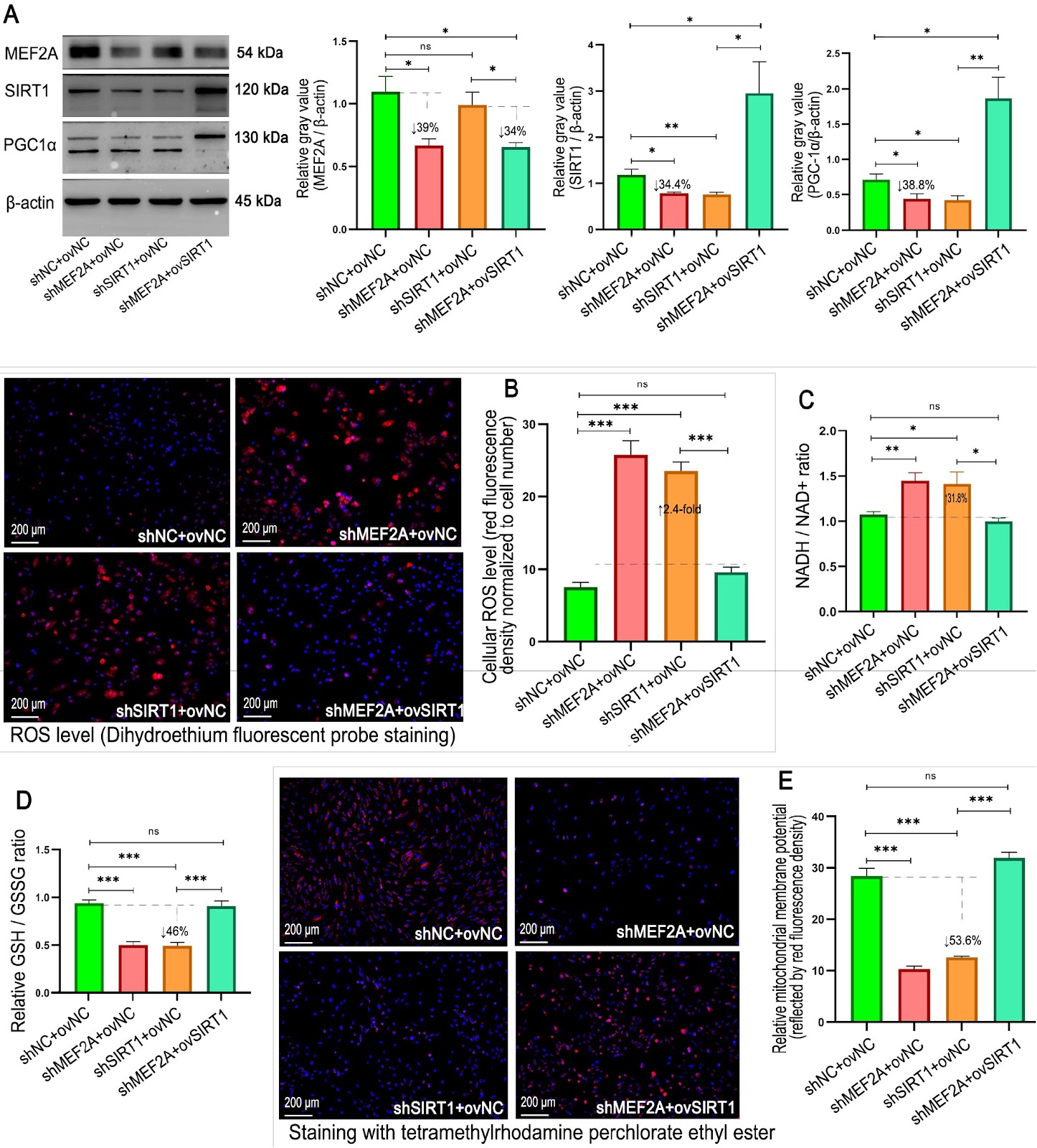
Cardiac PTN-SIRT1 axis alleviates oxidative stress and promotes mitochondrial energy reprogramming to mitigate doxorubicin-induced cardiotoxicity through AMPK/PGC1α signaling

Authors: Yuxiao Sun, Tianwen Wei, Hongping Xu, Hongda Li, Chang Zhou, Xianliang Liu, Yafei Li, Shangwei Huang, Qi Zhang, Xia Duan

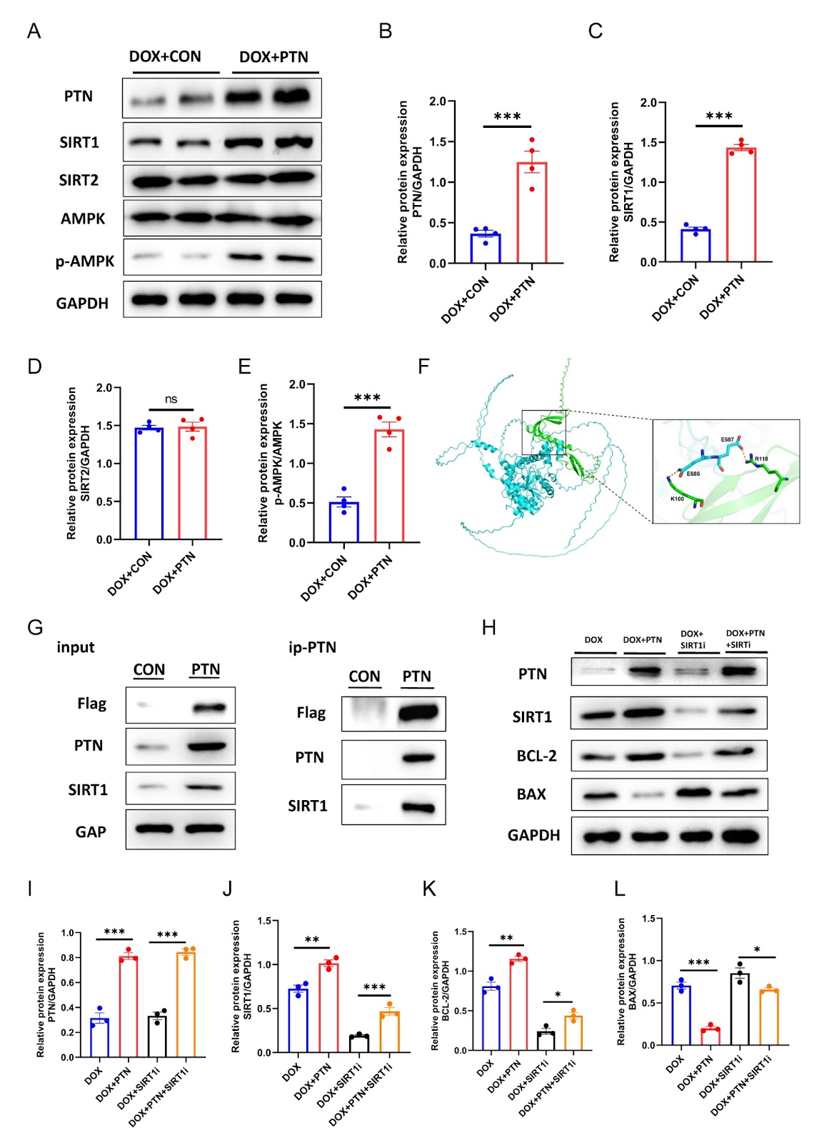
Doxorubicin (DOX) remains a cornerstone chemotherapeutic agent for malignancies, yet its clinical utility is severely limited by dose-dependent cardiotoxicity, which can lead to progressive left ventricular dysfunction and heart failure. Pleiotrophin (PTN), a heparin-binding growth factor with diverse physiological functions, regulates glucose and lipid metabolism and promotes oxidative energy pathways. However, whether PTN exerts protective effects against DOX-induced cardiotoxicity (DIC) remains unclear. In this study, we establish cellular and animal models of DIC. DOX administration induces pronounced myocardial injury in both models, characterized by impaired ventricular contractility, increased fibrotic remodeling, and reduced cell viability. Concurrently, PTN protein expression is significantly downregulated in cardiomyocytes under DOX treatment. Overexpression of PTN substantially alleviates these pathological changes. In vitro, PTN reduces mitochondrial oxidative stress and apoptosis while restoring energy production and cell viability. In vivo, PTN improves mitochondrial ultrastructure, decreases cardiomyocyte apoptosis, and enhances cardiac function. Mechanistically, PTN directly binds to SIRT1 and activates AMPK phosphorylation at Thr172, triggering a downstream cascade through the AMPK-PGC1α axis that reprograms mitochondrial energy metabolism and attenuates cardiotoxicity. In conclusion, the PTN-SIRT1 axis protects against DIC by reducing oxidative stress and promoting mitochondrial energy homeostasis via the AMPK/PGC1α pathway, highlighting its potential as a novel therapeutic target for preventing chemotherapy-related cardiac injury.

Cardiac PTN-SIRT1 axis alleviates oxidative stress and promotes mitochondrial energy reprogramming to mitigate doxorubicin-induced cardiotoxicity through AMPK/PGC1α signaling
Graphical Abstract
Original ResearchVol. 58, Issue 5 • pp. 1183-1186DOI: 10.3724/abbs.2025242

Expression characteristics of serum exosomal microRNAs in patients with liver injury induced by anti-tuberculosis drugs

Authors: Yinpeng Jin, Xiaofang Yu, Mingquan Guo, Li Li, Shuangshuang Sun, Liling Yang, Ying Yuan, Qingchun Fu, Rongfeng Shi, Meng Jin

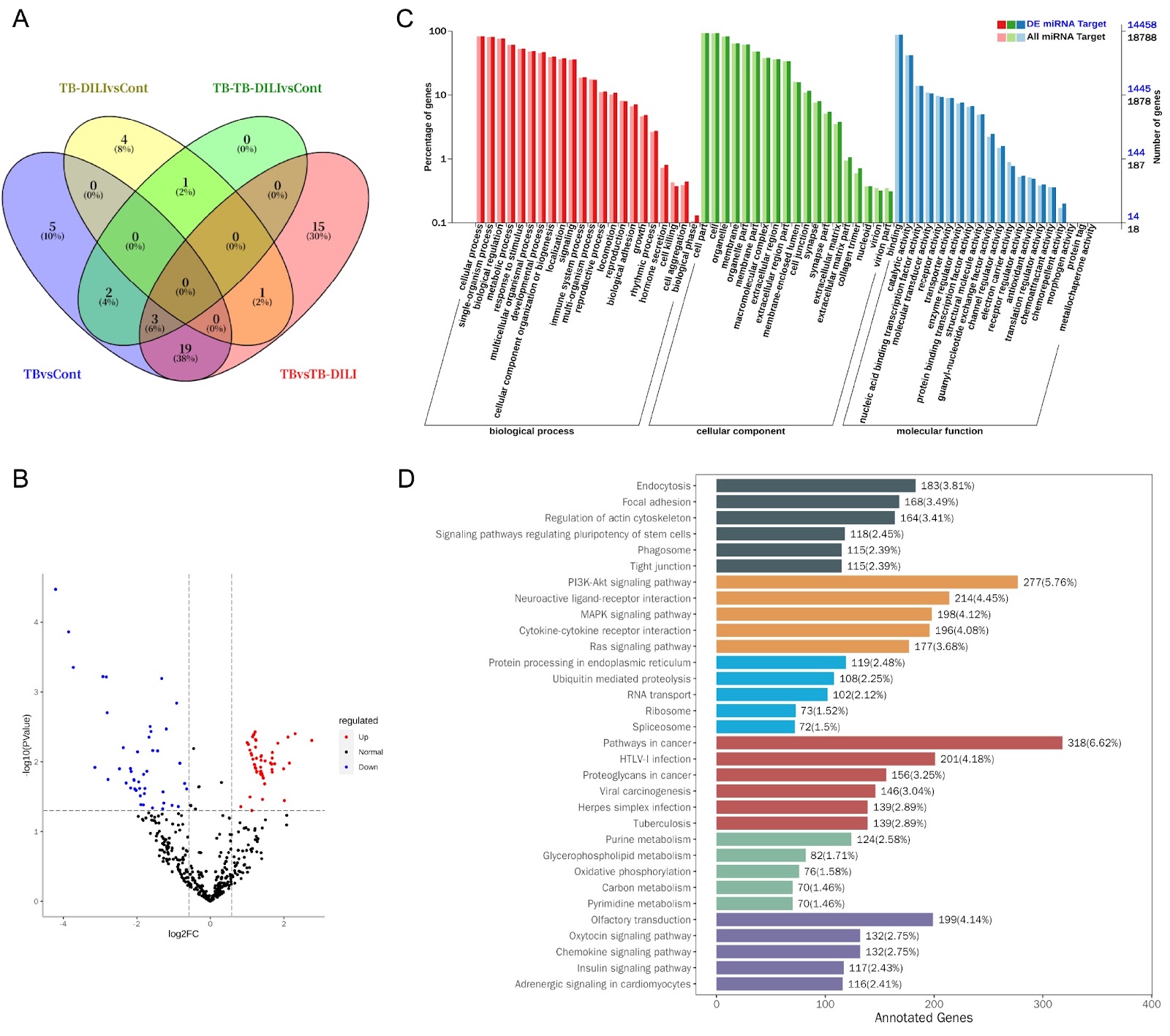
Drug-induced liver injury (DILI) caused by anti-tuberculosis drugs is a serious clinical problem that can lead to acute liver failure and even death. Current diagnosis relies on lagged indicators such as serum transaminase levels, which rise only 48–72 hours after liver injury. This study is the first to systematically analyze the microRNA expression profile of serum exosomes in patients with anti-tuberculosis drug-induced liver injury (TB-DILI) to discover early diagnostic markers. A total of 12 tuberculosis patients and 6 normal controls were included. Serum exosomes were isolated and characterized, and small RNA sequencing identified 701 miRNAs, with 128 differentially expressed between TB-DILI and TB groups. Notably, miR-122-5p was upregulated and has shown early warning value. Target gene prediction and enrichment analysis revealed involvement in GTPase activity regulation, cell migration, and BMP signaling. These findings suggest that exosomal miRNAs, particularly miR-122-5p, may serve as early diagnostic biomarkers for TB-DILI.

Expression characteristics of serum exosomal microRNAs in patients with liver injury induced by anti-tuberculosis drugs
Graphical Abstract
Original ResearchVol. 58, Issue 8 • pp. 1875-1886DOI: 10.3724/abbs.2025208

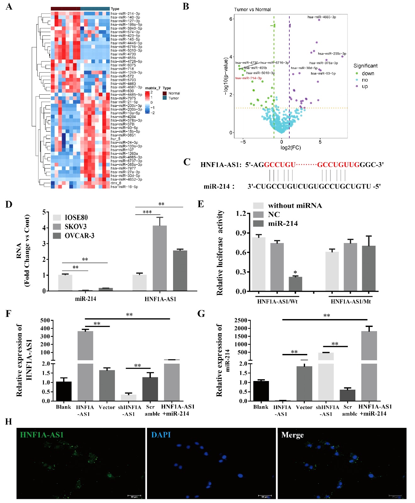
Long noncoding RNA HNF1A-AS1 promotes ovarian cancer growth and M2 macrophage polarization by counteracting miR-214-mediated suppression of semaphorin 4D signaling

Authors: Yang Liu, Yanzhi Wu, Yujie Gengxiao, Yan Li, Jiamei Song, Chunyi Sun

To determine whether lncRNA HNF1A-AS1 affects epithelial ovarian cancer (EOC) growth and macrophage polarization through miR-214/SEMA4D, the endogenous HNF1A-AS1 and miR-214 levels in human EOC cell lines are compared with those in normal ovarian epithelial IOSE80 cells. HNF1A-AS1 is overexpressed or silenced to investigate whether HNF1A-AS1 regulates miR-214/SEMA4D in SKOV3 cells and xenograft tumors, as well as the phenotypic switching of THP-1 cells. Compared with IOSE80 cells, EOC cells present significantly higher HNF1A-AS1 level and lower miR-214 level. Fluorescence in situ hybridization reveals predominant cytoplasmic localization of HNF1A-AS1, supporting its role as a competing endogenous RNA. HNF1A-AS1 and miR-214 antagonize each other in SKOV3 cells. In vitro, HNF1A-AS1 inhibits SKOV3 apoptosis and promotes migration and invasion. HNF1A-AS1 overexpression enhances miR-214 downstream of SEMA4D/PLEXIN-B1/TIAM1/RAC signaling, but miR-214 mimics significantly reverses this effect. Compared with control tumors, xenograft tumors derived from HNF1A-AS1-overexpressing SKOV3 cells present increased tumor growth, attenuated miR-214 expression, and activated SEMA4D/PLEXIN-B1/TIAM/RAC signaling. Knockdown of HNF1A-AS1 has the opposite effects. Additionally, HNF1A-AS1 promotes M2 phenotypic switching in THP-1 cells, which could be reversed by miR-214 overexpression or SEMA4D silencing. Our study suggests that by antagonizing miR-214, HNF1A-AS1 activates the SEMA4D/PLEXIN-B1/TIAM/RAC pathway, facilitating EOC growth and potentially promoting M2 macrophage polarization in the tumor microenvironment. HNF1A-AS1 represents a compelling therapeutic target for treating EOC.

Long noncoding RNA HNF1A-AS1 promotes ovarian cancer growth and M2 macrophage polarization by counteracting miR-214-mediated suppression of semaphorin 4D signaling
Graphical Abstract
Original ResearchVol. 58, Issue 8 • pp. 1842-1854DOI: 10.3724/abbs.2025205

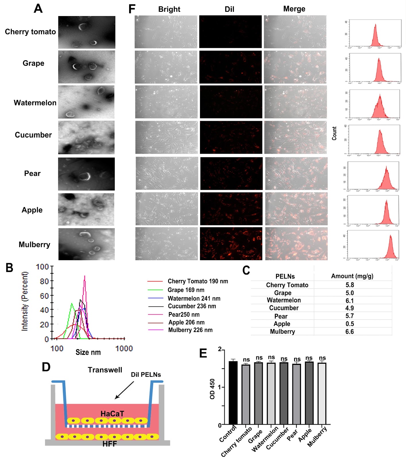
Effective resistance to UVB-induced skin damage through the encapsulation of chebulinic acid in mulberry-derived exosome-like nanoparticles

Authors: Weiwei Zhao, Ruobing Liu, Siqi Yang, Chaozhi Liu, Songlin Guo, Guihong Sun, Mingxiong Guo

Ultraviolet-B (UVB) radiation induces significant skin damage by penetrating into the dermal layer, leading to reactive oxygen species (ROS) generation and triggering cellular necrosis and apoptosis. Conventional sunscreens focus primarily on UVB blocking but are limited in their ability to repair dermal damage due to insufficient permeability. In this study, we discover that chebulinic acid (CA), one of the principal monomers in Terminalia chebula Retz., has superior efficacy in promoting recovery from UVB-induced skin damage compared with other major monomers. Mechanistically, CA’s anti-UVB function involves regulating the expression of IL-6 and IFN-β through activation of the MAPK pathway. To overcome the formidable barrier posed by the skin, we identify mulberry exosome-like nanoparticles (MELNs) as an efficient transdermal delivery system and develop CA@MELNs loaded with CA. Furthermore, we demonstrate that the dissociative CA within the CA@MELNs delivery system significantly enhances both transdermal penetration and anti-UVB efficiency in vitro and in vivo. Our findings suggest the substantial potential of CA as an effective ingredient and CA@MELNs as a robust and accessible platform for mitigating UVB damage.

Effective resistance to UVB-induced skin damage through the encapsulation of chebulinic acid in mulberry-derived exosome-like nanoparticles
Graphical Abstract
Original ResearchVol. 58, Issue 8 • pp. 1783-1792DOI: 10.3724/abbs.2025209

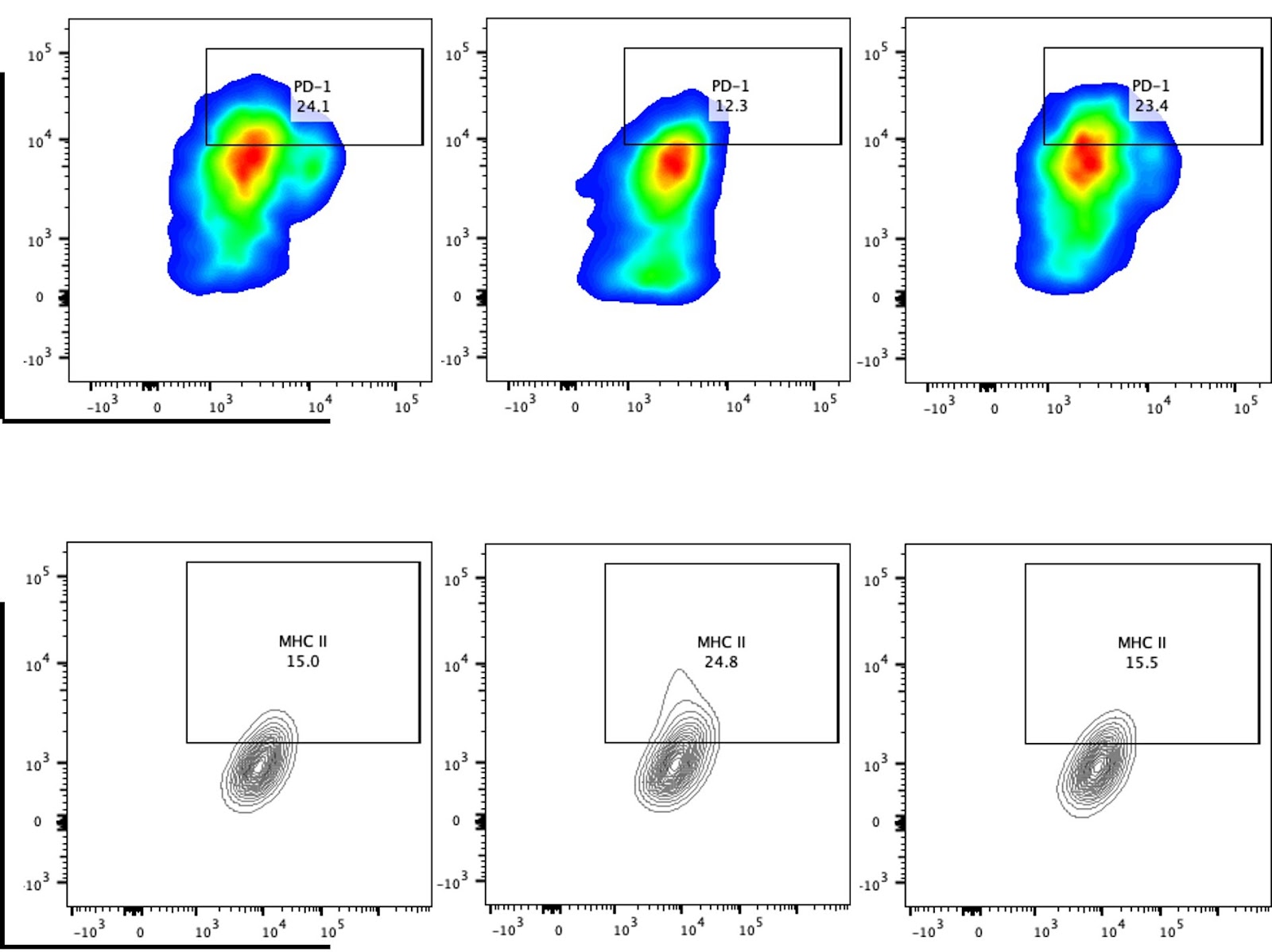
cGAS-STING pathway reprograms macrophage polarization and is highly expressed in responding tumors after neoadjuvant immunotherapy in head and neck carcinoma

Authors: Zhaohong An, Xiwei Zhang, Lin Li, Dilinaer Wusiman, Zhaoyang Wang, Fa Zhang, Xiaohui Zhao, Changming An, Zhenzhen Yin, Wei Gao

Given the critical role of the cGAS-STING pathway in antitumor immunity, this study investigates the functional role of STING in head and neck squamous cell carcinoma (HNSCC) to evaluate the therapeutic potential of STING agonists. Analysis of the TCGA-HNSC dataset reveals that elevated expression of the STING-encoding gene TMEM173 is significantly correlated with increased M1 macrophage infiltration and enrichment of macrophage polarization-related signaling pathways. In vitro experiments in which RAW 264.7 cells are co-cultured with tumor cell-conditioned medium demonstrate that the STING agonist MSA-2 effectively reprograms tumor-induced M2-polarized macrophages toward the M1 phenotype. This MSA-2-induced M1 polarization is accompanied by increased expressions of IFN-α, IFN-β, IFN-γ, TNF-α, and IL-6, while the STING inhibitor H-151 reverses these effects. Flow cytometry further reveals that MSA-2 treatment reduces PD-1 and increases MHC II expression on macrophages. Immunohistochemical analysis of clinical samples confirms that high STING expression is correlated with increased numbers of CD68⁺ and CD80⁺ (M1-like) macrophages. In support of translational relevance, analysis of single-cell RNA-seq data from HNSCC patients receiving neoadjuvant immunotherapy indicates that TMEM173 is expressed primarily in T cells and macrophages and that the cGAS-STING pathway score is significantly higher in patients who respond to treatment. Collectively, these findings provide systematic clinical and experimental evidence supporting the potential of STING agonists, such as MSA-2, to enhance antitumor immunity in HNSCC, particularly when combined with immunotherapy.

cGAS-STING pathway reprograms macrophage polarization and is highly expressed in responding tumors after neoadjuvant immunotherapy in head and neck carcinoma
Graphical Abstract
Original ResearchVol. 58, Issue 8 • pp. 1822-1833DOI: 10.3724/abbs.2025161

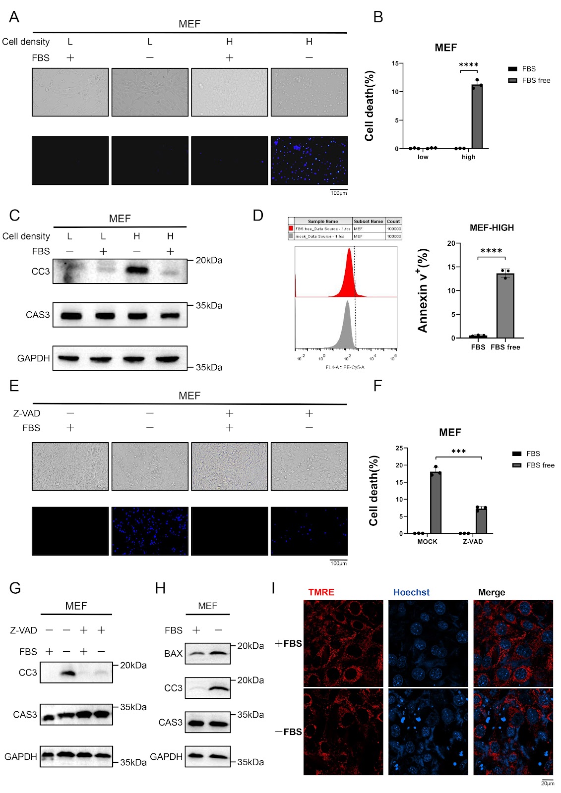
Serum starvation induces density-dependent apoptosis via HIF-1 activation and JNK suppression

Authors: Qifan Yang, Yaofeng Hu, Jiahui Lv, Jiaqi Xue, Jiaqi Chen, Changwan Wang, Fajian Hou

Serum deprivation is a well-established inducer of apoptosis, yet the molecular mechanisms governing this process remain incompletely understood. Here, we show that serum starvation selectively triggers intrinsic apoptosis in high-density murine embryonic fibroblasts (MEFs) through coordinated HIF-1α activation and JNK signaling suppression. Knockdown of HIF-1α abolishes caspase-3 activation and apoptosis induced by serum deprivation, whereas upregulation of HIF-1α in low-density cells recapitulates the apoptotic response observed in high-density cultures. Simultaneously, serum deprivation leads to the suppression of the JNK pathway, which contributes to apoptosis. Notably, combined HIF-1α activation and JNK inhibition in low-density cells fully mimics the apoptotic phenotype of high-density conditions, underscoring the interplay between these pathways. Together, these findings define a density-dependent apoptotic switch in which HIF-1α drives metabolic stress adaptation, whereas JNK suppression removes a critical survival signal, converging to promote mitochondrial-mediated cell death. This work provides a mechanistic framework for understanding nutrient stress-induced apoptosis and suggests potential therapeutic targets for diseases characterized by aberrant cell survival.

Serum starvation induces density-dependent apoptosis via HIF-1 activation and JNK suppression
Graphical Abstract
Original ResearchVol. 58, Issue 8 • pp. 1866-1874DOI: 10.3724/abbs.2025170

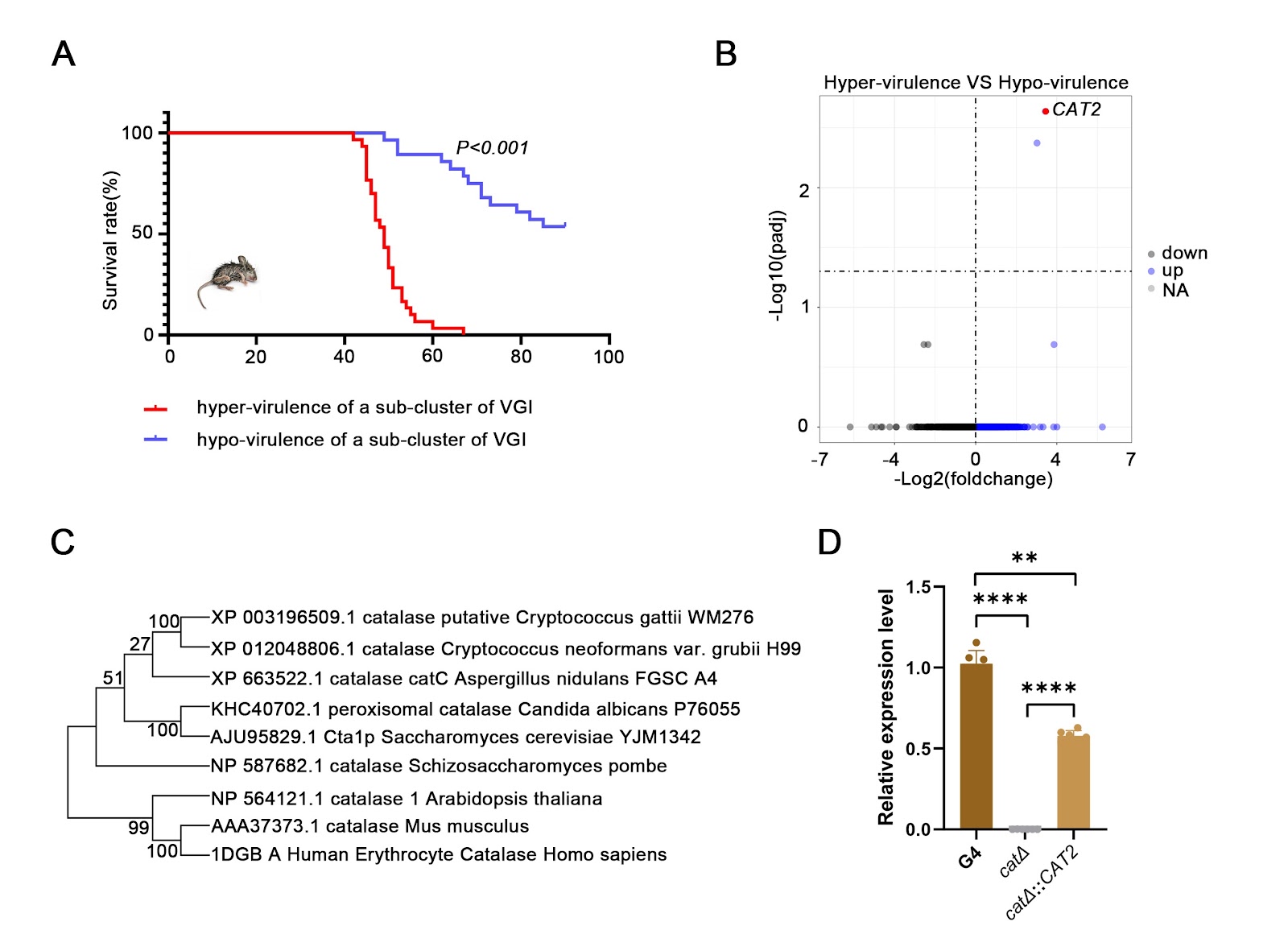
The catalase gene CAT2 and its role in the virulence of one sub-cluster of Cryptococcus gattii VGI clinical isolates

Authors: Yemei Huang, Hengyu Deng, Xuelei Zang, Enqi Zhao, Yan Zhuo, Wencong He, Chen Wang, Yangyu Zhou, Jiahui Huang, Rui Liu, Tingyue Tian, Chen Yang, Jing Liu, Ping Zhang, Liye Zhang, Xinying Xue

Cryptococcus gattii causes cryptococcosis and life-threatening cryptococcal meningitis. Currently, the pathogenic virulence mechanisms of C. gattii remain a significant area of ongoing research with considerable unexplored aspects. On the basis of our established research, a sub-cluster of strains with independent evolutionary relationships from WM276 in the phylogenetic analysis of VGI-type strains is identified. In vivo infection experiments on this sub-branch of strains reveal that there are hypervirulent strains and hypovirulent strains among these strains, and the virulence differences are significant (P < 0.001). Bioinformatic interrogation of differentially expressed genes reveals that the catalase-encoding gene CGB_J0620W, CAT2, is a pivotal virulence-associated gene. The hypervirulent clinical isolate G4 (G4-WT) is selected as the parental strain, from which an isogenic CAT2-knockout mutant (cat2Δ) is constructed via homologous recombination, which shows increased sensitivity to oxidative stress, as well as growth defects in response to hyperosmosis, 5-fluorocytosine, fluconazole and amphotericin B. The cat2Δ::CAT2 strain exhibits phenotypic restoration to wild type (WT). In the mouse experiments, significant differences in survival (P < 0.001), pulmonary fungal burden (P < 0.01), and alveolar structural damage are observed between the WT and cat2Δ strains, which are completely different from C. neoformans. Moreover, comparative transcriptome analysis is performed on the WT and cat2Δ strains, which reveals that enzymes encoded by CAT2 may be involved in oxidative stress, metabolism and sugar transport. In conclusion, this study may explain the differences in virulence among different genetic evolutionary processes of a sub-cluster of the VGI geneotype of C. gattii and provide a theoretical basis for targeted therapy in a specific genotype population in the future.

The catalase gene CAT2 and its role in the virulence of one sub-cluster of Cryptococcus gattii VGI clinical isolates
Graphical Abstract
Original ResearchVol. 58, Issue 8 • pp. 1855-1865DOI: 10.3724/abbs.2025164

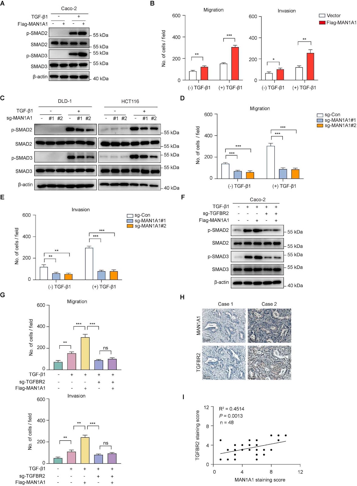
MAN1A1 promotes colorectal cancer liver metastasis by maintaining TGFBR2 protein stability

Authors: Yingxi Hu, Yinwen Xu, Kai Chen, Shihua Guan, Huiling Zhou, Tao Li, Rongrui Liang, Min Tao, Yiyi Yu, Xinxin Ge, Yuanyuan Ruan

Emerging biochemical and genetic evidence has firmly established aberrant protein glycosylation as a critical regulator of oncogenic transformation, with glycocalyx remodeling profoundly influencing tumor microenvironment dynamics and metastatic progression. Despite the well-documented association between metastatic dissemination and poor clinical outcomes in patients with colorectal cancer, the underlying molecular mechanisms remain incompletely characterized. Through integrative analysis of single-cell RNA sequencing data from a public database, we identify the Golgi-resident α-1,2-mannosidase MAN1A1 as a consistently upregulated enzyme in malignant epithelial cells derived from colorectal cancer liver metastases. Clinically, elevated MAN1A1 expression is correlated with reduced overall survival, suggesting that MAN1A1 is both a prognostic biomarker and therapeutic target for colorectal cancer liver metastases. Genetic manipulation of MAN1A1 in colorectal cancer cells demonstrates that although the proliferation capacity of colorectal cancer cells remains unchanged, MAN1A1 overexpression significantly enhances migratory and invasive capacities in transwell assays, suggesting its specific involvement in metastatic progression. Mechanistic investigations reveal that MAN1A1 exerts its pro-metastatic effects by significantly prolonging the TGFBR2 protein half-life. Together, our work identifies MAN1A1 as both a prognostic biomarker and a promising therapeutic target, highlighting the critical role of glycan remodeling in the metastatic progression of colorectal cancer.

MAN1A1 promotes colorectal cancer liver metastasis by maintaining TGFBR2 protein stability
Graphical Abstract
Original ResearchVol. 58, Issue 7 • pp. 1499-1508DOI: 10.3724/abbs.2025121

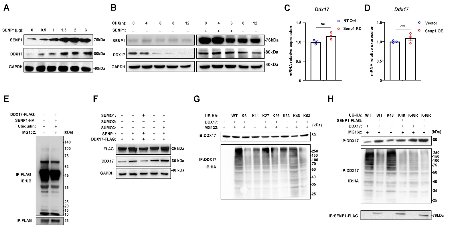
Carfilzomib triggers cardiotoxicity by suppressing SENP1-mediated deSUMOylation of DDX17

Authors: Sheng Wang, Jingjing Wang, Xin Li, Zhigao Dai, Tiantian Li, Yixuan Wang, Ziyi Peng, Mengqi Wang, Hao Cheng, Linchuang Jia, Danchen Su, Mu Qiao, Jingya Wang, Ying Xie, Jing Guo, Xiaozhi Liu, Tong Liu

Carfilzomib (Cfz) is a second-generation proteasome inhibitor approved for the treatment of relapsed/refractory multiple myeloma (RRMM). Previous studies have shown that Cfz is associated with a higher incidence of severe adverse cardiac effects than bortezomib (Btz); however, the underlying mechanisms remain to be elucidated. The aim of this study is to identify key regulators of cardiotoxicity induced by Cfz and to investigate the mechanisms by which these factors exert their effects. We establish a mouse model of cardiac toxicity induced by Cfz and confirm the phenotype through cardiac functional analysis, morphology assessment, myocardial fibrosis, and apoptosis analysis. We subsequently perform RNA sequencing to identify differentially expressed genes (DEGs) and further validate their functions and mechanisms. We find that Cfz induces myocardial hypertrophy and myocardial injury, along with the suppression of SENP1 expression in mouse heart tissues and in vitro cultured neonatal rat cardiomyocytes. Suppression of SENP1 exacerbates Cfz-induced injury and remodeling in cardiomyocytes by directly binding to and deconjugating the SUMO1-mediated SUMOylation of the RNA helicase DDX17. This process leads to a reduction in K-48 ubiquitin-linked polyubiquitination and degradation of DDX17, resulting in increased expressions of anti-apoptotic genes and maintenance of mitochondrial homeostasis. Therefore, the overexpression of SENP1 using AAV vectors alleviates Cfz-induced cardiotoxicity in mice. In summary, our findings reveal a previously unknown role of the SENP1-DDX17 axis in protecting against cardiotoxicity induced by Cfz, providing a potential foundation for developing therapeutic strategies to mitigate cardiac side effects in the clinical management of MM patients.

Carfilzomib triggers cardiotoxicity by suppressing SENP1-mediated deSUMOylation of DDX17
Graphical Abstract
Original ResearchVol. 58, Issue 7 • pp. 1667-1670DOI: 10.3724/abbs.2025193

Investigation of the cardioprotective potential of dantrolene in mitigating arsenic-induced cardiac dysfunction in rats

Authors: Chuncui Chen, Ruoxi Chen, Xueting Guo, Lei Huang, Kuican Liu, Wenrong Shi, Caiyun Zhang, Kunxuan Liu, Huan Liu, Shanshan Dong, Guilin Lu, Wenjuan Qin

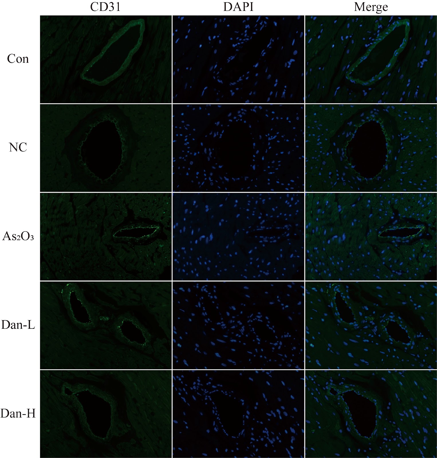
Arsenic, a toxic metalloid, exists in organic or inorganic states within the Earth’s seawater, river water, soil, atmosphere, food sources, and diverse biological tissues [1]. It poses a threat to the health of hundreds of millions of people globally [2]. Arsenic exposure has toxic effects on the cardiovascular system of organisms, thus endangering human health [3]. Research has indicated that the harmful effect of arsenic exposure on the heart is associated with abnormal calcium handling in myocardial cells [4]. The cardiac ryanodine receptor type 2 (RyR2) is a primary channel involved in the surface of the endoplasmic reticulum in cardiac myocytes that regulates the release of Ca2+ during the systolic phase [5]. The integrity of its function is crucial for maintaining calcium homeostasis in cardiac myocytes. However, when myocardial tissue is damaged and undergoes pathological changes, the spatial structure of the RyR2 protein becomes unstable and becomes excessively activated, thereby triggering Ca2+ leakage [6]. Dantrolene (Dan), which serves as a stabilizer of RyR1, is frequently employed in clinical settings for the treatment of malignant hyperpyrexia and relieves spastic muscle tension [7]. Previous studies have demonstrated that dantrolene also has a stabilizing effect on RyR2 [8]. Research has shown that dantrolene can prevent calcium leakage in myocardial cells by stabilizing the tertiary structure of the RyR2 protein and thereby inhibiting the pathological hyperactivity of RyR2 [9]. Therefore, this study hypothesizes that dantrolene, by virtue of this stabilizing effect, can alleviate myocardial injury caused by arsenic exposure to some extent and plays a role in protecting cardiac function. For this purpose, we established an arsenic exposure model and a Dan intervention arsenic exposure model to verify the protective effect of Dan on the myocardial tissue and cardiac function of arsenic-exposed rats.

Investigation of the cardioprotective potential of dantrolene in mitigating arsenic-induced cardiac dysfunction in rats
Graphical Abstract
Original ResearchVol. 58, Issue 7 • pp. 1473-1485DOI: 10.3724/abbs.2025165

Levosimendan ameliorates cardiomyocyte injury and mitochondrial dysfunction in an Nrf2-dependent manner in mice with sepsis-induced cardiomyopathy

Authors: Xinyuan Zhu, Hongyan Zhai, Huishuang Shao, Dawei Wu, Jun Ren, Daqing Sun, Sujuan Liu

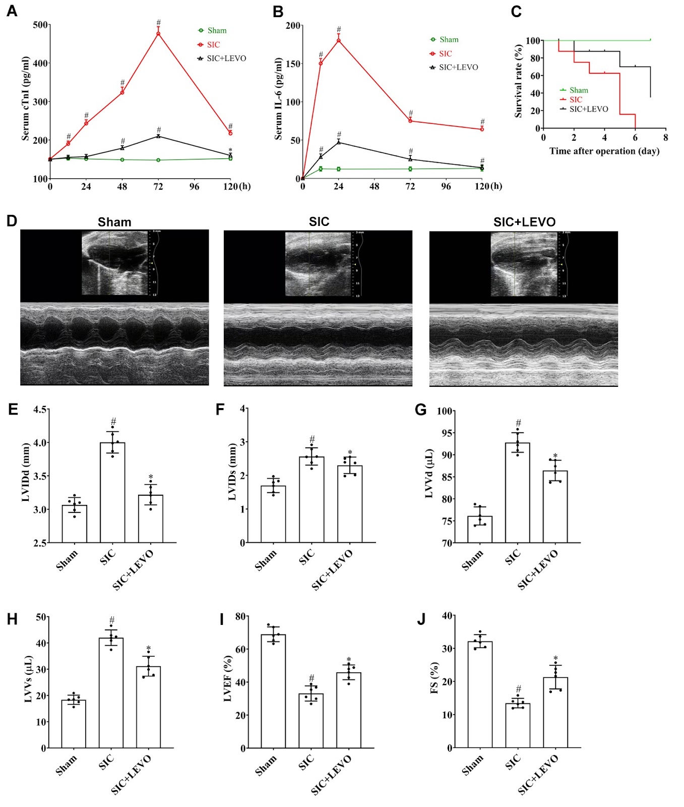
Sepsis-induced cardiomyopathy (SIC) is a severe complication of sepsis and septic shock and is characterized by cardiac dysfunction. Levosimendan (LEVO), a calcium sensitizer, has shown therapeutic potential in SIC, although its underlying mechanism remains unclear. Nrf2, a pivotal regulator of antioxidant and anti-inflammatory responses, may represent a potential target for SIC treatment. In this study, we examine the effects of LEVO on SIC and explore the mechanistic role of Nrf2 in mediating its cardioprotective effects. A murine SIC model is established via cecal ligation and puncture (CLP), and cardiomyocyte injury is induced in vitro via lipopolysaccharide (LPS) exposure in HL-1 cells. The CLP procedure significantly elevates serum cTnI and IL-6 levels and reduces the survival rates of mice. Echocardiographic analysis reveals impaired cardiac structure and function, accompanied by mitochondrial morphological and functional damage, in SIC mice. Interestingly, these pathological changes in SIC are markedly attenuated by LEVO treatment. Similarly, LEVO administration restores proliferative capacity; increases mitochondrial ATP, mitochondrial membrane potential (MMP) and NADH levels; and reduces ROS production and intracellular calcium overload. Notably, the protective effects of LEVO on cardiomyocyte viability and mitochondrial function are significantly diminished following Nrf2 inhibition or Nrf2 knockout (KO). Collectively, these findings demonstrate that LEVO mitigates cardiomyocyte injury and mitochondrial dysfunction in SIC through an Nrf2-dependent mechanism.

Levosimendan ameliorates cardiomyocyte injury and mitochondrial dysfunction in an Nrf2-dependent manner in mice with sepsis-induced cardiomyopathy
Graphical Abstract
Original ResearchVol. 58, Issue 7 • pp. 1458-1472DOI: 10.3724/abbs.2025199

Pregnancy-induced metabolic reprogramming in skeletal muscle: a multi-omics interrogation of transcriptional and metabolic adaptations

Authors: Zhongliang Lin, Kejing Zhu, Renke He, Xueying Liu, Qinyu Luo, Jianzhong Sheng, Jiexue Pan, Hefeng Huang

Pregnancy induces profound physiological adaptations to meet the dynamic nutritional demands of fetal development, including a deliberate reduction in maternal insulin sensitivity to ensure fetal glucose availability. However, excessive insulin resistance may precipitate gestational diabetes mellitus (GDM), increasing the risk of both obstetric complications and long-term metabolic disorders in mothers and offspring. Although the role of adipose tissue in pregnancy-associated metabolic adaptation has been extensively studied, the contribution of skeletal muscle remains poorly understood. Here, we systematically characterize pregnancy-induced molecular and metabolic changes in maternal skeletal muscle through multi-omics profiling. We use transcriptomic, metabolomic, computational single-cell deconvolution, and qPCR validation in an established C57BL/6J mouse pregnancy model (8-week-old females). Pregnancy triggers remarkable skeletal muscle remodelling, featuring histological reorganization with myofiber depletion and expanded endothelial compartments. Concurrent metabolic disturbances include insulin resistance, dysregulated TCA cycle activity, and impaired ubiquinone biosynthesis. This study represents a multi-omics-based systematic elucidation of pregnancy-induced maternal skeletal muscle adaptations. Our findings demonstrate that pregnancy induces profound structural reorganization and metabolic reprogramming in maternal skeletal muscle, characterized by prioritized fetal nutrient provision at the expense of maternal tissue utilization. These observations not only reveal previously unrecognized mechanisms of pregnancy-specific metabolic regulation but also, more importantly, establish a critical theoretical foundation for developing skeletal muscle-targeted intervention strategies to prevent gestational diabetes mellitus.

Pregnancy-induced metabolic reprogramming in skeletal muscle: a multi-omics interrogation of transcriptional and metabolic adaptations
Graphical Abstract
Original ResearchVol. 58, Issue 6 • pp. 1329-1341DOI: 10.3724/abbs.2025178

circDCBLD2 regulates the Th1/Th2 immune balance via the miR-26a-5p/PTEN axis

Authors: Yue Zhao, Guangling Liu, Rui Li, Shuting Yu, Beibei Zhu, Xunzhou Liu, Hanyi Jiang, Jinya Wang

Asthma is a prevalent chronic respiratory disease in children. Recently, adjusting the Th1/Th2 imbalance has become a significant focus in asthma immunotherapy. The present study aims to investigate the roles and mechanisms of circDCBLD2 in maintaining the Th1/Th2 immune balance. CircDCBLD2 is downregulated in CD4+ T cells from asthmatic patients and in CD4+ T cells from an OVA-induced asthmatic mouse model. Additionally, circDCBLD2 levels are significantly decreased in the PBMCs of asthmatic mice. The expression of circDCBLD2 is positively correlated with the Th1 cytokines IFN-γ and IL-2 but negatively correlated with the Th2 cytokines IL-4 and IL-13. Flow cytometry and ELISA analyses demonstrate that circDCBLD2 overexpression increases the proportion of Th1 cells (CD4+IFN-γ+) and the levels of Th1 cytokines while decreasing the proportion of Th2 cells (CD4+IL-4+) and the levels of Th2 cytokines. Furthermore, circDCBLD2 overexpression alleviates the asthma phenotype in OVA-induced mice, reduces the infiltration of inflammatory cells in the lungs, and corrects the Th1/Th2 imbalance. Mechanistically, circDCBLD2 is found to target miR-26a-5p. Rescue experiments indicate that circDCBLD2 regulates the Th1/Th2 immune balance by targeting miR-26a-5p. Additionally, PTEN has been identified as a direct target of miR-26a-5p. The overexpression of PTEN partially reverses the effects of miR-26a-5p on the Th1/Th2 immune balance. These findings indicate that circDCBLD2 increases the proportion of Th1 cells and decreases the proportion of Th2 cells via the miR-26a-5p/PTEN axis, providing a promising target for asthma treatment.

circDCBLD2 regulates the Th1/Th2 immune balance via the miR-26a-5p/PTEN axis
Graphical Abstract
Original ResearchVol. 58, Issue 6 • pp. 1374-1386DOI: 10.3724/abbs.2025152

A Mycobacterium tuberculosis multi-epitope DNA vaccine encoding adaptive immune antigens provokes IFNγ/Th1 immunity and confers potential protection

Authors: Jingyao Xue, Yumeng Li, Chi Li, Yu Zhang, Chiuan Yee Leow, Gaoqian Feng, Minjun Ji, Qiao Liu, Zhipeng Xu

Tuberculosis (TB), caused by Mycobacterium tuberculosis (MTB), remains a significant global health threat. However, the licensed Bacille Calmette-Guérin (BCG) vaccine provides only limited protection in adults, underscoring the urgent need for more effective preventive strategies. Recent studies have shown that multi-epitope DNA vaccines are superior to traditional vaccines in terms of immunogenicity, safety and stability. In this study, we develop a multi-epitope DNA vaccine that contains CD8+ T-cell epitopes, CD4+ T-cell epitopes, and B-cell epitopes using bioinformatics tools. These epitopes are derived from three genome-encoded proteins, ESAT-6, Rv2660c, and RpfB, which exhibit stage-specific immunodominance in the early, resting, and convalescent stages of MTB infection. Using reverse vaccinology and computational immunomodulation, we demonstrate that the multiepitope vaccine increases antigen-specific antibody titres, activates CD8+ T and CD4+ T cells, and enhances IFN-γ secretion. In vitro validation studies in HEK293T cells confirm high-yield expression of multi-epitope-encoded antigens, whereas in vivo immunization experiments reveal significant expansion of NK cells and Th1-polarized lymphocytes, with concomitant upregulation of pro-inflammatory mediators. Collectively, these results highlight the potent activation of adaptive immunity through Th1-driven mechanisms and IFN-γ-mediated mycobacterial clearance, which are crucial for defending against MTB.

A Mycobacterium tuberculosis multi-epitope DNA vaccine encoding adaptive immune antigens provokes IFNγ/Th1 immunity and confers potential protection
Graphical Abstract
Original ResearchVol. 58, Issue 6 • pp. 1413-1422DOI: 10.3724/abbs.2025176

The D826V point mutation in IREB2 causes early-onset neurodegeneration in mice

Authors: Zhenglong Guo, Yibing Lv, Jianmei Huang, Yingying Shao, Yuwei Zhang, Yibin Hao, Bingtao Hao, Zhenbo Cheng, Shixiu Liao

The iron regulatory protein IREB2 (Iron Responsive Element Binding Protein 2) plays a crucial role in maintaining cellular iron homeostasis through the posttranscriptional regulation of genes involved in iron metabolism. Mutations in the IREB2 gene have been linked to NDCAMA (OMIM#618451), a rare genetic neurological disorder characterized by early-onset neurodegeneration, choreoathetoid movements, and microcytic anemia. However, the absence of an IREB2-mutated animal model has left the underlying pathogenic mechanisms poorly understood. To investigate this, we establish a CRISPR-Cas9-mediated Ireb2D826V/D826V mouse model, which carries the c.2477A>T (p.D826V) pathogenic variant in IREB2 identified in a Chinese pedigree with NDCAMA. Behavioral studies, including the Morris water maze (MWM), open field test (OFT), and Y-maze, reveal significant neurobehavioral deficits, such as impaired spatial learning and memory and reduced motor activity, in Ireb2D826V/D826V mice. Furthermore, we observe increased microglial activation and decreased dendritic spine density in the hippocampus, along with impaired long-term potentiation (LTP) and elevated paired-pulse facilitation (PPF), indicating synaptic dysfunction. Mechanistically, Ireb2D826V/D826V mice present reduced Ireb2 protein levels, dysregulated iron metabolism, and an altered expression profile associated with neurological function. This study elucidates the molecular mechanisms underlying NDCAMA and establishes Ireb2D826V/D826V mice as a model for iron metabolism-driven neurodegeneration. This finding links the instability of IREB2 to synaptic failure and neuroinflammation, highlighting potential therapeutic implications for neurodegenerative diseases.

The D826V point mutation in IREB2 causes early-onset neurodegeneration in mice
Graphical Abstract
Original ResearchVol. 58, Issue 6 • pp. 1250-1264DOI: 10.3724/abbs.2025236

Biochemical and structural studies of NFIA and NFIC reveal a conserved mechanism for specific DNA recognition and provide insight into potential pathogenicity of disease-associated mutations

Authors: Shuli Pan, Wenjie Pei, Jin Zhang, Jinrong Min, Ke Liu

Nuclear factor I (NFI) transcription factors play essential roles in multiple aspects of nervous system development, including radial glia maturation, neurogenesis, gliogenesis, and brain morphogenesis. Numerous NFI variants have been identified in individuals with neurodevelopmental disorders, yet the molecular basis of their pathogenicity remains unclear. The absence of resolved NFI-DNA complex structures continues to impede mechanistic insights and therapeutic exploration. Here, we define the oligomeric states of NFIA and NFIC, and determine the crystal structures of the NFIC homodimer, as well as the NFIA and NFIC monomers lacking their dimerization region, in complexes with double-stranded DNAs. Structural analysis reveals the molecular mechanism underlying NFI dimerization and recognition of a dyad-symmetric TGGCA(N3)TGCCA sequence motif, and demonstrates that dimerization enhances both DNA-binding affinity and specificity of NFI proteins. The functional importance of key NFI residues and DNA bases involved in the protein-DNA interaction is further validated by mutagenesis and binding assays. Additionally, we systematically evaluate the effects of the neurodevelopmental disorders-associated NFI mutations on DNA binding of NFIA, providing insights into their potential pathogenic mechanisms. Together, our findings elucidate the structural basis of NFI dimerization and dyad-symmetric DNA recognition and highlight pathogenic variants for further mechanistic studies in neurodevelopmental disorders.

Biochemical and structural studies of NFIA and NFIC reveal a conserved mechanism for specific DNA recognition and provide insight into potential pathogenicity of disease-associated mutations
Graphical Abstract
Original ResearchVol. 58, Issue 6 • pp. 1313-1328DOI: 10.3724/abbs.2025151

Gut-brain axis and exosome-mediated communication in postoperative cognitive dysfunction associated with colorectal cancer

Authors: Ying Gao, Biao Xi, Yang Zhang, Mei Mei, Ming Zhai, Yunzhi Ling, Weiwei Chu

Postoperative cognitive dysfunction (POCD) is a serious complication in patients undergoing colorectal cancer (CRC) surgery. It is characterized by significant impairments in memory, information processing and attention, and may also result in mood and personality changes, thereby increasing the risk of postoperative mortality. Currently, there are no effective interventions available, highlighting the need for further investigation into its pathogenesis. While the current literature has identified an association between gut microbiota dysregulation and cognitive deficits, the precise mechanisms involved remain insufficiently understood. This study hypothesizes that exosome-like (Exos-like) nanoparticles derived from the gut microbiota contribute to POCD by modulating autophagy-dependent ferroptosis in hippocampal neurons. In a rat model of CRC, significant alterations in the gut microbiota composition, including reduced microbial diversity and changes in the abundance of key taxa, are observed. Exosomes derived from these microbiota enhance neuronal uptake and trigger markers of ferroptosis, as evidenced by increased expressions of ATG5 and COX2, along with decreased levels of GPX4 and FTH1. These findings establish a mechanistic link between microbial dysbiosis, ferroptosis, and cognitive decline in POCD, providing new insights into potential therapeutic targets for CRC-associated POCD.

Gut-brain axis and exosome-mediated communication in postoperative cognitive dysfunction associated with colorectal cancer
Graphical Abstract
Original ResearchVol. 58, Issue 6 • pp. 1342-1355DOI: 10.3724/abbs.2025172

Long noncoding RNA LINC02432 inhibits papillary thyroid cancer via promoting ferroptosis

Authors: Yilin Zhang, Chuimian Zeng, Junxin Chen, Weijian Ke, Yi Zhao, Niandong Yi, Xueying Chen, Jinmei Deng, Xianying Zhu, Yanbing Li, Hongyu Guan

Long noncoding RNAs (lncRNAs) are essential in regulating the development and progression of different types of cancer. However, our insights into their impact and mechanisms in papillary thyroid carcinoma (PTC) are still insufficient. In this study, we investigate the effects of the ferroptosis-associated long noncoding RNA LINC02432 on PTC, which recognizes ferroptosis as a critical mechanism in cancer biology and lncRNAs as significant factors in various malignancies. To identify lncRNAs associated with ferroptosis in PTC, we conduct bioinformatics analyses and perform functional assays to evaluate the biological impact of LINC02432 on PTC cells, as well as its relationship with ferroptosis. Mechanistic studies employ methods such as western blot analysis, flow cytometry, and real-time PCR. Our analysis of transcriptome data from TCGA reveals that LINC02432 is the only lncRNA consistently identified by all 10 machine learning methods used, and its expression is significantly downregulated in PTC. Overexpression of LINC02432 in PTC cells inhibits cell proliferation and migration while promoting ferroptosis through inactivation of the NRF2 pathway. LINC02432 knockdown in PTC cells yields the opposite result. These findings highlight the potential of LINC02432 as a tumor suppressor in PTC progression, offering new insights into the mechanisms underlying the development and progression of this malignancy.

Long noncoding RNA LINC02432 inhibits papillary thyroid cancer via promoting ferroptosis
Graphical Abstract
Original ResearchVol. 58, Issue 6 • pp. 1427-1430DOI: 10.3724/abbs.2025180

Yaf9 conditionally contributes to cell size control in Candida albicans

Authors: Wencheng Zhu, Baodi Dai, Yinxing Xu, Jiangye Chen

Candida albicans is an opportunistic fungal pathogen renowned for its ability to transition between distinct phenotypic states, such as the yeast-hyphae transition and the white-opaque switching. This morphological plasticity allows the organism to adapt to various host environments and evade immune responses. The white state is characterized by yeast-like cells with high proliferative capacity, whereas the opaque state features elongated cells with enhanced mating ability. The regulation of white-opaque switching is primarily controlled by a complex network of transcription factors. White-Opaque Regulator 1 (Wor1) serves as a master regulator crucial for the establishment and maintenance of the opaque state by activating the expression of genes required for opaque cell formation [1–3]. Conversely, the Mating-Type Like (MTL) locus in C. albicans acts as a critical barrier to white-opaque switching. The genes present at this locus strictly repress the white-to-opaque transition by the formation of a1/α2 complex; therefore, only MTLa/a or MTLα/α strains frequently switch to the opaque state [4,5]. Although the MTLa/α lab strain CAI4 is typically locked in the white state, some MTLa/α clinical isolates can switch to opaque [6]. Several genes were found to modulate this repression. For example, loss of HBR1, which is an activator of MTLALPHA1 and MTLALPHA2 gene expression, enables switching in MTLa/α cells [7]. Deletion of transcriptional repressors of the opaque state such as TUP1 also facilitates white-to-opaque switching [8]. The SWR1 complex incorporates H2A.Z into chromatin, and loss of Swr1 enhances switching and stabilizes the opaque state in MTL homozygous cells [9]. Our previous work revealed that the NuA4 histone acetyltransferase complex and the SWR1 complex merge into a supercomplex via Yaf9 in white-state yeast cells in C. albicans [10]. Here, we first tested whether Yaf9 is involved in white-to-opaque switching in MTLa/α heterozygous cells. The knockout of the YAF9 gene was validated by genotyping and qRT-PCR, confirming its loss at both the genomic and transcriptional levels (Supplementary Figure S1). The yaf9 null mutant cells were spread onto YPD plates and incubated in 20% CO2 at 25°C. After eight days of growth, sectors containing opaque cells were observed (Figure 1A). The frequency of opaque cell formation in the yaf9 mutant exceeded that in wild-type (WT) cells overexpressing WOR1 (Figure 1B). qRT-PCR analysis confirmed significant upregulation of opaque cell-specific markers, including WOR1 and OP4, in yaf9 mutant opaque cells, whereas the white cell marker WH11 was downregulated (Figure 1C). To examine whether YAF9 deletion affects the expression of MTL genes, we performed qRT-PCR for MTLA1 and MTLALPHA2 in white WT cells and in both white and opaque yaf9 mutant cells. The expression of both genes remained unchanged in yaf9 mutant cells (Figure 1D), suggesting that Yaf9-mediated repression of white-to-opaque switching occurs independently of MTL gene regulation. As Yaf9 is a component of the NuA4 and SWR1 complexes, we next investigated the roles of the NuA4 core enzyme Esa1 and the SWR1 core enzyme Swr1 in white-to-opaque switching in MTLa/α heterozygous cells. As shown in Figure 1E (upper panel), esa1 cells failed to switch to the opaque form under 20% CO2 stimulation, indicating that Esa1 activity is essential for opaque cell formation under the tested conditions. In contrast, swr1 cells readily underwent white-to-opaque switching (Figure 1E, lower panel), similar to the yaf9 mutant. These results indicate that Yaf9 functions as a repressor of white-to-opaque switching and that its deletion bypasses the repression imposed by the MTLa/α configuration. We then examined the role of YAF9 in white-to-opaque switching in MTLa/a cells, where MTL repression is removed. In air, yaf9 cells remained white; however, when exposed to 20% CO2, they frequently (> 50%) switched to the opaque form, which occurred at a significantly higher frequency than WT cells (Figure 2A,B). Notably, yaf9 cells exhibited a novel elongated opaque morphology, which we term e-Op cells. Quantification revealed that e-Op cells had similar width but were two to three times longer than WT opaque cells (Figure 2C). At the transcriptional level, e-Op cells displayed comparable upregulation of WOR1 and OP4 and downregulation of WH11 (Figure 2D). Notably, WH11 expression in yaf9 white cells was slightly higher than that in WT white cells. Like white cells, opaque cells are also capable of forming filaments under specific conditions [11]. To determine whether e-Op cells represent a filamentous form of opaque cells, we examined their gene expression and morphological stability. Multiple lines of evidence indicate that e-Op cells are distinct from these filamentous forms. First, when cultured on SOR medium, which promotes filamentous g

Yaf9 conditionally contributes to cell size control in Candida albicans
Graphical Abstract
Original ResearchVol. 58, Issue 5 • pp. 975-988DOI: 10.3724/abbs.2025207

COCA-seq: genome-wide mapping of O-GlcNAc-associated open chromatin

Authors: Chang Ge, Ran Zhao, Hongyu Jiang, Qingbin Chen, Zhentao Yu, Hankai Yang, Xuan Jiang, Qile Ma, Lirui Han, Kairan Yu, Guofang Li, Huang Huang, Wei Wang, Yubo Liu, Qingyue Zhang, Xing Jin

O-GlcNAcylation, a prevalent reversible post-translational modification, intricately alters non-histone proteins, influencing the organization of gene transcriptional regulation within the accessible chromatin environment. This nucleoplasmic landscape, characterized by histone-free regions, fundamentally enables O-GlcNAc-mediated modulation through dynamic accessibility. However, unraveling the O-GlcNAc-open chromatin interplay that governs sophisticated transcriptional regulatory networks remains constrained by current techniques, which lack the resolution to probe this spatiotemporal crosstalk. Here, we report a general strategy to systematically and chemoselectively profile O-GlcNAc-associated chromatin accessibility on a genome-wide scale (COCA-seq). Through comprehensive validation across low- and high-throughput levels, we demonstrate COCA-seq’s dual fidelity in both O-GlcNAc chemoselectivity and open chromatin specificity. We employ it to delve into doxorubicin resistance for breast cancer, scrutinizing pivotal regulatory genes and transcription factors implicated in this complex biological event. By integrating bulk RNA-seq with COCA-seq, we offer a multiomics perspective, shedding light on related biological processes and pathways like drug efflux and stress homeostasis, thereby uncovering potential mechanisms by which O-GlcNAc-associated open chromatin orchestrates tumor drug resistance. COCA-seq emerges as a general and versatile tool across various biological contexts, poised to reveal the landscape of O-GlcNAc-associated open chromatin regions across the genome and decipher the significance of glycosylation behind it.

COCA-seq: genome-wide mapping of O-GlcNAc-associated open chromatin
Graphical Abstract
Original ResearchVol. 58, Issue 5 • pp. 989-1007DOI: 10.3724/abbs.2025234

Cancer-specific bivalent promoters featuring low-level H3K27me3 signals favor active transcription and govern the cancer cell state transition

Authors: Fan Yang, Guanlan Fan, Jing Cao, Qiuyan Zhao, Kexiu Guo, Min Liu, Xin Yin, Hongying Zong, Feng Li, Fubing Wang, Jie Xiong

Bivalent chromatin maintains genes in low-expression, poised states in embryonic stem cells (ESCs). However, bivalent promoters correlate with the transcriptional activation of oncogenic programs in malignancies, a seemingly contradiction that remains to be resolved. Here, we identify a class of cancer-specific bivalent promoters (CSBPs) through the integration of a system-level longitudinal framework. Compared with ESCs, CSBPs are characterized by lower and narrower H3K27me3 deposition alongside abundant H3K4me3, thus permitting the persistent expression of genes critical for cancer stem cell (CSC) formation and maintenance, as exemplified by SOX9. The generation of CSBPs is essentially induced by the acquisition of H3K27me3 during cell state transition, which is mediated by specific binding of PRC2.1 and the de novo recruitment of PRC2.2. Notably, disrupting the bivalency of CSBPs significantly increases H3K4me3 levels, leading to hyperactivation of CSBPs and eventually inhibiting clonal expansion of CSCs and impairing tumorigenesis. Our study not only helps explain the puzzle of transcriptionally active bivalent genes in cancer but also provides insights into the development of therapies targeting phenotypic plasticity.

Cancer-specific bivalent promoters featuring low-level H3K27me3 signals favor active transcription and govern the cancer cell state transition
Graphical Abstract
Original ResearchVol. 58, Issue 5 • pp. 1102-1118DOI: 10.3724/abbs.2025136

Metabolic analysis and identification of potential biomarkers of early-stage melanoma lung metastasis

Authors: Chunying Gu, Hongyu Liu, Guangyu Jiang, Ying Lv, Jiafu Liu

Tumor cells exhibit a notable ability to adapt to constantly changing microenvironments and possess distinct metabolic traits during metastasis. This study aims to establish a melanoma lung metastasis model in mice to elucidate the metabolic mechanisms involved in early-stage metastasis prior to treatment. The male C57BL/6 mice are divided into five groups based on time intervals of 6, 24, 72, and 120 h post-injection (SKCM-M groups) of melanoma cells, as well as a normal control group (NOR group). Our results demonstrate that platelet activation mainly occurs in the initial phases of metastasis to help tumor cells survive. NMR-based metabolomics analysis of mouse lung tissues identifies distinct metabolites and pathways associated with early-stage metastasis, revealing significant alterations in energy and amino acid metabolism during tumor progression. Further analysis indicates that methylxanthine and allantoin could serve as potential biomarkers for monitoring the early progression of tumor metastasis in cancer patients, providing novel insights into early diagnostic strategies for lung metastasis.
Metabolic analysis and identification of potential biomarkers of early-stage melanoma lung metastasis
Graphical Abstract
Original ResearchVol. 58, Issue 5 • pp. 1045-1054DOI: 10.3724/abbs.2025168

The DNA-PKcs-primary cilia axis maintains ionizing radiation-induced senescence in tumor cells

Authors: Xiuzhu Liu, Li Wei, Rong Zhang, Jiaxin Chen, Tongshan Zhang, Junrui Hua, Jufang Wang, Jinpeng He, Xiaodong Xie

Senescence is a cellular response closely associated with genotoxic stress and plays a critical role in determining cell fate following irradiation exposure. Primary cilia, which are sensory organelles on the cell surface, detect and transmit diverse signaling cues. However, the relationship between primary cilia and senescence in long-term cell fate decisions after ionizing radiation remains poorly understood. Here, we show that the DNA-dependent protein kinase catalytic subunit (DNA-PKcs) colocalizes with centromeres during various stages of mitosis, whereas during interphase, phosphorylated DNA-PKcs (p-DNA-PKcs) is confined to the nucleus in tumor cells. Following irradiation exposure, primary cilia are formed and persistently maintained at high levels in senescent tumor cells. Inhibition of DNA-PKcs enhances primary cilia formation, whereas combined inhibition with siDNA-PKcs and irradiation reduces cilia generation. Moreover, chloral hydrate-induced primary cilia removal results in senescent cell death and decreases p-DNA-PKcs protein expression. Notably, treatment with the apoptosis inducer ABT263 also leads to increased cell death and decreased incidence of primary cilia. Inhibition of either primary cilia or DNA-PKcs further enhances the radiosensitivity of tumor cells. These findings suggest that DNA-PKcs contributes to primary cilia formation after irradiation and plays a critical role in both the induction and maintenance of cellular senescence.

The DNA-PKcs-primary cilia axis maintains ionizing radiation-induced senescence in tumor cells
Graphical Abstract
Original ResearchVol. 58, Issue 5 • pp. 1069-1082DOI: 10.3724/abbs.2025163

Myocyte enhancer factor 2A orchestrates vascular redox homeostasis via direct transcriptional activation of SIRT1

Authors: Benrong Liu, Lei Fang, Chunxia Miao, Xinyu Wen, Xiumiao Zheng, Minxing Xu, Junli Lin, Yujuan Xiong, Shi-Ming Liu

Myocyte enhancer factor 2A (MEF2A), a transcription factor implicated in coronary artery disease, remains unexplored in vascular redox regulation. To address this gap and overcome the limitations of current antioxidant therapies, we investigate the role of MEF2A in oxidative defense via human umbilical vein endothelial cells (HUVECs) and murine models. Adenoviral vectors encoding MEF2A-specific shRNAs or mRNAs are used to silence or overexpress MEF2A in HUVECs. For in vivo validation, endothelial-targeted MEF2A knockdown is achieved via AAV1-shRNA delivery in mice fed with a high-fat diet. Systemic redox status is assessed by measuring reactive oxygen species (ROS), glutathione homeostasis (GSH/GSSG ratio), the NADH/NAD+ balance, the mitochondrial membrane potential (ΔΨm), and 8-hydroxy-2′-deoxyguanosine (8-OHdG). Mechanistic insights are derived from immunofluorescence, qPCR, western blotting, and dual-luciferase reporter assays. MEF2A silencing induces redox imbalance, characterized by elevated ROS, a reduced GSH/GSSG ratio, and ΔΨm collapse. Conversely, MEF2A overexpression synergizes with SIRT1 to restore the glutathione pool, maintain NAD+ homeostasis, and suppress ROS under oxidative stress. Chromatin immunoprecipitation confirms that MEF2A directly binds to two cis-elements in the SIRT1 promoter, driving transcriptional activation. In vivo, MEF2A-deficient mice present increased vascular oxidative damage, as indicated by elevated DNA damage marker (8-OHdG) and ROS levels. The downregulation of SIRT1/PGC-1α in MEF2A-silenced cells is verified in vivo. Our findings establish MEF2A as a master regulator of endothelial redox defense via the SIRT1-PGC-1α axis, providing a mechanistic foundation for the treatment of oxidative cardiovascular disorders. This work suggests that pharmacological MEF2A activation is a novel strategy for precision antioxidant therapy in vascular medicine.
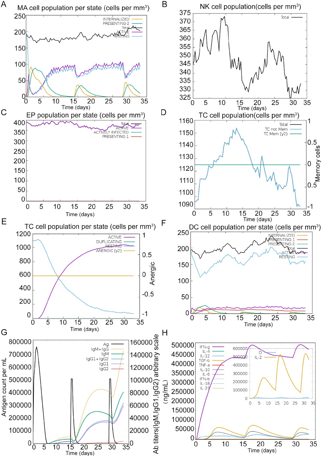
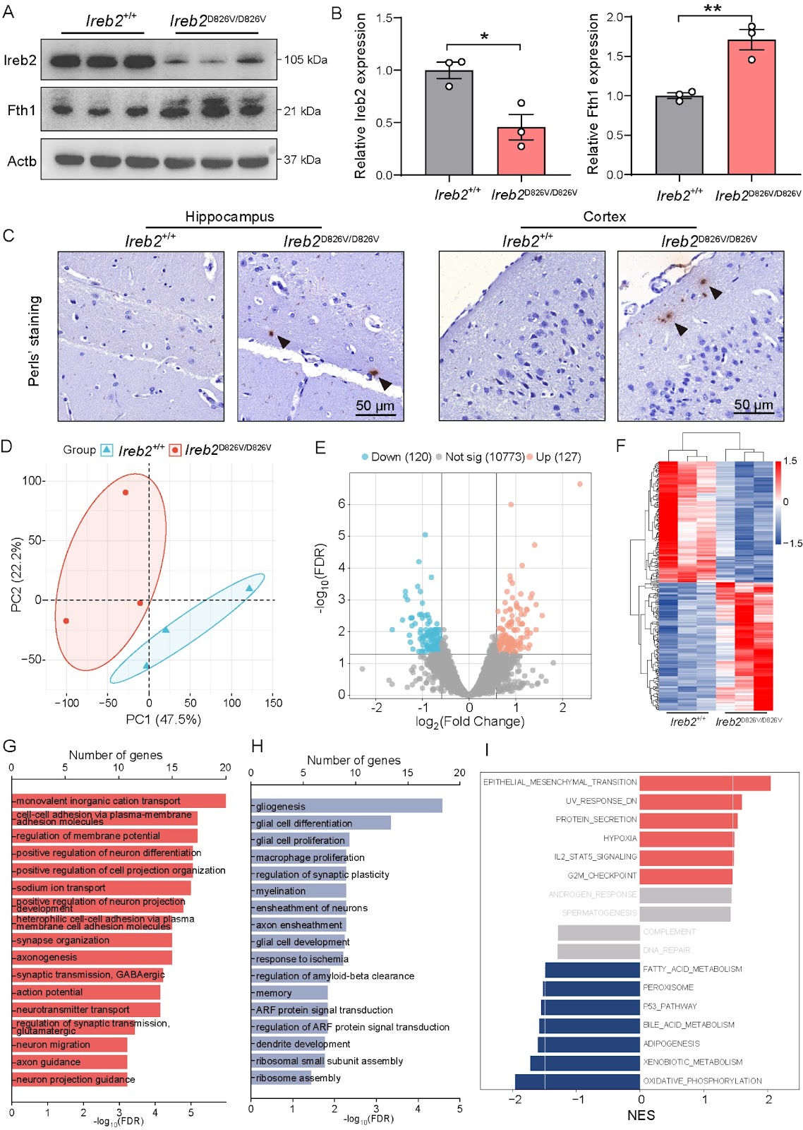
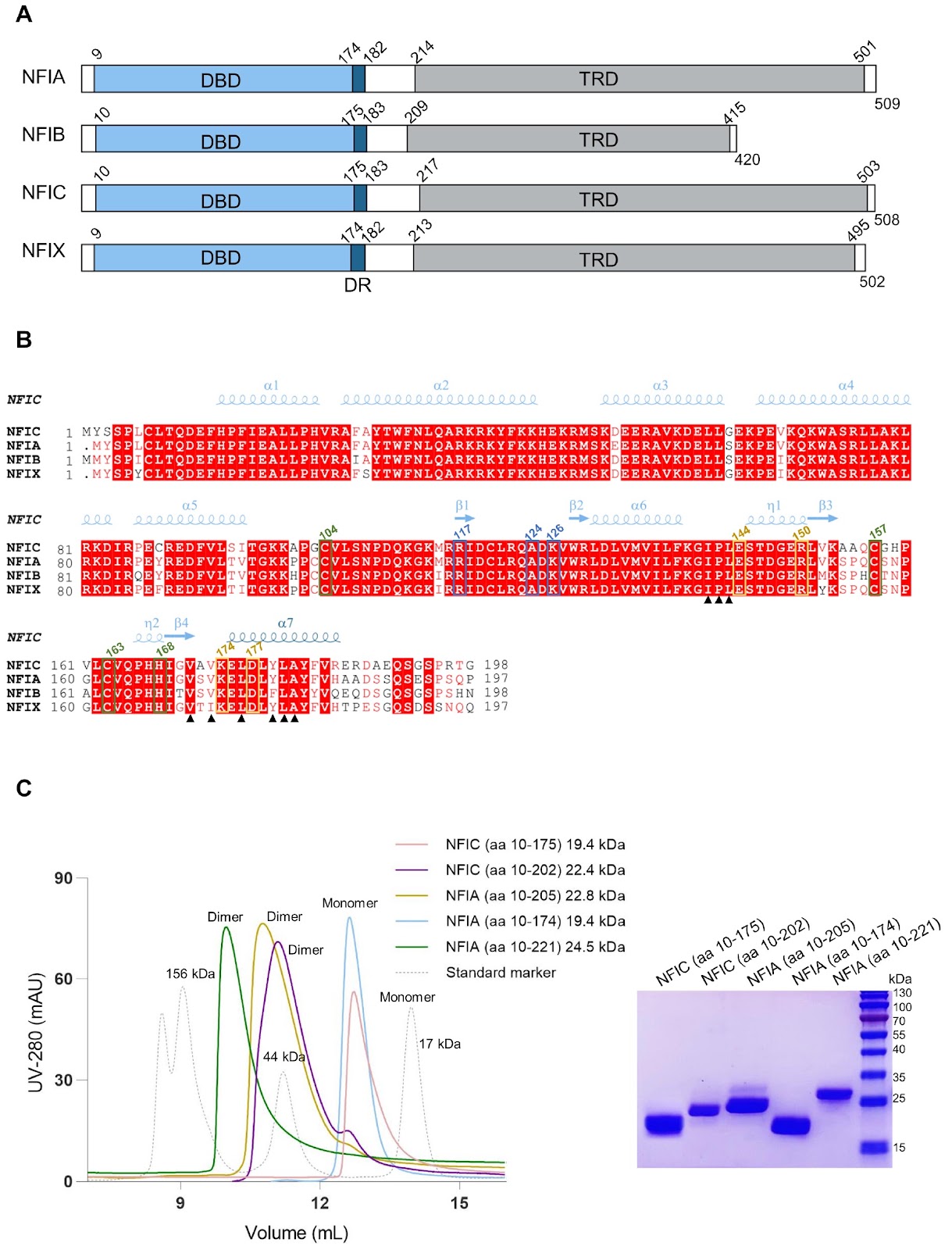
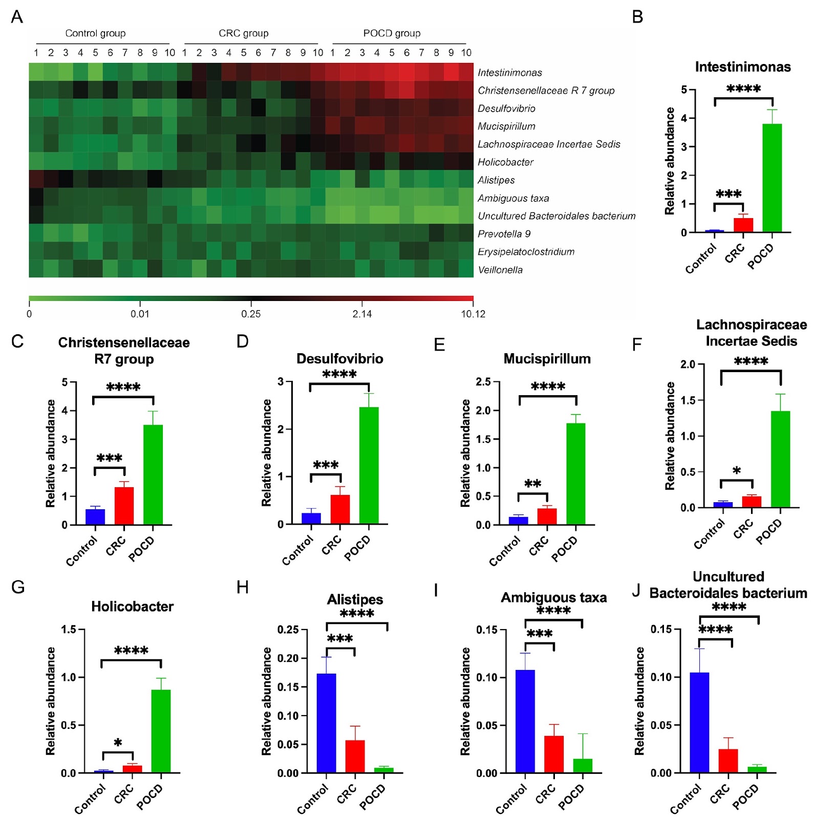
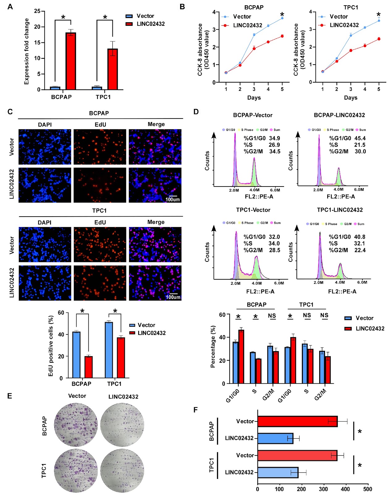
Myocyte enhancer factor 2A orchestrates vascular redox homeostasis via direct transcriptional activation of SIRT1
Graphical Abstract
Original ResearchVol. 58, Issue 5 • pp. 947-962DOI: 10.3724/abbs.2025212

Antibody-oligonucleotide conjugates for spatial proteomics: principles, applications, and challenges

Authors: Yinghui Qiu, Chunlan Li, Peiying Ye, Haiyun Zhang, Yanxiu Liu, Weiyan Ma, Chen Lin, Rongqin Ke

Spatial biology aims to elucidate cellular organization, function, and interactions within native tissue contexts, offering key insights into both normal physiology and disease. Spatial proteomics complements this by enabling high-resolution mapping of protein localization and abundance, directly reflecting functional cellular states. Unlike transcriptomics, which infers potential activity, proteomics captures actual molecular functions, including post-translational modifications and dynamic interactions. However, in situ protein profiling poses significant challenges, as proteins cannot be directly sequenced or easily targeted via nucleic acid hybridization. Antibody-oligonucleotide conjugates (AOCs) address this limitation by converting protein recognition into a DNA-based readout, thereby enabling sensitive and scalable detection. In this review, we outline the core principles of AOC-based spatial proteomic technologies, including multiplexed protein analysis, in situ protein-protein interactions, and integration with other biomolecular data. We highlight their applications in decoding tissue complexity and disease pathology and examine key technical challenges that remain. Overall, AOCs offer distinct advantages, including DNA-mediated signal amplification, spatially resolved proteomic profiling, and compatibility with multi-omics approaches, positioning them as powerful platforms in the advancement of spatial biology.

Antibody-oligonucleotide conjugates for spatial proteomics: principles, applications, and challenges
Graphical Abstract
Original ResearchVol. 58, Issue 4 • pp. 819-832DOI: 10.3724/abbs.2025110

Directly targeting G-quadruplexes contributes to the anti-multiple myeloma efficacy of Epimedokoreanin B

Authors: Pingting Jia, Shangzhao Wang, Wanting Huang, Ye Fang, Jian Gao

Multiple myeloma (MM) is a hematological malignancy for which novel therapeutic strategies are urgently needed. Epimedokoreanin B (EKB), an isoprenylated flavonoid compound derived from the medicinal plant Epimedium koreanum, has demonstrated promising antitumor activity. However, its effects on MM have not been previously investigated. This study explores the anti-MM activity and the molecular interaction mechanisms between EKB and G-quadruplexes (G4) through a combination of biological activity assessments and computer-aided methodologies. EKB exhibits potent cytotoxicity against the MM cell lines U266 and RPMI-8226, with IC50 values of 5.28 μM and 6.81 μM, respectively. It induces apoptosis in a concentration-dependent manner and specifically stabilizes the G4 structures of oncogenes such as c-Myc, c-KIT, Bcl-2, and k-RAS, as confirmed by BG4 immunofluorescence staining and fluorescence resonance energy transfer (FRET) assays. Additionally, EKB significantly suppresses the mRNA and protein expression levels of these genes in myeloma cells. Computational studies, including molecular docking, molecular dynamics (MD) simulations, and MM/GBSA calculations, confirm the strong binding affinity and stabilizing effects of EKB on G4s, revealing a mechanism involving π-π stacking and hydrogen bonding. This discovery underscores the unique ability of EKB to increase the stability of G4 structures, which are critical for regulating gene expression and inhibiting cancer cell proliferation. This research highlights the therapeutic potential of EKB in targeting these specific molecular structures, thereby offering a more effective approach to managing MM.

Directly targeting G-quadruplexes contributes to the anti-multiple myeloma efficacy of Epimedokoreanin B
Graphical Abstract
Original ResearchVol. 58, Issue 5 • pp. 1023-1031DOI: 10.3724/abbs.2025183

Gut microbiota and bile acids changes in MASLD mice model with hepatic PLD1 knockout

Authors: Yushang Zhao, Huan Wang, Wanling Lin, Hui Wang, Lin-Lin Cao

Hepatocyte phospholipase D1 (PLD1) knockout alleviates metabolic dysfunction-associated steatotic liver disease (MASLD) in mice, but the underlying mechanism is largely unknown. In this study, the mice are divided into four groups: Con (wild-type mice with normal control diet), HFHC (wild-type mice with high-fat diet), Con_KO (hepatocyte PLD1-knockout mice with normal control diet), and HFHC_KO (hepatocyte PLD1-knockout mice with high-fat diet). Intestinal contents of mice are analyzed via metagenomics and metabolomics, and the liver bile acids are assessed by mass spectrometry imaging. The results show that at the phylum level the abundance of Bacillota in the intestines of MASLD model mice is significantly increased, whereas that of Bacteroidota significantly is decreased. However, after the deletion of hepatocyte PLD1, Pseudomonadota and Candidatus Bathyarchaeota are significantly decreased in the MASLD model mice. At the species level, compared with that in the Con group, the abundance of Faecalibaculum rodentium is significantly increased in the HFHC group, whereas hepatocyte PLD1 knockout causes the abundances of Desulfovibrionaceae bacterium LT0009 and Lachnospiraceae bacterium 10-1 to be significantly decreased. In terms of intestinal bile acids, the levels of two bile acids (hyodeoxycholic acid and glycolithocholic acid) differ between the HFHC_KO group and the HFHC group. Association analysis shows that Faecalibaculum co-occurs with DCA, βMCA, ΩMCA and αMCA, while probiotic Bacteroides uniformis is significantly correlated with UDCA, 12-KetoLCA, and 7-KetoLCA. Finally, mass spectrometry imaging reveals that the TCA and TDCA contents in the liver are significantly decreased after PLD1 knockout in hepatocytes. These findings demonstrate that hepatocyte PLD1 knockout alters the gut microbiota and bile acids profiles, suggesting that PLD1 deficiency may modulate MASLD progression by changing intestinal microbiota-bile acid homeostasis.

Gut microbiota and bile acids changes in MASLD mice model with hepatic PLD1 knockout
Graphical Abstract
Original ResearchVol. 58, Issue 4 • pp. 709-724DOI: 10.3724/abbs.2025230

Explore antibody repertoire in the era of AI

Authors: Yudi Zhang, Hefei Wang, Chencheng Liu, Fei-Long Meng

The diverse antibodies of adaptive immunity comprise an antibody repertoire that combats various pathogens. This repertoire is shaped by both intrinsic antibody gene diversification and extrinsic cellular selection. Conversely, an antibody repertoire contains multiple layers of immunological information, including the history of pathogen exposure. High-throughput sequencing-based antibody repertoire cloning approaches have revealed unexpected features of adaptive immunity. However, our understanding of antibody repertoire data is still in its infancy. In this review, we introduce the emerging concepts and discuss the application of deep learning approaches to understanding antibody repertoires. First, we introduce the definition and functional features of antibody clonotype. Next, we review the evolution of antibody clonotypes and discuss potential antibody repertoire-directed vaccination approaches. Lastly, we summarize the application of deep learning in predicting antibody binding, generating specific antibodies, and making immunologic diagnoses. Recently, artificial intelligence (AI) has made revolutionary progress in biology. Leveraging high-dimensional antibody repertoire information, deep learning models have the potential to transform our understanding of antibody repertoire.

Explore antibody repertoire in the era of AI
Graphical Abstract
Original ResearchVol. 58, Issue 3 • pp. 663-680DOI: 10.3724/abbs.2025114

The TCF7L2/miR-206/Cofilin1 axis promotes the metastasis of bladder cancer cells by regulating the formation of invadopodia

Authors: Yuzhen Jie, Yinggui Yang, Chengyan Guo, Qinghui Wu, Zhewen Ou, Weifu Wang, Ning Xu, Wei Peng, Yingguang Wu, Jiangfan Peng, Shengchao Ma, Shufang Zhang, Fei Wang

Bladder cancer (BCa) is one of the most common malignant tumors of the urinary system, but its pathogenesis is still unclear. T1G3 BCa is particularly invasive and relapses readily after treatment, with progression to invasive cancer or distant metastasis. Therefore, identification of the molecular mechanism by which it invades and metastasizes to guide treatment and predict patient prognosis is needed. Cofilin1 plays an important role in regulating gene expression and the invasiveness of tumors. In this study, we show that Cofilin1 is highly expressed in BCa and lymph nodes with metastasis, which is positively related to the grade of BCa, and is significantly related to clinicopathological parameters and cancer-specific survival. Phenotypic analysis reveals that Cofilin1 knockout inhibits the proliferation and migration of BCa cells, whereas Cofilin1 overexpression promotes the opposite phenotype. Cofilin1 binds to cortactin, thereby reducing the expression of F-actin and promoting the formation of invadopodia in BCa cells. Further experiments reveal that TCF7L2 can bind to the promoter of Cofilin1 and transactivate it, promoting a malignant phenotype. TCF7L2 may also reverse the inhibitory effect of miR-206 on the binding of Cofilin1 and cortactin and promote the metastasis of BCa by inhibiting the transcription maturation of miR-206. This study confirms that Cofilin1 is an oncogene in T1G3 BCa, and the TCF7L2/miR-206/Cofilin1 signaling pathway plays an important role in the formation of invadopodia in BCa.

The TCF7L2/miR-206/Cofilin1 axis promotes the metastasis of bladder cancer cells by regulating the formation of invadopodia
Graphical Abstract
Original ResearchVol. 58, Issue 3 • pp. 516-529DOI: 10.3724/abbs.2025112

Scutellarin inhibits ferroptosis by promoting cellular antioxidant capacity through regulating Nrf2 signaling

Authors: Haiyan Yang, Onkei Chan, Xiaodi Huang, Liang Yan, Nuo Sun, Yaping Li, Zijian Shi, Qingbing Zha, Dongyun Ouyang, Jinhua Li, Xianhui He

Ferroptosis is a lytic form of regulated cell death that is driven by iron-dependent lipid peroxidation and has been implicated in various diseases, including acute kidney injury (AKI). Scutellarin is a flavonoid isolated from Erigeron breviscapus (Vant.) Hand.-Mazz. and possesses various pharmacological activities, including anti-inflammatory and antioxidative properties. However, it is unclear whether scutellarin can inhibit ferroptosis and mitigate related diseases. In this study, we show that scutellarin can inhibit ferroptosis in both human HK-2 cells and mouse bone marrow-derived macrophages stimulated with RSL3 or erastin. Mitochondrial dysfunction and reactive oxygen species generation are counteracted by scutellarin treatment, suggesting the involvement of its antioxidative activity. Furthermore, scutellarin increases the nuclear levels of Nrf2 and the expressions of its target genes, including HO-1 and GPX4. Scutellarin-mediated inhibition of ferroptosis and increases in these proteins are abrogated by co-treatment with brusatol, an Nrf2 inhibitor, indicating an essential role for Nrf2 in this process. In a mouse model of folic acid-induced AKI, scutellarin mitigates acute renal damage, as revealed by histopathological analysis and serum blood urea nitrogen and creatinine assays. Folic acid-induced acute renal injury is associated with increased ferroptosis, as revealed by elevated level of 4-hydroxynonenal (4-HNE), a surrogate marker of ferroptosis, which is diminished by scutellarin co-treatment. Specifically, the elevated 4-HNE levels in macrophages (MAC-2 positive) and other renal cells are suppressed by scutellarin. Overall, scutellarin can inhibit ferroptosis both in cultured cells and in a mouse model of AKI by regulating Nrf2 signaling.

Scutellarin inhibits ferroptosis by promoting cellular antioxidant capacity through regulating Nrf2 signaling
Graphical Abstract
Original ResearchVol. 58, Issue 4 • pp. 911-919DOI: 10.3724/abbs.2025182

Structural basis for suramin binding to the C-terminal domain of the SARS-CoV-2 nucleocapsid protein

Authors: Chenyun Guo, Xiao Li, Hao Xu, Jiaxin Yu, Jia Li, Donghai Lin

The global threat posed by COVID-19 persists, largely due to the high mutability of SARS-CoV-2 and the limited availability of effective antiviral therapeutics. The nucleocapsid (N) protein of SARS-CoV-2 is an attractive drug target because of its high degree of sequence conservation and essential role in viral replication. In this study, we show that suramin, a polysulfonated antiviral compound, binds to the C-terminal domain (N-CTD) of the N protein and interferes with its interaction with RNA. Biolayer interferometry (BLI) shows that suramin has a higher binding affinity for N-CTD (Kd, 3.30 μM) than for RNA (Kd, 10.12 μM). Electrophoretic mobility shift assays (EMSAs) further confirms that suramin effectively displaces RNA from N-CTD. NMR titration experiments and site-directed mutagenesis identify the α1-η1 helix (residues 248–262) as the primary suramin binding region, with residues K256, R259 and R262 playing critical roles in ligand recognition. In addition, NMR relaxation and model-free analyses reveal that the α1-η1 helix is highly flexible on the picosecond to nanosecond timescale, a dynamic feature that likely facilitates ligand binding. Furthermore, ITC and EMSA experiments demonstrate that suramin can bind to the full-length N protein at multiple sites and dissociate RNA from the N protein. Taken together, these findings provide structural and biophysical insights into the mechanism of action of suramin and establish a rational basis for the development of targeted antiviral therapies against SARS-CoV-2.

Structural basis for suramin binding to the C-terminal domain of the SARS-CoV-2 nucleocapsid protein
Graphical Abstract
Original ResearchVol. 58, Issue 3 • pp. 530-540DOI: 10.3724/abbs.2025142

Combined ex vivo and in vivo evaluation of dolutegravir embryotoxicity: NTDs and yolk sac vascular abnormalities

Authors: Ruifang Ao, Ran Li, Zelin Li, Guicai Wu, Haoran Xu, Xuecong Wang, Jiayi Du, Xiaozheng Zhang, Jun Xie

Dolutegravir (DTG) disrupts mouse embryonic development in a dose-dependent manner, culminating in neural-tube defects (NTDs). Using whole embryo culture (WEC), mouse embryos at embryonic day 8.5 (E8.5) are cultured for 24–48 h with 8, 10, or 12 μM DTG. The results reveal that higher DTG concentrations dose-dependently disrupt yolk sac development and markedly increase the frequency of NTDs. In vivo NTD models are generated by intraperitoneally injecting DTG at a dose of 7.5 mg/kg, and the resulting embryos exhibit disrupted yolk sac blood circulation, embryonic growth restriction, and malformations. Mechanistic studies suggest that DTG contributes to NTDs by inducing apoptosis: DTG exposure activates the Nrf2-SOD1/CAT antioxidant axis, yet it culminates in increased apoptosis and suppressed proliferation, ultimately impairing yolksac vasculogenesis and neuralepithelial closure, thereby producing NTDs. This study provides new evidence for assessing the potential risk of DTG in embryonic development and highlights the need to re-evaluate its clinical safety in future applications.

Combined ex vivo and in vivo evaluation of dolutegravir embryotoxicity: NTDs and yolk sac vascular abnormalities
Graphical Abstract
Original ResearchVol. 58, Issue 3 • pp. 649-662DOI: 10.3724/abbs.2025144

Exploring DNA topoisomerase II alpha in adrenocortical carcinoma through multi-omics analysis: a potential biomarker and therapeutic target

Authors: Jianming Lu, Pei Deng, Zhenjie Wu, Yuxiang Liang, Yangjia Zhuo, Yongding Wu, Yingke Liang, Jianheng Ye, Wenjie Xie, Zhouda Cai, Chao Cai, Jiahong Chen, Le Zhang, Junhong Deng, Weide Zhong, Jiaojiao Tang, Zhaodong Han

Adrenocortical carcinoma (ACC) is a rare but aggressive cancer. Recent studies identified DNA Topoisomerase II Alpha (TOP2A) as a potential biomarker for ACC, which can provide new avenues for targeted therapy and improve clinical outcomes. This study aims to elucidate the role of TOP2A in ACC by exploring its prognostic value and identifying inhibitors for ACC therapy. Utilizing RNA sequencing data, mutation data, and clinical information from The Cancer Genome Atlas (TCGA-ACC) and additional datasets from the Gene Expression Omnibus (GEO), differential expression and prognostic analyses are conducted to assess the significance of TOP2A in ACC. Immunohistochemistry and cell assays, including cell viability, colony formation, and transwell assays, are conducted to validate the oncogenic effects of TOP2A. The “IOBR” R package is used to examine the relationship between TOP2A expression and CD8+ T-cell infiltration. The CMap platform is used to identify potential TOP2A inhibitors. In vivo assays verify the therapeutic effect of TOP2A inhibitors on ACC. Our findings indicate that TOP2A is significantly overexpressed in ACC and is associated with poor prognosis. Immunohistochemistry and cell assays confirm the oncogenic role of TOP2A. Furthermore, distinct gene expression patterns related to different TOP2A expression levels are identified, influencing the response to immunotherapy. Potential inhibitors targeting TOP2A are discovered, and the therapeutic effects of resminostat and etoposide are confirmed via in vivo assays, suggesting new therapeutic strategies for ACC treatment. In conclusion, TOP2A serves as a crucial biomarker in ACC and is associated with adverse clinical outcomes and a diminished immune response. The identification of potential inhibitors against TOP2A opens new avenues for the development of targeted therapies for ACC patients.

Exploring DNA topoisomerase II alpha in adrenocortical carcinoma through multi-omics analysis: a potential biomarker and therapeutic target
Graphical Abstract
Original ResearchVol. 58, Issue 4 • pp. 792-805DOI: 10.3724/abbs.2025150

Rapid detection of Escherichia coli in bloodstream infection via CRISPR-Cas9 engineered reporter phage T7::Nluc and microfluidic chip platform

Authors: Minwei Li, Zhiyun Hao, Jing Yan, Ximeng Chen, Hangyi Li, Chengbin Wang, Chi Wang

Rapid identification of pathogens responsible for bloodstream infection is critical for early intervention and effective treatment. Reporter phages, which are known for their exceptional sensitivity and specificity in pathogen detection, have garnered significant interest. In this study, we systematically evaluate phage genome editing strategies that combine homologous recombination with the CRISPR-Cas9 system. We investigate the impacts of homologous arm length, sgRNA activity, target site, and plasmid interactions on editing efficiency. Our results demonstrate that successful genome editing depends on both sufficient cleavage pressure and optimal homologous arm length, particularly when using low-activity sgRNAs. On the basis of these findings, we develop a highly efficient gene editing strategy TPMSR (triple-plasmid-mediated synchronous recombination) that overcomes the limitations of conventional methods that rely on high-activity sgRNA and restricted editing sites. Using the TPMSR strategy, we integrate the Nluc gene into phage T7, generating the reporter phage T7::Nluc, which is then incorporated into a microfluidic chip. Validation with 51 clinical isolates demonstrates outstanding sensitivity, specificity, and accuracy in detecting Escherichia coli in blood within 1.5 h at concentrations less than 30 CFU/mL. This study presents a robust strategy for phage genome engineering and develops a promising method for the rapid diagnosis of bloodstream infections caused by E. coli.

Rapid detection of Escherichia coli in bloodstream infection via CRISPR-Cas9 engineered reporter phage T7::Nluc and microfluidic chip platform
Graphical Abstract
Original ResearchVol. 58, Issue 3 • pp. 574-583DOI: 10.3724/abbs.2025135

Targeting USP2 induces degradation of PML-RARα with or without drug-resistant mutations in acute promyelocytic leukemia

Authors: Jie Zhang, Wenxuan Wu, Yun Wang, Youping Zhang, Yingying Wang, Wenhui Bai, Zhenge Zhang, Chujiao Zhu, Yunzhao Wu, Ziwei Zhang, Li Yang, Hu Lei, Hanzhang Xu, Li Zhou, Yingli Wu

Despite the high efficacy of all-trans retinoic acid (ATRA) and arsenic trioxide (ATO) in treating acute promyelocytic leukemia (APL), approximately 10%–20% of patients develop drug resistance due to mutations in PML-RARα and other factors. Here, we find that inhibition of USP2 with ML364 or USP2 silencing reduces PML-RARα protein levels in both ATRA-sensitive and ATRA-resistant APL cells, and this effect is reversed by proteasome inhibition. Conversely, USP2 overexpression enhances PML-RARα stability. Mechanistically, USP2 interacts with and deubiquitinates PML-RARα, including its drug-resistant mutants. Consistent with PML-RARα degradation, ML364 treatment significantly induces apoptosis in APL cell lines and primary leukemia cells. In conclusion, this study identifies USP2 as a novel deubiquitinating enzyme for PML-RARα and highlights USP2 inhibition as a potential therapeutic strategy for APL with PML-RARα mutations.

Targeting USP2 induces degradation of PML-RARα with or without drug-resistant mutations in acute promyelocytic leukemia
Graphical Abstract
Original ResearchVol. 58, Issue 3 • pp. 700-703DOI: 10.3724/abbs.2025104

A novel method to increase transgene expression and the stability of gene therapy-associated episomal vectors

Authors: Xi Zhang, Rui Liu, Zimeng Han, Zihan Guo, Mengying Ji, Wen Wang, Yanlong Jia, Tianyun Wang, Xiaoyin Wang

Non-viral episomal vectors offer a safe and attractive alternative to viral and integrated vectors by avoiding insertional mutagenesis and position effects, making them ideal expression vectors for gene therapy. The first non-viral episomal vector, pEPI-1, which is based on the full-length scaffold/matrix attachment region (S/MAR), was established by Piechaczek et al. The full-length S/MAR element interacts with the nuclear matrix via the matrix protein, e.g. SAF-A, thereby maintaining mitotic stability and transgene expression. Several strategies, including optimization of the vector backbone and promoter and incorporation of chromatin-modifying elements, have been used to increase expression levels and stability. In our previous work, we constructed the novel vector pEGFP-C1-M on the basis of S/MAR characteristic motifs (only 375 bp). This vector, which is shorter than the prototype episomal vector pEPI-1, resulted in relatively higher transgene expression. Building on the pEGFP-C1-M vector, we further constructed the episomal vector pEMEα with the EF-1α promoter and demonstrated that pEMEα maintained higher transgene expression, stability and copy number. The transgene expression levels of episomal vectors are correlated with gene copy number, that is, the number of plasmid episomes on the host cell chromosome. Previous studies have shown that the episomal maintenance of pEPI-1 vectors is mediated primarily by SAF-A. While the role of SAF-A in maintaining mammalian pEPI-1 episomal vectors has been well established, it remains unknown whether the overexpression of SAF-A promotes transgene expression and stability and whether the 375 bp MAR characteristic sequence retains its interaction with SAF-A. In the present study, we first evaluated whether transgene expression is positively correlated with the expression level of SAF-A. The non-viral episomal vector pEMEα was used as the gene of interest (GOI) vector and was subsequently transfected into CHO-K1 cells using the Lipofectamine 2000 reagent. The cells were cultured in medium containing 800 μg/mL geneticin (G418) 48 h post-transfection, and the G418 concentration was then reduced to 400 μg/mL to obtain monoclonal cell lines using the limiting dilution method. Five monoclonal cell clones were selected, and the eGFP expression levels, measured as the mean fluorescence intensity (MFI), were (6.5 ± 1.0) × 104, (6.8 ± 0.9) × 104, (7.0 ± 1.4) × 104, (23.5 ± 1.2) × 104 and (14.9 ± 0.17) × 104 for Clones 1–5, respectively. qPCR analysis of Clones 1–5 revealed that the relative mRNA levels of SAF-A and eGFP were 0.16 ± 0.13, 0.43 ± 0.11, 0.46 ± 0.23, 2.17 ± 0.41, 1.78 ± 0.15 and 0.51 ± 0.16, 0.71 ± 0.12, 1.05 ± 0.09, 2.47 ± 0.14, and 1.86 ± 0.10, respectively. Our results indicated that eGFP mRNA and protein expression levels are positively correlated with SAF-A mRNA level. To further verify the relationship between SAF-A expression and transgene expression, two shRNA plasmids targeting SAF-A (shRNA1: 5′-GCCACCTGTTGAAGAAGAAGA-3′, and shRNA2: 5′-GCTGGAGGAAGAGCTTCTTAT-3′) which were obtained from Shanghai GenePharma Co., Ltd. were designed and transfected into stable cell pools with the pEMEα vector. qPCR analysis revealed that the relative SAF-A mRNA levels in the shRNA1 and shRNA2 vectors were 0.47 ± 0.01 and 0.19 ± 0.02, respectively, indicating successful downregulation of SAF-A expression. Moreover, flow cytometry and qPCR revealed that, compared with those in the control group, the relative protein and mRNA levels of eGFP were reduced by 0.47- and 0.23-fold, and 0.47- and 0.40-fold in the pools of cells transfected with the shRNA1 and shRNA2 vectors, respectively. On the basis of the above results, the SAF-A overexpression vector pIRES-SAF-A was constructed and transfected into CHO-K1 cells, and the cells were cultured in blasticidin-containing medium 48 h after transfection to obtain stable cell pools. The stable cell pools overexpressing SAF-A were subsequently transfected with the pEMEα vector. Stable cell pools coexpressing SAF-A and GOI were selected, and the relative mRNA levels of SAF-A and eGFP were analyzed. qPCR analysis revealed that the relative mRNA levels of SAF-A and eGFP in the pools of cells overexpressing SAF-A were 2.69-fold and 2.05-fold higher than those in the control group, respectively. Flow cytometry also revealed a 2.07-fold increase in MFI in stable cell pools overexpressing SAF-A compared with the control group. To assess the long-term stability of transgene expression, we measured the MFI in st

A novel method to increase transgene expression and the stability of gene therapy-associated episomal vectors
Graphical Abstract
Original ResearchVol. 58, Issue 3 • pp. 584-594DOI: 10.3724/abbs.2025146

FSCN1-mediated hepatic gluconeogenesis is indispensable for neonatal mice survival

Authors: Xiangxiang Liu, Yuanzhao Hu, Liangwei Wu, Yiwen Zhang, Lei Sang, Yake Gao, Lei He, Wenyong Xiong, Shengyu Yang, Jianwei Sun

Actin-bundling protein Fascin1 (FSCN1) is encoded by the Fscn1 gene and is crucial for cytoskeletal remodeling and cellular migration. Although a previous study linked Fscn1 deficiency to neonatal lethality in mice, the underlying metabolic mechanism remains unclear. In this study, we report that systemic knockout (KO) of Fscn1 leads to 52.2% mortality within 24 h post-birth, accompanied by severe hypoglycemia in KO pups compared with their littermates. Remarkably, this lethality is fully rescued by oral glucose administration, indicating a glucose supply-dependent survival mechanism. Surviving Fscn1-KO neonates display persistent developmental deficits, including growth retardation and depleted lipid stores, despite intact canonical insulin-regulated hepatic gluconeogenic pathways. Transcriptomic profiling of P0 livers reveals that Fscn1 loss predominantly disrupts metabolic pathways, with the glycerol phosphate shuttle being the most significantly downregulated module. Mechanistically, Fscn1-KO livers exhibit markedly reduced protein levels of glycerol-3-phosphate dehydrogenase isoforms (GPD1/GPD2), key enzymes bridging glycolysis and gluconeogenesis. Consistently, glycerol tolerance tests demonstrate impaired glycerol-to-glucose conversion in Fscn1-KO mice, confirming defective glycerol-driven gluconeogenesis. Our findings establish FSCN1 as a novel cytoskeletal-metabolic integrator essential for neonatal survival by sustaining hepatic glucose production from glycerol, thus revealing an unexpected role of actin dynamics in coordinating metabolic adaptation during early postnatal development.

FSCN1-mediated hepatic gluconeogenesis is indispensable for neonatal mice survival
Graphical Abstract
Original ResearchVol. 58, Issue 3 • pp. 595-609DOI: 10.3724/abbs.2025130

Autophagy-dependent sensitization effects of PARP inhibitors on recurrent nasopharyngeal carcinoma treated with carbon ion and photon irradiation

Authors: Ziyu Le, Haojiong Zhang, Li Chen, Wanzun Lin, Qingting Huang, Shikai Geng, Wei Hu, Huaiyuan Chen, Fangzhu Wan, Xingyu Liu, Jiyi Hu, Fengtao Su, Jiade J. Lu, Lin Kong

Tumor radioresistance and severe toxicity make reirradiation for recurrent nasopharyngeal carcinoma (NPC) a significant clinical challenge. This study aims to investigate the ability of the poly(ADP-ribose) polymerase (PARP) inhibitor olaparib to sensitize recurrent NPC cells irradiated with photon or carbon ion (C-ion), and to explore the underlying mechanism of the synergistic promotion of cell death by olaparib and ionizing radiation. The results show that olaparib has significant X-ray and C-ion radiosensitization effects on recurrent NPC cells and the associated HK-RR photon-resistant model. Radiation, particularly C-ion exposure, induces a homologous recombination (HR)-deficient gene signature in HR-proficient NPC cells, potentially increasing their sensitivity to PARP inhibition. C-ion and X-ray irradiation induces similar modes of cell death, and multiple cell death pathways [including apoptosis, necrosis, ferroptosis, senescence, and autophagic cell death (ACD)] contribute to the cytotoxic effects of radiation combined with olaparib, with ACD being the dominant pathway. Both the pharmacological and genetic inhibition of autophagy significantly attenuate the radiosensitization effect of olaparib. In conclusion, olaparib effectively sensitizes recurrent NPC cells to both X-ray irradiation and C-ion irradiation, with autophagy playing a central role in mediating this effect.

Autophagy-dependent sensitization effects of PARP inhibitors on recurrent nasopharyngeal carcinoma treated with carbon ion and photon irradiation
Graphical Abstract
Original ResearchVol. 58, Issue 3 • pp. 541-550DOI: 10.3724/abbs.2025179

Pervasive environmental contaminant acrolein compromises myocardial geometry and function through the induction of cuproptosis

Authors: Xiaohu Wang, Shuyi Zhu, Yipin Zhao, Xinlong Di, Lingfei Cao, Russel J. Reiter, Emily Y. He, Yuan Zhou, Bonglee Kim, Yong Cheng, Jun Ren

Acrolein, a highly reactive α,β-unsaturated aldehyde found in cigarette smoke, automobile exhaust, industrial emissions, combustion byproducts, cooking and cyclophosphamide chemotherapy, has raised serious health concerns, although the precise mechanism remains unclear. This study is designed to examine the impact of this pervasive environmental pollutant on myocardial geometry and function alongside the underlying cellular mechanisms. Adult C57BL/6 mice are challenged with acrolein (2.5 mg/kg/day, i.p., for 20 days) prior to the evaluation of myocardial geometry and function. Acrolein exposure evokes evident cardiac remodeling (interstitial fibrosis), compromised echocardiographic (enlarged LVESD, compromised ejection fraction and fractional shortening), cardiomyocyte contractile and intracellular Ca2+ capacities [decreased peak shortening, maximal velocity of shortening and relengthening (±dL/dt), and electrically stimulated rise in Fura-2 fluorescence intensity (ΔFFI), prolonged time-to-90% relengthening (TR90) and intracellular Ca2+ decay], accompanied by overt mitochondrial damage (ultrastructure, aconitase and mitochondrial protein contents), free radical buildup, apoptosis (Bax, Caspase-3, and Bcl2) and cuproptosis (upregulated SLC31A1, DLAT and FDX1), downregulated the Fe-S cluster proteins ACO2 and NDUFS8 alongside unchanged ATP7A and the ferroptosis markers GPX4 and SLC7A11. The levels of copper-sensing protein metal response element binding transcription factor 2 (MTF2), but not MTF1, are increased by acrolein insult. CB-DOCK2 analysis predicts an interaction between acrolein and the MTF2 dimer within its DNA-binding regions. In vivo administration of the cuproptosis inhibitor tetrathiomolybdate (TTM), the mitochondrial antioxidant mitoTEMPO or the nonselective MTF2 inhibitor actinomycin D alleviates acrolein-evoked cardiomyocyte dysfunction (decreased PS, ±dL/dt, and prolonged TR90). These findings indicate that acrolein evoked cardiac functional anomalies possibly through MTF2-related control of cuproptosis.

Pervasive environmental contaminant acrolein compromises myocardial geometry and function through the induction of cuproptosis
Graphical Abstract
Original ResearchVol. 58, Issue 3 • pp. 681-690DOI: 10.3724/abbs.2025228

SOX2 transactivates NRF2 to promote carboplatin resistance in lung squamous cell carcinoma

Authors: Hanfei Gao, Chaomei Li, Jie Sun, Liyuan Deng, Jia Li, Zhiqiang Wu, Hu Chen

Lung squamous cell carcinoma (LUSC) remains a major therapeutic challenge because of its pronounced resistance to chemotherapy, particularly carboplatin. In this study, we investigate the role of SOX2, a lineage-survival oncogene, in mediating carboplatin resistance in LUSC. We demonstrate that SOX2 is highly expressed in LUSC and is significantly associated with poor prognosis. Our results show that SOX2 directly transactivates the expression of NRF2, a master regulator of cellular redox homeostasis, thereby increasing glutathione (GSH) synthesis and protecting cells from carboplatin-induced oxidative stress. Pharmacological or genetic inhibition of NRF2 effectively abrogates SOX2-mediated carboplatin resistance both in vitro and in vivo, resensitizing LUSC cells to chemotherapy. These findings highlight SOX2 as a critical redox regulator that modulates NRF2 signaling to promote carboplatin resistance in LUSC. The identification of the SOX2-NRF2 axis as a potential therapeutic target suggests that NRF2 inhibition may represent a promising strategy to overcome chemoresistance in LUSC.

SOX2 transactivates NRF2 to promote carboplatin resistance in lung squamous cell carcinoma
Graphical Abstract
Original ResearchVol. 58, Issue 2 • pp. 216-230DOI: 10.3724/abbs.2025123

Unveiling the multifaceted roles of extracellular vesicles in cancer: insights from molecular imaging and engineering strategies

Authors: Yuqin Teng, Gang Huang, Hao Yang

Extracellular vesicles (EVs), a class of nanoscale, membrane-bound vesicles secreted by various cell types, have emerged as rapidly advancing fields of research in recent years. This heterogeneous vesicle is a versatile carrier system for a variety of biomolecules, including proteins, nucleic acids, and metabolites. EVs play pivotal roles in intercellular communication, immune regulation, and disease pathogenesis, with particular implications for cancer biology. On the one hand, EVs promote tumor progression and metastasis by facilitating communication between cancer cells and their microenvironment. On the other hand, EVs carry noncoding RNAs, such as miRNAs and other regulatory RNAs, which directly modulate immune cell function or exert antitumor effects by influencing cancer cell proliferation and apoptosis. In addition to their biological roles, EVs show great potential as drug delivery systems because of their ability to be effectively taken up by target cells and stably deliver therapeutic payloads. In the context of cancer therapy, natural EVs demonstrate inherent therapeutic potential, particularly in targeting highly metabolically active organs. Furthermore, engineered EVs, which serve as both therapeutic vehicles and molecular imaging probes, have demonstrated significant potential for cancer theranostics. This review focuses on elucidating the dynamic changes and biological functions of EVs in vivo, with the aim of exploring the translational potential of EV-based molecular imaging and tracing technologies in cancer treatment. This work seeks to provide critical insights that may enhance the precision and efficacy of tumor therapies, offering a foundation for future clinical applications.

Unveiling the multifaceted roles of extracellular vesicles in cancer: insights from molecular imaging and engineering strategies
Graphical Abstract
Original ResearchVol. 58, Issue 2 • pp. 353-368DOI: 10.3724/abbs.2025108

METTL3-mediated m6A modification facilitates Nectin-4-induced VNN1 upregulation and promotion of ESCC progression

Authors: Yuanfeng Long, Hang Yang, Ruolan Zhang, Quanneng Zhao, Mi Yang, Guiqin Song, Kang Liu

Esophageal squamous cell carcinoma (ESCC) is a highly aggressive malignancy with poor prognosis and limited therapeutic options. N6-methyladenosine (m6A) RNA modification plays a role in tumorigenesis, but its contributions to ESCC and the regulation of cell adhesion molecules such as Nectin-4 are not fully elucidated. In this study, we investigate the role and the regulatory mechanisms of Nectin-4 in ESCC, particularly regarding the influence of m6A modification and its downstream metabolic effects. Our study demonstrates that methyltransferase-like protein 3 (METTL3) enhances Nectin-4 mRNA stability and expression through m6A methylation in ESCC, as validated by actinomycin D assay, MeRIP-qPCR, and dual-luciferase reporter assay. Both METTL3 and Nectin-4 are highly expressed in ESCC tissues and promote malignant phenotypes such as proliferation, migration, and invasion. Further analysis identifies pantothenate esterase 1 (VNN1) as a downstream target of Nectin-4, mediating the oncogenic effects of the METTL3/Nectin-4 axis and promoting the biosynthesis of pantothenic acid and coenzyme A, thus driving ESCC progression. By integrating transcriptomic data, this study elucidates a key pathogenic mechanism in which the METTL3/Nectin-4/VNN1 axis regulates metabolic reprogramming to promote ESCC development. These findings provide new insights into the molecular pathology of ESCC and offer potential biomarkers and therapeutic targets for early screening, prognosis, and precision treatment for ESSC.

METTL3-mediated m6A modification facilitates Nectin-4-induced VNN1 upregulation and promotion of ESCC progression
Graphical Abstract
Original ResearchVol. 58, Issue 2 • pp. 437-452DOI: 10.3724/abbs.2025197

The prognostic marker NRIP1 is associated with tumor progression and immune infiltration in acute myeloid leukemia

Authors: Xunxun Zhu, Mingyan Zhang, Jingjing Zhang, Yanling Tao, Hao Zhang

Acute myeloid leukemia (AML) is a clinically aggressive hematologic malignancy characterized by high relapse rates and treatment resistance, highlighting the need for novel biomarkers to improve clinical outcomes. In this study, we explore the roles of nuclear receptor-interacting protein 1 (NRIP1) in AML, focusing on its associations with tumor progression and immune infiltration. Analysis of public AML gene expression datasets reveals that NRIP1 expression is significantly increased in AML patients. Those with high NRIP1 expression have markedly shorter overall survival than those with low expression. Furthermore, NRIP1 expression is significantly associated with the infiltration of diverse immune cells, including B cells, dendritic cells, T cells, mast cells, eosinophils, and T helper cells, suggesting that NRIP1 may be a regulator of immune cell infiltration. Functional enrichment analysis indicates that NRIP1 and its interacting partners are involved in tumorigenesis, immune microenvironment remodeling, and metabolic reprogramming. Survival analysis confirms the prognostic value of NRIP1. Importantly, functional validation in AML cell lines confirms that NRIP1 knockdown suppresses proliferation and induces apoptosis. Our study identifies NRIP1 as a multifaceted regulator that promotes AML by driving tumor progression, regulating immune cell infiltration, and modulating ferroptosis, highlighting its role as a novel prognostic biomarker.

The prognostic marker NRIP1 is associated with tumor progression and immune infiltration in acute myeloid leukemia
Graphical Abstract
Original ResearchVol. 58, Issue 2 • pp. 258-274DOI: 10.3724/abbs.2025075

FGF8 promotes lipid droplet accumulation via the FGFR1/p-p38 axis in chondrocytes

Authors: Minglei Huang, Haoran Chen, Jieya Wei, Caixia Pi, Mengmeng Duan, Xiaohua Pu, Zhixing Niu, Siqun Xu, Shasha Tu, Sijun Liu, Jiazhou Li, Li Zhang, Yang Liu, Hao Chen, Chunming Xu, Jing Xie

Chondrocytes store lipids in the form of lipid droplets (LDs) and maintain cartilage lipid metabolic homeostasis by consuming or regenerating LDs. This modulation is largely mediated by a series of biochemical factors. Fibroblast growth factor 8 (FGF8) is one of the most important factors involved in the proliferation, differentiation, and migration of chondrocytes and has attracted increasing attention in the physiology and pathology of cartilage. However, the effect of FGF8 on LD accumulation in chondrocytes remains unclear. This study aims to elucidate the role of FGF8 in LDs and explore the underlying biomechanism involved. The results reveal that FGF8 promotes LD accumulation in chondrocytes by upregulating perilipin1 (Plin1) expression. FGF8 activates the cytoplasmic p-p38 signaling pathway via fibroblast growth factor receptor 1 (FGFR1) to increase LD accumulation in chondrocytes. Subsequent experiments with siRNAs and specific inhibitors further confirm the importance of the FGFR1/p38 axis for LD accumulation in chondrocytes exposed to FGF8. The results increase our understanding of the role of FGF8 in the lipid metabolic homeostasis of chondrocytes and provide insights into the physiology and pathology of cartilage.

FGF8 promotes lipid droplet accumulation via the FGFR1/p-p38 axis in chondrocytes
Graphical Abstract
Original ResearchVol. 58, Issue 2 • pp. 396-405DOI: 10.3724/abbs.2025159

MSCs attenuate airway remodeling in HDM-induced asthma by inhibiting the Timp1-Wnt2b axis

Authors: Kai Yu, Xinyu Feng, Rong Zhang, Jian Fan, Jiaying Yuan, Yan Shang, Jiayi Zhao

MSCs have demonstrated their unique therapeutic potential in early clinical trials for a variety of respiratory diseases in recent years, but their use in the treatment of asthma has rarely been reported. In this study, a chronic murine asthma model that is more similar to clinical asthma is constructed via sustained HDM induction for 70 days, followed by treatment via tail vein injection of MSCs after modeling. The mechanism by which MSCs alleviate airway remodeling is investigated via RNA-seq. The airways on the day following treatment are used to screen for transcriptomic changes resulting from the MSC treatment under study, filtering for differentially expressed genes (DEGs), identifying their enrichment pathways, and finally confirming the DEGs gained via western blot analysis. After HDM treatment, airway remodeling is reversed, asthma and the HIF-1 signaling pathway are inhibited, and the expression levels of Timp1 and Wnt2b in the fibrosis pathway are also significantly decreased. STRING analysis reveals a reciprocal interaction in their expression, which is also confirmed by western blot analysis. To verify whether MSCs alleviate airway remodeling by inhibiting Timp1, we construct MSCs overexpressing Timp1 and evaluate their effects in vitro and in vivo. The ability of MSCs to alleviate airway remodeling is reversed after Timp1 is overexpressed. These findings demonstrate that MSCs alleviate asthma-induced airway remodeling by inhibiting the Timp1-Wnt2b axis.

MSCs attenuate airway remodeling in HDM-induced asthma by inhibiting the Timp1-Wnt2b axis
Graphical Abstract
Original ResearchVol. 58, Issue 2 • pp. 231-244DOI: 10.3724/abbs.2025175

The dual role of whole-genome duplication: biological mechanisms, functional consequences, and detection advances

Authors: Yawei Song, Jiajie Yang, Shuheng Wu, Wei Wu

Whole-genome duplication (WGD) represents an evolutionarily conserved process occurring in prokaryotes, eukaryotes, and somatic mammalian tissues. While developmentally programmed WGD supports normal tissue regeneration, unscheduled WGD drives chromosomal instability and oncogenic progression in cancer. Recent studies have clarified dual roles of WGD across physiological homeostasis and disease pathogenesis. Here, we review the prevalence of WGD, the molecular mechanisms driving its major causes and its biological consequences. In addition, we highlight recent advancements in WGD detection, including both conventional cytogenetic techniques and newly developed high-throughput sequencing approaches. The integration of multi-omics and machine learning further improves ploidy analysis, particularly in cancer research. Together, these insights establish WGD as a critical regulator of development, regeneration, and disease and underscore the importance of emerging computational and sequencing tools for its precise characterization.

The dual role of whole-genome duplication: biological mechanisms, functional consequences, and detection advances
Graphical Abstract
Original ResearchVol. 58, Issue 2 • pp. 245-257DOI: 10.3724/abbs.2025086

Gankyrin-deficiency reprograms intrahepatic glucose and lipid metabolism to delay liver regeneration

Authors: Yitian Liu, Yiwei Sun, Lv Jin, Ying Xu, Bibo Wang, Ting Yu, Xiaofei Wei, Jing Xu, Yating Wei, Shuai Yang, Min Yu, Hongyang Wang, Yao Chen

Liver regeneration is a critical adaptive response to hepatic injury, requiring precise metabolic reprogramming to meet the energetic and biosynthetic demands of proliferating hepatocytes. While the oncoprotein Gankyrin is well-established as a promoter of liver fibrosis and hepatocarcinogenesis, its role in metabolic adaptations underlying liver regeneration remains unclear. In this study, we demonstrate that Gankyrin deficiency in the liver (Gank△Hep/Y) induces hepatic hypertrophy and aberrant glycogen accumulation. Gankyrin expression is significantly upregulated after partial hepatectomy (PHx), whereas Gank△Hep/Y -PHx mice exhibit impaired liver regeneration. This impairment is marked by a delayed restoration of the liver-to-body weight ratio, blunted glycogenolysis, and reduced fatty acid uptake. Mechanistically, Gankyrin activates Pygl and Cd36, key regulators of glycogenolysis and lipid uptake, respectively. Pharmacological inhibition of PYGL activity retards liver regeneration. Furthermore, we identify a novel interaction between Gankyrin and FOXO1, wherein Gankyrin promotes FOXO1 ubiquitination and subsequent proteasomal degradation. This Gankyrin-dependent suppression of FOXO1 leads to the transcriptional upregulation of Pygl and Cd36, thereby fueling hepatocyte proliferation. Collectively, our findings reveal Gankyrin as a master regulator of liver regeneration, integrating metabolic reprogramming with proliferative signaling through the FOXO1-PYGL/CD36 axis. These insights not only elucidate the mechanistic underpinnings of liver regeneration but also unveil the therapeutic potential of targeting the Gankyrin/FOXO1 pathway to mitigate hepatic insufficiency and enhance regenerative capacity in clinical settings.

Gankyrin-deficiency reprograms intrahepatic glucose and lipid metabolism to delay liver regeneration
Graphical Abstract
Original ResearchVol. 58, Issue 2 • pp. 369-382DOI: 10.3724/abbs.2025103

IKZF3 promotes gastric cancer progression via Hedgehog signaling activation and is targetable by SANT-1

Authors: Muhammad Ali, Shantanu Baral, Jun Ren, Liuhua Wang, Bin Liu, Sen Wang, Daorong Wang

Elevated expression of Aiolos family zinc finger 3 (IKZF3), a transcription factor crucial for lymphocyte maturation, is observed in hematological cancers. However, its role in gastric cancer (GC) remains unclear. We detect the increased IKZF3 levels in GC tissues using immunohistochemical, qRT-PCR and western blot analysis. The function of IKZF3 in GC cells is further studied through CCK-8, Transwell, colony formation, scratch wound healing, and flow cytometry assays. IKZF3 overexpression significantly promotes GC cell invasion, migration, and proliferation, whereas IKZF3 knockdown induces cell cycle arrest at the G1/S phase. Flow cytometry confirms these alterations in cell cycle dynamics. Using the JASPAR database, we determine that IKZF3 binds to the SMO promoter region, thereby activating SMO expression. Notably, the SMO inhibitor SANT-1 effectively reverses IKZF3-mediated effects. Furthermore, IKZF3 promotes GC tumor growth in xenograft models. Our findings highlight the pivotal role of IKZF3 in GC progression by modulating SMO expression and activating the Hedgehog signaling pathway. Therapeutically, targeting IKZF3 with SANT-1 is promising for mitigating GC proliferation and invasion. This study provides insights into potential therapeutic approaches targeting IKZF3 for GC treatment.

IKZF3 promotes gastric cancer progression via Hedgehog signaling activation and is targetable by SANT-1
Graphical Abstract
Original ResearchVol. 58, Issue 2 • pp. 458-462DOI: 10.3724/abbs.2025118

Zinc finger protein 154 inhibits the growth and metastasis of cervical cancer cells through inhibiting Wnt/β-catenin signaling by upregulating NLK

Authors: Chulan Yang, Hongyu Zhao, Jing Tuo, Wei Zhao, Zhiwei Zhang, Zemin Pan, Lianghai Wang, Haixuan Zhao, Songhua Zhao, Hongtao Li

Cervical cancer represents a significant global health concern affecting women. The global cancer burden data published by the World Health Organization’s International Agency for Research on Cancer (IARC) indicated that the incidence and mortality of cervical cancer were the fourth most common malignancy in females worldwide in 2022 [1]. DNA methylation is recognized as a pivotal epigenetic mechanism for gene silencing, which may accumulate with disease severity [2]. Hypermethylation has been discovered in several tumor suppressor gene (TSG) promoters in human cancers, and further understanding of gene silencing mechanisms has led more studies to consider epigenetic disruption as an important mechanism leading to the silencing of tumor suppressor genes in tumor development [3]. Recent studies have reported that methylation of the zinc finger protein 154 (ZNF154) gene plays an oncogenic role in the development of several cancers [4]. ZNF154 has been shown to inhibit tumor cell proliferation in nasopharyngeal carcinoma by altering the expression of E-cadherin through the Wnt/β-catenin pathway, thereby inhibiting epithelial-to-mesenchymal transition (EMT) [5]. He et al. [6] demonstrated that ZNF154 could transcriptionally regulate the expressions of tumor suppressor genes involved in the cell cycle, the p53 signaling pathway, and the Wnt/β-catenin signaling pathway in esophageal squamous cell carcinoma. Thus, ZNF154 can be considered a novel cancer biomarker of clinical significance. However, the role of ZNF154 in cervical cancer remains unclear. In the present study, we analyzed ZNF154 expression and its potential biological functions and molecular mechanisms in cervical cancer. ZNF154 was found to be downregulated by promoter methylation in cervical cancer tissue. Its overexpression in cervical cancer cells inhibited cell proliferation and migration. Mechanistically, ZNF154 inhibits the Wnt/β-catenin signaling pathway by directly targeting and positively modulating Nemo-like kinase (NLK) activity. Collectively, our findings indicate the crucial role of ZNF154 in the proliferation and migration of cervical cancer cells, indicating that ZNF154 may serve as a promising target for future therapeutic development.

Zinc finger protein 154 inhibits the growth and metastasis of cervical cancer cells through inhibiting Wnt/β-catenin signaling by upregulating NLK
Graphical Abstract
Original ResearchVol. 58, Issue 2 • pp. 463-465DOI: 10.3724/abbs.2025140

ADD domain added new binding partners for the nuclear hub protein ATRX

Authors: Yan Chen, Yang Luo, Jielin Sun, Shouhua Wang, Bingbing Wan

ATRX is a large, multi-domain nuclear protein that functions as a crucial ATP-dependent chromatin remodeler, transcriptional regulator, and guardian of telomeric and genomic integrity. As a member of the SWI/SNF family of chromatin remodeling proteins, a primary and well-defined function of ATRX is to facilitate the replication-independent deposition of the histone variant H3.3 at specific genomic loci, predominantly repetitive sequences such as telomeres, pericentromeric heterochromatin, and ribosomal DNA (rDNA). Mutations in the ATRX gene are associated with a severe X-linked neurodevelopmental disorder and alpha-thalassemia. Moreover, ATRX dysfunction can lead to genomic instability, contributing to the development and progression of various cancers, including gliomas and pancreatic neuroendocrine tumors (PanNETs). ATRX orchestrates chromatin dynamics through its modular domains. Its N-terminal ADD domain and a PxVxL-like motif recognize histone H3K9me3 and interact with the heterochromatin-binding protein HP1α, respectively. Collaborating with the histone chaperone DAXX, ATRX then utilizes its C-terminal ATPase/Helicase domain to provide the energy needed to remodel chromatin and deposit the histone variant H3.3 into repetitive DNA regions. The Chen lab and other two groups previously elucidated the minimal elements for DAXX interaction, demonstrating that a short 1260‒1289 residues motif (DAXX-binding motif, DBM) of ATRX is solely responsible for ATRX-DAXX heterodimer formation. Beyond its role in chromatin remodeling, ATRX employs its RBR (RNA-binding region) to engage the telomeric long non-coding RNA (lncRNA) TERRA, thereby regulating TERRA-mediated R-loops and telomeric G-quadruplex (G4) structures. Interestingly, this same RBR also binds the muscle-specific lncRNA ChRO1 to coordinate constitutive heterochromatin reorganization and regulate cell differentiation. Notably, nearly half of the disease-causing mutations in ATRX, leading to a severe neurodevelopmental disorder, are clustered within the ADD domain, highlighting this relatively small domain’s critical functional importance and warranting more intense investigation. This Research Highlight discusses recent findings by Yan et al. that the histone variant macroH2A binds the ATRX ADD domain, expanding the known binding partners of this domain and providing structural insights into the interaction.

ADD domain added new binding partners for the nuclear hub protein ATRX
Graphical Abstract
Original ResearchVol. 58, Issue 2 • pp. 201-215DOI: 10.3724/abbs.2025106

Energy stress and adaptation strategy of tumor cells in different microenvironments: from primary tumors to distant metastases

Authors: Mingzhe Xu, Junjie Fei, Zhi-Xiong Xiao, Yong Yi

Since the Warburg effect was first described in the 1920s, tumor energy metabolism has been a central focus of cancer research, emerging as a potential therapeutic target. The tumor microenvironment—including blood vessels, immune cells, stromal components, and other cell types—profoundly influences tumor cell metabolism. Variations in energy supply, oxygen availability, nutrient composition, and the accumulation of metabolic waste across different microenvironments challenge tumor cell survival and progression. In response, tumor cells adapt through flexible regulation and reprogramming of metabolic pathways. Although recent studies have explored metabolic adaptation mechanisms in various tumor microenvironments, the full spectrum from primary tumors to distant metastases remains unexplored. This review summarizes energy stress and adaptation maneuvers in tumor cells across different stages of tumor progression and offers a new perspective for comprehensive research to explore therapeutic strategies targeting tumor metabolism.

Energy stress and adaptation strategy of tumor cells in different microenvironments: from primary tumors to distant metastases
Graphical Abstract
Original ResearchVol. 58, Issue 2 • pp. 303-310DOI: 10.3724/abbs.2025120

Structural insight into Vibrio cholerae EIIC sugar transporter dimer captured in a substrate-free inward-facing state

Authors: Hanhan Guo, Qiaoshuo Zhang, Zhao Wang, Kuo Zhang, Yang Fu

The phosphoenolpyruvate-dependent sugar phosphotransferase system (PTS) is a central pathway for carbohydrate transport in bacteria and plays a critical role in nutrient acquisition, metabolism, and virulence. In Vibrio cholerae, the glucose-specific EIIC transporter is a key component of the PTS system, mediating the transport of sugars into the bacterial cell, coupled with phosphorylation during translocation. Here, we present the 3.68 Å cryo-electron microscopy (cryo-EM) structure of the dimeric EIIC transporter from Vibrio cholerae in its inward-facing, substrate-free conformation. The structure reveals a detailed arrangement of the scaffold and transport domains, stabilized by extensive inter- and intraprotomer interactions. Comparative analysis with substrate-bound inward-facing structures of EIIC from E. coli highlights conformational changes, providing insights into substrate release and the structural transitions required for alternating access. Notably, the observed substrate-free inward-facing conformation features a larger substrate-binding pocket, which is consistent with a state poised for glucose release into the cytoplasm. The formation of a unique intraprotomer disulfide bond between residues C240 and C254 stabilizes the interface between the scaffold and transport domains, potentially regulating transporter dynamics. These findings elucidate the structural basis for substrate release in the PTS system and underscore the dynamic nature of EIIC-mediated sugar transport. Our study enhances the understanding of PTS system function in Vibrio cholerae and highlights the EIIC transporter as a promising target for antimicrobial drug development. Disruption of sugar transport in this essential pathway could impair bacterial growth and virulence, suggesting a novel therapeutic strategy against cholera. These results provide a foundation for future investigations into the structural and functional dynamics of bacterial sugar transporters.

Structural insight into Vibrio cholerae EIIC sugar transporter dimer captured in a substrate-free inward-facing state
Graphical Abstract
Original ResearchVol. 58, Issue 1 • pp. 1-2DOI: 10.3724/abbs.2025247

Special issue: advances in immunology and its applications

Authors: Mingshun Han, Hongyan Wang

Innate immunity and adaptive immunity play crucial roles in regulating inflammation-related diseases such as tumors, infections, and autoimmune diseases. The interplay between innate immunity and adaptive immunity ensures the maintenance of tissue homeostasis and effective clearance of invading pathogens or tumor cells. However, dysregulation in each type of immune cells or their communications leads to pathological conditions, ranging from chronic inflammation to malignant progression. In recent years, the field of immunology has witnessed a paradigm shift, moving from merely describing immune cell signaling or phenotypes to actively exploring strategies that reshape immune functions for therapeutic benefit. Exploring effectors or strategies to reshape T cell function for the improved anti-tumor and anti-infection efficacy has emerged as a critical research direction. This includes the development and optimization of chimeric antigen receptor T-cell (CAR-T) therapies for clinical applications. While CAR-T therapy has revolutionized the treatment of hematologic malignancies, its success in solid tumors remains limited. Chen et al. [1] dissect the distinct signaling mechanisms of chimeric antigen receptors (CARs) compared to T cell receptors (TCRs). They elucidate how CAR-T cells, despite overcoming MHC restrictions, encounter significant hurdles such as inefficient tumor infiltration and the hostility of the immunosuppressive tumor microenvironment (TME). To address these challenges, the authors underscore several innovative strategies, such as optimizing receptor clustering to facilitate immune synapse formation and integrating novel co-stimulatory domains to augment therapeutic efficacy in solid tumors. Moving beyond engineering, fundamental insights into microenvironmental stress and T cell intrinsic signaling are pivotal. While oxidative stress is a well-established concept in the TME, Ji and Xiao [2] draw attention to the phenomenon of “reductive stress”. They explain how a surplus of intracellular reducing agents disrupts redox balance, creating a reductive environment that significantly influences immune cell differentiation and tumor survival. In a complementary study, Shi et al. [3] investigate the intrinsic role of pattern recognition receptor (PRR) signaling within T cells. Although PRRs are historically classified as innate sensors, this review highlights their critical function in adaptive immunity, discussing how T cells utilize PRRs to interpret endogenous danger signals and microbial cues to regulate cytokine release and proliferation. Additionally, Ma et al. [4] provide a comprehensive update on T cell immunoglobulin and mucin-containing molecule 3 (TIM-3), an important immune checkpoint. By detailing its expression profile across T cells, NK cells, and myeloid lineages, they propose that rational combination therapies targeting TIM-3 alongside other checkpoints offer a promising avenue to surmount current resistance mechanisms. This issue also highlights the regulation of immunity by neurotransmitters and biological rhythms, illustrating the profound integration of the immune system with physiological networks. Fan and Zhao [5] summarize the neurotransmitter-receptor landscape in T cell tumor immunology. They elaborate on how specific neurotransmitters, including glutamate, acetylcholine, GABA, and serotonin, could dictate T cell activation and differentiation within the TME. The authors advocate for targeting these neuro-immune axes, such as through β-blockers or glutamate receptor inhibitors, as a new frontier for enhancing cancer immunotherapy. From a different perspective, Sun et al. [6] assess the influence of circadian rhythms on the TME and immunotherapy outcomes. They present evidence indicating that the efficacy of treatments like immune checkpoint blockade depends on administration timing, suggesting that future clinical protocols should incorporate chronobiology to maximize patient benefit. Further exploring this dimension, Zhao et al. [7] examine the bidirectional circadian dialogue between the host and the gut microbiota. They describe how modern lifestyle factors, such as shift work and irregular dietary habits, disrupt this synchronization, resulting in compromised barrier integrity and systemic metabolic disorders. This issue also focuses on the function of various innate immune cells, including ILCs, macrophages, and dendritic cells (DCs), as well as their crosstalk with T cells in maintaining homeostasis across lung, gut, and aging contexts. Chen et al. [8] concentrate on Group 2 innate lymphoid cells (ILC2s), which functionally mirror Th2 cells. They underscore the context-specific roles of ILC2s in pulmonary diseases, explaining how these cells react to environmental alarmins such as IL-33 and thymic stromal lymphopoietin (TSLP). The review also discusses targeting ILC2 plasticity as a potential therapeutic intervention for lung inflammation. Recent findings suggest lipid metabolism as a crucial determinant of innate immune function. Huang et al. [9] offer an updated survey of lipid-regulated immunobiology in macrophages, examining how

Special issue: advances in immunology and its applications
Graphical Abstract
Original ResearchVol. 58, Issue 1 • pp. 49-66DOI: 10.3724/abbs.2025235

Immune checkpoint TIM-3 in tumor immunotherapy

Authors: Shuaiya Ma, Mengyao Zhu, Chunhong Ma, Chunyang Li

Over the past decade, immunotherapy has emerged as a pivotal therapeutic strategy in cancer treatment. Immune checkpoint inhibitors (ICIs), such as CTLA-4 and PD-1 monoclonal antibodies, have demonstrated remarkable clinical efficacy in different types of cancer. However, the overall success rate of immune checkpoint therapies remains low. Investigating alternative immune checkpoint molecules is imperative. T-cell immunoglobulin and mucin-containing molecule-3 (TIM-3), which is expressed in T cells, natural killer (NK) cells, macrophages, and dendritic cells, has gained recognition as a promising candidate for tumor immunotherapy. Targeting TIM-3 represents a promising approach for cancer immunotherapy, particularly through the rational design of novel combination therapies with other ICIs. In this review, we present a comprehensive summary of the research advancements concerning the role of TIM-3 in regulating immune responses in different cell types and explore theoretical frameworks for targeting TIM-3 to achieve more effective immunotherapeutic breakthroughs.

Immune checkpoint TIM-3 in tumor immunotherapy
Graphical Abstract
Original ResearchVol. 58, Issue 1 • pp. 137-155DOI: 10.3724/abbs.2025239

An updated overview of lipid-regulated immunobiology in macrophages

Authors: Ziyang Huang, He Xu, Han Lin, Quan D. Zhou

Macrophages are well known for their widespread distribution, diverse roles, and involvement in multiple pathophysiological contexts, thereby constructing an immunological front line. Meanwhile, constant efforts over the past few decades have unveiled diverse reprogramming patterns of lipid metabolism as crucial, response- and context-specific drivers of macrophage functions and fate. Here, we take a bird’s-eye view of major fields across the research landscape of lipid-regulated macrophages; review the latest advances in understanding how alterations in several lipid subclasses, especially their fatty acyl composition and oxidative status, direct macrophage-mediated responses and pathology outcomes; and summarize representative insights that have deciphered the lipidome composition of macrophages or profiled specific lipid species under different scenarios. We hope that this review provides readers with a handy grip to learn and explore the field of lipid-regulated immunobiology, exemplified by but not limited to macrophages.

An updated overview of lipid-regulated immunobiology in macrophages
Graphical Abstract
Original ResearchVol. 58, Issue 1 • pp. 39-48DOI: 10.3724/abbs.2025227

T cell-intrinsic PRR signaling in immunity and pathology

Authors: Yixuan Shi, Meng Wang, Baodi Dai, Xinliang Lu, Sirui Li

The immune system orchestrates a delicate balance between robust defense against pathogens and restraint to prevent tissue damage, with T cells serving as central mediators of adaptive immunity. The canonical pathway for T-cell activation hinges on the precise recognition of peptide antigens presented by major histocompatibility complex (MHC) molecules via the T-cell receptor (TCR), which is complemented by essential co-stimulatory signals. However, this model alone cannot fully explain the nuanced contextualization of immune responses, particularly how T cells integrate signals related to the nature of the threat. Pattern recognition receptors (PRRs), which are traditionally studied in innate immune cells, are recognized as critical regulators of T cell function, challenging the conventional dichotomy between innate and adaptive immunity. T cell-intrinsic PRR signaling integrates endogenous danger signals and microbes to modulate critical processes, including cytokine production, proliferation, and polarization, thereby shaping immune responses and disease outcomes in contexts ranging from viral infections to chronic inflammation and cancer. However, the molecular mechanisms underlying PRR-mediated T cell regulation and their contributions to immune homeostasis or pathology remain incompletely understood. This study investigates the role of T cell-intrinsic PRR signaling in shaping immune responses and its implications for disease. By elucidating key signaling pathways and their impact on T cell function, we aim to offer novel insights into the complex regulation of T cell-mediated immunity and uncover an underappreciated paradigm for immune-related disorders, providing new insights into the pathogenesis of inflammatory and neoplastic diseases.

T cell-intrinsic PRR signaling in immunity and pathology
Graphical Abstract
Original ResearchVol. 58, Issue 1 • pp. 90-105DOI: 10.3724/abbs.2025203

Circadian rhythm in immunotherapy and cellular therapy: impacts on the tumor microenvironment

Authors: Xiaoyang Sun, Lulu Qin, Xinghua Liang, Dongrui Wang

Immunotherapy, including cellular therapy, has emerged as a crucial pillar in cancer treatment, complementing established modalities such as surgery, chemotherapy and radiotherapy. The clinical observation that immunotherapy is effective in only a limited proportion of patients inspires mechanistic research on the complicated regulatory network within the tumor microenvironment (TME). Circadian regulation significantly affects immune cell behavior, including the activity of immune cells and cytokine production, and emerging evidence suggests the key role of circadian regulation in the TME, which subsequently affects the effectiveness of immunotherapy. Results from preclinical and clinical studies indicate that appropriate timing of adoptive cellular therapy and immune checkpoint blockade therapy improves their efficacy. Therefore, understanding the molecular mechanism of the circadian rhythm together with its role in immunotherapy is essential for optimizing cellular function, proliferation and persistence in the TME. Here, we review how circadian rhythms influence immunotherapy and the TME across different stages of tumor progression. Future clinical protocols may integrate concepts of circadian rhythm and immunotherapy to enhance treatment response.

Circadian rhythm in immunotherapy and cellular therapy: impacts on the tumor microenvironment
Graphical Abstract
Original ResearchVol. 58, Issue 1 • pp. 120-136DOI: 10.3724/abbs.2025243

The context-dependent role of group 2 innate lymphoid cells in lung diseases

Authors: Yue Chen, Xiaojuan Ji, Jinxin Qiu, Ju Qiu

Group 2 innate lymphoid cells (ILC2s), a subset of innate lymphoid cells (ILCs) lacking antigen-specific receptors and functionally mirroring T helper 2 (Th2) cells, are indispensable components of the innate immune system that lack antigen-specific receptors but phenotypically and functionally mirror T helper 2 (Th2) cells, particularly in their expression of the transcription factor GATA3 and the secretion of type 2 cytokines for mediating type 2 immune responses. ILC2s are tissue-resident cells in mucosal tissues, including the lung, where they play crucial roles in maintaining tissue homeostasis and regulating immune responses. ILC2s are poised to respond to environmental signals such as IL-25, IL-33, and TSLP, which activate and expand ILC2s. Their functions are highly context-dependent and influenced by interactions with other immune cells. In this review, we summarize recent findings on the roles of ILC2s in lung diseases, highlighting their typical characteristics and their responsiveness to environmental signals in the context of pulmonary pathology. We also discuss potential therapeutic strategies targeting ILC2s, which may offer new avenues for the treatment of inflammatory lung diseases. Understanding the mechanisms by which ILC2s contribute to lung disease progression will provide valuable insights for the development of novel diagnostic (e.g., ILC2 phenotypic markers) and therapeutic approaches (e.g., targeting ILC2 plasticity or alarmin-ILC2 signaling axes).

The context-dependent role of group 2 innate lymphoid cells in lung diseases
Graphical Abstract
Original ResearchVol. 58, Issue 1 • pp. 183-200DOI: 10.3724/abbs.2025157

Intestinal aging-related immune dysfunction: mechanisms and interventions

Authors: Xin Shen, Xianzhi Gao, Lie Wang

Intestinal immunosenescence, a hallmark of organismal aging, has emerged as a critical biological process impacting the health of elderly individuals. This review systematically examines the core mechanisms underlying intestinal immunosenescence, including immune cell dysfunction, imbalances in immune-microbiota interactions, and impaired barrier function. We analyze its associations with infectious diseases, chronic inflammation, and neurodegenerative disorders, summarizing recent advances in dietary interventions, microecological therapy, and other emerging strategies. By integrating cutting-edge technologies, we prospect the development of precision interventions aimed at delaying intestinal immunosenescence, thereby providing a theoretical basis for improving the healthspan of the aging population.

Intestinal aging-related immune dysfunction: mechanisms and interventions
Graphical Abstract
Original ResearchVol. 58, Issue 1 • pp. 156-168DOI: 10.3724/abbs.2025231

Metabolic crosstalk between intestinal microbiota and dendritic cells: from homeostasis to inflammation

Authors: Mengjie Wang, Haibi Su, Juan Liu

The intestinal microbiota plays critical roles in regulating immunity and inflammation through intricate interactions between microbial metabolites and diverse immune cells. Dendritic cells (DCs), the most potent professional antigen-presenting cells, are essential for sensing the complicated microbiota environment and subsequently initiating and regulating adaptive immune responses. While the commensal microbiota typically mediates DC-triggered immune tolerance and thus the maintenance of intestinal homeostasis, epithelial injury or pathogenic infection generally drives the proinflammatory function of DCs, contributing to harmful inflammation and intestinal disorders. Various microbiota metabolites (such as short-chain fatty acids, bile acids, and tryptophan derivatives) play critical roles in modulating the developmental and functional diversity of DCs through metabolic, epigenetic, or signaling reprogramming. In this review, we discuss the metabolic crosstalk between the intestinal microbiota and DCs and its pivotal function in orchestrating the balance between intestinal homeostasis and pathogenic inflammation. We also discuss future directions to better elucidate the microbiota-DC dialog in intestinal immunity and develop therapeutic approaches for manipulating the microbiota-DC axis against inflammatory disorders.

Metabolic crosstalk between intestinal microbiota and dendritic cells: from homeostasis to inflammation
Graphical Abstract
Original ResearchVol. 58, Issue 1 • pp. 169-182DOI: 10.3724/abbs.2025192

Dysregulated immunometabolism in gut inflammation

Authors: Mengqi Zheng, Qiuheng Tian, Jing Shen, Shiyang Li

Gut inflammatory diseases, including inflammatory bowel disease (IBD), infectious enteritis, and other inflammatory conditions, are among the most common non-neoplastic intestinal disorders. Their pathogenesis is often driven by an imbalance between pro-inflammatory and anti-inflammatory signals, with immune cells playing pivotal roles in maintaining this equilibrium. Immune cells in the gut exhibit complex, multifaceted functions: they eliminate pathogens, promote tissue repair, and counteract tumors, but excessive immune activation can exacerbate tissue damage and disease progression. Notably, metabolic reprogramming in inflammatory contexts serves as a key regulator of immune cell function and phenotypic switching. This includes alterations in cellular energy metabolism (e.g., macrophage polarization via disrupted glycolysis or fatty acid oxidation) and the modulation of immune responses by microenvironmental metabolites (e.g., bile acid-mediated Th17/Treg balance). While alterations in immune cell function and composition within the inflammatory milieu are well-established, the significance of disease-associated metabolic reprogramming—specifically how metabolism regulates immune cell function—has garnered increasing attention. This review explores how cellular metabolic reprogramming, changes in the metabolic microenvironment, and gut dysbiosis collectively influence the differentiation, proliferation, and function of immune cells in various intestinal inflammatory diseases, as well as their impact on disease progression.

Dysregulated immunometabolism in gut inflammation
Graphical Abstract
Original ResearchVol. 58, Issue 4 • pp. 854-864DOI: 10.3724/abbs.2026015

P300-mediated H3K18 acetylation triggers necroptosis via modulation of KRT18 transcription in diabetic nephropathy

Authors: Qiao Zhao, Qinqin Cai, Aynigar Nizam, Qingxia Yang, Xu Liu, Fufen Meng, Zhipeng Meng

Diabetic nephropathy (DN) is a major cause of end-stage renal disease. While glomerular damage is a known aspect of its pathology, tubular epithelial cell necroptosis also plays a crucial role in disease progression. Epigenetic modifications, particularly histone acetylation, have garnered attention for their role in the regulation of kidney damage-related gene expression. This study explores whether the histone acetyltransferase P300 regulates KRT18 expression via histone H3 lysine 18 acetylation (H3K18ac), driving tubular epithelial cell necroptosis and accelerating DN progression. We establish an STZ-induced diabetic nephropathy mouse model and a high glucose-treated HK-2 cell model. Western blot analysis, qPCR, immunohistochemistry, and AO/PI staining are employed to assess the expression levels of P300, H3K18ac, KRT18, and necroptosis-related proteins (RIPK1 and MLKL). Functional validation of the P300-KRT18 axis is performed using shRNA interference, overexpression, and the small molecule inhibitor C646. Both in vivo and in vitro models show significant upregulation of P300, H3K18ac, and KRT18, coupled with RIPK1/MLKL pathway activation and increased cell death. P300 knockdown or C646 treatment effectively inhibits H3K18ac and KRT18 expression, reducing necroptosis; KRT18 knockdown also alleviates P300 overexpression-induced cell death. Co-transfection with P300 overexpression and KRT18 interference demonstrates that KRT18 is a key downstream effector of P300-mediated necroptosis. In conclusion, P300 upregulates KRT18 expression through H3K18 acetylation, subsequently activating the RIPK1/MLKL pathway and promoting tubular epithelial cell necroptosis. The P300-KRT18 axis may serve as a novel epigenetic therapeutic target for DN, suggesting that epigenetic regulation could be a viable intervention strategy to delay DN progression.

P300-mediated H3K18 acetylation triggers necroptosis via modulation of KRT18 transcription in diabetic nephropathy
Graphical Abstract