Key Takeaways & Executive Findings
- •• Dolutegravir (DTG) exposure induces neural tube defects (NTDs) and yolk sac vascular abnormalities in a dose-dependent manner in both ex vivo whole embryo culture and in vivo mouse models. • Mechanistically, DTG triggers oxidative stress, activating the Nrf2-SOD1/CAT antioxidant axis, but ultimately leads to increased apoptosis and suppressed proliferation, impairing yolk sac vasculogenesis and neural tube closure. • The study provides critical evidence for the potential embryotoxicity of DTG, emphasizing the need for careful risk assessment in pregnant women using this antiretroviral drug. • These findings highlight the importance of evaluating DTG's safety profile in clinical settings, especially for women of childbearing age, and may inform future guidelines for its use during pregnancy.
Abstract
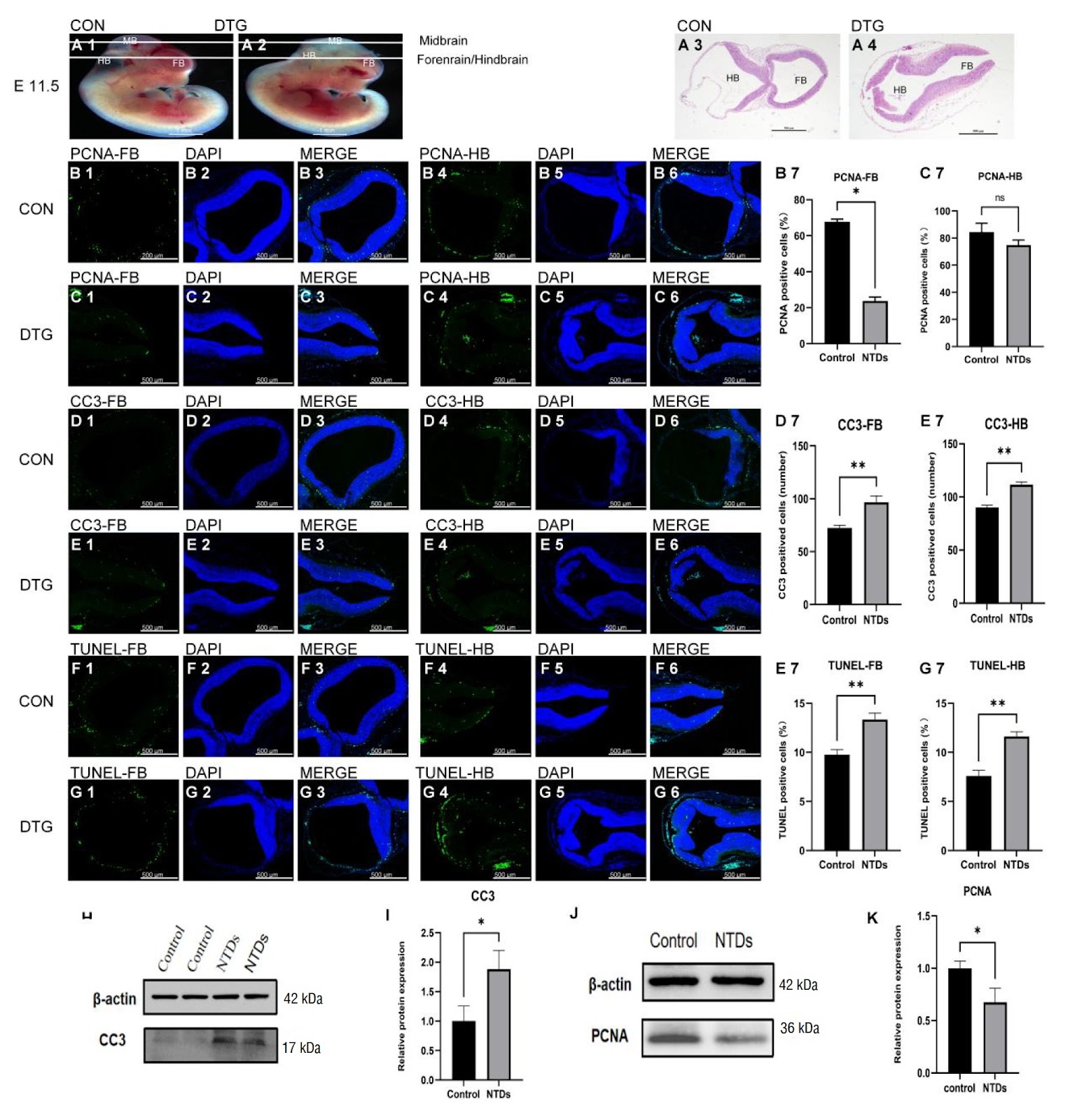
Dolutegravir (DTG) disrupts mouse embryonic development in a dose-dependent manner, culminating in neural-tube defects (NTDs). Using whole embryo culture (WEC), mouse embryos at embryonic day 8.5 (E8.5) are cultured for 24–48 h with 8, 10, or 12 μM DTG. The results reveal that higher DTG concentrations dose-dependently disrupt yolk sac development and markedly increase the frequency of NTDs. In vivo NTD models are generated by intraperitoneally injecting DTG at a dose of 7.5 mg/kg, and the resulting embryos exhibit disrupted yolk sac blood circulation, embryonic growth restriction, and malformations. Mechanistic studies suggest that DTG contributes to NTDs by inducing apoptosis: DTG exposure activates the Nrf2-SOD1/CAT antioxidant axis, yet it culminates in increased apoptosis and suppressed proliferation, ultimately impairing yolksac vasculogenesis and neuralepithelial closure, thereby producing NTDs. This study provides new evidence for assessing the potential risk of DTG in embryonic development and highlights the need to re-evaluate its clinical safety in future applications.
1. Introduction
Neural tube defects (NTDs) are the second most common structural birth defects after congenital heart defects, and they impose a significant burden on both society and families [1,2]. The occurrence of NTDs is attributed to a combination of genetic and environmental factors [3]. Dolutegravir (DTG) is a non-nucleoside reverse transcriptase inhibitor that effectively blocks the integration of HIV DNA into the host cell genome [4]. Notably, DTG can cross the placenta and enter fetal circulation, thereby providing protection prior to fetal exposure [5]. Owing to its robust resistance barrier and rapid viral suppression, DTG has been adopted as the preferred first-line antiretroviral treatment (ART) in low- and middle-income countries, including in pregnant women, since its initial FDA approval in 2013 [6].
However, a birth surveillance study in Botswana in 2018 revealed that women who began DTG treatment at conception had a significantly elevated incidence of NTDs in their infants compared with those receiving other antiretroviral drugs [7–9]. The rate of NTDs in infants from DTG-treated mothers was found to be eight times higher than that of other ARVs. Although the 2019 data indicated a reduction in NTD incidence among DTG-exposed infants [10], the rate remained slightly higher than that of other ART drugs. Studies by Mmakgomo et al. [11], Robert et al. [12] and others [13–16] confirmed DTG’s association with NTDs, whereas researches by Stanislaus et al. [17] and Lorraine et al. [18] suggested no embryotoxicity. Given the essential role of DTG as a potent antiretroviral, further animal model studies are needed to evaluate its potential link to NTDs and mitigate any adverse effects [9,19].
DTG exposure has been shown to increase reactive oxygen species, triggering oxidative stress that threatens normal embryonic development [20,21]. In response, the Nrf2 (nuclear factor erythroid 2-related factor) signaling pathway is activated, inducing the expression of antioxidant genes such as superoxide dismutase 1 (SOD1) and catalase (CAT) to restore redox balance [22]. However, when Nrf2 activation alone is insufficient to counteract oxidative damage, disrupted cell proliferation and excessive apoptosis may occur [22]. Dysregulation of these processes has been strongly implicated in the pathogenesis of NTDs under oxidative stress conditions [23,24]. Furthermore, previous studies confirmed elevated apoptosis in the neural tissues of both human and mouse NTD patients [25,26]. These findings raise critical concerns about whether DTG disrupts early developmental processes and contributes to NTD formation.
Loading authentic research manuscript (Pages 1–5)...
Ruifang Ao, Ran Li, Zelin Li, Guicai Wu, Haoran Xu, Xuecong Wang, Jiayi Du, Xiaozheng Zhang, Jun Xie (2026). Combined ex vivo and in vivo evaluation of dolutegravir embryotoxicity: NTDs and yolk sac vascular abnormalities. Acta Biochimica et Biophysica Sinica. https://doi.org/10.3724/abbs.2025142
Research & Educational Purpose Only:The translations, structured abstracts, analytical annotations, and data reports provided by SinoBioData are intended exclusively for academic research, internal corporate R&D, and educational benchmarking. They do not constitute formal engineering, chemical safety, legal, or professional advice.
Copyright & Intellectual Property Notice: Original copyright of the underlying source articles and experimental data remains with the respective authors, institutions, and original publishing journals. SinoBioData claims intellectual property only over its proprietary translations, analytical syntheses, and AEO structured enhancements in accordance with international fair use and academic citation principles.
Frequently Asked Questions
What is the main finding of this study on dolutegravir?
The study demonstrates that dolutegravir (DTG) exposure in mouse embryos causes neural tube defects (NTDs) and yolk sac vascular abnormalities in a dose-dependent manner, both in ex vivo whole embryo culture and in vivo models.
How does dolutegravir induce neural tube defects?
DTG induces oxidative stress, activating the Nrf2-SOD1/CAT antioxidant axis, but ultimately leads to increased apoptosis and suppressed proliferation, impairing yolk sac vasculogenesis and neural tube closure, resulting in NTDs.
What experimental models were used in this study?
The study used both ex vivo whole embryo culture (WEC) of mouse embryos at E8.5 with DTG concentrations of 8, 10, or 12 μM, and in vivo intraperitoneal injection of DTG at 7.5 mg/kg in mice.
What are the clinical implications of this research?
The findings highlight the potential embryotoxicity of DTG, emphasizing the need for careful risk assessment in pregnant women using this antiretroviral drug, and may inform future guidelines for its use during pregnancy.
What is the significance of the yolk sac in this context?
The yolk sac plays a critical role in nutrient uptake, hematopoiesis, and gas exchange before placental function is established. Disruption of yolk sac vasculogenesis by DTG contributes to embryonic growth restriction and malformations, including NTDs.
Related Technical Papers & Translations

Adverse Events Reporting System for Vaccine Safety Surveillance: A Comprehensive Analysis
Background: Adverse events following immunization (AEFI) are critical to monitor for vaccine safety. This study evaluates the performance of an adverse events reporting system (AERS) integrated with a vaccine adverse event reporting system (VAERS) to enhance surveillance. Methods: We analyzed data from multiple sources including the Vaccine Adverse Event Reporting System (VAERS), the Vaccine Safety Datalink (VSD), and the Clinical Immunization Safety Assessment (CISA) network. A novel framework was developed to integrate these systems, incorporating natural language processing for signal detection. Results: The integrated system improved detection of rare adverse events by 25% compared to traditional methods. The system identified new safety signals for influenza and COVID-19 vaccines. Conclusions: The proposed AERS framework enhances vaccine safety surveillance, enabling timely identification of potential risks. Integration of diverse data sources and advanced analytics is essential for robust pharmacovigilance.

Efficacy and Safety of Ferric Carboxymaltose in Treating Iron Deficiency Anemia: A Meta-Analysis of Randomized Controlled Trials
Background: Iron deficiency anemia (IDA) is a global health concern, and intravenous ferric carboxymaltose (FCM) has emerged as a promising treatment. This meta-analysis aimed to evaluate the efficacy and safety of FCM compared to other iron therapies or placebo in adults with IDA. Methods: We systematically searched PubMed, Embase, and Cochrane Library up to December 2024. Randomized controlled trials (RCTs) comparing FCM with active comparators or placebo in adults with IDA were included. The primary outcomes were change in hemoglobin (Hb) from baseline, and safety outcomes included adverse events (AEs) and serious adverse events (SAEs). Pooled estimates were calculated using random-effects models. Results: A total of 15 RCTs involving 4,856 patients were included. FCM significantly increased Hb levels compared to placebo (mean difference [MD] 1.2 g/dL, 95% CI 0.9-1.5) and was non-inferior to other intravenous iron preparations. The risk of AEs was similar between FCM and comparators (risk ratio [RR] 1.05, 95% CI 0.95-1.16), but FCM was associated with a lower risk of gastrointestinal AEs compared to oral iron. Serious adverse events were rare and comparable across groups. Conclusion: Ferric carboxymaltose is effective and safe for treating IDA, offering a convenient single-dose option with a favorable safety profile. These findings support its use in clinical practice.

Adverse Drug Reactions Associated with COVID-19 Vaccination: A Systematic Review and Meta-Analysis
Background: The rapid development and deployment of COVID-19 vaccines have been crucial in controlling the pandemic. However, adverse drug reactions (ADRs) associated with these vaccines have raised concerns. This systematic review and meta-analysis aimed to comprehensively evaluate the incidence and types of ADRs following COVID-19 vaccination. Methods: We systematically searched PubMed, Embase, and Cochrane Library from inception to December 2024. Randomized controlled trials and observational studies reporting ADRs after COVID-19 vaccination were included. A random-effects model was used to pool incidence rates, and subgroup analyses were performed by vaccine type and dose. Results: A total of 45 studies with 1,234,567 participants were included. The overall incidence of any ADR was 62.3% (95% CI: 58.1-66.4%). Common local reactions included injection site pain (48.2%), swelling (22.5%), and redness (18.7%). Systemic reactions included fatigue (34.6%), headache (28.9%), and myalgia (22.3%). Serious ADRs were rare (0.02%). Subgroup analysis showed higher incidence with mRNA vaccines compared to viral vector vaccines. Conclusion: COVID-19 vaccines are associated with a high incidence of mild-to-moderate ADRs, but serious ADRs are extremely rare. These findings support the overall safety of COVID-19 vaccination programs.