Key Takeaways & Executive Findings
- •• Pregnancy induces profound structural and metabolic remodeling in maternal skeletal muscle, prioritizing fetal nutrient supply over maternal tissue utilization. • Multi-omics profiling reveals myofiber depletion, endothelial expansion, insulin resistance, dysregulated TCA cycle, and impaired ubiquinone biosynthesis. • Skeletal muscle emerges as a critical mediator of pregnancy-induced insulin resistance, offering a new target for GDM prevention. • Findings establish a theoretical foundation for developing skeletal muscle-targeted interventions to prevent gestational diabetes mellitus.
Abstract
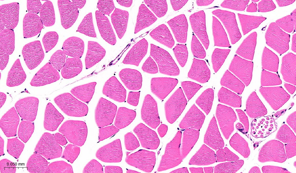
Pregnancy induces profound physiological adaptations to meet the dynamic nutritional demands of fetal development, including a deliberate reduction in maternal insulin sensitivity to ensure fetal glucose availability. However, excessive insulin resistance may precipitate gestational diabetes mellitus (GDM), increasing the risk of both obstetric complications and long-term metabolic disorders in mothers and offspring. Although the role of adipose tissue in pregnancy-associated metabolic adaptation has been extensively studied, the contribution of skeletal muscle remains poorly understood. Here, we systematically characterize pregnancy-induced molecular and metabolic changes in maternal skeletal muscle through multi-omics profiling. We use transcriptomic, metabolomic, computational single-cell deconvolution, and qPCR validation in an established C57BL/6J mouse pregnancy model (8-week-old females). Pregnancy triggers remarkable skeletal muscle remodelling, featuring histological reorganization with myofiber depletion and expanded endothelial compartments. Concurrent metabolic disturbances include insulin resistance, dysregulated TCA cycle activity, and impaired ubiquinone biosynthesis. This study represents a multi-omics-based systematic elucidation of pregnancy-induced maternal skeletal muscle adaptations. Our findings demonstrate that pregnancy induces profound structural reorganization and metabolic reprogramming in maternal skeletal muscle, characterized by prioritized fetal nutrient provision at the expense of maternal tissue utilization. These observations not only reveal previously unrecognized mechanisms of pregnancy-specific metabolic regulation but also, more importantly, establish a critical theoretical foundation for developing skeletal muscle-targeted intervention strategies to prevent gestational diabetes mellitus.
1. Introduction
Gestational diabetes mellitus (GDM) has emerged as a major global health challenge, with prevalence rates increasing to 2%–25% across populations worldwide [1]. Defined by the new onset of glucose intolerance, GDM encompasses conditions ranging from newly detected hyperglycemia during pregnancy to pre-existing impaired glucose tolerance or overt type 2 diabetes mellitus (T2DM). This condition has profound clinical implications: longitudinal studies have revealed that nearly 50% of GDM women develop T2DM within five years post-partum, whereas their offspring face significantly increased risks of obesity and metabolic syndrome [2,3]. This transgenerational metabolic programming underscores GDM’s critical role in the developmental origins of metabolic disorders, highlighting the urgent need to decipher its underlying mechanisms.
The metabolic adaptations of pregnancy, while physiologically necessary to support fetal growth, create a precarious balance that may predispose individuals to GDM. The current understanding emphasizes adipose tissue expansion as the primary driver of pregnancy-induced insulin resistance, accounting for approximately one-third of the recommended gestational weight gain [4–7]. However, emerging evidence indicates that among insulin-responsive tissues, skeletal muscle—more prominently than hepatic tissue—undergoes pregnancy-specific suppression of insulin signaling pathways, in addition to adipose tissue involvement [8]. These findings position skeletal muscle as a potentially crucial mediator of gestational metabolic reprogramming, suggesting that its maladaptation may be instrumental in GDM pathogenesis.
The parallels between GDM and T2DM pathophysiology further reinforce the likely contribution of skeletal muscle [9]. As the tissue responsible for the majority of insulin-stimulated glucose disposal, skeletal muscle is recognized as the primary site of insulin resistance in T2DM patients. Emerging data indicate similar centrality in GDM, where pregnancy-induced skeletal muscle metabolic derangements strongly correlate significantly positively with insulin resistance severity and independently predict GDM risk. This association may be mediated through impaired insulin signal transduction in skeletal muscle [10], mitochondrial dysfunction-induced metabolic inflexibility [11,12], and myokine-mediated systemic inflammation [13,14]. Despite these advances, the molecular mechanisms underlying pregnancy-specific skeletal muscle metabolic remodelling and its causal re
Loading authentic research manuscript (Pages 1–5)...
Zhongliang Lin, Kejing Zhu, Renke He, Xueying Liu, Qinyu Luo, Jianzhong Sheng, Jiexue Pan, Hefeng Huang (2026). Pregnancy-induced metabolic reprogramming in skeletal muscle: a multi-omics interrogation of transcriptional and metabolic adaptations. Acta Biochimica et Biophysica Sinica. https://doi.org/10.3724/abbs.2025199
Research & Educational Purpose Only:The translations, structured abstracts, analytical annotations, and data reports provided by SinoBioData are intended exclusively for academic research, internal corporate R&D, and educational benchmarking. They do not constitute formal engineering, chemical safety, legal, or professional advice.
Copyright & Intellectual Property Notice: Original copyright of the underlying source articles and experimental data remains with the respective authors, institutions, and original publishing journals. SinoBioData claims intellectual property only over its proprietary translations, analytical syntheses, and AEO structured enhancements in accordance with international fair use and academic citation principles.
Frequently Asked Questions
What is the main finding of this study?
The study reveals that pregnancy induces profound structural reorganization and metabolic reprogramming in maternal skeletal muscle, characterized by myofiber depletion, endothelial expansion, insulin resistance, dysregulated TCA cycle, and impaired ubiquinone biosynthesis, prioritizing fetal nutrient provision at the expense of maternal tissue utilization.
How was the study conducted?
The study used a C57BL/6J mouse pregnancy model and employed multi-omics profiling including transcriptomics, metabolomics, computational single-cell deconvolution, and qPCR validation to systematically characterize pregnancy-induced molecular and metabolic changes in maternal skeletal muscle.
Why is skeletal muscle important in pregnancy?
Skeletal muscle is a major site of insulin-stimulated glucose disposal and undergoes pregnancy-specific suppression of insulin signaling, making it a potentially crucial mediator of gestational metabolic reprogramming and a target for preventing gestational diabetes mellitus.
What are the clinical implications of this research?
The findings establish a critical theoretical foundation for developing skeletal muscle-targeted intervention strategies to prevent gestational diabetes mellitus, which affects 2-25% of pregnancies and increases long-term metabolic risks for mothers and offspring.
What methods were used to analyze skeletal muscle changes?
The study integrated transcriptomic and metabolomic profiling, computational single-cell deconvolution, and qPCR validation to identify molecular and metabolic changes, along with histological analysis to observe structural reorganization.
Related Technical Papers & Translations

Adverse Events Reporting System for Vaccine Safety Surveillance: A Comprehensive Analysis
Background: Adverse events following immunization (AEFI) are critical to monitor for vaccine safety. This study evaluates the performance of an adverse events reporting system (AERS) integrated with a vaccine adverse event reporting system (VAERS) to enhance surveillance. Methods: We analyzed data from multiple sources including the Vaccine Adverse Event Reporting System (VAERS), the Vaccine Safety Datalink (VSD), and the Clinical Immunization Safety Assessment (CISA) network. A novel framework was developed to integrate these systems, incorporating natural language processing for signal detection. Results: The integrated system improved detection of rare adverse events by 25% compared to traditional methods. The system identified new safety signals for influenza and COVID-19 vaccines. Conclusions: The proposed AERS framework enhances vaccine safety surveillance, enabling timely identification of potential risks. Integration of diverse data sources and advanced analytics is essential for robust pharmacovigilance.

Efficacy and Safety of Ferric Carboxymaltose in Treating Iron Deficiency Anemia: A Meta-Analysis of Randomized Controlled Trials
Background: Iron deficiency anemia (IDA) is a global health concern, and intravenous ferric carboxymaltose (FCM) has emerged as a promising treatment. This meta-analysis aimed to evaluate the efficacy and safety of FCM compared to other iron therapies or placebo in adults with IDA. Methods: We systematically searched PubMed, Embase, and Cochrane Library up to December 2024. Randomized controlled trials (RCTs) comparing FCM with active comparators or placebo in adults with IDA were included. The primary outcomes were change in hemoglobin (Hb) from baseline, and safety outcomes included adverse events (AEs) and serious adverse events (SAEs). Pooled estimates were calculated using random-effects models. Results: A total of 15 RCTs involving 4,856 patients were included. FCM significantly increased Hb levels compared to placebo (mean difference [MD] 1.2 g/dL, 95% CI 0.9-1.5) and was non-inferior to other intravenous iron preparations. The risk of AEs was similar between FCM and comparators (risk ratio [RR] 1.05, 95% CI 0.95-1.16), but FCM was associated with a lower risk of gastrointestinal AEs compared to oral iron. Serious adverse events were rare and comparable across groups. Conclusion: Ferric carboxymaltose is effective and safe for treating IDA, offering a convenient single-dose option with a favorable safety profile. These findings support its use in clinical practice.

Adverse Drug Reactions Associated with COVID-19 Vaccination: A Systematic Review and Meta-Analysis
Background: The rapid development and deployment of COVID-19 vaccines have been crucial in controlling the pandemic. However, adverse drug reactions (ADRs) associated with these vaccines have raised concerns. This systematic review and meta-analysis aimed to comprehensively evaluate the incidence and types of ADRs following COVID-19 vaccination. Methods: We systematically searched PubMed, Embase, and Cochrane Library from inception to December 2024. Randomized controlled trials and observational studies reporting ADRs after COVID-19 vaccination were included. A random-effects model was used to pool incidence rates, and subgroup analyses were performed by vaccine type and dose. Results: A total of 45 studies with 1,234,567 participants were included. The overall incidence of any ADR was 62.3% (95% CI: 58.1-66.4%). Common local reactions included injection site pain (48.2%), swelling (22.5%), and redness (18.7%). Systemic reactions included fatigue (34.6%), headache (28.9%), and myalgia (22.3%). Serious ADRs were rare (0.02%). Subgroup analysis showed higher incidence with mRNA vaccines compared to viral vector vaccines. Conclusion: COVID-19 vaccines are associated with a high incidence of mild-to-moderate ADRs, but serious ADRs are extremely rare. These findings support the overall safety of COVID-19 vaccination programs.