Key Takeaways & Executive Findings
- •• Cryo-EM structure of cyanophage Pam5 TerS reveals a nonameric ring with a spiral architecture, providing the first structural insight into TerS from cyanophages. • Pam5 TerS recognizes a specific 21-bp DNA sequence via its N-terminal HTH domain, whereas Pam1 TerS binds DNA sequence-independently, demonstrating two distinct DNA recognition strategies. • The study highlights the evolutionary plasticity of viral genome packaging mechanisms, even among phages infecting the same host. • These findings advance our understanding of TerS-mediated DNA recognition and may inform the design of phage-based therapies or biotechnological applications.
Abstract
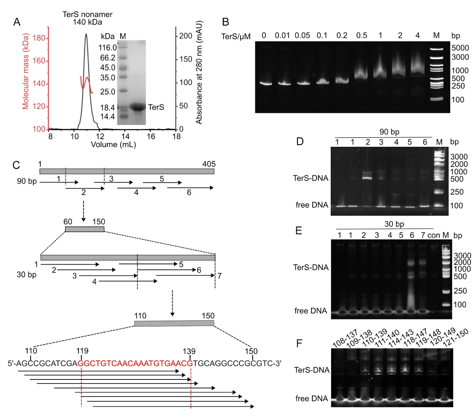
Efficient genome packaging is a critical step in the phage life cycle, directly influencing the viral maturation and infectivity. In tailed phages, this process is driven by a packaging motor composed of a portal protein and a terminase complex. The terminase complex usually consists of a large subunit (TerL) and a small subunit (TerS), which cooperate to recognize, cleave, and translocate genomic DNA into the capsid. However, due to the remarkable diversity and complexity of phage packaging systems, the molecular mechanisms governing TerS-mediated DNA recognition remain poorly understood. Here, we report the 3.51 Å cryo-electron microscopy structure of the TerS from the short-tailed cyanophage Pam5, which infects the host Pseudanabaena mucicola Chao 1806. Pam5 TerS assembles into a nonameric ring with a radially symmetric spiral architecture. Biochemical assays show that Pam5 TerS recognizes the genomic DNA via a specific interaction between the N-terminal helix-turn-helix (HTH) domain of TerS and a 21-bp DNA sequence within the terS gene. In contrast, the TerS from another short-tailed cyanophage, Pam1, which infects the same host, binds to DNA in a sequence-independent manner. These findings reveal that cyanophages, even infecting the same host, could adopt two distinct DNA recognition strategies: HTH-mediated sequence-dependent or sequence-independent modes. This work provides structural and mechanistic insights into the diverse DNA-recognition strategies of TerS and advances our understanding of the evolutionary plasticity of viral genome packaging mechanisms.
1. Introduction
Efficient genome packaging is a pivotal event in the bacteriophage life cycle, directly determining the viral maturation and infectivity [1]. In tailed bacteriophages, genome packaging is powered by a highly efficient molecular motor composed of a portal protein and a terminase complex capable of generating forces exceeding 50 pN [2]. The terminase complex typically comprises two components, the large subunit (TerL) and the small subunit (TerS). They usually assemble into ring-like oligomers that interact with each other, although their precise stoichiometry within the complex remains unclear [3].
The phage capsid contains a specialized vertex occupied by a dodecameric portal that plays essential roles in procapsid assembly, genome packaging, and viral maturation [4]. During DNA packaging, the terminase complex docks onto the portal, enabling the translocation of genomic DNA through the central channel of the portal into the capsid [5,6]. Once genome packaging is completed, the terminase complex dissociates from the portal, and the tail apparatus attaches to the portal vertex, thereby generating the mature virions [3]. TerL functions as the catalytic core of the packaging machinery, which cleaves the genomic DNA and drives its translocation into the capsid [7]. TerL comprises an N-terminal ATPase domain and a C-terminal nuclease domain, which are linked by a flexible linker [8]. In contrast, TerS was proposed to recognize the genomic DNA that ensures specific initiation of genome packaging [9]. The TerS proteins typically assemble into octameric to dodecameric rings [10,11], which transiently associate with TerL and modulate its ATPase and nuclease activities [11–13]. Despite a similar oligomeric architecture, TerS proteins display substantial sequence diversity, and in many phages, it remains unclear whether a terS gene is present at all [14,15].
Previous studies have suggested that phages have evolved distinct genome packaging strategies, such as the cos and pac (headful) modes, which primarily differ in how the terminase recognizes and cleaves genomic DNA [16]. In cos phages such as λ and HK97, TerS specifically recognizes cohesive-end sequences, termed cos sites, enabling TerL to introduce precise nicks that ensure the incorporation of a single complete unit of genome per capsid [17,18]. Structural studies have revealed that HK97 TerS forms an asymmetric complex with DNA, in which DNA bends along one side of the central channel and interacts with the α-helices of two neighboring subunits [19]. In contrast, the pac phages initiate genome packaging at a specific pac site but terminate in a nonspecific manner when the capsid interior is fully occupied, yielding genomes that are ~110% of the unit length [16]. The pac site serves as the recognition element for TerS; for instance, it corresponds to a 22-bp asymmetric sequence located within the terS gene in phage P22 [13,20]. In contrast, the pac site in SPP1 comprises multiple sequence motifs surrounding the cleavage origin [21,22]. The interactions between TerS and DNA in pac phages are relatively weak and difficult to characterize in vitro. For instance, the phage P74-26 TerS exhibits a low DNA binding affinity, p
Loading authentic research manuscript (Pages 1–5)...
De-Qin Dong, Feng Yang, Kang Du, Kang Xu, Wen-Bin Cheng, Yuxing Chen, Cong-Zhao Zhou, Yong-Liang Jiang (2026). Structural and functional insights into the distinct DNA recognition mechanisms of the terminase small subunit TerS from cyanophages. Acta Biochimica et Biophysica Sinica. https://doi.org/10.3724/abbs.2026042
Research & Educational Purpose Only:The translations, structured abstracts, analytical annotations, and data reports provided by SinoBioData are intended exclusively for academic research, internal corporate R&D, and educational benchmarking. They do not constitute formal engineering, chemical safety, legal, or professional advice.
Copyright & Intellectual Property Notice: Original copyright of the underlying source articles and experimental data remains with the respective authors, institutions, and original publishing journals. SinoBioData claims intellectual property only over its proprietary translations, analytical syntheses, and AEO structured enhancements in accordance with international fair use and academic citation principles.
Frequently Asked Questions
What is the main finding of this study?
The study reveals that cyanophages infecting the same host can adopt two distinct DNA recognition strategies: one using a sequence-dependent HTH-mediated mechanism (Pam5) and the other using a sequence-independent mode (Pam1).
What technique was used to determine the structure of Pam5 TerS?
Cryo-electron microscopy (cryo-EM) was used to determine the 3.51 Å structure of Pam5 TerS.
How does Pam5 TerS recognize DNA?
Pam5 TerS recognizes a specific 21-bp DNA sequence within the terS gene via its N-terminal helix-turn-helix (HTH) domain.
What is the oligomeric state of Pam5 TerS?
Pam5 TerS assembles into a nonameric ring with a radially symmetric spiral architecture.
Why is this study significant?
It provides structural and mechanistic insights into the diverse DNA-recognition strategies of TerS, advancing our understanding of the evolutionary plasticity of viral genome packaging mechanisms.
Related Technical Papers & Translations

Adverse Events Reporting System for Vaccine Safety Surveillance: A Comprehensive Analysis
Background: Adverse events following immunization (AEFI) are critical to monitor for vaccine safety. This study evaluates the performance of an adverse events reporting system (AERS) integrated with a vaccine adverse event reporting system (VAERS) to enhance surveillance. Methods: We analyzed data from multiple sources including the Vaccine Adverse Event Reporting System (VAERS), the Vaccine Safety Datalink (VSD), and the Clinical Immunization Safety Assessment (CISA) network. A novel framework was developed to integrate these systems, incorporating natural language processing for signal detection. Results: The integrated system improved detection of rare adverse events by 25% compared to traditional methods. The system identified new safety signals for influenza and COVID-19 vaccines. Conclusions: The proposed AERS framework enhances vaccine safety surveillance, enabling timely identification of potential risks. Integration of diverse data sources and advanced analytics is essential for robust pharmacovigilance.

Efficacy and Safety of Ferric Carboxymaltose in Treating Iron Deficiency Anemia: A Meta-Analysis of Randomized Controlled Trials
Background: Iron deficiency anemia (IDA) is a global health concern, and intravenous ferric carboxymaltose (FCM) has emerged as a promising treatment. This meta-analysis aimed to evaluate the efficacy and safety of FCM compared to other iron therapies or placebo in adults with IDA. Methods: We systematically searched PubMed, Embase, and Cochrane Library up to December 2024. Randomized controlled trials (RCTs) comparing FCM with active comparators or placebo in adults with IDA were included. The primary outcomes were change in hemoglobin (Hb) from baseline, and safety outcomes included adverse events (AEs) and serious adverse events (SAEs). Pooled estimates were calculated using random-effects models. Results: A total of 15 RCTs involving 4,856 patients were included. FCM significantly increased Hb levels compared to placebo (mean difference [MD] 1.2 g/dL, 95% CI 0.9-1.5) and was non-inferior to other intravenous iron preparations. The risk of AEs was similar between FCM and comparators (risk ratio [RR] 1.05, 95% CI 0.95-1.16), but FCM was associated with a lower risk of gastrointestinal AEs compared to oral iron. Serious adverse events were rare and comparable across groups. Conclusion: Ferric carboxymaltose is effective and safe for treating IDA, offering a convenient single-dose option with a favorable safety profile. These findings support its use in clinical practice.

Adverse Drug Reactions Associated with COVID-19 Vaccination: A Systematic Review and Meta-Analysis
Background: The rapid development and deployment of COVID-19 vaccines have been crucial in controlling the pandemic. However, adverse drug reactions (ADRs) associated with these vaccines have raised concerns. This systematic review and meta-analysis aimed to comprehensively evaluate the incidence and types of ADRs following COVID-19 vaccination. Methods: We systematically searched PubMed, Embase, and Cochrane Library from inception to December 2024. Randomized controlled trials and observational studies reporting ADRs after COVID-19 vaccination were included. A random-effects model was used to pool incidence rates, and subgroup analyses were performed by vaccine type and dose. Results: A total of 45 studies with 1,234,567 participants were included. The overall incidence of any ADR was 62.3% (95% CI: 58.1-66.4%). Common local reactions included injection site pain (48.2%), swelling (22.5%), and redness (18.7%). Systemic reactions included fatigue (34.6%), headache (28.9%), and myalgia (22.3%). Serious ADRs were rare (0.02%). Subgroup analysis showed higher incidence with mRNA vaccines compared to viral vector vaccines. Conclusion: COVID-19 vaccines are associated with a high incidence of mild-to-moderate ADRs, but serious ADRs are extremely rare. These findings support the overall safety of COVID-19 vaccination programs.