Key Takeaways & Executive Findings
- •• FGF8 promotes lipid droplet accumulation in chondrocytes by upregulating perilipin1 (Plin1) expression. • FGF8 activates the FGFR1/p-p38 signaling axis to enhance lipid droplet accumulation. • siRNA and inhibitor experiments confirm the critical role of the FGFR1/p38 pathway in FGF8-induced lipid droplet formation. • These findings provide new insights into cartilage lipid metabolism and potential therapeutic targets for cartilage-related diseases.
Abstract
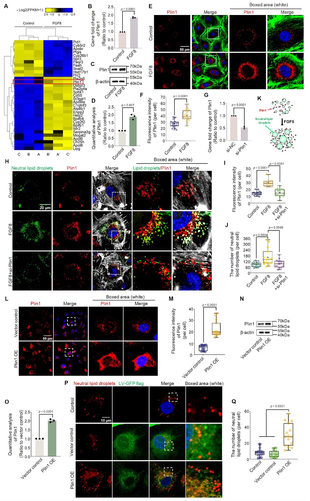
Chondrocytes store lipids in the form of lipid droplets (LDs) and maintain cartilage lipid metabolic homeostasis by consuming or regenerating LDs. This modulation is largely mediated by a series of biochemical factors. Fibroblast growth factor 8 (FGF8) is one of the most important factors involved in the proliferation, differentiation, and migration of chondrocytes and has attracted increasing attention in the physiology and pathology of cartilage. However, the effect of FGF8 on LD accumulation in chondrocytes remains unclear. This study aims to elucidate the role of FGF8 in LDs and explore the underlying biomechanism involved. The results reveal that FGF8 promotes LD accumulation in chondrocytes by upregulating perilipin1 (Plin1) expression. FGF8 activates the cytoplasmic p-p38 signaling pathway via fibroblast growth factor receptor 1 (FGFR1) to increase LD accumulation in chondrocytes. Subsequent experiments with siRNAs and specific inhibitors further confirm the importance of the FGFR1/p38 axis for LD accumulation in chondrocytes exposed to FGF8. The results increase our understanding of the role of FGF8 in the lipid metabolic homeostasis of chondrocytes and provide insights into the physiology and pathology of cartilage.
1. Introduction
The lipid droplet (LD), a principal hub organelle for intracellular lipid storage, consists of a neutral lipid core enveloped by a phospholipid monolayer membrane [1–3]. LD biogenesis begins mostly in the endoplasmic reticulum (ER), the main organelle involved in neutral lipid synthesis [4]. The first step in LD biosynthesis is the synthesis of neutral lipids such as triacylglycerols (TGs) and cholesterol esters (CEs) in the ER [4,5]. These neutral lipids are dispersed in the ER at low concentrations. With continuous neutral lipid synthesis, biophysical processes result in the formation of a neutral lipid lens (also called the oil lens) [5]. A widely recognized model states that neutral lipids nucleate an oil phase that minimizes the entropy costs involved in disrupting the ER bilayer membrane [1,4,5]. Subsequently, the oil phase transitions to the oil lens. The oil lens accommodates more neutral lipids and promotes the aggregation of neutral lipids to form LDs [5]. After being released into the cytoplasm, these nascent LDs gradually develop into larger mature LDs by storing more lipids or merging with each other [3–5]. In addition, LDs interact with other organelles, such as mitochondria and lysosomes, to regulate their metabolism and function. For example, Meng et al. [6] reported that phosphofructokinase, a glycolytic enzyme, promotes LD-mitochondrion tethering to increase β-oxidation. Menon et al. [7] reported that ARL8B, a GTPase, promotes LD-lysosome contact and induces lysosomal lipolysis of LDs. More importantly, lipophagy, an autophagy process that specifically targets LDs, plays an important role in maintaining the cellular energy supply and alleviating metabolic diseases such as atherosclerosis [8]. Lin et al. [9] reported that the recovery of lipophagy via the inhibition of PPAR/PI3K/AKT signaling relieves atherosclerosis-related symptoms, including lipid accumulation, apoptosis, and inflammation.
The primary function of intracellular LDs is to serve as storage reservoirs for neutral lipids and supply essential lipid precursors whenever the cellular lipid level decreases [10]. These neutral lipids can be used for cell energy supply through mitochondrial β-oxidation and for structural composition, such as lipid membrane expansion [10]. In addition, LDs can assist cells in preventing lipid toxicity [11]. Free lipids such as fatty acids can act as detergents to disrupt the cell membrane structure. Synthesizing fatty acids into triglycerides and storing them in LDs effectively prevents intracellular lipid toxicity. However, despite the numerous positive effects of LDs within cells, excessive LD accumulation can result in the development of various diseases, such as hepatic steatosis [10,11]. As organelles with important functions in cells, LDs are also regulated by a variety of proteins [12]. These proteins, including a series of enzymes that regulate the dynamic process of LD biogenesis and linker proteins that connect LDs to other organelles, regulate LD activities [12]. These proteins are roughly divided into two types: integral and peripheral proteins. Integral proteins are inserted into the monolayer membrane of LDs and can recruit peripheral proteins to LDs [12]. Integral proteins play vital roles in the generation, maintenance, and lipolysis of LDs and are composed of two functional subclasses according to their trafficking pathways. One is a Class I LD protein that originates in the ER [12]. These proteins are then transferred to
Loading authentic research manuscript (Pages 1–5)...
Minglei Huang, Haoran Chen, Jieya Wei, Caixia Pi, Mengmeng Duan, Xiaohua Pu, Zhixing Niu, Siqun Xu, Shasha Tu, Sijun Liu, Jiazhou Li, Li Zhang, Yang Liu, Hao Chen, Chunming Xu, Jing Xie (2026). FGF8 promotes lipid droplet accumulation via the FGFR1/p-p38 axis in chondrocytes. Acta Biochimica et Biophysica Sinica. https://doi.org/10.3724/abbs.2025075
Research & Educational Purpose Only:The translations, structured abstracts, analytical annotations, and data reports provided by SinoBioData are intended exclusively for academic research, internal corporate R&D, and educational benchmarking. They do not constitute formal engineering, chemical safety, legal, or professional advice.
Copyright & Intellectual Property Notice: Original copyright of the underlying source articles and experimental data remains with the respective authors, institutions, and original publishing journals. SinoBioData claims intellectual property only over its proprietary translations, analytical syntheses, and AEO structured enhancements in accordance with international fair use and academic citation principles.
Frequently Asked Questions
What is the role of FGF8 in chondrocytes?
FGF8 promotes lipid droplet accumulation in chondrocytes by upregulating perilipin1 (Plin1) expression and activating the FGFR1/p-p38 signaling axis.
How does FGF8 affect lipid droplet accumulation?
FGF8 activates the FGFR1/p-p38 pathway, which increases the expression of Plin1, a protein that coats lipid droplets and promotes their accumulation.
What is the significance of the FGFR1/p38 axis in this study?
The FGFR1/p38 axis is critical for FGF8-induced lipid droplet accumulation, as confirmed by siRNA and inhibitor experiments that blocked this pathway and reduced lipid droplet formation.
What are the potential implications of this research?
This research provides insights into cartilage lipid metabolism and may inform therapeutic strategies for cartilage-related diseases where lipid homeostasis is disrupted.
What methods were used to confirm the findings?
The study used siRNA knockdown and specific inhibitors to validate the involvement of FGFR1 and p38 in FGF8-mediated lipid droplet accumulation.
Related Technical Papers & Translations

Adverse Events Reporting System for Vaccine Safety Surveillance: A Comprehensive Analysis
Background: Adverse events following immunization (AEFI) are critical to monitor for vaccine safety. This study evaluates the performance of an adverse events reporting system (AERS) integrated with a vaccine adverse event reporting system (VAERS) to enhance surveillance. Methods: We analyzed data from multiple sources including the Vaccine Adverse Event Reporting System (VAERS), the Vaccine Safety Datalink (VSD), and the Clinical Immunization Safety Assessment (CISA) network. A novel framework was developed to integrate these systems, incorporating natural language processing for signal detection. Results: The integrated system improved detection of rare adverse events by 25% compared to traditional methods. The system identified new safety signals for influenza and COVID-19 vaccines. Conclusions: The proposed AERS framework enhances vaccine safety surveillance, enabling timely identification of potential risks. Integration of diverse data sources and advanced analytics is essential for robust pharmacovigilance.

Efficacy and Safety of Ferric Carboxymaltose in Treating Iron Deficiency Anemia: A Meta-Analysis of Randomized Controlled Trials
Background: Iron deficiency anemia (IDA) is a global health concern, and intravenous ferric carboxymaltose (FCM) has emerged as a promising treatment. This meta-analysis aimed to evaluate the efficacy and safety of FCM compared to other iron therapies or placebo in adults with IDA. Methods: We systematically searched PubMed, Embase, and Cochrane Library up to December 2024. Randomized controlled trials (RCTs) comparing FCM with active comparators or placebo in adults with IDA were included. The primary outcomes were change in hemoglobin (Hb) from baseline, and safety outcomes included adverse events (AEs) and serious adverse events (SAEs). Pooled estimates were calculated using random-effects models. Results: A total of 15 RCTs involving 4,856 patients were included. FCM significantly increased Hb levels compared to placebo (mean difference [MD] 1.2 g/dL, 95% CI 0.9-1.5) and was non-inferior to other intravenous iron preparations. The risk of AEs was similar between FCM and comparators (risk ratio [RR] 1.05, 95% CI 0.95-1.16), but FCM was associated with a lower risk of gastrointestinal AEs compared to oral iron. Serious adverse events were rare and comparable across groups. Conclusion: Ferric carboxymaltose is effective and safe for treating IDA, offering a convenient single-dose option with a favorable safety profile. These findings support its use in clinical practice.

Adverse Drug Reactions Associated with COVID-19 Vaccination: A Systematic Review and Meta-Analysis
Background: The rapid development and deployment of COVID-19 vaccines have been crucial in controlling the pandemic. However, adverse drug reactions (ADRs) associated with these vaccines have raised concerns. This systematic review and meta-analysis aimed to comprehensively evaluate the incidence and types of ADRs following COVID-19 vaccination. Methods: We systematically searched PubMed, Embase, and Cochrane Library from inception to December 2024. Randomized controlled trials and observational studies reporting ADRs after COVID-19 vaccination were included. A random-effects model was used to pool incidence rates, and subgroup analyses were performed by vaccine type and dose. Results: A total of 45 studies with 1,234,567 participants were included. The overall incidence of any ADR was 62.3% (95% CI: 58.1-66.4%). Common local reactions included injection site pain (48.2%), swelling (22.5%), and redness (18.7%). Systemic reactions included fatigue (34.6%), headache (28.9%), and myalgia (22.3%). Serious ADRs were rare (0.02%). Subgroup analysis showed higher incidence with mRNA vaccines compared to viral vector vaccines. Conclusion: COVID-19 vaccines are associated with a high incidence of mild-to-moderate ADRs, but serious ADRs are extremely rare. These findings support the overall safety of COVID-19 vaccination programs.