Key Takeaways & Executive Findings
- •• DNA-PKcs localizes to centromeres during mitosis and is phosphorylated in the nucleus during interphase, linking it to primary cilia dynamics. • Ionizing radiation induces persistent primary cilia formation in senescent tumor cells, which is dependent on DNA-PKcs activity. • Disruption of primary cilia or DNA-PKcs enhances tumor cell radiosensitivity and promotes senescent cell death. • The DNA-PKcs-primary cilia axis represents a potential therapeutic target to overcome radioresistance in cancer treatment.
Abstract
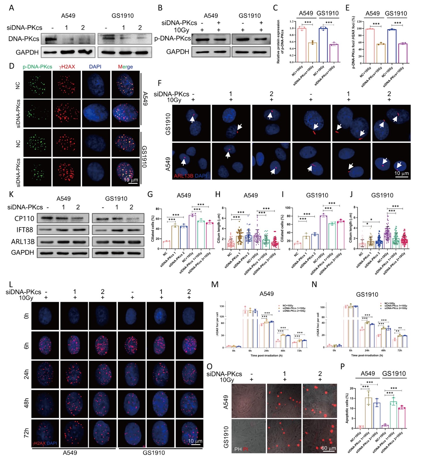
Senescence is a cellular response closely associated with genotoxic stress and plays a critical role in determining cell fate following irradiation exposure. Primary cilia, which are sensory organelles on the cell surface, detect and transmit diverse signaling cues. However, the relationship between primary cilia and senescence in long-term cell fate decisions after ionizing radiation remains poorly understood. Here, we show that the DNA-dependent protein kinase catalytic subunit (DNA-PKcs) colocalizes with centromeres during various stages of mitosis, whereas during interphase, phosphorylated DNA-PKcs (p-DNA-PKcs) is confined to the nucleus in tumor cells. Following irradiation exposure, primary cilia are formed and persistently maintained at high levels in senescent tumor cells. Inhibition of DNA-PKcs enhances primary cilia formation, whereas combined inhibition with siDNA-PKcs and irradiation reduces cilia generation. Moreover, chloral hydrate-induced primary cilia removal results in senescent cell death and decreases p-DNA-PKcs protein expression. Notably, treatment with the apoptosis inducer ABT263 also leads to increased cell death and decreased incidence of primary cilia. Inhibition of either primary cilia or DNA-PKcs further enhances the radiosensitivity of tumor cells. These findings suggest that DNA-PKcs contributes to primary cilia formation after irradiation and plays a critical role in both the induction and maintenance of cellular senescence.
1. Introduction
Cellular senescence refers to stable growth arrest accompanied by an antiapoptotic state that occurs in response to irreparable stress [1,2]. Research has demonstrated that ionizing radiation (IR) can induce cellular senescence, which is common during tumor radiotherapy [3]. Furthermore, cellular senescence triggered by tumor treatments is closely associated with treatment resistance and poor prognosis. Current evidence indicates that activation of the p16-pRB and p53-p21 signaling pathways, mitochondrial dysfunction [4], and cyclic GMP-AMP synthase stimulation are major mechanisms underlying senescence. However, the specific molecular basis of radiation-induced senescence remains incompletely defined. Previous findings demonstrated that Aurora A acts as a key downstream effector of p21 in radiation-induced senescent tumor cells [5] and that its degradation facilitates the formation of primary cilia [6]. These observations suggest that primary cilia may be critical in mediating the cellular senescence induced by IR.
Primary cilia are hair-like, nonmotile organelles that extend from the cell surface [7] and function as central hubs for sensing physical, chemical, and biological cues from the extracellular environment. By transmitting these signals into the cell, the PC plays a critical role in regulating various cellular processes [8]. Structural or functional defects in primary cilia have been implicated in the development and progression of multiple diseases, including cancer [9]. Numerous studies have demonstrated a strong association between primary cilia dynamics and cellular senescence under physiological conditions. Cilium length increases significantly in aged human fibroblasts and in the kidneys and pancreas of aged mice [10,11]. Similar elongation has also been observed in the hippocampal region of aged rats [12,13]. Additionally, silencing of ciliogenesis-related genes in primary cilia can induce senescence in renal epithelial cells from mouse models of cystic kidney disease [14]. Notably, further investigations have indicated that primary cilia contribute to the senescence of tumor cells. For example, persistent primary cilia have been found to induce senescence in human cervical cancer cells [15], and primary cilia play a key role in etoposide-induced senescence in adrenal cortical tumor cells [16]. Although the involvement of primary cilia in cellular senescence has been partially clarified, the precise regulatory mechanisms remain to be fully defined. In normal cells, recent studies have reported a transient increase in cilia formation following irradiation exposure, followed by a gradual decrease, and this transient cilia formation plays an essential role in initiating cellular senescence postirradiation [17]. However, no studies have reported whether primary cilia exhibit similar functions or expression patterns during IR-induced tumor cellular senescence.
Loading authentic research manuscript (Pages 1–5)...
Xiuzhu Liu, Li Wei, Rong Zhang, Jiaxin Chen, Tongshan Zhang, Junrui Hua, Jufang Wang, Jinpeng He, Xiaodong Xie (2026). The DNA-PKcs-primary cilia axis maintains ionizing radiation-induced senescence in tumor cells. Acta Biochimica et Biophysica Sinica. https://doi.org/10.3724/abbs.2025168
Research & Educational Purpose Only:The translations, structured abstracts, analytical annotations, and data reports provided by SinoBioData are intended exclusively for academic research, internal corporate R&D, and educational benchmarking. They do not constitute formal engineering, chemical safety, legal, or professional advice.
Copyright & Intellectual Property Notice: Original copyright of the underlying source articles and experimental data remains with the respective authors, institutions, and original publishing journals. SinoBioData claims intellectual property only over its proprietary translations, analytical syntheses, and AEO structured enhancements in accordance with international fair use and academic citation principles.
Frequently Asked Questions
What is the role of primary cilia in radiation-induced senescence?
The study shows that primary cilia are persistently formed and maintained at high levels in senescent tumor cells after ionizing radiation, and they are essential for the induction and maintenance of senescence.
How does DNA-PKcs influence primary cilia formation?
Inhibition of DNA-PKcs enhances primary cilia formation, while combined inhibition with siDNA-PKcs and irradiation reduces cilia generation, indicating that DNA-PKcs contributes to primary cilia formation after irradiation.
What happens when primary cilia are removed in senescent tumor cells?
Chloral hydrate-induced primary cilia removal results in senescent cell death and decreases p-DNA-PKcs protein expression, suggesting that primary cilia are critical for senescent cell survival.
Can targeting primary cilia or DNA-PKcs enhance tumor radiosensitivity?
Yes, inhibition of either primary cilia or DNA-PKcs further enhances the radiosensitivity of tumor cells, indicating potential therapeutic strategies to overcome radioresistance.
What is the clinical significance of the DNA-PKcs-primary cilia axis?
The axis may serve as a novel target to improve radiotherapy outcomes by promoting senescent cell death and increasing tumor cell sensitivity to radiation.
Related Technical Papers & Translations

Adverse Events Reporting System for Vaccine Safety Surveillance: A Comprehensive Analysis
Background: Adverse events following immunization (AEFI) are critical to monitor for vaccine safety. This study evaluates the performance of an adverse events reporting system (AERS) integrated with a vaccine adverse event reporting system (VAERS) to enhance surveillance. Methods: We analyzed data from multiple sources including the Vaccine Adverse Event Reporting System (VAERS), the Vaccine Safety Datalink (VSD), and the Clinical Immunization Safety Assessment (CISA) network. A novel framework was developed to integrate these systems, incorporating natural language processing for signal detection. Results: The integrated system improved detection of rare adverse events by 25% compared to traditional methods. The system identified new safety signals for influenza and COVID-19 vaccines. Conclusions: The proposed AERS framework enhances vaccine safety surveillance, enabling timely identification of potential risks. Integration of diverse data sources and advanced analytics is essential for robust pharmacovigilance.

Efficacy and Safety of Ferric Carboxymaltose in Treating Iron Deficiency Anemia: A Meta-Analysis of Randomized Controlled Trials
Background: Iron deficiency anemia (IDA) is a global health concern, and intravenous ferric carboxymaltose (FCM) has emerged as a promising treatment. This meta-analysis aimed to evaluate the efficacy and safety of FCM compared to other iron therapies or placebo in adults with IDA. Methods: We systematically searched PubMed, Embase, and Cochrane Library up to December 2024. Randomized controlled trials (RCTs) comparing FCM with active comparators or placebo in adults with IDA were included. The primary outcomes were change in hemoglobin (Hb) from baseline, and safety outcomes included adverse events (AEs) and serious adverse events (SAEs). Pooled estimates were calculated using random-effects models. Results: A total of 15 RCTs involving 4,856 patients were included. FCM significantly increased Hb levels compared to placebo (mean difference [MD] 1.2 g/dL, 95% CI 0.9-1.5) and was non-inferior to other intravenous iron preparations. The risk of AEs was similar between FCM and comparators (risk ratio [RR] 1.05, 95% CI 0.95-1.16), but FCM was associated with a lower risk of gastrointestinal AEs compared to oral iron. Serious adverse events were rare and comparable across groups. Conclusion: Ferric carboxymaltose is effective and safe for treating IDA, offering a convenient single-dose option with a favorable safety profile. These findings support its use in clinical practice.

Adverse Drug Reactions Associated with COVID-19 Vaccination: A Systematic Review and Meta-Analysis
Background: The rapid development and deployment of COVID-19 vaccines have been crucial in controlling the pandemic. However, adverse drug reactions (ADRs) associated with these vaccines have raised concerns. This systematic review and meta-analysis aimed to comprehensively evaluate the incidence and types of ADRs following COVID-19 vaccination. Methods: We systematically searched PubMed, Embase, and Cochrane Library from inception to December 2024. Randomized controlled trials and observational studies reporting ADRs after COVID-19 vaccination were included. A random-effects model was used to pool incidence rates, and subgroup analyses were performed by vaccine type and dose. Results: A total of 45 studies with 1,234,567 participants were included. The overall incidence of any ADR was 62.3% (95% CI: 58.1-66.4%). Common local reactions included injection site pain (48.2%), swelling (22.5%), and redness (18.7%). Systemic reactions included fatigue (34.6%), headache (28.9%), and myalgia (22.3%). Serious ADRs were rare (0.02%). Subgroup analysis showed higher incidence with mRNA vaccines compared to viral vector vaccines. Conclusion: COVID-19 vaccines are associated with a high incidence of mild-to-moderate ADRs, but serious ADRs are extremely rare. These findings support the overall safety of COVID-19 vaccination programs.