Key Takeaways & Executive Findings
- •• Serum starvation triggers apoptosis selectively in high-density MEFs via coordinated HIF-1α activation and JNK suppression. • HIF-1α is necessary and sufficient for serum deprivation-induced apoptosis, while JNK inhibition mimics the high-density apoptotic phenotype. • The study reveals a density-dependent apoptotic switch integrating metabolic stress and survival signaling. • These findings offer potential therapeutic targets for diseases involving aberrant cell survival, such as cancer.
Abstract
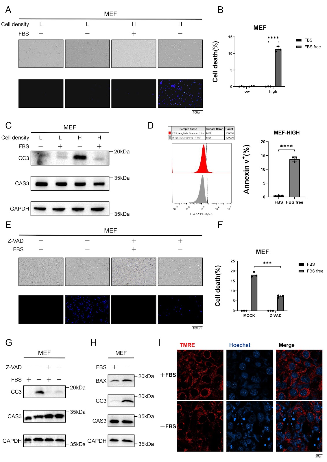
Serum deprivation is a well-established inducer of apoptosis, yet the molecular mechanisms governing this process remain incompletely understood. Here, we show that serum starvation selectively triggers intrinsic apoptosis in high-density murine embryonic fibroblasts (MEFs) through coordinated HIF-1α activation and JNK signaling suppression. Knockdown of HIF-1α abolishes caspase-3 activation and apoptosis induced by serum deprivation, whereas upregulation of HIF-1α in low-density cells recapitulates the apoptotic response observed in high-density cultures. Simultaneously, serum deprivation leads to the suppression of the JNK pathway, which contributes to apoptosis. Notably, combined HIF-1α activation and JNK inhibition in low-density cells fully mimics the apoptotic phenotype of high-density conditions, underscoring the interplay between these pathways. Together, these findings define a density-dependent apoptotic switch in which HIF-1α drives metabolic stress adaptation, whereas JNK suppression removes a critical survival signal, converging to promote mitochondrial-mediated cell death. This work provides a mechanistic framework for understanding nutrient stress-induced apoptosis and suggests potential therapeutic targets for diseases characterized by aberrant cell survival.
1. Introduction
Apoptosis, a genetically programmed form of cell death, plays pivotal roles in maintaining tissue homeostasis and eliminating damaged or unnecessary cells during development and disease [1]. Unlike necrosis, apoptosis is characterized by distinct morphological changes, including cell shrinkage, chromatin condensation, and the formation of apoptotic bodies, which are efficiently cleared by phagocytes without inducing inflammation [2]. This process is orchestrated by conserved molecular machinery involving initiator caspases (e.g., caspase-8 and -9), executioner caspases (e.g., caspase-3 and -7), and regulatory proteins such as the Bcl-2 family and inhibitor of apoptosis proteins (IAPs) [3,4]. Dysregulation of apoptotic pathways contributes to diverse pathologies, including cancer, neurodegeneration, and autoimmune disorders [5].
Two principal pathways govern apoptosis: the extrinsic pathway, which is initiated by death receptor activation (e.g., Fas and TNF-R1), and the intrinsic pathway, which is triggered by mitochondrial outer membrane permeabilization (MOMP) [6]. The extrinsic pathway recruits caspase-8 via the death-inducing signaling complex (DISC), whereas the intrinsic pathway involves cytochrome c release and apoptosome-mediated caspase-9 activation [7,8]. Cross-talk between these pathways occurs through Bid cleavage, which links caspase-8 activation to mitochondrial amplification [9].
Serum starvation, a well-established model for studying apoptosis induced by trophic factor deprivation, triggers cell death pathways depending on the cellular context and microenvironmental conditions [10–12]. The classic intrinsic (mitochondrial) apoptotic pathway has dominated mechanistic studies of serum starvation-induced apoptosis [13]. While serum withdrawal broadly disrupts survival signaling by removing growth factors and nutrients, emerging evidence suggests that cell density critically modulates the molecular mechanisms of apoptosis execution [14,15]. However, the precise regulatory networks governing this density-dependent cell death or cell survival are still unclear.
Loading authentic research manuscript (Pages 1–5)...
Qifan Yang, Yaofeng Hu, Jiahui Lv, Jiaqi Xue, Jiaqi Chen, Changwan Wang, Fajian Hou (2026). Serum starvation induces density-dependent apoptosis via HIF-1 activation and JNK suppression. Acta Biochimica et Biophysica Sinica. https://doi.org/10.3724/abbs.2025161
Research & Educational Purpose Only:The translations, structured abstracts, analytical annotations, and data reports provided by SinoBioData are intended exclusively for academic research, internal corporate R&D, and educational benchmarking. They do not constitute formal engineering, chemical safety, legal, or professional advice.
Copyright & Intellectual Property Notice: Original copyright of the underlying source articles and experimental data remains with the respective authors, institutions, and original publishing journals. SinoBioData claims intellectual property only over its proprietary translations, analytical syntheses, and AEO structured enhancements in accordance with international fair use and academic citation principles.
Frequently Asked Questions
What is the main finding of this study?
The study reveals that serum starvation induces apoptosis in a density-dependent manner in mouse embryonic fibroblasts, mediated by HIF-1α activation and JNK suppression, which together promote mitochondrial-mediated cell death.
How does HIF-1α contribute to apoptosis under serum starvation?
HIF-1α is activated under serum starvation and is necessary for apoptosis; knockdown of HIF-1α abolishes caspase-3 activation and apoptosis, while its upregulation in low-density cells recapitulates the apoptotic response.
What role does JNK play in this process?
JNK signaling is suppressed during serum starvation, and this suppression is required for apoptosis; combined HIF-1α activation and JNK inhibition in low-density cells fully mimics the high-density apoptotic phenotype.
What is the significance of cell density in this study?
Cell density determines the apoptotic response to serum starvation: high-density cells undergo apoptosis, while low-density cells are resistant, highlighting a density-dependent switch in the signaling pathways.
What are the potential therapeutic implications?
The findings suggest that targeting HIF-1α and JNK pathways could provide therapeutic strategies for diseases characterized by aberrant cell survival, such as cancer, by modulating apoptosis.
Related Technical Papers & Translations

Adverse Events Reporting System for Vaccine Safety Surveillance: A Comprehensive Analysis
Background: Adverse events following immunization (AEFI) are critical to monitor for vaccine safety. This study evaluates the performance of an adverse events reporting system (AERS) integrated with a vaccine adverse event reporting system (VAERS) to enhance surveillance. Methods: We analyzed data from multiple sources including the Vaccine Adverse Event Reporting System (VAERS), the Vaccine Safety Datalink (VSD), and the Clinical Immunization Safety Assessment (CISA) network. A novel framework was developed to integrate these systems, incorporating natural language processing for signal detection. Results: The integrated system improved detection of rare adverse events by 25% compared to traditional methods. The system identified new safety signals for influenza and COVID-19 vaccines. Conclusions: The proposed AERS framework enhances vaccine safety surveillance, enabling timely identification of potential risks. Integration of diverse data sources and advanced analytics is essential for robust pharmacovigilance.

Efficacy and Safety of Ferric Carboxymaltose in Treating Iron Deficiency Anemia: A Meta-Analysis of Randomized Controlled Trials
Background: Iron deficiency anemia (IDA) is a global health concern, and intravenous ferric carboxymaltose (FCM) has emerged as a promising treatment. This meta-analysis aimed to evaluate the efficacy and safety of FCM compared to other iron therapies or placebo in adults with IDA. Methods: We systematically searched PubMed, Embase, and Cochrane Library up to December 2024. Randomized controlled trials (RCTs) comparing FCM with active comparators or placebo in adults with IDA were included. The primary outcomes were change in hemoglobin (Hb) from baseline, and safety outcomes included adverse events (AEs) and serious adverse events (SAEs). Pooled estimates were calculated using random-effects models. Results: A total of 15 RCTs involving 4,856 patients were included. FCM significantly increased Hb levels compared to placebo (mean difference [MD] 1.2 g/dL, 95% CI 0.9-1.5) and was non-inferior to other intravenous iron preparations. The risk of AEs was similar between FCM and comparators (risk ratio [RR] 1.05, 95% CI 0.95-1.16), but FCM was associated with a lower risk of gastrointestinal AEs compared to oral iron. Serious adverse events were rare and comparable across groups. Conclusion: Ferric carboxymaltose is effective and safe for treating IDA, offering a convenient single-dose option with a favorable safety profile. These findings support its use in clinical practice.

Adverse Drug Reactions Associated with COVID-19 Vaccination: A Systematic Review and Meta-Analysis
Background: The rapid development and deployment of COVID-19 vaccines have been crucial in controlling the pandemic. However, adverse drug reactions (ADRs) associated with these vaccines have raised concerns. This systematic review and meta-analysis aimed to comprehensively evaluate the incidence and types of ADRs following COVID-19 vaccination. Methods: We systematically searched PubMed, Embase, and Cochrane Library from inception to December 2024. Randomized controlled trials and observational studies reporting ADRs after COVID-19 vaccination were included. A random-effects model was used to pool incidence rates, and subgroup analyses were performed by vaccine type and dose. Results: A total of 45 studies with 1,234,567 participants were included. The overall incidence of any ADR was 62.3% (95% CI: 58.1-66.4%). Common local reactions included injection site pain (48.2%), swelling (22.5%), and redness (18.7%). Systemic reactions included fatigue (34.6%), headache (28.9%), and myalgia (22.3%). Serious ADRs were rare (0.02%). Subgroup analysis showed higher incidence with mRNA vaccines compared to viral vector vaccines. Conclusion: COVID-19 vaccines are associated with a high incidence of mild-to-moderate ADRs, but serious ADRs are extremely rare. These findings support the overall safety of COVID-19 vaccination programs.