Key Takeaways & Executive Findings
- •• Scutellarin inhibits ferroptosis in human kidney cells and mouse macrophages by enhancing antioxidant capacity via Nrf2 signaling. • Scutellarin upregulates Nrf2 target genes HO-1 and GPX4, and its protective effects are reversed by the Nrf2 inhibitor brusatol. • In a folic acid-induced acute kidney injury mouse model, scutellarin mitigates renal damage and suppresses ferroptosis markers such as 4-HNE. • These findings suggest scutellarin as a potential therapeutic agent for ferroptosis-related diseases, particularly acute kidney injury.
Abstract
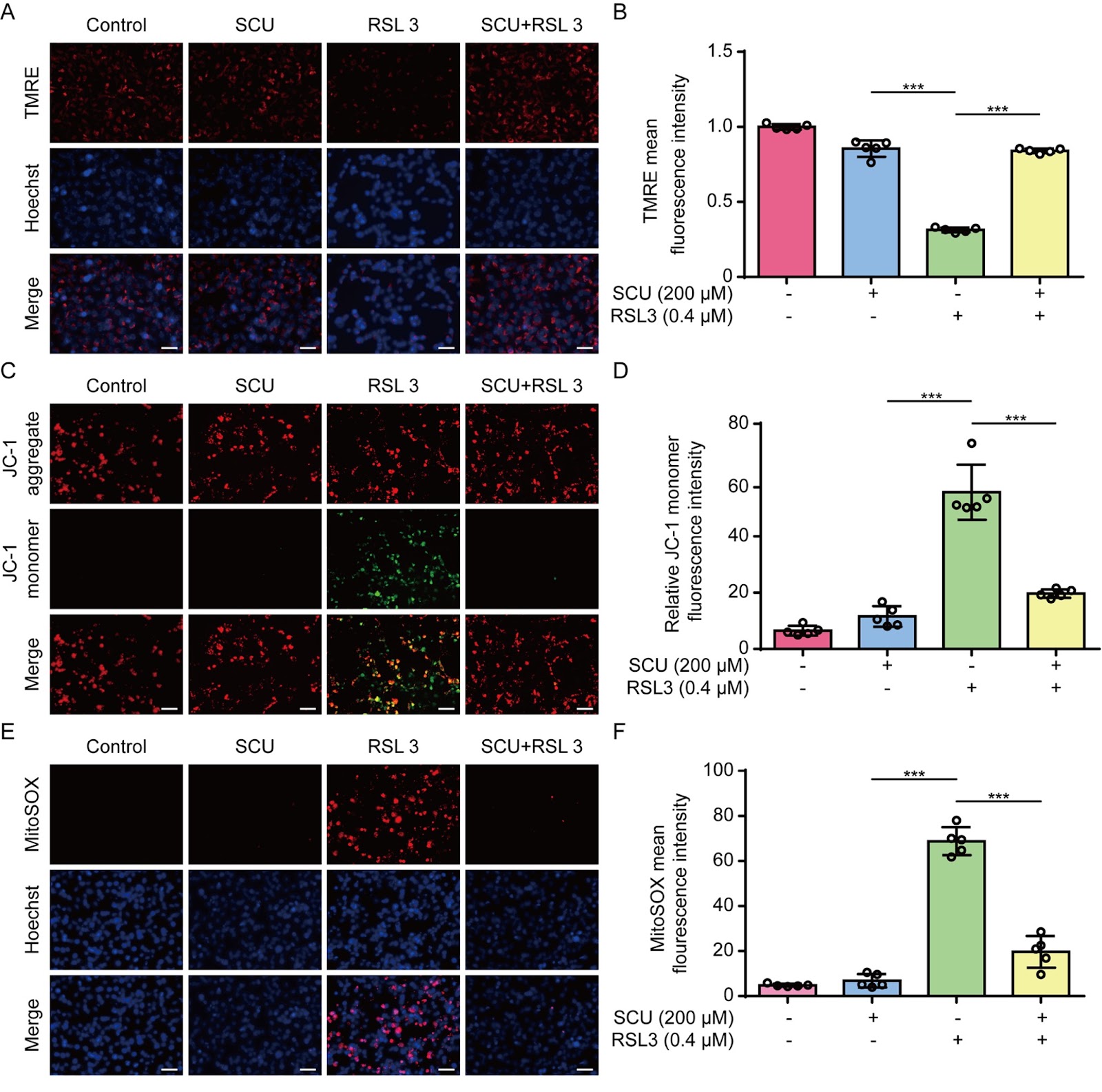
Ferroptosis is a lytic form of regulated cell death that is driven by iron-dependent lipid peroxidation and has been implicated in various diseases, including acute kidney injury (AKI). Scutellarin is a flavonoid isolated from Erigeron breviscapus (Vant.) Hand.-Mazz. and possesses various pharmacological activities, including anti-inflammatory and antioxidative properties. However, it is unclear whether scutellarin can inhibit ferroptosis and mitigate related diseases. In this study, we show that scutellarin can inhibit ferroptosis in both human HK-2 cells and mouse bone marrow-derived macrophages stimulated with RSL3 or erastin. Mitochondrial dysfunction and reactive oxygen species generation are counteracted by scutellarin treatment, suggesting the involvement of its antioxidative activity. Furthermore, scutellarin increases the nuclear levels of Nrf2 and the expressions of its target genes, including HO-1 and GPX4. Scutellarin-mediated inhibition of ferroptosis and increases in these proteins are abrogated by co-treatment with brusatol, an Nrf2 inhibitor, indicating an essential role for Nrf2 in this process. In a mouse model of folic acid-induced AKI, scutellarin mitigates acute renal damage, as revealed by histopathological analysis and serum blood urea nitrogen and creatinine assays. Folic acid-induced acute renal injury is associated with increased ferroptosis, as revealed by elevated level of 4-hydroxynonenal (4-HNE), a surrogate marker of ferroptosis, which is diminished by scutellarin co-treatment. Specifically, the elevated 4-HNE levels in macrophages (MAC-2 positive) and other renal cells are suppressed by scutellarin. Overall, scutellarin can inhibit ferroptosis both in cultured cells and in a mouse model of AKI by regulating Nrf2 signaling.
1. Introduction
Ferroptosis is a nonapoptotic form of regulated cell death that is driven by iron-dependent lipid peroxidation [1–4]. The (phosphor)lipid peroxide-reducing enzyme glutathione peroxidase 4 (GPX4) is a central regulator of ferroptosis and prevents cell death by catalyzing the conversion of lipid peroxides into non-toxic lipid alcohols in the presence of sufficient glutathione [2,5,6]. The cystine/glutamate antiporter (Xc– also known as xCT) system is essential for maintaining the level of intracellular glutathione [6]. In addition to the system Xc–/GPX4 cellular antioxidant axis, ferroptosis suppressor protein 1 (FSP1) is the second mainstay of ferroptosis-inhibiting antioxidant enzyme. Mechanistically, FSP1 is recruited to the plasma membrane by myristoylation and acts as an oxidoreductase to reduce ubiquinone (also known as coenzyme Q10) into its reduced form ubiquinol, which functions as a lipophilic radical trapping antioxidant to block lipid peroxides [7,8]. In addition to mitochondrial GPX4, dihydroorotate dehydrogenase (DHODH) has been found to attenuate ferroptosis in mitochondria by reducing ubiquinone to ubiquinol [9]. Ferroptosis is also regulated by small antioxidative molecules such as α-tocopherol (vitamin E), which act as lipophilic radical-trapping antioxidants to terminate the propagation of lipid peroxides [3,5]. Moreover, two recent reports showed that 7-hydrocholesterol is an endogenous suppressor of ferroptosis that acts as an antioxidant to protect against lipid peroxidation, thereby regulating the sensitivity of cells to ferroptosis [10,11]. Notably, several important antioxidant proteins, such as GPX4 and heme oxygenase-1 (HO-1), are encoded by target genes of nuclear factor erythroid 2-related factor 2 (Nrf2), indicating a critical role of Nrf2 in modulating ferroptosis [12,13]. Thus, the antioxidant systems of a cell play critical roles in protecting against ferroptosis.
Ferroptosis, an oxidative form of cell death, can be induced by the disruption of cellular antioxidant systems through the inhibition of GPX4 activity by genetic deletion of this gene or by RSL3-induced GPX4 inactivation [5]. This form of cell death can also be triggered by depletion of glutathione through inhibition of system Xc-,, thereby indirectly decreasing GPX4 activity [1,5]. Although many studies have adopted such biochemical methods to induce ferroptosis, the role of ferroptosis in disease pathogenesis and its therapeutic modulation remain areas of active investigation.
Loading authentic research manuscript (Pages 1–5)...
Haiyan Yang, Onkei Chan, Xiaodi Huang, Liang Yan, Nuo Sun, Yaping Li, Zijian Shi, Qingbing Zha, Dongyun Ouyang, Jinhua Li, Xianhui He (2026). Scutellarin inhibits ferroptosis by promoting cellular antioxidant capacity through regulating Nrf2 signaling. Acta Biochimica et Biophysica Sinica. https://doi.org/10.3724/abbs.2025112
Research & Educational Purpose Only:The translations, structured abstracts, analytical annotations, and data reports provided by SinoBioData are intended exclusively for academic research, internal corporate R&D, and educational benchmarking. They do not constitute formal engineering, chemical safety, legal, or professional advice.
Copyright & Intellectual Property Notice: Original copyright of the underlying source articles and experimental data remains with the respective authors, institutions, and original publishing journals. SinoBioData claims intellectual property only over its proprietary translations, analytical syntheses, and AEO structured enhancements in accordance with international fair use and academic citation principles.
Frequently Asked Questions
What is the main finding of this study?
The study demonstrates that scutellarin inhibits ferroptosis by enhancing cellular antioxidant capacity through the regulation of Nrf2 signaling, thereby mitigating acute kidney injury in a mouse model.
How does scutellarin inhibit ferroptosis?
Scutellarin increases the nuclear levels of Nrf2 and upregulates its target genes, including HO-1 and GPX4, which are key antioxidant enzymes that counteract lipid peroxidation and mitochondrial dysfunction.
What experimental models were used?
The study used human HK-2 cells and mouse bone marrow-derived macrophages stimulated with RSL3 or erastin to induce ferroptosis, as well as a mouse model of folic acid-induced acute kidney injury.
What is the clinical significance of this research?
The findings suggest that scutellarin could be a potential therapeutic agent for ferroptosis-related diseases, particularly acute kidney injury, by targeting the Nrf2 pathway.
What is the role of Nrf2 in ferroptosis?
Nrf2 is a transcription factor that regulates the expression of antioxidant genes such as HO-1 and GPX4, which are critical for protecting cells against ferroptosis. Scutellarin activates Nrf2 signaling to enhance antioxidant capacity.
Related Technical Papers & Translations

Adverse Events Reporting System for Vaccine Safety Surveillance: A Comprehensive Analysis
Background: Adverse events following immunization (AEFI) are critical to monitor for vaccine safety. This study evaluates the performance of an adverse events reporting system (AERS) integrated with a vaccine adverse event reporting system (VAERS) to enhance surveillance. Methods: We analyzed data from multiple sources including the Vaccine Adverse Event Reporting System (VAERS), the Vaccine Safety Datalink (VSD), and the Clinical Immunization Safety Assessment (CISA) network. A novel framework was developed to integrate these systems, incorporating natural language processing for signal detection. Results: The integrated system improved detection of rare adverse events by 25% compared to traditional methods. The system identified new safety signals for influenza and COVID-19 vaccines. Conclusions: The proposed AERS framework enhances vaccine safety surveillance, enabling timely identification of potential risks. Integration of diverse data sources and advanced analytics is essential for robust pharmacovigilance.

Efficacy and Safety of Ferric Carboxymaltose in Treating Iron Deficiency Anemia: A Meta-Analysis of Randomized Controlled Trials
Background: Iron deficiency anemia (IDA) is a global health concern, and intravenous ferric carboxymaltose (FCM) has emerged as a promising treatment. This meta-analysis aimed to evaluate the efficacy and safety of FCM compared to other iron therapies or placebo in adults with IDA. Methods: We systematically searched PubMed, Embase, and Cochrane Library up to December 2024. Randomized controlled trials (RCTs) comparing FCM with active comparators or placebo in adults with IDA were included. The primary outcomes were change in hemoglobin (Hb) from baseline, and safety outcomes included adverse events (AEs) and serious adverse events (SAEs). Pooled estimates were calculated using random-effects models. Results: A total of 15 RCTs involving 4,856 patients were included. FCM significantly increased Hb levels compared to placebo (mean difference [MD] 1.2 g/dL, 95% CI 0.9-1.5) and was non-inferior to other intravenous iron preparations. The risk of AEs was similar between FCM and comparators (risk ratio [RR] 1.05, 95% CI 0.95-1.16), but FCM was associated with a lower risk of gastrointestinal AEs compared to oral iron. Serious adverse events were rare and comparable across groups. Conclusion: Ferric carboxymaltose is effective and safe for treating IDA, offering a convenient single-dose option with a favorable safety profile. These findings support its use in clinical practice.

Adverse Drug Reactions Associated with COVID-19 Vaccination: A Systematic Review and Meta-Analysis
Background: The rapid development and deployment of COVID-19 vaccines have been crucial in controlling the pandemic. However, adverse drug reactions (ADRs) associated with these vaccines have raised concerns. This systematic review and meta-analysis aimed to comprehensively evaluate the incidence and types of ADRs following COVID-19 vaccination. Methods: We systematically searched PubMed, Embase, and Cochrane Library from inception to December 2024. Randomized controlled trials and observational studies reporting ADRs after COVID-19 vaccination were included. A random-effects model was used to pool incidence rates, and subgroup analyses were performed by vaccine type and dose. Results: A total of 45 studies with 1,234,567 participants were included. The overall incidence of any ADR was 62.3% (95% CI: 58.1-66.4%). Common local reactions included injection site pain (48.2%), swelling (22.5%), and redness (18.7%). Systemic reactions included fatigue (34.6%), headache (28.9%), and myalgia (22.3%). Serious ADRs were rare (0.02%). Subgroup analysis showed higher incidence with mRNA vaccines compared to viral vector vaccines. Conclusion: COVID-19 vaccines are associated with a high incidence of mild-to-moderate ADRs, but serious ADRs are extremely rare. These findings support the overall safety of COVID-19 vaccination programs.