🧬 SinoBioData Academic Portal
🏛️ Indexed Academic JournalOriginal: 干细胞研究与转化

Stem Cell Research & Therapy

Premier Chinese Biomedical Journal indexed in SinoBioData: Stem Cell Research & Therapy (干细胞研究与转化).

Total Research Papers: 200
Access: 100% Free Open Access
Browse by Publication Year & VolumeReset All Filters ✕
Select Specific Issue:

Published Research PapersFiltered: Year 2024 • 15

Showing 57 of 200 peer-reviewed papers with full Graphical Abstracts.

Original ResearchVol. 15, None • pp. 274DOI: 10.1186/s13287-024-03886-y

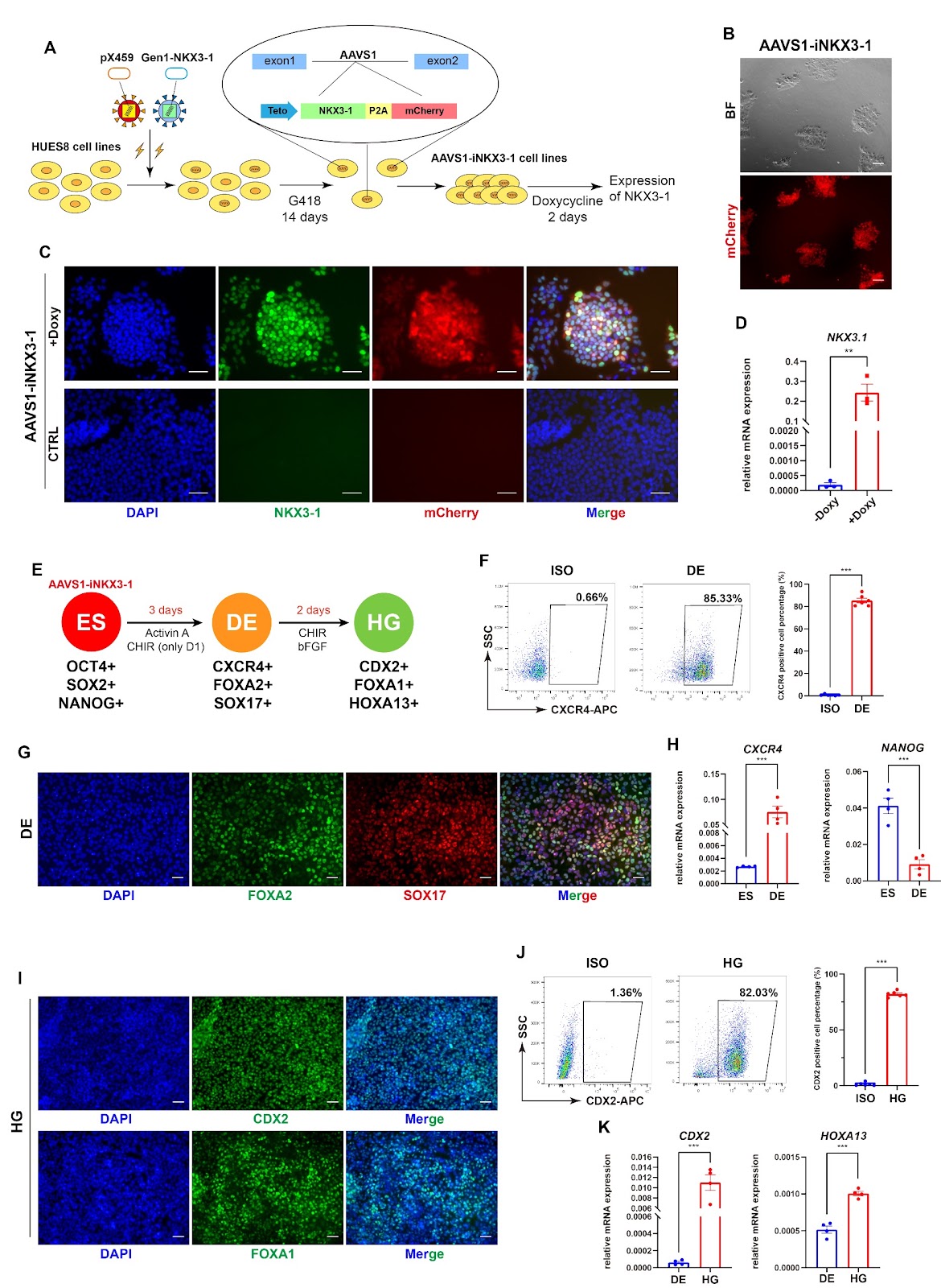
Prostatic lineage differentiation from human embryonic stem cells through inducible expression of NKX3-1

Authors: Songwei Wang, Yangyang Yu, Yinglei Li, Tianzhe Zhang, Wei Jiang, Xinghuan Wang, Ran Liu

Background Understanding the lineage differentiation of human prostate not only is crucial for basic research on human developmental biology but also significantly contributes to the management of prostate-related disorders. Current knowledge mainly relies on studies on rodent models, lacking human-derived alternatives despite clinical samples may provide a snapshot at certain stage. Human embryonic stem cells can generate all the embryonic lineages including the prostate, and indeed a few studies demonstrate such possibility based on co-culture or co-transplantation with urogenital mesenchyme into mouse renal capsule. Methods To establish a stepwise protocol to obtain prostatic organoids in vitro from human embryonic stem cells, we apply chemicals and growth factors by mimicking the regulation network of transcription factors and signal transduction pathways, and construct cell lines carrying an inducible NKX3-1 expressing cassette, together with three-dimensional culture system. Unpaired t test was applied for statistical analyses. Results We first successfully generate the definitive endoderm, hindgut, and urogenital sinus cells. The embryonic stem cell-derived urogenital sinus cells express prostatic key transcription factors AR and FOXA1, but fail to express NKX3-1. Therefore, we construct NKX3-1-inducible cell line by homologous recombination, which is eventually able to yield AR, FOXA1, and NKX3-1 triple-positive urogenital prostatic lineage cells through stepwise differentiation. Finally, combined with 3D culture we successfully derive prostate-like organoids with certain structures and prostatic cell populations. Conclusions This study reveals the crucial role of NKX3-1 in prostatic differentiation and offers the inducible NKX3-1 cell line, as well as provides a stepwise differentiation protocol to generate human prostate-like organoids, which should facilitate the studies on prostate development and disease pathogenesis.
Prostatic lineage differentiation from human embryonic stem cells through inducible expression of NKX3-1
Graphical Abstract
Original ResearchVol. 15, Issue 1 • pp. 135DOI: 10.1186/s13287-024-03745-w

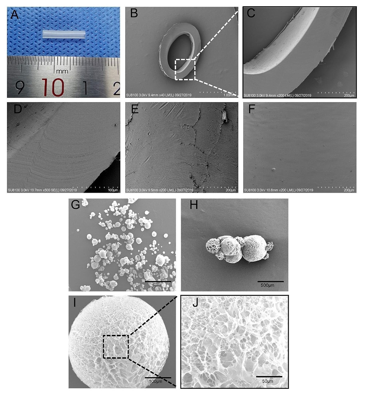
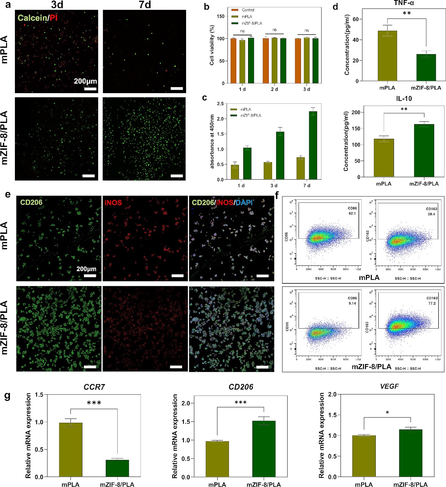
Osteoinductive micro-nano guided bone regeneration membrane for in situ bone defect repair

Authors: Bingqian Wang, Xinfang Xie, Wenbin Jiang, Yichen Zhan, Yifan Zhang, Yaqi Guo, Zhenxing Wang, Nengqiang Guo, Ke Guo, Jiaming Sun

Background Biomaterials used in bone tissue engineering must fulfill the requirements of osteoconduction, osteoinduction, and osseointegration. However, biomaterials with good osteoconductive properties face several challenges, including inadequate vascularization, limited osteoinduction and barrier ability, as well as the potential to trigger immune and inflammatory responses. Therefore, there is an urgent need to develop guided bone regeneration membranes as a crucial component of tissue engineering strategies for repairing bone defects. Methods The mZIF-8/PLA membrane was prepared using electrospinning technology and simulated body fluid external mineralization method. Its ability to induce biomimetic mineralization was evaluated through TEM, EDS, XRD, FT-IR, zeta potential, and wettability techniques. The biocompatibility, osteoinduction properties, and osteo-immunomodulatory effects of the mZIF-8/PLA membrane were comprehensively evaluated by examining cell behaviors of surface-seeded BMSCs and macrophages, as well as the regulation of cellular genes and protein levels using PCR and WB. In vivo, the mZIF-8/PLA membrane's potential to promote bone regeneration and angiogenesis was assessed through Micro-CT and immunohistochemical staining. Results The mineralized deposition enhances hydrophilicity and cell compatibility of mZIF-8/PLA membrane. mZIF-8/PLA membrane promotes up-regulation of osteogenesis and angiogenesis related factors in BMSCs. Moreover, it induces the polarization of macrophages towards the M2 phenotype and modulates the local immune microenvironment. After 4-weeks of implantation, the mZIF-8/PLA membrane successfully bridges critical bone defects and almost completely repairs the defect area after 12-weeks, while significantly improving the strength and vascularization of new bone. Conclusions The mZIF-8/PLA membrane with dual osteoconductive and immunomodulatory abilities could pave new research paths for bone tissue engineering.

Osteoinductive micro-nano guided bone regeneration membrane for in situ bone defect repair
Graphical Abstract
Original ResearchVol. 15, None • pp. 292DOI: 10.1186/s13287-024-03898-8

Targeting NPM1 inhibits proliferation and promotes apoptosis of hepatic progenitor cells via suppression of mTOR signalling pathway

Authors: Ping Wang, Min Wang, Lin Liu, Hongyi Li, Helin Liu, Jiangbo Ren, Tianhui Liu, Min Cong, Zhijun Zhu, Xinyan Zhao, Liying Sun, Jidong Jia

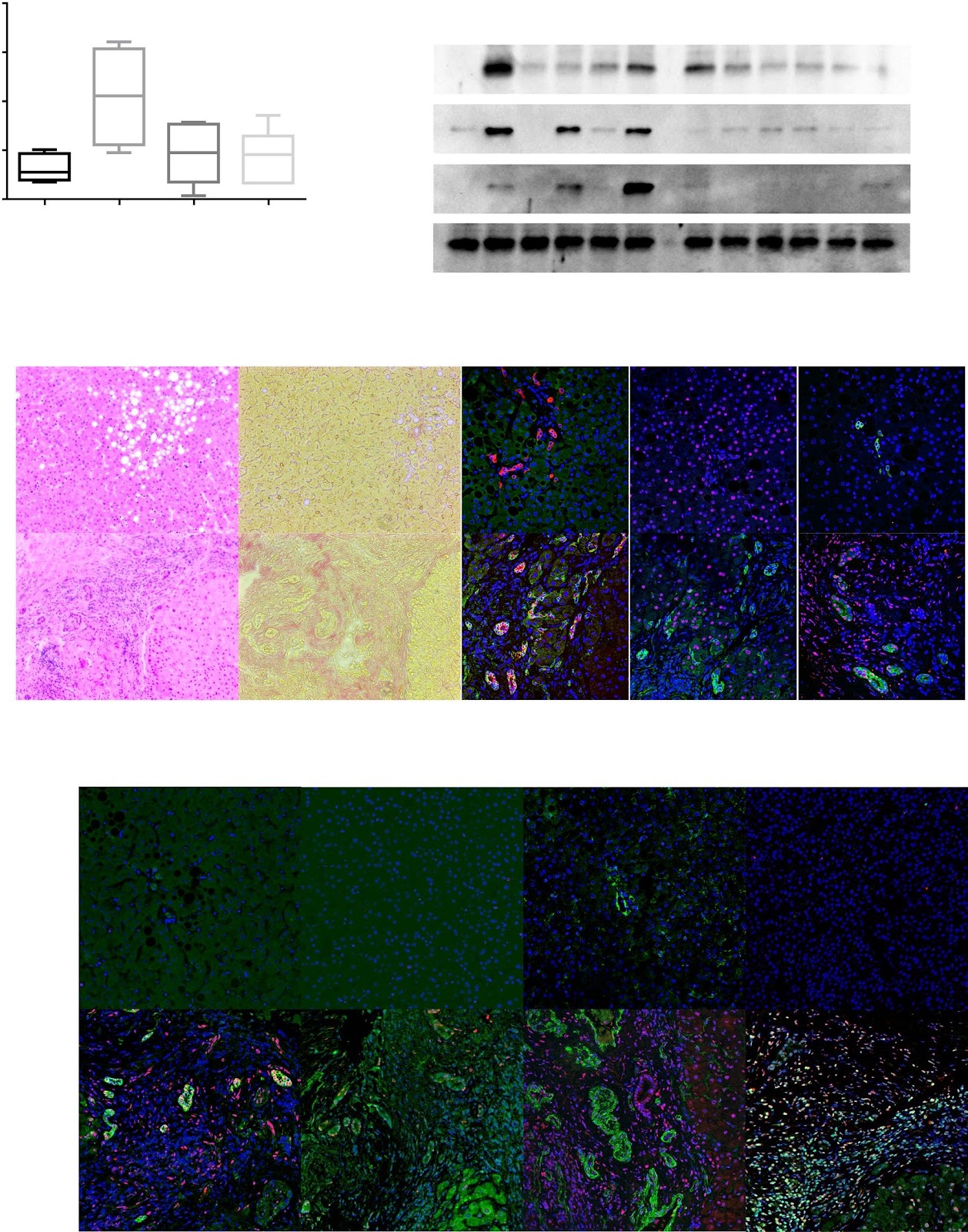
Background Hepatic progenitor cells serve not only as the origin of combined hepatocellular cholangiocarcinoma (cHCC-CCA) but are also responsible for malignancy recurrence after surgical resection. Nucleophosmin 1 (NPM1) has been implicated in cancer metastasis and poor prognosis. This study aimed to determine the expression of NPM1 by hepatic progenitor cells in cHCC-CCA and the effects of targeting NPM1 on hepatic progenitor cells and BEL-7402 cells with characteristics of both progenitor cells and cHCC-CCA. Methods First, NPM1 was detected by RT‒PCR, western blotting, and double-immunofluorescence staining in cHCC-CCA tissues. NPM1 expression was subsequently analysed in rat hepatic progenitor cells cultured in vitro and in interleukin 6 (IL6)-treated cells. The effects and mechanism of NPM1 on hepatic progenitor cells were determined by knocking down NPM1 and performing RNA sequencing analysis. Finally, NSC348884, a small-molecule inhibitor that disrupts NPM1 dimer formation, was used to confirm the function of NPM1 in BEL-7402 cells. Results Both human hepatic progenitor cells in cHCC-CCA tissues and rat in vitro cultured hepatic progenitor cells highly expressed NPM1. IL6, a cytokine involved in the malignant transformation of hepatic progenitor cells, dose-dependently increased NPM1 and PCNA expression. Knocking down NPM1 reduced IL6R transcription (P < 0.0001) and inhibited the proliferation (P = 0.0065) of hepatic progenitor cells by suppressing the mTOR signalling pathway and activating the apoptosis pathway. Furthermore, knocking down NPM1 in hepatic progenitor cells resulted in more apoptotic cells (7.33 ± 0.09% vs. 3.76 ± 0.13%, P < 0.0001) but fewer apoptotic cells in the presence of NSC348884 (47.57 ± 0.49% vs. 63.40 ± 0.05%, P = 0.0008) than in the control cells, suggesting that low-NPM1-expressing cells are more resistant to NSC348884. In addition, NSC348884 induced the apoptosis of BEL-7402 cells with an IC50 of 2.77 μmol/L via the downregulation of the IL-6R and mTOR signalling pathways and inhibited the growth of BEL-7402 cells in a subcutaneous xenograft tumour model (P = 0.0457). Conclusions Targeting NPM1 inhibits proliferation and induces apoptosis in hepatic progenitor cells and BEL-7402 cells, thus serving as a potential therapy for cHCC-CCA.

Targeting NPM1 inhibits proliferation and promotes apoptosis of hepatic progenitor cells via suppression of mTOR signalling pathway
Graphical Abstract
Original ResearchVol. 15, None • pp. 156DOI: 10.1186/s13287-024-03761-w

TGF-β signaling regulates differentiation of MSCs in bone metabolism: disputes among viewpoints

Authors: Erfan Wei, Menglong Hu, Likun Wu, Xingtong Pan, Qiyue Zhu, Hao Liu, Yunsong Liu

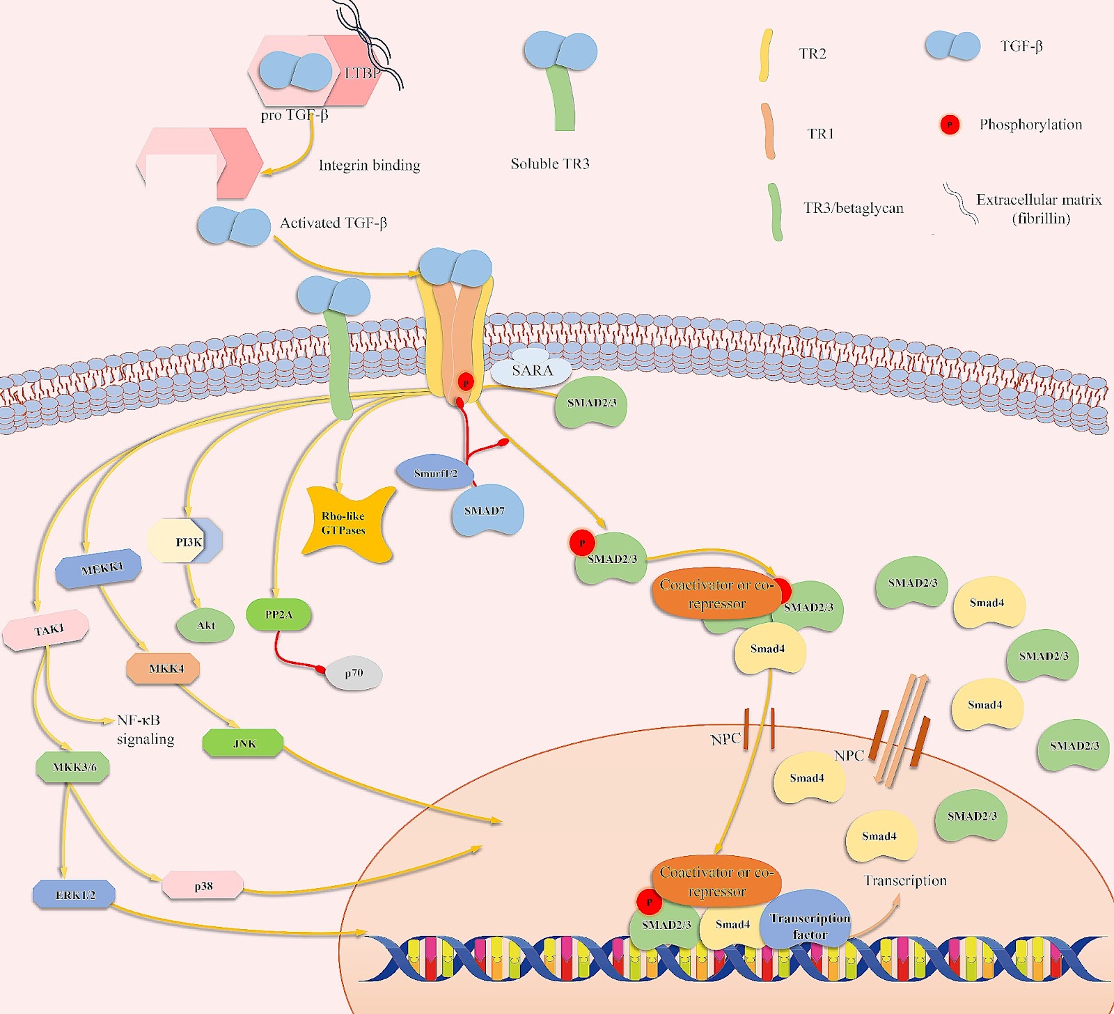
Mesenchymal stem cells (MSCs) are multipotent cells that can differentiate into cells of different lineages to form mesenchymal tissues, which are promising in regard to treatment for bone diseases. Their osteogenic differentiation is under the tight regulation of intrinsic and extrinsic factors. Transforming growth factor β (TGF-β) is an essential growth factor in bone metabolism, which regulates the differentiation of MSCs. However, published studies differ in their views on whether TGF-β signaling regulates the osteogenic differentiation of MSCs positively or negatively. The controversial results have not been summarized systematically and the related explanations are required. Therefore, we reviewed the basics of TGF-β signaling and summarized how each of three isoforms regulates osteogenic differentiation. Three isoforms of TGF-β (TGF-β1/β2/β3) play distinct roles in regulating osteogenic differentiation of MSCs. Additionally, other possible sources of conflicts are summarized here. Further understanding of TGF-β signaling regulation in MSCs may lead to new applications to promote bone regeneration and improve therapies for bone diseases.

TGF-β signaling regulates differentiation of MSCs in bone metabolism: disputes among viewpoints
Graphical Abstract
Original ResearchVol. 15, None • pp. 95DOI: 10.1186/s13287-024-03708-1

Manufacturing, quality control, and GLP-grade preclinical study of nebulized allogenic adipose mesenchymal stromal cells-derived extracellular vesicles

Authors: Jing Wang, Zhong-jin Chen, Ze-yi Zhang, Mei-ping Shen, Bo Zhao, Wei Zhang, Ye Zhang, Ji-gang Lei, Cheng-jie Ren, Jing Chang, Cui-li Xu, Meng Li, Yang-yang Pi, Tian-lun Lu, Cheng-xiang Dai, Su-ke Li, Ping Li

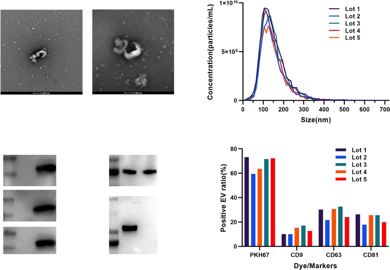
Background Human adipose stromal cells-derived extracellular vesicles (haMSC-EVs) have been shown to alleviate inflammation in acute lung injury (ALI) animal models. However, there are few systemic studies on clinical-grade haMSC-EVs. Our study aimed to investigate the manufacturing, quality control (QC) and preclinical safety of clinical-grade haMSC-EVs. Methods haMSC-EVs were isolated from the conditioned medium of human adipose MSCs incubated in 2D containers. Purification was performed by PEG precipitation and differential centrifugation. Characterizations were conducted by nanoparticle tracking analysis, transmission electron microscopy (TEM), Western blotting, nanoflow cytometry analysis, and the TNF-α inhibition ratio of macrophage [after stimulated by lipopolysaccharide (LPS)]. RNA-seq and proteomic analysis with liquid chromatography tandem mass spectrometry (LC–MS/MS) were used to inspect the lot-to-lot consistency of the EV products. Repeated toxicity was evaluated in rats after administration using trace liquid endotracheal nebulizers for 28 days, and respiratory toxicity was evaluated 24 h after the first administration. In vivo therapeutic effects were assessed in an LPS-induced ALI/ acute respiratory distress syndrome (ARDS) rat model. Results The quality criteria have been standardized. In a stability study, haMSC-EVs were found to remain stable after 6 months of storage at − 80°C, 3 months at − 20 °C, and 6 h at room temperature. The microRNA profile and proteome of haMSC-EVs demonstrated suitable lot-to-lot consistency, further suggesting the stability of the production processes. Intratracheally administered 1.5 × 10^8 particles/rat/day for four weeks elicited no significant toxicity in rats. In LPS-induced ALI/ARDS model rats, intratracheally administered haMSC-EVs alleviated lung injury, possibly by reducing the serum level of inflammatory factors. Conclusion haMSC-EVs, as an off-shelf drug, have suitable stability and lot-to-lot consistency. Intratracheally administered haMSC-EVs demonstrated excellent safety at the tested dosages in systematic preclinical toxicity studies. Intratracheally administered haMSC-EVs improved the lung function and exerted anti-inflammatory effects on LPS-induced ALI/ARDS model rats.

Manufacturing, quality control, and GLP-grade preclinical study of nebulized allogenic adipose mesenchymal stromal cells-derived extracellular vesicles
Graphical Abstract
Original ResearchVol. 15, None • pp. 131DOI: 10.1186/s13287-024-03725-0

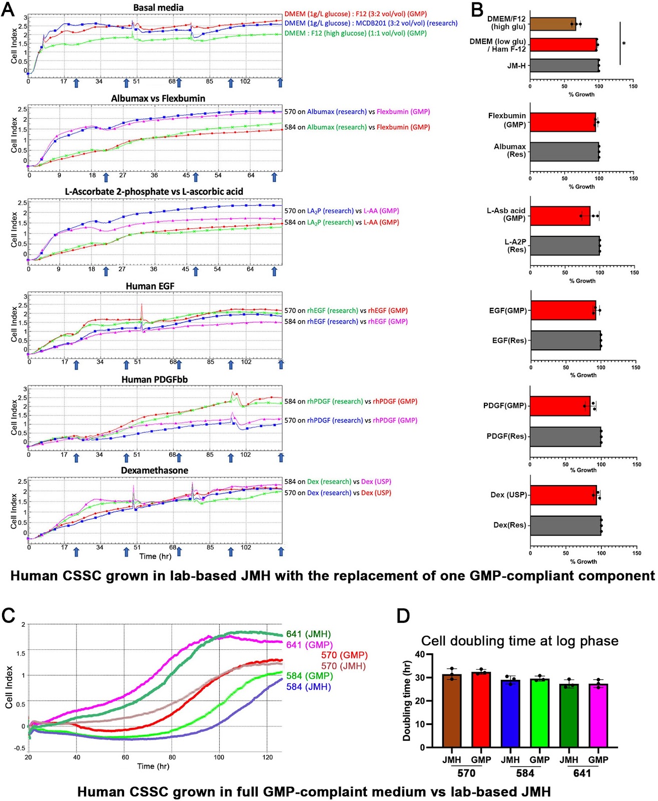
A GMP-compliant manufacturing method for Wharton’s jelly-derived mesenchymal stromal cells

Authors: Wanglong Chu, Fen Zhang, Xiuping Zeng, Fangtao He, Guanyan Shang, Tao Guo, Qingfang Wang, Jianfu Wu, Tongjing Li, Zhen Zhong Zhong, Xiao Liang, Junyuan Hu, Muyun Liu

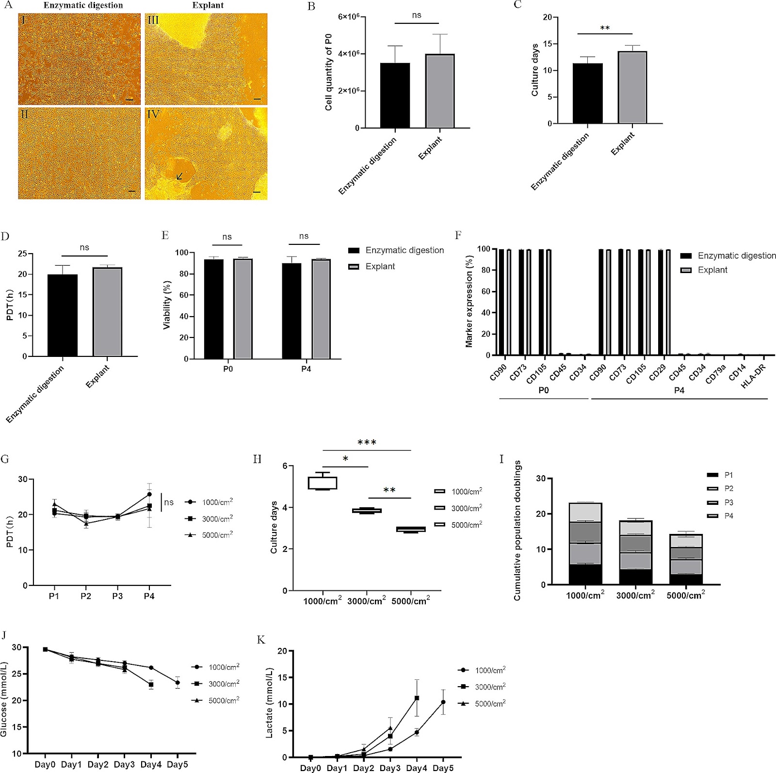
Background Wharton’s jelly-derived mesenchymal stem cells (WJ-MSCs) hold great therapeutic potential in regenerative medicine. Therefore, it is crucial to establish a Good Manufacturing Practice (GMP)-compliant methodology for the isolation and culture of WJ-MSCs. Through comprehensive research, encompassing laboratory-scale experiments to pilot-scale studies, we aimed to develop standardized protocols ensuring the high yield and quality of WJ-MSCs manufacturing. Methods Firstly, optimization of parameters for the enzymatic digestion method used to isolate WJ-MSCs was conducted. These parameters included enzyme concentrations, digestion times, seeding densities, and culture media. Additionally, a comparative analysis between the explant method and the enzymatic digestion method was performed. Subsequently, the consecutive passaging of WJ-MSCs, specifically up to passage 9, was evaluated using the optimized method. Finally, manufacturing processes were developed and scaled up, starting from laboratory-scale flask-based production and progressing to pilot-scale cell factory-based production. Furthermore, a stability study was carried out to assess the storage and use of drug products (DPs). Results The optimal parameters for the enzymatic digestion method were a concentration of 0.4 PZ U/mL Collagenase NB6 and a digestion time of 3 h, resulting in a higher yield of P0 WJ-MSCs. In addition, a positive correlation between the weight of umbilical cord tissue and the quantities of P0 WJ-MSCs has been observed. Evaluation of different concentrations of human platelet lysate revealed that 2% and 5% concentrations resulted in similar levels of cell expansion. Comparative analysis revealed that the enzymatic digestion method exhibited faster outgrowth of WJ-MSCs compared to the explant method during the initial passage. Passages 2 to 5 exhibited higher viability and proliferation ability throughout consecutive passaging. Moreover, scalable manufacturing processes from the laboratory scale to the pilot scale were successfully developed, ensuring the production of high-quality WJ-MSCs. Multiple freeze-thaw cycles of the DPs led to reduced cell viability and viable cell concentration. Subsequent thawing and dilution of the DPs resulted in a significant decrease in both metrics, especially when stored at 20–27 °C.

A GMP-compliant manufacturing method for Wharton’s jelly-derived mesenchymal stromal cells
Graphical Abstract
Original ResearchVol. 15, None • pp. 335DOI: 10.1186/s13287-024-03952-5

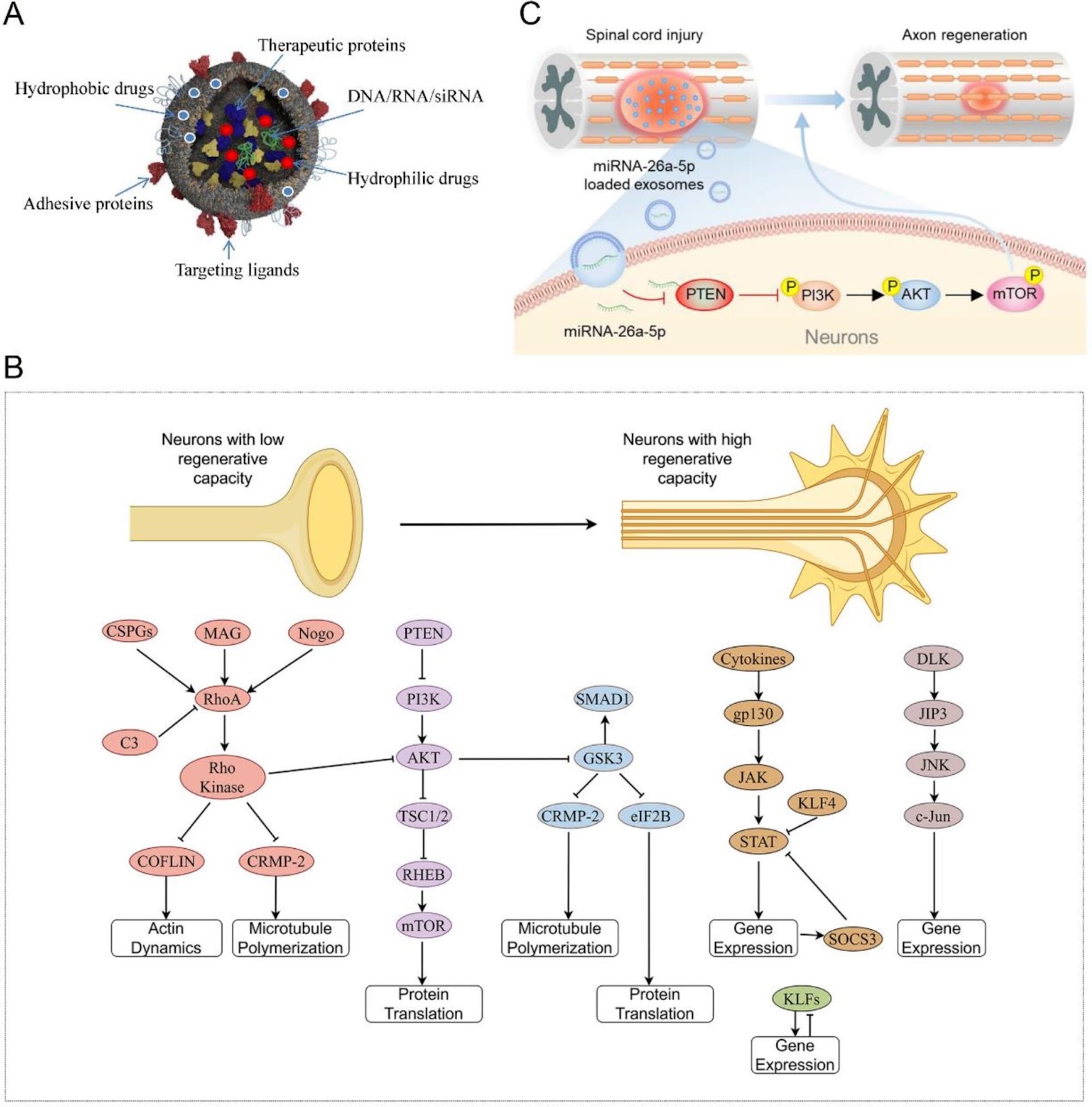
Exosomes as promising bioactive materials in the treatment of spinal cord injury

Authors: Yueying Li, Wenqi Luo, Chuikai Meng, Kaiyuan Shi, Rui Gu, Shusen Cui

Patients with spinal cord injury (SCI) have permanent devastating motor and sensory disabilities. Secondary SCI is known for its complex progression and presents with sophisticated aberrant inflammation, vascular changes, and secondary cellular dysfunction, which aggravate the primary damage. Since their initial discovery, the potent neuroprotective effects and powerful delivery abilities of exosomes (Exos) have been reported in different research fields, including SCI. In this study, we summarize therapeutic advances related to the application of Exos in preclinical animal studies. Subsequently, we discuss the mechanisms of action of Exos derived from diverse cell types, including neurogenesis, angiogenesis, blood–spinal cord barrier preservation, anti-apoptosis, and anti-inflammatory potential. We also evaluate the relationship between the Exo delivery cargo and signaling pathways. Finally, we discuss the challenges and advantages of using Exos to offer innovative insights regarding the development of efficient clinical strategies for SCI.

Exosomes as promising bioactive materials in the treatment of spinal cord injury
Graphical Abstract
Original ResearchVol. 15, Issue 1 • pp. 344DOI: 10.1186/s13287-024-03945-4

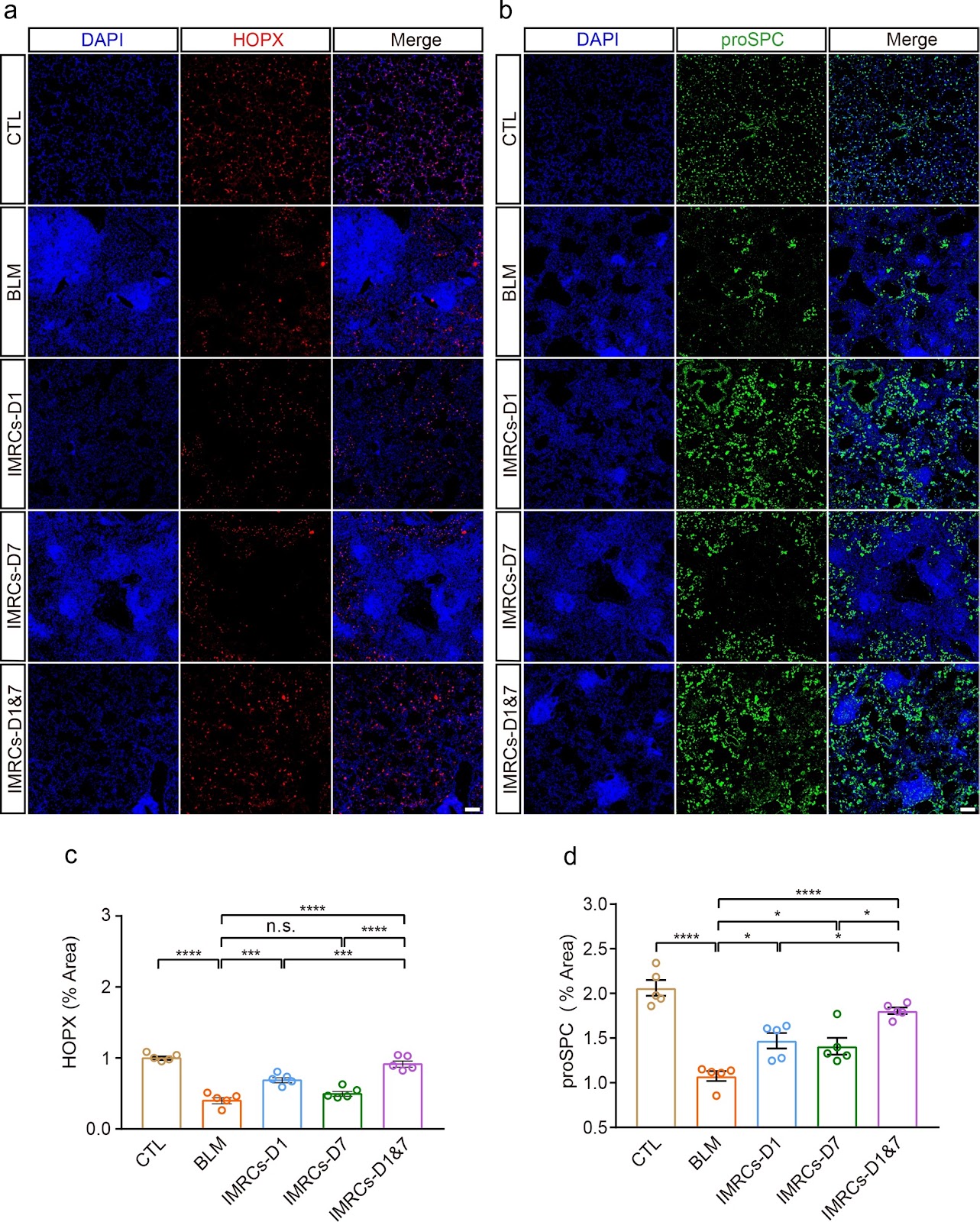
Optimized administration of human embryonic stem cell-derived immunity-and-matrix regulatory cells for mouse lung injury and fibrosis

Authors: Dingyun Song, Zhongwen Li, Faguo Sun, Kaiwei Wu, Kan Zhang, Wenjing Liu, Kaidi Liu, Bin An, Zai Wang, Tiemei Zhao, Huaiyong Chen, Li Xiao, Liu Wang, Lixin Xie, Wei Li, Liang Peng, Jie Hao, Jun Wu, Huaping Dai

Background Lung injury and pulmonary fibrosis (PF), frequently arising as sequelae of severe and acute lung disease, currently face a dearth of effective therapeutic potions. Mesenchymal stem cells (MSCs) with immunomodulatory and tissue repair functions have immense potential to treat lung injury and PF. However, the optimal route of administration, timing, and frequency of dosing remain elusive. Human embryonic stem cell-derived immunity-and-matrix-regulatory cells (IMRCs) have shown therapeutic potential for lung injury and PF. Methods To ascertain the optimal therapeutic regimen for IMRCs in PF, we conducted an experimental study. Utilizing a mouse model of PF induced by bleomycin (BLM), IMRCs were administered via either a single or double intravenous (IV) or intratracheal (IT) injection on the first and seventh days post-BLM induction. Results Our findings revealed that IV infusion of IMRCs surpassed IT infusion in enhancing survival rates, facilitating body weight recovery, and optimizing Ashcroft and Szapiel scores among the model mice. Notably, IV administration exhibited a more profound ability to mitigate lung inflammation and fibrosis. Moreover, earlier and more frequent administrations of IMRCs were found to be advantageous in enhancing their therapeutic effects. Specifically, early administration with two IV infusions significantly improved body weight, lung organ coefficient, pulmonary ventilation and diffusion functions, and PF. This was accompanied by an increase in alveolar type I and II epithelial cells and a suppression of macrophage infiltration via CD24. Conclusion Collectively, these results suggested that IMRCs infusion ameliorated lung injury by promoting lung regeneration and inhibiting macrophage infiltration in a route, time, and frequency-dependent manner.

Optimized administration of human embryonic stem cell-derived immunity-and-matrix regulatory cells for mouse lung injury and fibrosis
Graphical Abstract
Original ResearchVol. 15, None • pp. 189DOI: 10.1186/s13287-024-03813-1

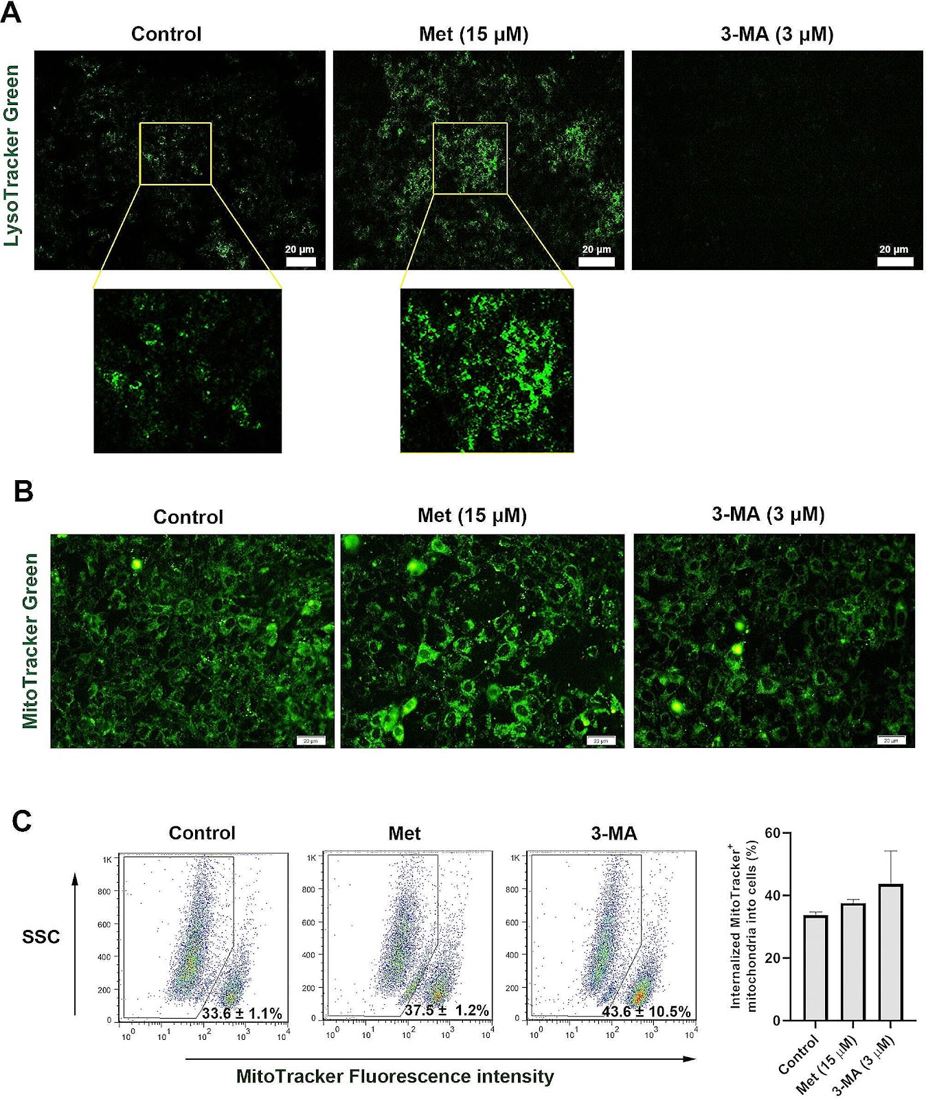
Autophagy modulation effect on homotypic transfer of intracellular components via tunneling nanotubes in mesenchymal stem cells

Authors: Fatemeh Sadeghsoltani, Çığır Biray Avci, Parisa Hassanpour, Sanya Haiaty, Mohamad Rahmati, Ali Mota, Reza Rahbarghazi, Maryam Nemati, Mahdi Mahdipour, Mehdi Talebi, Leila Sabour Takanlou, Maryam Sabour Takanlou, Amir Mehdizadeh

Background Recent studies have proved the role of autophagy in mesenchymal stem cell (MSCs) function and regenerative properties. How and by which mechanism autophagy modulation can affect the juxtacrine interaction of MSCs should be addressed. Here, the role of autophagy was investigated in the formation of tunneling nanotubes (TNTs) and homotypic mitochondrial donation. Methods MSCs were incubated with 15 µM Metformin (Met) and/or 3 µM 3-methyladenine (3-MA) for 48 h. The formation of TNTs was assessed using bright-field and SEM images. The mitochondria density and ΔΨ values were monitored using flow cytometry analysis. Using RT-PCR and protein array, the close interaction and shared mediators between autophagy, apoptosis, and Wnt signaling pathways were also monitored. The total fatty acid profile was assessed using gas chromatography. Result Data indicated the increase of TNT length and number, along with other cell projections after the induction of autophagy while these features were blunted in 3-MA-treated MSCs (p < 0.05). Western blotting revealed the significant reduction of Rab8 and p-FAK in 3-MA-treated MSCs (p < 0.05), indicating the inhibition of TNT assembly and vesicle transport. Likewise, the stimulation of autophagy increased autophagic flux and mitochondrial membrane integrity compared to 3-MA-treated MSCs. Despite these findings, protein levels of mitochondrial membrane Miro1 and 2 were unchanged after autophagy inhibition/stimulation (p > 0.05). We found that the inhibition/stimulation of autophagy can affect the protein, and transcription levels of several mediators related to Wnt and apoptosis signaling

Autophagy modulation effect on homotypic transfer of intracellular components via tunneling nanotubes in mesenchymal stem cells
Graphical Abstract
Original ResearchVol. 15, None • pp. 377DOI: 10.1186/s13287-024-03997-6

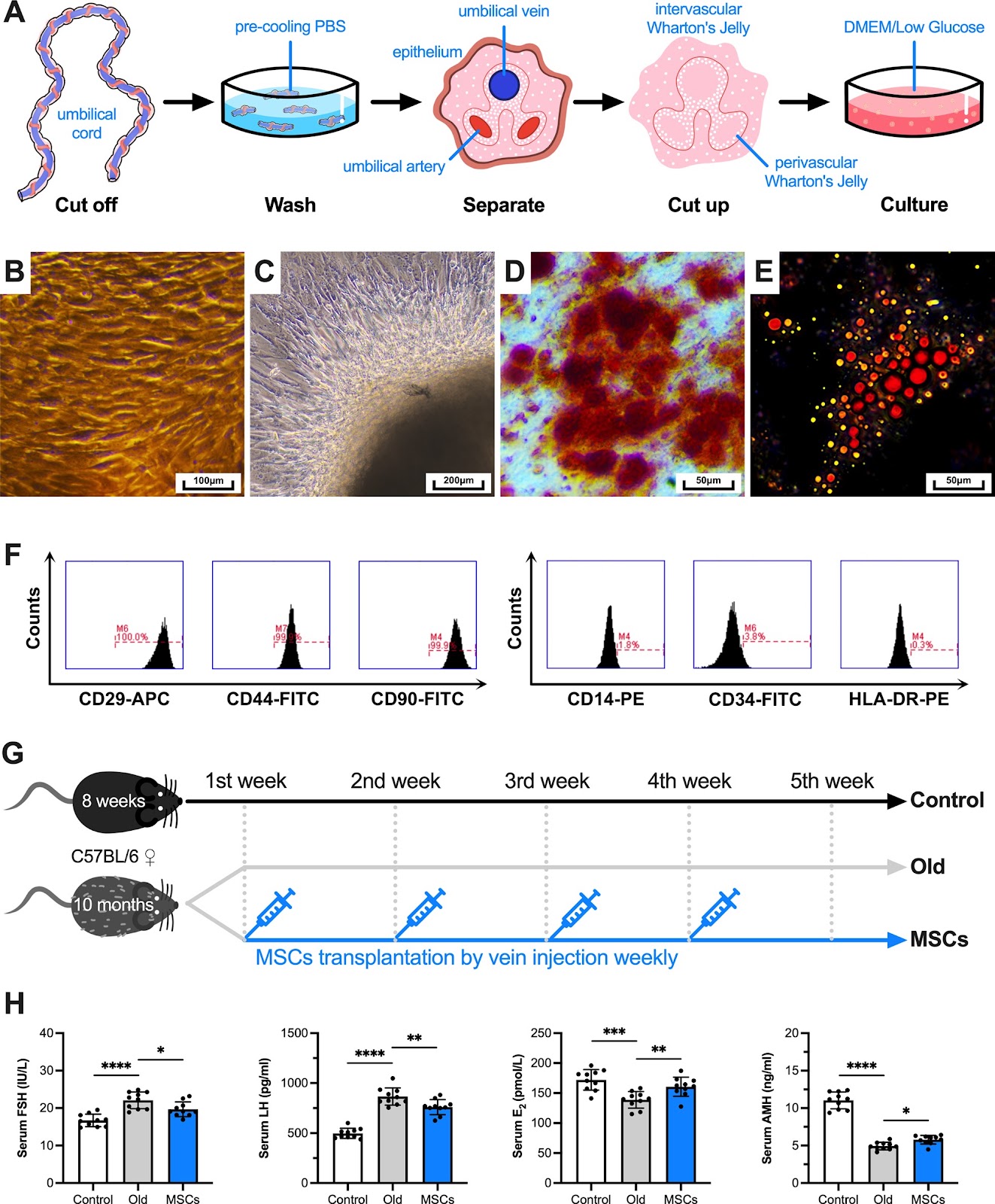
Transplantation of human umbilical cord-derived mesenchymal stem cells improves age-related ovarian functional decline via regulating the local renin–angiotensin system on inflammation and oxidative stress

Authors: Lun Wei, Le Bo, Chao Luo, Na Yin, Wangtao Jiang, Fei Qian, Anwen Zhou, Xuanping Lu, Huiping Guo, Caiping Mao

Background Age-related reproductive aging is a natural and irreversible physiological process, and delaying childbearing is increasingly common all over the world. Transplantation of mesenchymal stem cells (MSCs) is considered a new and effective therapy to restore ovarian function, but the relevant mechanisms remain unclear. Recently, it has been found that there is a local Renin–angiotensin system (RAS) in human ovary and it plays a key role. Methods After collecting follicular fluid from women who received oocyte retrieval for pure male factor infertility, the level of RAS components in it were detected, and the correlation analysis by linear regression. Then, the in vivo experiments on female C57BL/6 mice were designed to measure ovarian function, and the transcription and translation levels of RAS pathway were detected by molecular biology methods. Moreover, the role of RAS in regulating inflammation and oxidative stress in the co-culture system were explored in in vitro experiments on KGN cells. Results First, a total of 139 samples of analyzable follicular fluid were obtained. The local RAS of ovary, which is independent of systemic RAS (P > 0.05), is affected by age (Pearson r < 0, P < 0.05) and related to ovarian function, inflammation, oxidative stress indexes and assisted reproduction laboratory outcomes (P < 0.05). Next, the ovary/body weight of aging mice decreased significantly and serum sex hormones levels changed significantly (P < 0.01). The number of functional follicles decreased, while the atresia follicles increased (P < 0.05). After MSCs transplantation, all the above measures have been partially recovered (P < 0.05). Although several RAS components in aging ovary changed, MSCs only improved the expression level of AT1R (P < 0.05). Furthermore, the secretion ability and mitochondrial membrane potential of aging KGN cells decreased, while the intracellular ROS level and the aging cells ratio increased (P < 0.01). All the above measures have been partially recovered when co-cultured with MSCs (P < 0.05). After Ang(1–7) were added into the co-culture system, the above have been more significantly restored compared with Ang II (P < 0.05). Nevertheless, there was no statistical difference in estradiol level no matter which one was added (P > 0.05). Conclusions Together, our findings indicate that a novel possible mechanism to explain how stem cells restore age-related ovarian functional decline.

Transplantation of human umbilical cord-derived mesenchymal stem cells improves age-related ovarian functional decline via regulating the local renin–angiotensin system on inflammation and oxidative stress
Graphical Abstract
Original ResearchVol. 15, Issue 1 • pp. 33DOI: 10.1186/s13287-024-03639-x

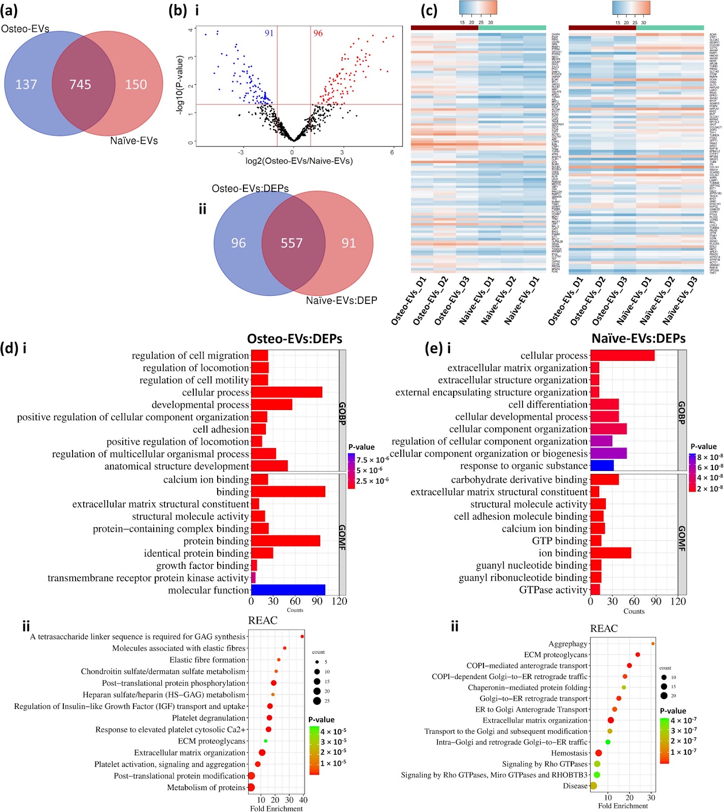
Osteogenic human MSC-derived extracellular vesicles regulate MSC activity and osteogenic differentiation and promote bone regeneration in a rat calvarial defect model

Authors: Niyaz Al-Sharabi, Samih Mohamed-Ahmed, Siddharth Shanbhag, Carina Kampleitner, Rammah Elnour, Shuntaro Yamada, Neha Rana, Even Birkeland, Stefan Tangl, Reinhard Gruber, Kamal Mustafa

Background There is growing evidence that extracellular vesicles (EVs) play a crucial role in the paracrine mechanisms of transplanted human mesenchymal stem cells (hMSCs). Little is known, however, about the influence of microenvironmental stimuli on the osteogenic effects of EVs. This study aimed to investigate the properties and functions of EVs derived from undifferentiated hMSC (Naïve-EVs) and hMSC during the early stage of osteogenesis (Osteo-EVs). A further aim was to assess the osteoinductive potential of Osteo-EVs for bone regeneration in rat calvarial defects. Methods EVs from both groups were isolated using size-exclusion chromatography and characterized by size distribution, morphology, flow cytometry analysis and proteome profiling. The effects of EVs (10 µg/ml) on the proliferation, migration, and osteogenic differentiation of cultured hMSC were evaluated. Osteo-EVs (50 µg) or serum-free medium (SFM, control) were combined with collagen membrane scaffold (MEM) to repair critical-sized calvarial bone defects in male Lewis rats and the efficacy was assessed using µCT, histology and histomorphometry. Results Although Osteo- and Naïve-EVs have similar characteristics, proteomic analysis revealed an enrichment of bone-related proteins in Osteo-EVs. Both groups enhance cultured hMSC proliferation and migration, but Osteo-EVs demonstrate greater efficacy in promoting in vitro osteogenic differentiation, as evidenced by increased expression of osteogenesis-related genes, and higher calcium deposition. In rat calvarial defects, MEM with Osteo-EVs led to greater and more consistent bone regeneration than MEM loaded with SFM. Conclusions This study discloses differences in the protein profile and functional effects of EVs obtained from naïve hMSC and hMSC during the early stage of osteogenesis, using different methods. The significant protein profile and cellular function of EVs derived from hMSC during the early stage of osteogenesis were further verified

Osteogenic human MSC-derived extracellular vesicles regulate MSC activity and osteogenic differentiation and promote bone regeneration in a rat calvarial defect model
Graphical Abstract
Original ResearchVol. 15, NoneDOI: 10.1186/s13287-024-04102-7

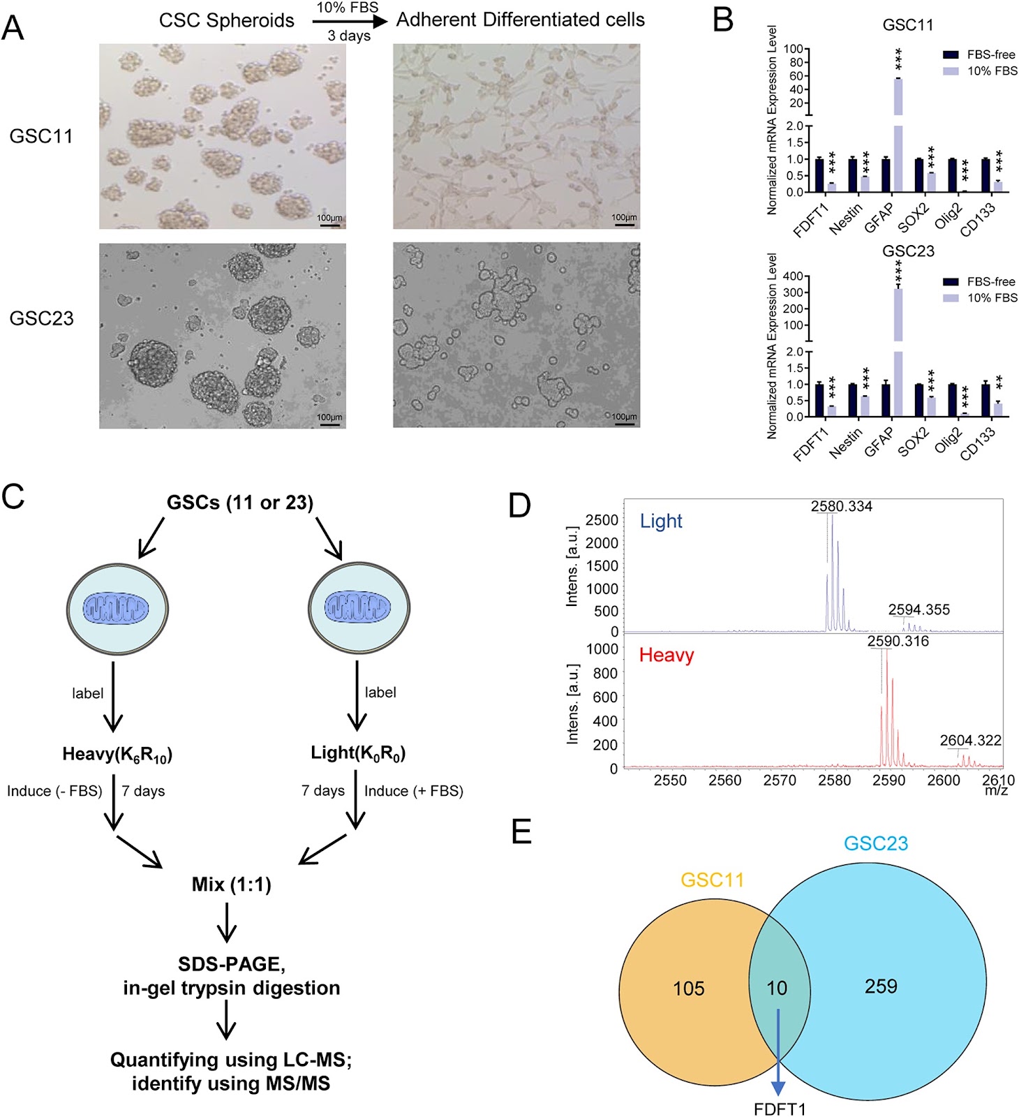
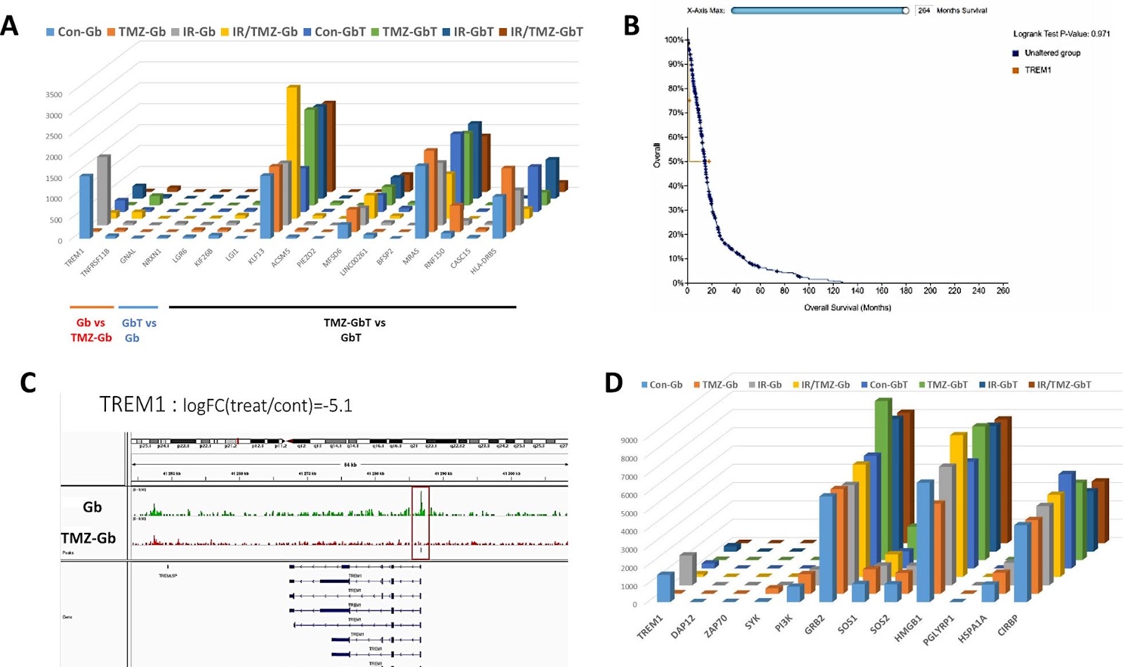
FDFT1 maintains glioblastoma stem cells through activation of the Akt pathway

Authors: Hui Mo, Jiajia Shao, Zhun Li, Peiting Zeng, Xinke Yin, Yongsheng Huang, Peng Wang, Jianwei Liao

Background Cancer stem cells (CSCs) have unique metabolic characteristics and are hypothesized to contribute significantly to the recurrence and drug resistance of glioblastoma multiforme (GBM). However, the reliance on mitochondrial metabolism and the underlying mechanism of glioblastoma stem cells (GSCs) remains to be elucidated. Methods To quantify differential mitochondrial protein expression between GSCs and differentiated cells, a mass spectrum screen was applied by the Stable Isotope Labeling with Amino Acids in Cell Culture (SILAC) technique. Functional experiments including CCK8, neurosphere formation, flow cytometry, transwell, and wound healing assays were conducted to evaluate GBM cell malignant phenotype. The potential molecular mechanism of FDFT1 was screened by RNA-seq analyses. The candidate target genes were validated through RT-qPCR and western blot analyses. Results As a top candidate, FDFT1 protein expression in GSCs was elevated relative to their differentiated counterparts. Functionally, the knockdown of FDFT1 suppressed the GBM cell proliferation and migration, while simultaneously enhancing sensitivity to temozolomide. Treatment with both the FDFT1 inhibitor (YM-53601) and simvastatin (an HMG-CoA reductase inhibitor) induced apoptosis in GSCs. Mechanistically, FDFT1 was transcriptionally regulated by SREBP2 but not SREBP1. Furthermore, FDFT1 activates the AKT pathway to regulate tumor metabolism and maintain the stemness of tumor cells. Conclusions GSCs exhibit a dependency on FDFT1-mediated mevalonate metabolism. Inhibition of FDFT1 could represent a potent strategy to eliminate GSCs.

FDFT1 maintains glioblastoma stem cells through activation of the Akt pathway
Graphical Abstract
Original ResearchVol. 15, Issue 1 • pp. 166DOI: 10.1186/s13287-024-03776-3

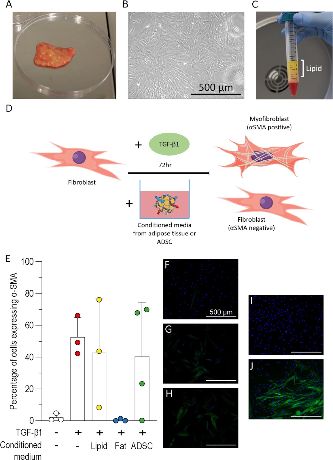
Inhibition and reversal of a TGF‑β1 induced myofibroblast phenotype by adipose tissue‑derived paracrine factors

Authors: S. Higginbotham, V. L. Workman, A-V. Giblin, N. H. Green, D. W. Lambert, V. Hearnden

Background Hypertrophic scarring results from myofibroblast differentiation and persistence during wound healing. Currently no effective treatment for hypertrophic scarring exists however, autologous fat grafting has been shown to improve scar elasticity, appearance, and function. The aim of this study was to understand how paracrine factors from adipose tissues and adipose-derived stromal cells (ADSC) affect fibroblast to myofibroblast differentiation. Methods The transforming growth factor-β1 (TGF-β1) induced model of myofibroblast differentiation was used to test the effect of conditioned media from adipose tissue, ADSC or lipid on the proportion of fibroblasts and myofibroblasts. Results Adipose tissue conditioned media inhibited the differentiation of fibroblasts to myofibroblasts but this inhibition was not observed following treatment with ADSC or lipid conditioned media. Hepatocyte growth factor (HGF) was readily detected in the conditioned medium from adipose tissue but not ADSC. Cells treated with HGF, or fortinib to block HGF, demonstrated that HGF was not responsible for the inhibition of myofibroblast differentiation. Conditioned media from adipose tissue was shown to reduce the proportion of myofibroblasts when added to fibroblasts previously treated with TGF-β1, however, conditioned media treatment was unable to significantly reduce the proportion of myofibroblasts in cell populations isolated from scar tissue. Conclusions Cultured ADSC or adipocytes have been the focus of most studies, however, this work highlights the importance of considering whole adipose tissue to further our understanding of fat grafting. This study supports the use of autologous fat grafts for scar treatment and highlights the need for further investigation to determine the mechanism.

Inhibition and reversal of a TGF‑β1 induced myofibroblast phenotype by adipose tissue‑derived paracrine factors
Graphical Abstract
Original ResearchVol. 15, Issue 1 • pp. 23DOI: 10.1186/s13287-024-03636-0

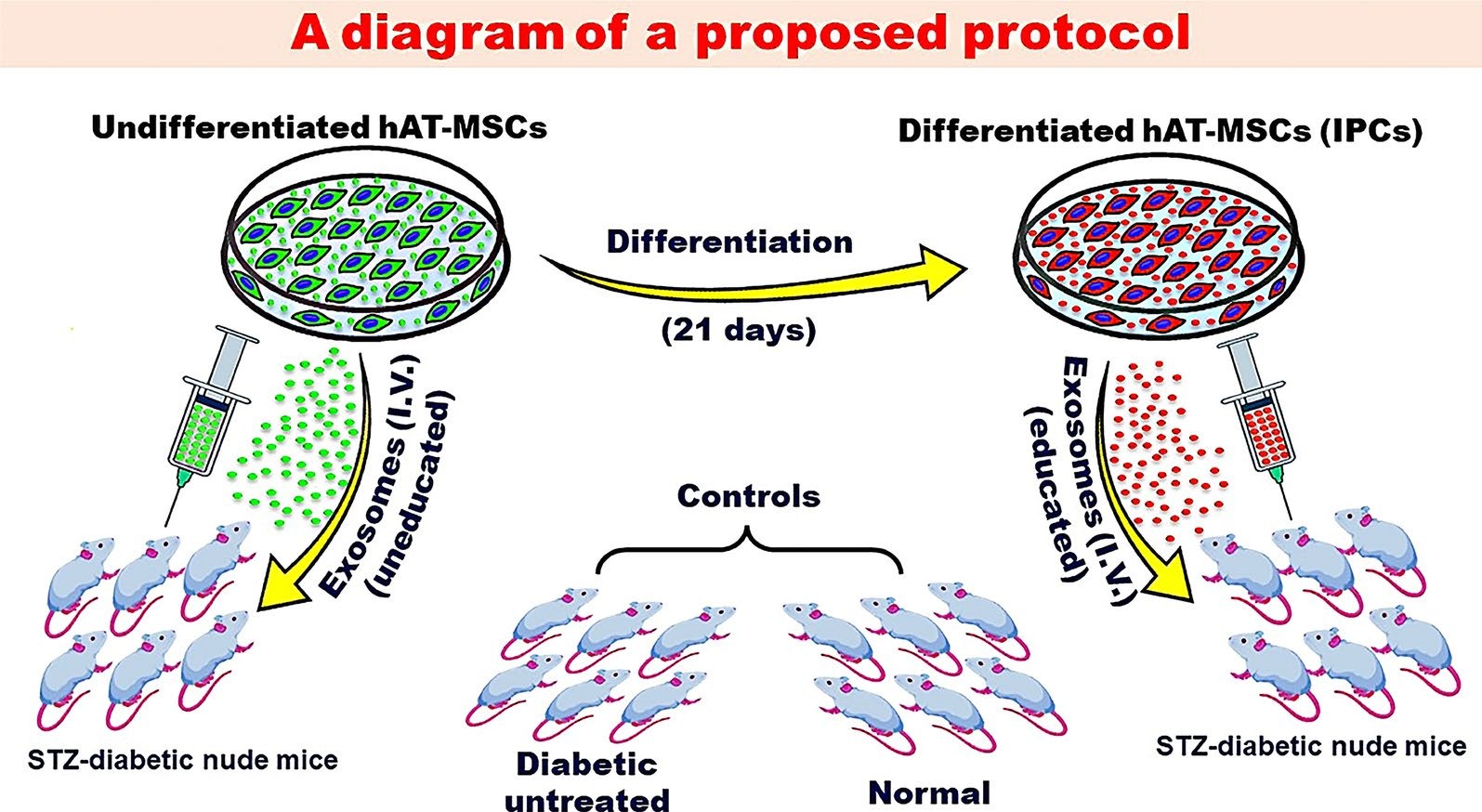
Current status of stem cell therapy for type 1 diabetes: a critique and a prospective consideration

Authors: Mohamed A. Ghoneim, Mahmoud M. Gabr, Sawsan M. El-Halawani, Ayman F. Refaie

Over the past decade, there had been progress in the development of cell therapy for insulin-dependent diabetes. Nevertheless, important hurdles that need to be overcome still remain. Protocols for the differentiation of pluripotent stem cells into pancreatic progenitors or fully differentiated β-cells have been developed. The resulting insulin-producing cells can control chemically induced diabetes in rodents and were the subject of several clinical trials. However, these cells are immunogenic and possibly teratogenic for their transplantation, and an immunoisolation device and/or immunosuppression is needed. A growing number of studies have utilized genetic manipulations to produce immune evasive cells. Evidence must be provided that in addition to the expected benefit, gene manipulations should not lead to any unforeseen complications. Mesenchymal stem/stromal cells (MSCs) can provide a viable alternative. MSCs are widely available from many tissues. They can form insulin-producing cells by directed differentiation. Experimentally, evidence has shown that the transplantation of allogenic insulin-producing cells derived from MSCs is associated with a muted allogeneic response that does not interfere with their functionality. This can be explained by the immunomodulatory functions of the MSC subpopulation that did not differentiate into insulin-producing cells. Recently, exosomes derived from naive MSCs have been used in the experimental domain to treat diabetes in rodents with varying degrees of success. Several mechanisms for their beneficial functions were proposed including a reduction in insulin resistance, the promotion of autophagy, and an increase in the T regulatory population. However, euglycemia was not achieved in any of these experiments. We suggest that exosomes derived from β-cells or insulin-producing cells (educated) can provide a better therapeutic effect than those derived from undifferentiated cells.

Current status of stem cell therapy for type 1 diabetes: a critique and a prospective consideration
Graphical Abstract
Original ResearchVol. 15, None • pp. 251DOI: 10.1186/s13287-024-03872-4

Characteristics of HPC(A) product obtained from a donor with SARS-CoV-2 infection and outcome of autologous transplant

Authors: Pranav S. Renavikar, Phyllis I. Warkentin, Shelly M. Williams, Krishna Gundabolu, Charles Branson, Scott A. Koepsell

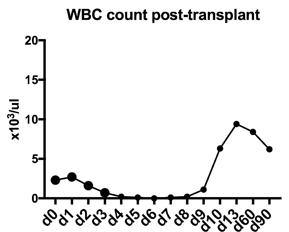
Collection of hematopoietic progenitor cell products [HPC(A)] is deferred if the donor is symptomatic and tests positive for Covid-19. However, donor questionnaires are subjective and may miss minimally symptomatic donors. Alternatively, myalgia associated with Covid-19 infection can be falsely dismissed as an adverse effect of granulocyte stimulating factor (Filgrastim) administered prior to product collection. The likelihood of donors with an underlying acute but minimally symptomatic infection undergoing successful product collection is significant. In these circumstances, it is less known whether Covid-19 infection results in product viremia or alters the clinical outcome of transplant. We aimed to evaluate the above question by studying a donor whose product was collected during acute Covid-19 infection. Aliquots of the product tested negative for SARS-CoV-2 RNA by reverse-transcriptase polymerase chain reaction assay (RT-PCR). Importantly, the donor received an autologous stem cell transplant using the product collected at the time of infection, and their case will be described in this report. We describe one of the very few reports of successful transplant of HPC(A) product collected during acute Covid-19 infection.

Characteristics of HPC(A) product obtained from a donor with SARS-CoV-2 infection and outcome of autologous transplant
Graphical Abstract
Original ResearchVol. 15, None • pp. 373DOI: 10.1186/s13287-024-03990-z

The role of biophysical cues and their modulated exosomes in dental diseases: from mechanism to therapy

Authors: Bilun Jin, Yuxin Liao, Zhaojing Ding, Rui Zou, Feng Xu, Ye Li, Bo Cheng, Lin Niu

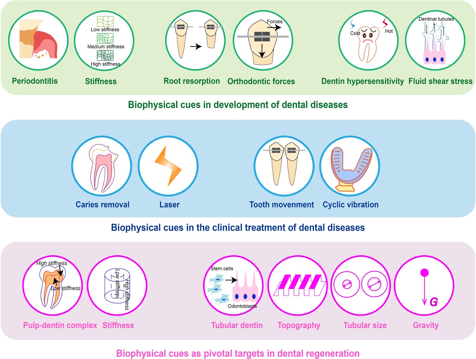
Dental diseases such as caries and periodontitis have been common public health problems. Dental disease treatment can be achieved through stem cell-based dental regeneration. Biophysical cues determine the fate of stem cells and govern the success of dental regeneration. Some studies have manifested exosomes derived from stem cells could not only inherit biophysical signals in microenvironment but also evade some issues in the treatment with stem cells. Nowadays, biophysical cue-regulated exosomes become another promising therapy in dental regenerative medicine. However, methods to improve the efficacy of exosome therapy and the underlying mechanisms are still unresolved. In this review, the association between biophysical cues and dental diseases was summarized. We retrospected the role of exosomes regulated by biophysical cues in curing dental diseases and promoting dental regeneration. Our research also delved into the mechanisms by which biophysical cues control the biogenesis, release, and uptake of exosomes, as well as potential methods to enhance the effectiveness of exosomes. The aim of this review was to underscore the important place biophysical cue-regulated exosomes occupy in the realm of dentistry, and to explore novel targets for dental diseases.

The role of biophysical cues and their modulated exosomes in dental diseases: from mechanism to therapy
Graphical Abstract
Original ResearchVol. 15, Issue 1 • pp. 427DOI: 10.1186/s13287-024-04025-3

Astilbin improves the therapeutic effects of mesenchymal stem cells in AKI-CKD mice by regulating macrophage polarization through PTGS2-mediated pathway

Authors: Xiaodong Geng, Zhangning Fu, Guangrui Geng, Kun Chi, Chao Liu, Haijuan Hong, Guangyan Cai, Xiangmei Chen, Quan Hong

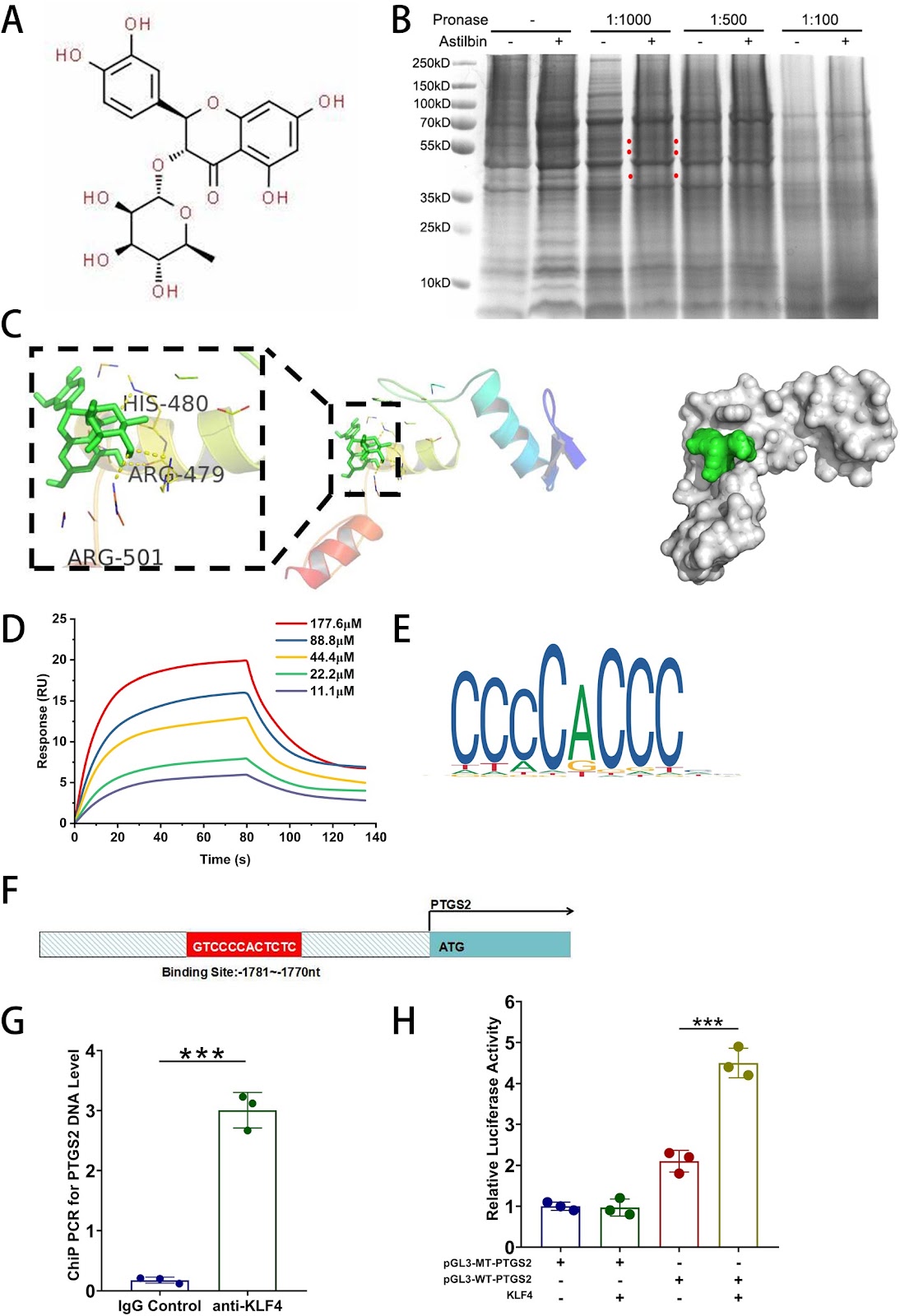
Background Although mesenchymal stem cells (MSCs) have been proven to be appropriate candidates for the treatment of AKI-CKD, their efficacy is limited and variable. Astilbin (AST) had a protective effect on MSCs from oxidative stress via ROS-scavenging, however, whether it can improve MSCs’ renoprotection and the underlying mechanism need to be elucidated. Methods AST-pretreated MSCs were administered intravenously into the ischemia–reperfusion injury mice models and the renal function, pathological changes and inflammation. Were evaluated. In addition, DARTS, molecular docking, surface plasma resonance(SPR), dual-luciferase reporter gene assay and the ChIP-PCR were utilized to explore the potential signaling pathways through which AST exert renal protective effects on MSCs. Results AST-pretreated MSCs markedly improved kidney function, reduced kidney pathological injury and inflammation in AKI and AKI-CKD mice. RNA-seq results showed that PTGS2 related pathway was significantly up-regulated in MSCs after AST pretreatment. DARTS assay, molecular docking and SPR assay revealed that AST could bind with the transcriptional factor of Kruppel-Like Factor 4(KLF4) protein. The promoter of PTGS2 had the binding and transcriptional activation by KLF4. Furthermore, AST pretreatment promoted the secretion of PGE2 in MSCs. And then the westren blot results showed that the protein levels of CD163 and CD206 were upregulated after coculture in AST-pretreated MSCs, indicating that the polarization of RAW264.7 cells towards M2-like macrophages was induced. Knockdown of PTGS2 reversed the ability of AST-pretreated MSCs in converting macrophages to M2 phenotype and reducing their therapeutic effects on AKI-CKD mice. Conclusion AST pretreatment enhances the efficacy of MSCs on AKI and AKI-CKD mice by inducing of M2-like phenotype polarization in macrophages through the PTGS2-mediated pathway. This approach not only provides a novel strategy to strengthen the capability of MSCs but also helps elucidate the beneficial effects of the Chinese herbal medicine AST.

Astilbin improves the therapeutic effects of mesenchymal stem cells in AKI-CKD mice by regulating macrophage polarization through PTGS2-mediated pathway
Graphical Abstract
Original ResearchVol. 15, Issue 1 • pp. 87DOI: 10.1186/s13287-024-03689-1

Identification of potential biomarkers for aging diagnosis of mesenchymal stem cells derived from the aged donors

Authors: Miao Hao, Hongyu Jiang, Yuan Zhao, Chunyi Li, Jinlan Jiang

Background The clinical application of human bone-marrow derived mesenchymal stem cells (MSCs) for the treatment of refractory diseases has achieved remarkable results. However, there is a need for a systematic evaluation of the quality and safety of MSCs sourced from donors. In this study, we sought to assess one potential factor that might impact quality, namely the age of the donor. Methods We downloaded two data sets from each of two Gene Expression Omnibus (GEO), GSE39035 and GSE97311 databases, namely samples form young (<65 years of age) and old (>65) donor groups. Through, bioinformatics analysis and experimental validation to these retrieved data, we found that MSCs derived from aged donors can lead to differential expression of gene profiles compared with those from young donors, and potentially affect the function of MSCs, and may even induce malignant tumors. Results We identified a total of 337 differentially expressed genes (DEGs), including two upregulated and eight downregulated genes from the databases of both GSE39035 and GSE97311. We further identified 13 hub genes. Six of them, TBX15, IGF1, GATA2, PITX2, SNAI1 and VCAN, were highly expressed in many human malignancies in Human Protein Atlas database. In the MSCs in vitro senescent cell model, qPCR analysis validated that all six hub genes were highly expressed in senescent MSCs. Our findings confirm that aged donors of MSCs have a significant effect on gene expression profiles. The MSCs from old donors have the potential to cause a variety of malignancies. These TBX15, IGF1, GATA2, PITX2, SNAI1, VCAN genes could be used as potential biomarkers to diagnosis aging state of donor MSCs, and evaluate whether MSCs derived from an aged donor could be used for therapy in the clinic. Our findings provide a diagnostic basis for the clinical use of MSCs to treat a variety of diseases. Conclusions Therefore, our findings not only provide guidance for the safe and standardized use of MSCs in the clinic for the treatment of various diseases, but also provide insights into the use of cell regeneration approaches to reverse aging and support rejuvenation.

Identification of potential biomarkers for aging diagnosis of mesenchymal stem cells derived from the aged donors
Graphical Abstract
Original ResearchVol. 15, None • pp. 203DOI: 10.1186/s13287-024-03803-3

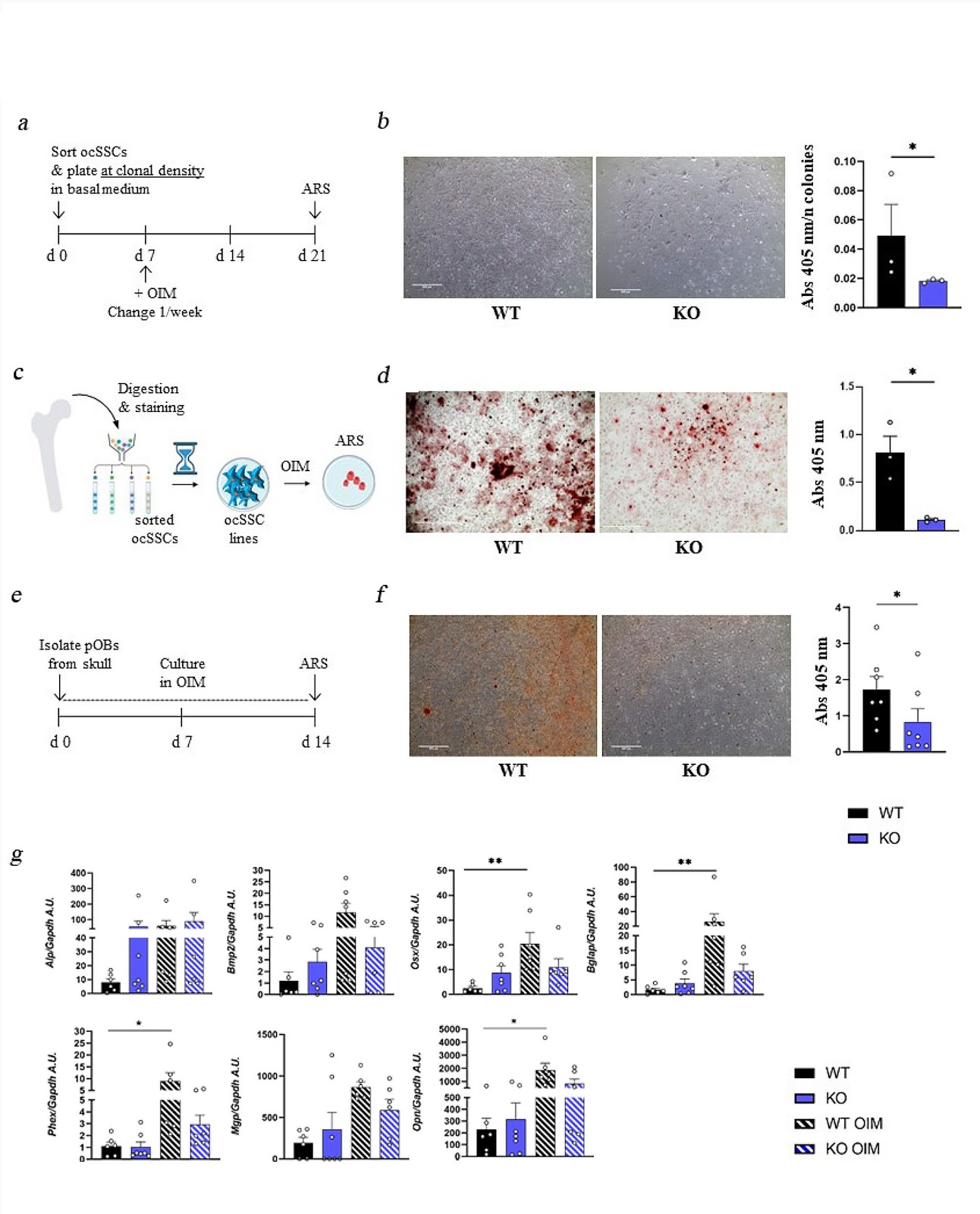
Rankl genetic deficiency and functional blockade undermine skeletal stem and progenitor cell differentiation

Authors: M. L. Schiavone, L. Crisafulli, C. Camisaschi, G. De Simone, F. R. Liberati, E. Palagano, N. Rucci, F. Ficara, Cristina Sobacchi

Background Skeletal Stem Cells (SSCs) are required for skeletal development, homeostasis, and repair. The perspective of their wide application in regenerative medicine approaches has supported research in this field, even though so far results in the clinic have not reached expectations, possibly due also to partial knowledge of intrinsic, potentially actionable SSC regulatory factors. Among them, the pleiotropic cytokine RANKL, with essential roles also in bone biology, is a candidate deserving deep investigation. Methods To dissect the role of the RANKL cytokine in SSC biology, we performed ex vivo characterization of SSCs and downstream progenitors (SSPCs) in mice lacking Rankl (Rankl−/−) by means of cytofluorimetric sorting and analysis of SSC populations from different skeletal compartments, gene expression analysis, and in vitro osteogenic differentiation. In addition, we assessed the effect of the pharmacological treatment with the anti-RANKL blocking antibody Denosumab (approved for therapy in patients with pathological bone loss) on the osteogenic potential of bone marrow-derived stromal cells from human healthy subjects (hBMSCs). Results We found that, regardless of the ossification type of bone, osteochondral SSCs had a higher frequency and impaired differentiation along the osteochondrogenic lineage in Rankl−/− mice as compared to wild-type. Rankl−/− mice also had increased frequency of committed osteochondrogenic and adipogenic progenitor cells deriving from perivascular SSCs. These changes were not due to the peculiar bone phenotype of increased density caused by lack of osteoclast resorption (defined osteopetrosis); indeed, they were not found in another osteopetrotic mouse model, i.e., the oc/oc mouse, and were therefore not due to osteopetrosis per se. In addition, Rankl−/− SSCs and primary osteoblasts showed reduced mineralization capacity. Of note, hBMSCs treated in vitro with Denosumab had reduced osteogenic capacity compared to control cultures. Conclusions We provide for the first time the characterization of SSPCs from mouse models of severe recessive osteopetrosis. We demonstrate that Rankl genetic deficiency in murine SSCs and functional blockade in hBMSCs reduce their osteogenic potential. Therefore, we propose that RANKL is an important regulatory factor of SSC features with translational relevance.

Rankl genetic deficiency and functional blockade undermine skeletal stem and progenitor cell differentiation
Graphical Abstract
Original ResearchVol. 15, None • pp. 277DOI: 10.1186/s13287-024-03901-2

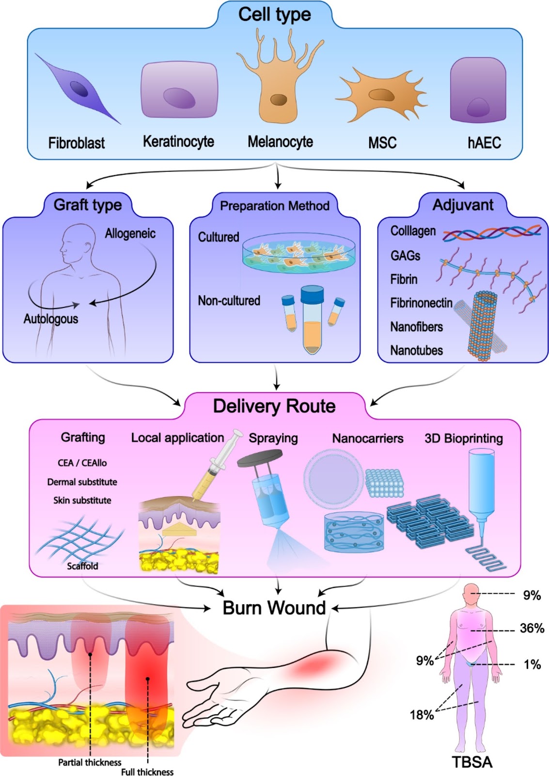
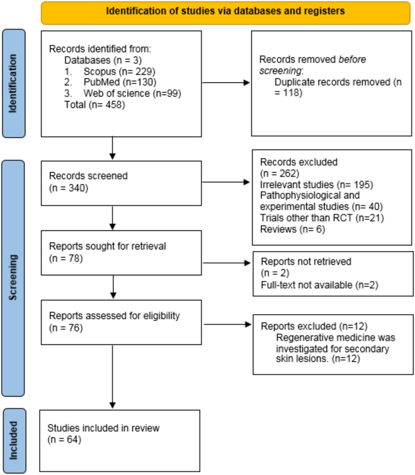
Advancements in cell-based therapies for thermal burn wounds: a comprehensive systematic review of clinical trials outcomes

Authors: Younes Yassaghi, Yasaman Nazerian, Feizollah Niazi, Hassan Niknejad

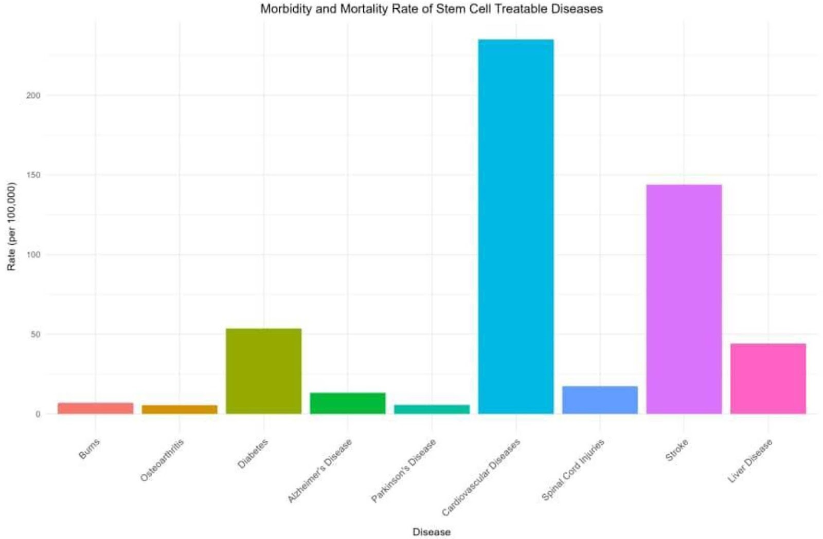
Background Burn trauma is one of the major causes of morbidity and mortality worldwide. The standard management of burn wounds consists of early debridement, dressing changes, surgical management, and split-thickness skin autografts (STSGs). However, there are limitations for the standard management that inclines us to find alternative treatment approaches, such as innovative cell-based therapies. We aimed to systematically review the different aspects of cell-based treatment approaches for burn wounds in clinical trials. Methods A systematic search through PubMed, Medline, Embase, and Cochrane Library databases was carried out using a combination of keywords, including “Cell transplantation”, “Fibroblast”, “Keratinocyte”, “Melanocyte”, or “Stem Cell” with “Burn”, “Burn wound”, or “Burn injury”. Firstly, titles and abstracts of the studies existing in these databases until “February 2024” were screened. Then, the selected studies were read thoroughly, and considering the inclusion and exclusion criteria, final articles were included in this systematic review. Moreover, a manual search was performed through the reference lists of the included studies to minimize the risk of missing reports. Results Overall, 30 clinical trials with 970 patients were included in our study. Considering the type of cells, six studies used keratinocytes, nine used fibroblasts, eight used combined keratinocytes and fibroblasts, one study used combined keratinocytes and melanocytes, five used combined keratinocytes and fibroblasts and melanocytes, and one study used mesenchymal stem cells (MSCs). Evaluation of the preparation type in these studies showed that cultured method was used in 25 trials, and non-cultured method in 5 trials. Also, the graft type of 17 trials was allogeneic, and of 13 other trials was autologous. Conclusions Our study showed that employing cell-based therapies for the treatment of burn wounds have significant results in clinical studies and are promising approaches that can be considered as alternative treatments in many cases. However, choosing appropriate cell-based treatment for each burn wound is essential and depends on the situation of each patient.

Advancements in cell-based therapies for thermal burn wounds: a comprehensive systematic review of clinical trials outcomes
Graphical Abstract
Original ResearchVol. 15, None • pp. 115DOI: 10.1186/s13287-024-03718-z

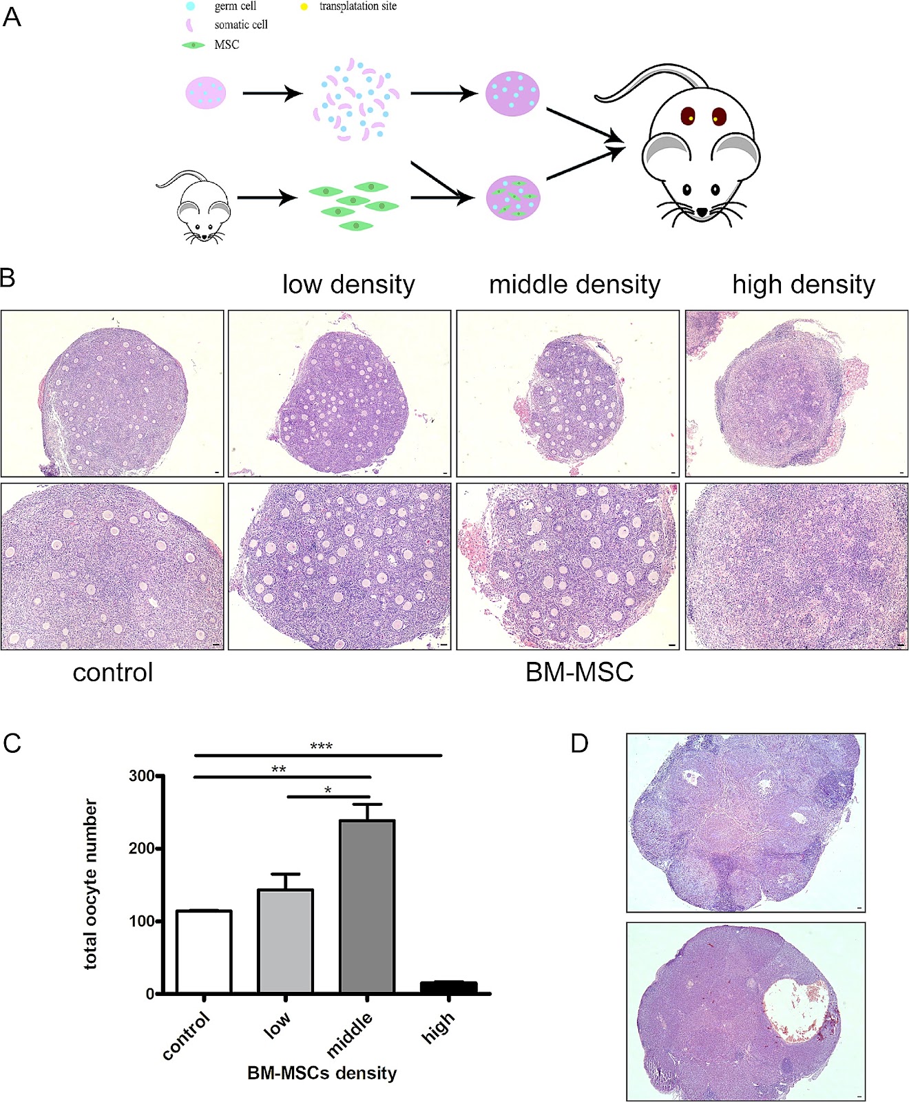
Mesenchymal stem cells promote ovarian reconstruction in mice

Authors: Jiazhao Li, Haonan Fan, Wei Liu, Jing Zhang, Yue Xiao, Yue Peng, Weijie Yang, Wenwen Liu, Yuanlin He, Lianju Qin, Xiang Ma, Jing Li

Background Studies have shown that chemotherapy and radiotherapy can cause premature ovarian failure and loss of fertility in female cancer patients. Ovarian cortex cryopreservation is a good choice to preserve female fertility before cancer treatment. Following the remission of the disease, the thawed ovarian tissue can be transplanted back and restore fertility of the patient. However, there is a risk to reintroduce cancer cells in the body and leads to the recurrence of cancer. Given the low success rate of current in vitro culture techniques for obtaining mature oocytes from primordial follicles, an artificial ovary with primordial follicles may be a good way to solve this problem. Methods In the study, we established an artificial ovary model based on the participation of mesenchymal stem cells (MSCs) to evaluate the effect of MSCs on follicular development and oocyte maturation. P2.5 mouse ovaries were digested into single cell suspensions and mixed with bone marrow derived mesenchymal stem cells (BM-MSCs) at a 1:1 ratio. The reconstituted ovarian model was then generated by using phytohemagglutinin. The phenotype and mechanism studies were explored by follicle counting, immunohistochemistry, immunofluorescence, in vitro maturation (IVM), in vitro fertilization (IVF), real-time quantitative polymerase chain reaction (RT-PCR), and Terminal-deoxynucleotidyl transferase mediated nick end labeling(TUNEL) assay. Results Our study found that the addition of BM-MSCs to the reconstituted ovary can enhance the survival of oocytes and promote the growth and development of follicles. After transplanting the reconstituted ovaries under kidney capsules of the recipient mice, we observed normal folliculogenesis and oocyte maturation. Interestingly, we found that BM-MSCs did not contribute to the formation of follicles in ovarian aggregation, nor did they undergo proliferation during follicle growth. Instead, the cells were found to be located around growing follicles in the reconstituted ovary. When theca cells were labeled with CYP17a1, we found some overlapped staining with green fluorescent protein(GFP)-labeled BM-MSCs. The results suggest that BM-MSCs may participate in directing the differentiation of theca layer in the reconstituted ovary. Conclusions The presence of BM-MSCs in the artificial ovary was found to promote the survival of ovarian cells, as well as facilitate follicle formation and development. Since the cells didn’t proliferate in the reconstituted ovary, this

Mesenchymal stem cells promote ovarian reconstruction in mice
Graphical Abstract
Original ResearchVol. 15, None • pp. 177DOI: 10.1186/s13287-024-03793-2

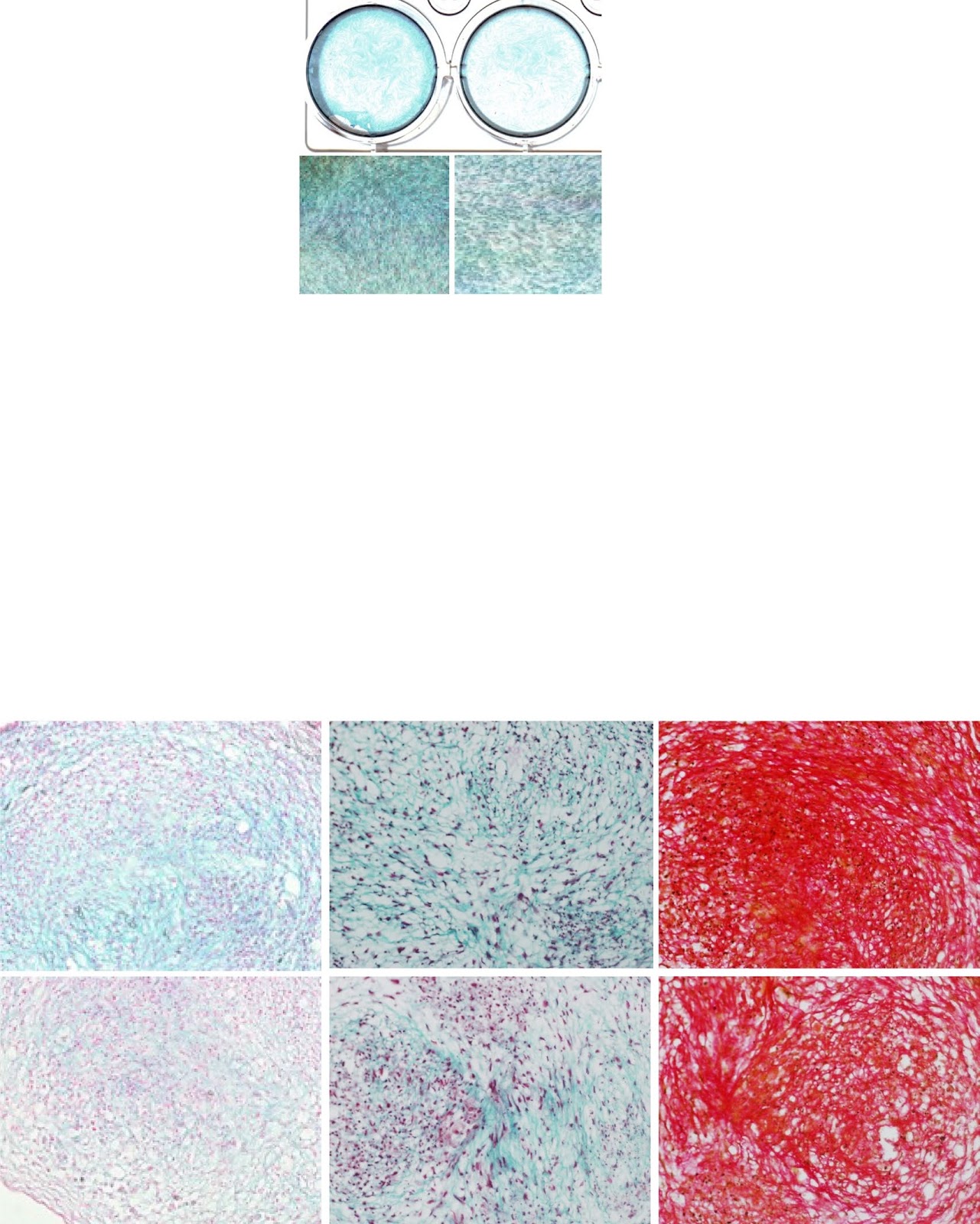
LncRNA SNHG1 enhances cartilage regeneration by modulating chondrogenic differentiation and angiogenesis potentials of JBMMSCs via mitochondrial function regulation

Authors: Hua Liu, Huina Liu, Qiubo Yang, Zhipeng Fan

Background Cartilage is a kind of avascular tissue, and it is difficult to repair itself when it is damaged. In this study, we investigated the regulation of chondrogenic differentiation and vascular formation in human jaw bone marrow mesenchymal stem cells (h-JBMMSCs) by the long-chain noncoding RNA small nucleolar RNA host gene 1 (SNHG1) during cartilage tissue regeneration. Methods JBMMSCs were isolated from the jaws via the adherent method. The effects of lncRNA SNHG1 on the chondrogenic differentiation of JBMMSCs in vitro were detected by real-time fluorescence quantitative polymerase chain reaction (RT-qPCR), Pellet experiment, Alcian blue staining, Masson’s trichrome staining, and modified Sirius red staining. RT-qPCR, matrix gel tube formation, and coculture experiments were used to determine the effect of lncRNA SNHG1 on the angiogenesis in JBMMSCs in vitro. A model of knee cartilage defects in New Zealand rabbits and a model of subcutaneous matrix rubber suppositories in nude mice were constructed for in vivo experiments. Changes in mitochondrial function were detected via RT-qPCR, dihydroethidium (DHE) staining, MitoSOX staining, tetramethyl rhodamine methyl ester (TMRM) staining, and adenosine triphosphate (ATP) detection. Western blotting was used to detect the phosphorylation level of signal transducer and activator of transcription 3 (STAT3). Results Alcian blue staining, Masson’s trichrome staining, and modified Sirius Red staining showed that lncRNA SNHG1 promoted chondrogenic differentiation. The lncRNA SNHG1 promoted angiogenesis in vitro and the formation of microvessels in vivo. The lncRNA SNHG1 promoted the repair and regeneration of rabbit knee cartilage tissue. Western blot and alcian blue staining showed that the JAK inhibitor reduced the increase of STAT3 phosphorylation level and staining deepening caused by SNHG1. Mitochondrial correlation analysis revealed that the lncRNA SNHG1 led to a decrease in reactive oxygen species (ROS) levels, an increase in mitochondrial membrane potential

LncRNA SNHG1 enhances cartilage regeneration by modulating chondrogenic differentiation and angiogenesis potentials of JBMMSCs via mitochondrial function regulation
Graphical Abstract
Original ResearchVol. 15, Issue 1 • pp. 63DOI: 10.1186/s13287-024-03676-6

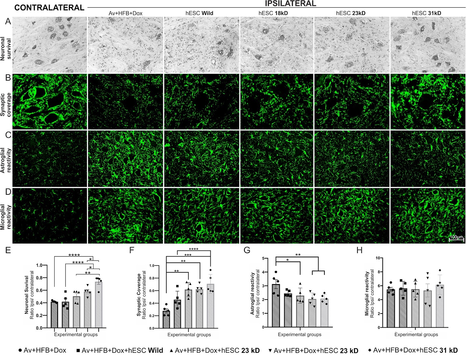
Embryonic stem cells overexpressing high molecular weight FGF2 isoform enhance recovery of pre-ganglionic spinal root lesion in combination with fibrin biopolymer mediated root repair

Authors: B. H. M. Lima, L. P. Cartarozzi, S. Kyrylenko, R. S. Ferreira Jr., B. Barraviera, Alexandre L. R. Oliveira

Background Spinal ventral root avulsion results in massive motoneuron degeneration with poor prognosis and high costs. In this study, we compared different isoforms of basic fibroblast growth factor 2 (FGF2), overexpressed in stably transfected Human embryonic stem cells (hESCs), following motor root avulsion and repair with a heterologous fibrin biopolymer (HFB). Methods In the present work, hESCs bioengineered to overexpress 18, 23, and 31 kD isoforms of FGF2, were used in combination with reimplantation of the avulsed roots using HFB. Statistical analysis was conducted using GraphPad Prism software with one-way or two-way ANOVA, followed by Tukey’s or Dunnett’s multiple comparison tests. Significance was set at *p < 0.05, **p < 0.01, ***p < 0.001, and ****p < 0.0001. Results For the first set of experiments, rats underwent avulsion of the ventral roots with local administration of HFB and engraftment of hESCs expressing the above-mentioned FGF2 isoforms. Analysis of motoneuron survival, glial reaction, and synaptic coverage, two weeks after the lesion, indicated that therapy with hESCs overexpressing 31 kD FGF2 was the most effective. Consequently, the second set of experiments was performed with that isoform, so that ventral root avulsion was followed by direct spinal cord reimplantation. Motoneuron survival, glial reaction, synaptic coverage, and gene expression were analyzed 2 weeks post-lesion; while the functional recovery was evaluated by the walking track test and von Frey test for 12 weeks. We showed that engraftment of hESCs led to significant neuroprotection, coupled with immunomodulation, attenuation of astrogliosis, and preservation of inputs to the rescued motoneurons. Behaviorally, the 31 kD FGF2 - hESC therapy enhanced both motor and sensory recovery. Conclusion Transgenic hESCs were an effective delivery platform for neurotrophic factors, rescuing axotomized motoneurons and modulating glial response after proximal spinal cord root injury, while the 31 kD isoform of FGF2 showed superior regenerative properties over other isoforms in addition to the significant functional recovery.

Embryonic stem cells overexpressing high molecular weight FGF2 isoform enhance recovery of pre-ganglionic spinal root lesion in combination with fibrin biopolymer mediated root repair
Graphical Abstract
Original ResearchVol. 15, None • pp. 224DOI: 10.1186/s13287-024-03846-6

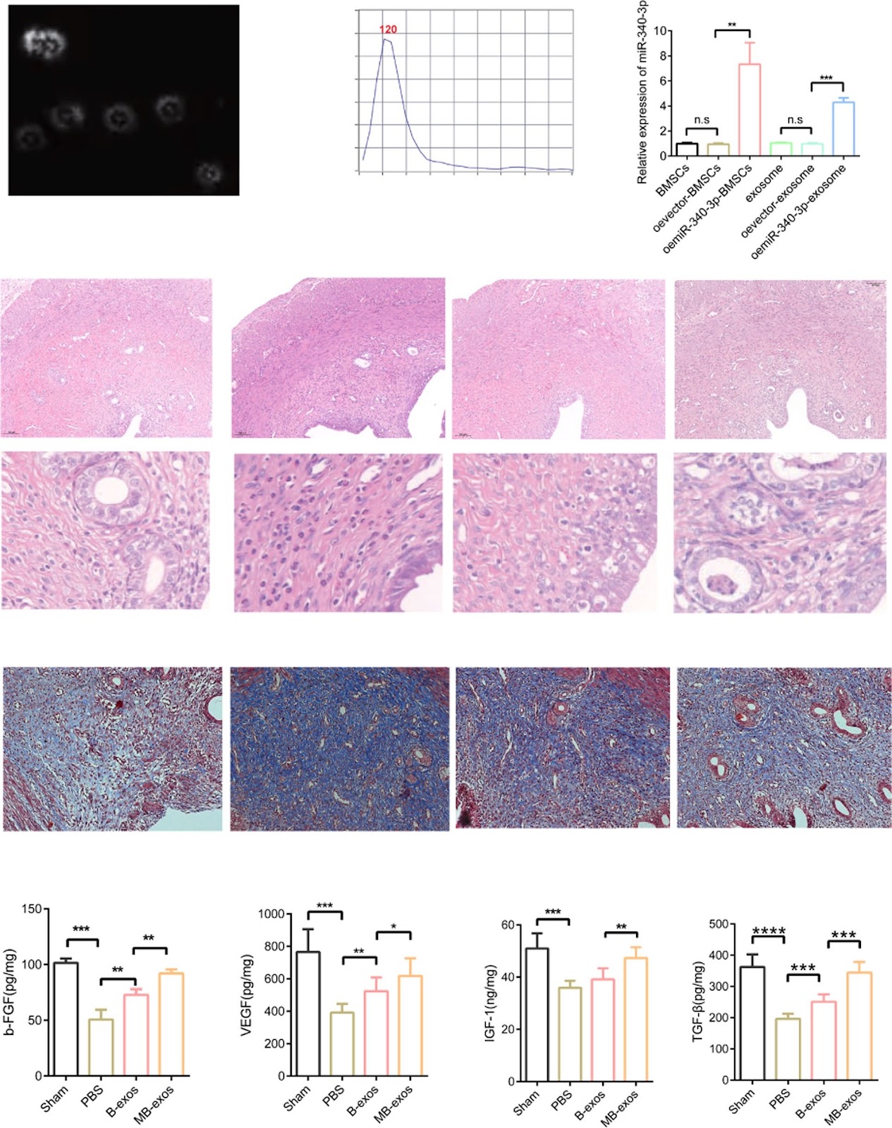
miR-340-3p-modified bone marrow mesenchymal stem cell-derived exosomes inhibit ferroptosis through METTL3-mediated m6A modification of HMOX1 to promote recovery of injured rat uterus

Authors: Bang Xiao, Yiqing Zhu, Meng Liu, Meiting Chen, Chao Huang, Dabing Xu, Fang Wang, Shuhan Sun, Jinfeng Huang, Ningxia Sun, Fu Yang

Background Ferroptosis is associated with the pathological progression of hemorrhagic injury and ischemia–reperfusion injury. According to our previous study, exosomes formed through bone marrow mesenchymal stem cells modified with miR-340-3p (MB-exos) can restore damaged endometrium. However, the involvement of ferroptosis in endometrial injury and the effect of MB-exos on ferroptosis remain elusive. Methods The endometrial injury rat model was developed. Exosomes were obtained from the supernatants of bone marrow mesenchymal stromal cells (BMSCs) and miR-340/BMSCs through differential centrifugation. We conducted RNA-seq analysis on endometrial tissues obtained from the PBS and MB-exos groups. Ferroptosis was induced in endometrial stromal cells (ESCs) by treating them with erastin or RSL3, followed by treatment with B-exos or MB-exos. We assessed the endometrial total m6A modification level after injury and subsequent treatment with B-exos or MB-exos by methylation quantification assay. We performed meRIP-qPCR to analyze m6A modification-regulated endogenous mRNAs. Results We reveal that MB-exos facilitate the injured endometrium to recover by suppressing ferroptosis in endometrial stromal cells. The injured endometrium showed significantly upregulated N6-methyladenosine (m6A) modification levels; these levels were attenuated by MB-exos through downregulation of the methylase METTL3. Intriguingly, METTL3 downregulation appears to repress ferroptosis by stabilizing HMOX1 mRNA, thereby potentially elucidating the mechanism through which MB-exos inhibit ferroptosis in ESCs. We identified YTHDF2 as a critical m6A reader protein that contributes to HMOX1 mRNA degradation. YTHDF2 facilitates HMOX1 mRNA degradation by identifying the m6A binding site in the 3′-untranslated regions of HMOX1. In a rat model, treatment with MB-exos ameliorated endometrial injury-induced fibrosis by inhibiting ferroptosis in ESCs. Moreover, METTL3 short hairpin RNA-mediated inhibition of m6A modification enhanced the inhibitory effect of MB-exos on ferroptosis in endometrial injury. Conclusions Thus, these observations provide new insights regarding the molecular mechanisms responsible for endometrial recovery promotion by MB-exos and highlight m6A modification-dependent ferroptosis inhibition as a prospective therapeutic target to attenuate endometrial injury.

miR-340-3p-modified bone marrow mesenchymal stem cell-derived exosomes inhibit ferroptosis through METTL3-mediated m6A modification of HMOX1 to promote recovery of injured rat uterus
Graphical Abstract
Original ResearchVol. 15, NoneDOI: 10.1186/s13287-024-04092-6

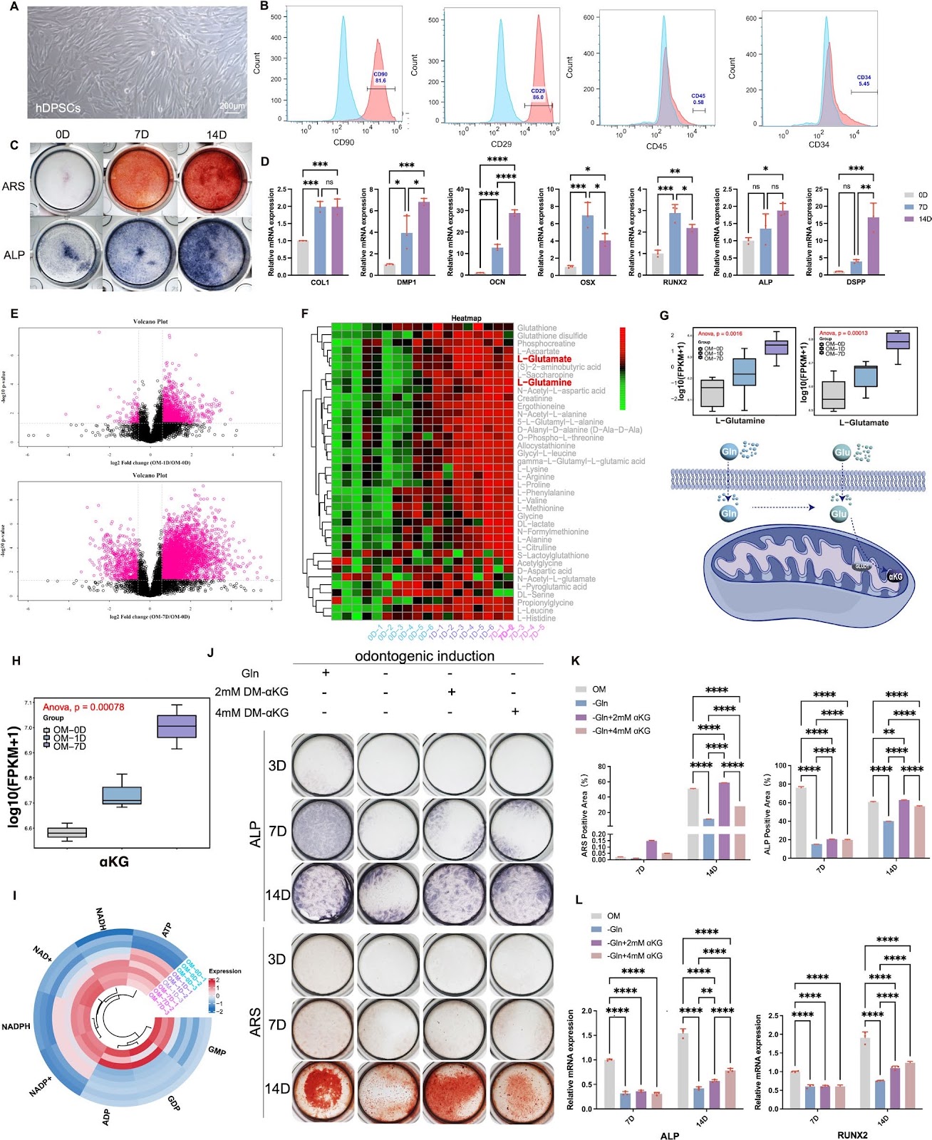
Glutamine-αKG axis affects dentin regeneration and regulates osteo/odontogenic differentiation of mesenchymal adult stem cells via IGF2 m6A modification

Authors: Qinglu Tian, Shiqi Gao, Siying Li, Mian Wan, Xin Zhou, Wei Du, Xuedong Zhou, Liwei Zheng, Yachuan Zhou

Background Multi-lineage differentiation of mesenchymal adult stem cells (m-ASCs) is crucial for tissue regeneration and accompanied with metabolism reprogramming, among which dental-pulp-derived m-ASCs has obvious advantage of easy accessibility. Stem cell fate determination and differentiation are closely related to metabolism status in cell microenvironment, which could actively interact with epigenetic modification. In recent years, glutamine-α-ketoglutarate (αKG) axis was proved to be related to aging, tumorigenesis, osteogenesis etc., while its role in m-ASCs still lack adequate research evidence. Methods We employed metabolomic analysis to explore the change pattern of metabolites during dental-pulp-derived m-ASCs differentiation. A murine incisor clipping model was established to investigate the influence of αKG on dental tissue repairment. shRNA technique was used to knockdown the expression of related key enzyme-dehydrogenase 1(GLUD1). RNA-seq, m6A evaluation and MeRIP-qPCR were used to dig into the underlying epigenetic mechanism. Results Here we found that the glutamine-αKG axis displayed an increased tendency along with the osteo/odontogenic differentiation of dental-pulp-derived m-ASCs, same as expression pattern of GLUD1. Further, the key metabolite αKG was found able to accelerate the repairment of clipped mice incisor and promote dentin formation. Exogenous DM-αKG was proved able to promote osteo/odontogenic differentiation of dental-pulp-derived m-ASCs, while the inhibition of glutamine-derived αKG level via GLUD1 knockdown had the opposite effect. Under the circumstance of GLUD1 knockdown, extracellular matrix (ECM) function and PI3k-Akt signaling pathway was screened out to be widely involved in the process with insulin-like growth factor 2 (IGF2) participation via RNA-seq. Inhibition of glutamine-αKG axis may affect IGF2 translation efficiency via m6A methylation and can be significantly rescued by αKG supplementation.

Glutamine-αKG axis affects dentin regeneration and regulates osteo/odontogenic differentiation of mesenchymal adult stem cells via IGF2 m6A modification
Graphical Abstract
Original ResearchVol. 15, Issue 1 • pp. 9DOI: 10.1186/s13287-023-03577-0

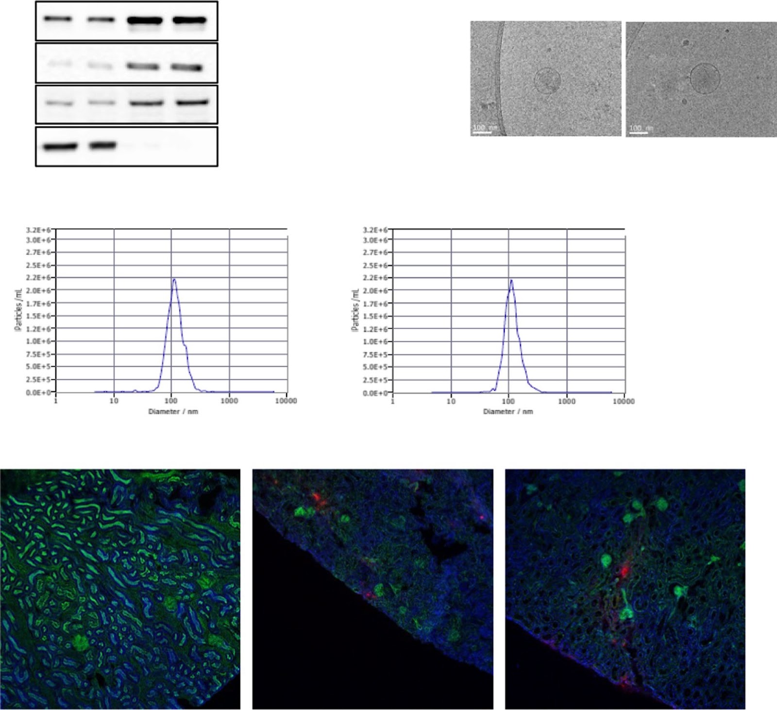
Pan PPAR agonist stimulation of induced MSCs produces extracellular vesicles with enhanced renoprotective effect for acute kidney injury

Authors: Hongduk Kim, Seul Ki Lee, Sungok Hong, Tae Sub Park, Jimin Kim, Soo Kim, Tae Min Kim

Background Acute kidney injury (AKI) has a complex pathophysiology and imposes serious health concerns worldwide. Extracellular vesicles (EVs) derived from induced mesenchymal stem cells (iMSCs) have been recognized as novel cell-free therapeutics for various inflammatory and degenerative disorders. In this study, we investigated whether iMSCs stimulated with a pan-peroxisome proliferator-activated receptor (PPAR) agonist could enhance the therapeutic efficacy of EVs against AKI. Methods Human iMSCs were primed with or without lanifibranor, a PPAR agonist for 24 h, and EVs were collected after an additional 24 h. The basic characteristics of EVs were evaluated using cryo-transmission electron microscopy imaging, immunoblot detection of EV markers, nanoparticle tracking analysis, and localization in AKI kidneys. In vitro, the potential of the EVs to promote the growth and survival of HK-2 cells undergoing cisplatin-induced apoptosis and anti-inflammatory effects in M1-polarized THP-1 was compared. Subsequently, AKI was induced in BALB/c mice using cisplatin. After 8 and 24 h of cisplatin treatment, iMSC-EVs or pan-PPAR-iMSC-EVs were injected intravascularly. At 96 h after cisplatin administration, the renoprotective effects of iMSC-EVs or pan-PPAR-iMSC-EVs in inhibiting inflammation and apoptosis were compared using serum biochemistry, histology, immunohistochemistry, and gene expression analysis by qPCR. Results Both EV types expressed EV markers and had typical EV morphology, and their localization in the renal tissue was confirmed. The proliferation and survival of HK-2 cells were higher in pan-PPAR-iMSC-EVs than those in iMSC-EVs. In M1-polarized THP-1 cells, the reduction in the mRNA expression of inflammatory cytokines was more significant in pan-PPAR-iMSC-EVs than that in iMSC-EVs. In the mouse model of cisplatin-induced AKI, pan-PPAR-iMSC-EVs markedly enhanced renoprotective effects compared to iMSC-EVs. Specifically, pan-PPAR-iMSC-EVs reduced tissue inflammation, immune cell infiltration, and apoptosis. Pan-PPAR-iMSC-EVs also increased renal capillary density. Conclusion Priming iMSCs with a PPAR agonist significantly improved the therapeutic potential of EVs by reducing inflammation and apoptosis. The reported strategy may contribute to the development of a novel cell-free option for AKI treatment. Trial registration: Not applicable.

Pan PPAR agonist stimulation of induced MSCs produces extracellular vesicles with enhanced renoprotective effect for acute kidney injury
Graphical Abstract
Original ResearchVol. 15, None • pp. 351DOI: 10.1186/s13287-024-03963-2

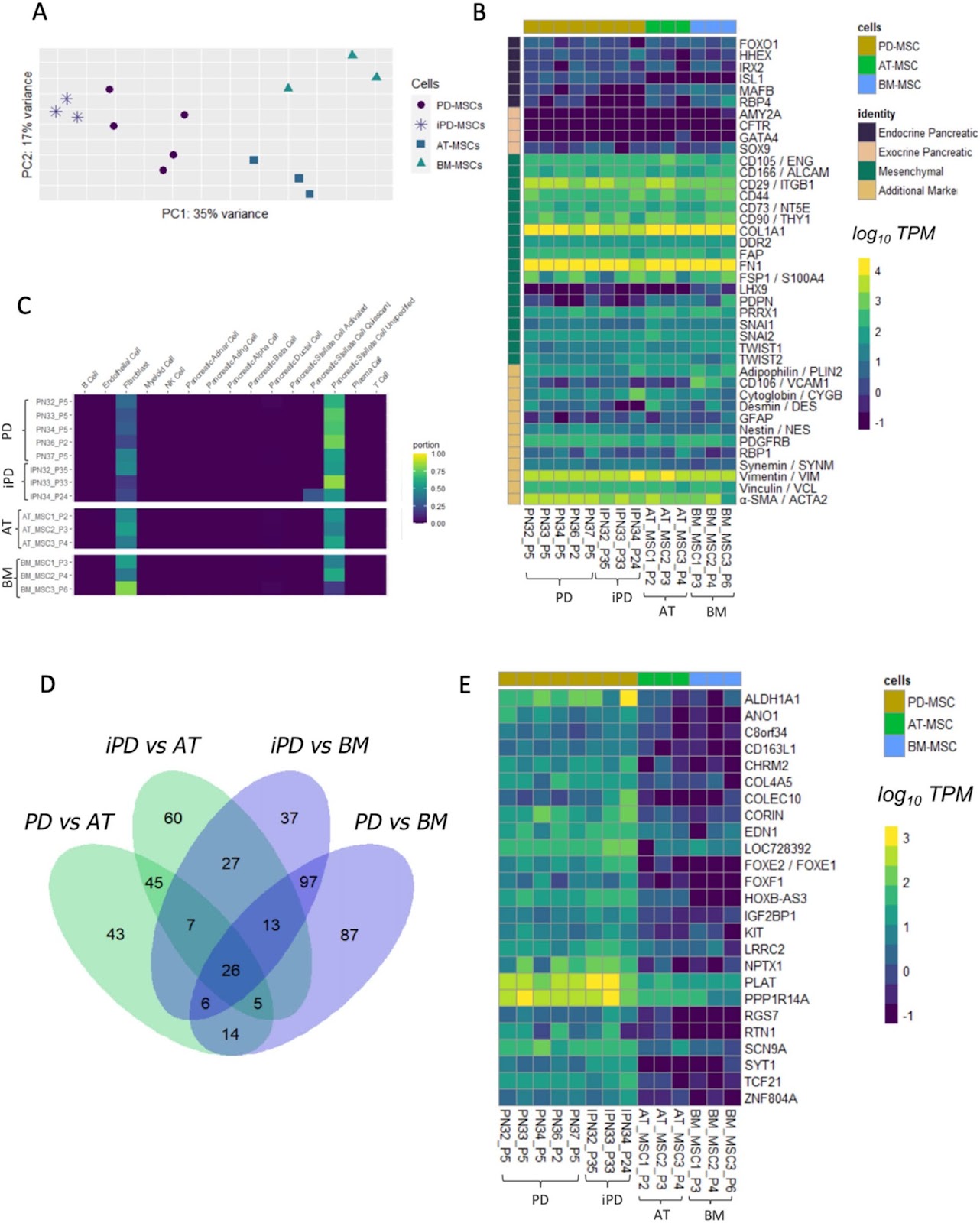
Human pancreatic islet-derived stromal cells reveal combined features of mesenchymal stromal cells and pancreatic stellate cells

Authors: Nour Ebrahim, Nikolay Kondratyev, Alexander Artyuhov, Alexei Timofeev, Nadya Gurskaya, Alexey Andrianov, Roman Izrailov, Egor Volchkov, Tatyana Dyuzheva, Elena Kopantseva, Ekaterina Kiseleva, Vera Golimbet, Erdem Dashinimaev

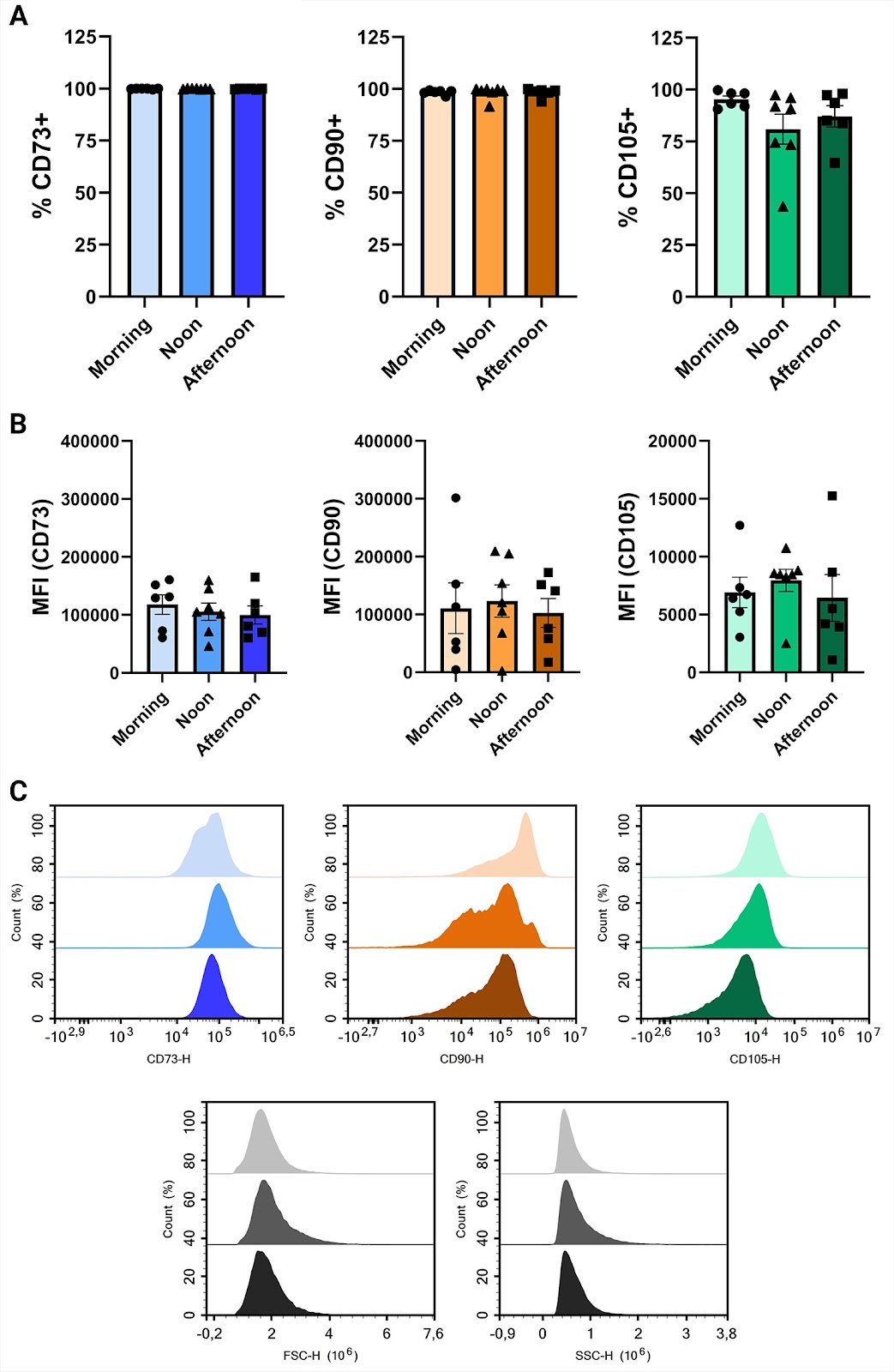
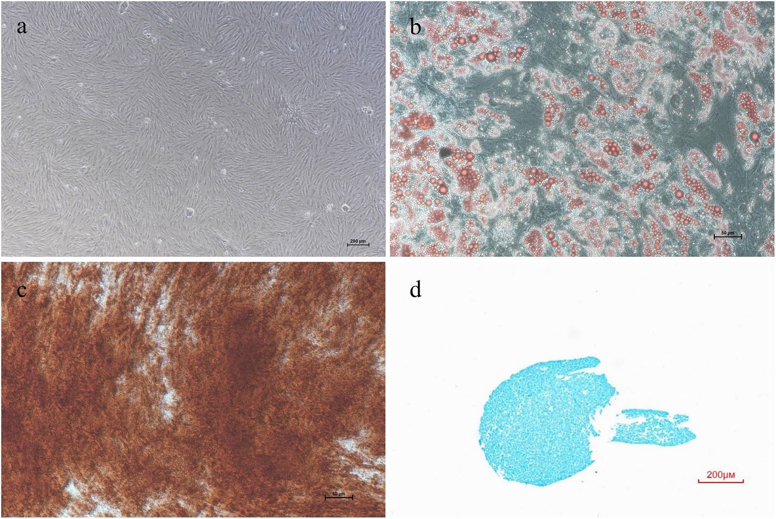
Background Mesenchymal stromal cells (MSCs) are recognized for their potential in regenerative medicine, attributed to their multipotent differentiation capabilities and immunomodulatory properties. Despite this potential, the classification and detailed characterization of MSCs, especially those derived from specific tissues like the pancreas, remains challenging leading to a proliferation of terminology in the literature. This study aims to address these challenges by providing a thorough characterization of human pancreatic islets-derived mesenchymal stromal cells (hPD-MSCs). Methods hPD-MSCs were isolated from donor islets using enzymatic digestion, immortalized through lentiviral transduction of human telomerase reverse transcriptase (hTERT). Cells were characterized by immunostaining, flow cytometry and multilineage differentiation potential into adipogenic and osteogenic lineages. Further a transcriptomic analysis was done to compare the gene expression profiles of hPD-MSCs with other mesenchymal cells. Results We show that hPD-MSCs express the classical MSC features, including morphological characteristics, surface markers expression (CD90, CD73, CD105, CD44, and CD106) and the ability to differentiate into both adipogenic and osteogenic lineages. Furthermore, transcriptomic analysis revealed distinct gene expression profiles, showing notable similarities between hPD-MSCs and pancreatic stellate cells (PSCs). The study also identified specific genes that distinguish hPD-MSCs from MSCs of other origins, including genes associated with pancreatic function (e.g., ISL1) and neural development (e.g., NPTX1, ZNF804A). A novel gene with an unknown function (ENSG00000286190) was also discovered. Conclusions This study enhances the understanding of hPD-MSCs, demonstrating their unique characteristics and potential applications in therapeutic strategies. The identification of specific gene expression profiles differentiates hPD-MSCs from other mesenchymal cells and opens new avenues for research into their role in pancreatic function and neural development.

Human pancreatic islet-derived stromal cells reveal combined features of mesenchymal stromal cells and pancreatic stellate cells
Graphical Abstract
Original ResearchVol. 15, Issue 1 • pp. 73DOI: 10.1186/s13287-024-03690-8

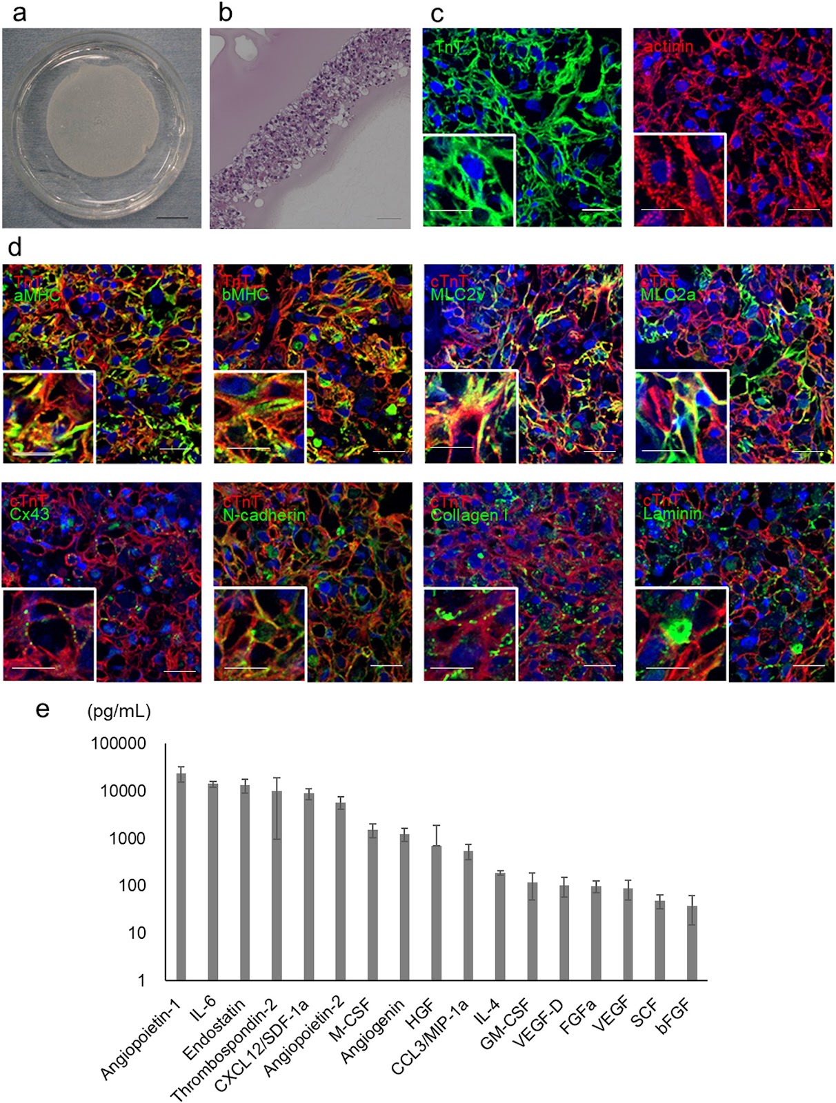
Pre-clinical evaluation of the efficacy and safety of human induced pluripotent stem cell-derived cardiomyocyte patch

Authors: Shigeru Miyagawa, Takuji Kawamura, Emiko Ito, Maki Takeda, Hiroko Iseoka, Junya Yokoyama, Akima Harada, Noriko Mochizuki-Oda, Yukiko Imanishi-Ochi, Junjun Li, Masao Sasai, Fumiyo Kitaoka, Masaki Nomura, Naoki Amano, Tomoko Takahashi, Hiromi Dohi, Eiichi Morii, Yoshiki Sawa

Background Cell- or tissue-based regenerative therapy is an attractive approach to treat heart failure. A tissue patch that can safely and effectively repair damaged heart muscle would greatly improve outcomes for patients with heart failure. In this study, we conducted a preclinical proof-of-concept analysis of the efficacy and safety of clinical-grade human induced pluripotent stem cell-derived cardiomyocyte (hiPSC-CM) patches. Methods A clinical-grade hiPSC line was established using peripheral blood mononuclear cells from a healthy volunteer that was homozygous for human leukocyte antigens. The hiPSCs were differentiated into cardiomyocytes. The obtained hiPSC-CMs were cultured on temperature-responsive culture dishes for patch fabrication. The cellular characteristics, safety, and efficacy of hiPSCs, hiPSC-CMs, and hiPSC-CM patches were analyzed. Results The hiPSC-CMs expressed cardiomyocyte-specific genes and proteins, and electrophysiological analyses revealed that hiPSC-CMs exhibit similar properties to human primary myocardial cells. In vitro and in vivo safety studies indicated that tumorigenic cells were absent. Moreover, whole-genome and exome sequencing revealed no genomic mutations. General toxicity tests also showed no adverse events posttransplantation. A porcine model of myocardial infarction demonstrated significantly improved cardiac function and angiogenesis in response to cytokine secretion from hiPSC-CM patches. No lethal arrhythmias were observed. Conclusions hiPSC-CM patches are promising for future translational research and may have clinical application potential for the treatment of heart failure.

Pre-clinical evaluation of the efficacy and safety of human induced pluripotent stem cell-derived cardiomyocyte patch
Graphical Abstract
Original ResearchVol. 15, None • pp. 243DOI: 10.1186/s13287-024-03856-4

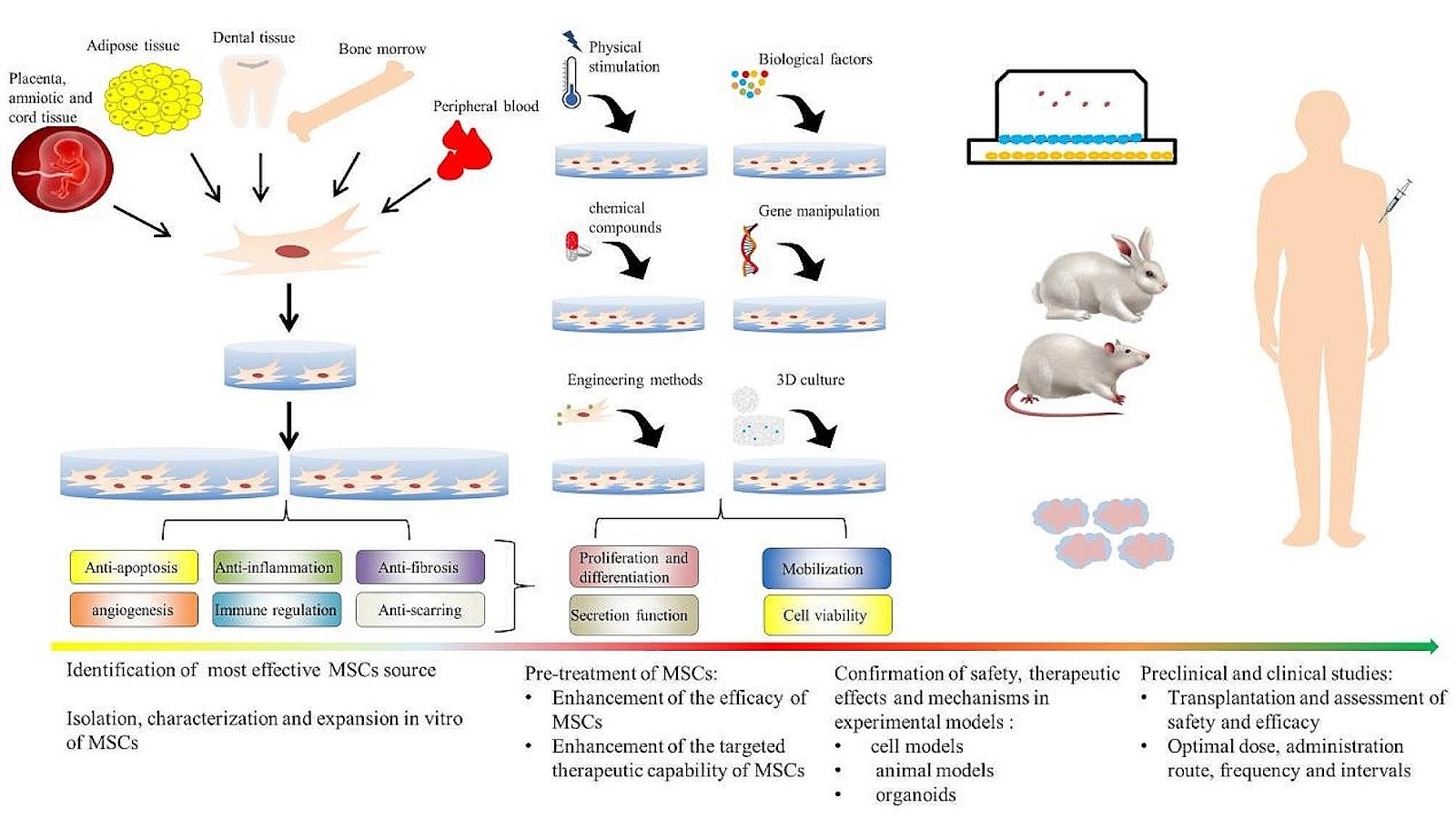
Natural small molecules synergize mesenchymal stem cells for injury repair in vital organs: a comprehensive review

Authors: Yanling Qu, Zhe Wang, Lingjuan Dong, Dan Zhang, Fengqing Shang, Afeng Li, Yanni Gao, Qinhua Bai, Dan Liu, Xiaodong Xie, Leiguo Ming

Mesenchymal stem cells (MSCs) therapy is a highly researched treatment that has the potential to promote immunomodulation and anti-inflammatory, anti-apoptotic, and antimicrobial activities. It is thought that it can enhance internal organ function, reverse tissue remodeling, and achieve significant organ repair and regeneration. However, the limited infusion, survival, and engraftment of transplanted MSCs diminish the effectiveness of MSCs-based therapy. Consequently, various preconditioning methods have emerged as strategies for enhancing the therapeutic effects of MSCs and achieving better clinical outcomes. In particular, the use of natural small molecule compounds (NSMs) as a pretreatment strategy is discussed in this narrative review, with a focus on their roles in regulating MSCs for injury repair in vital internal organs. Additionally, the discussion focuses on the future directions and challenges of transforming mesenchymal stem cell research into clinical applications.

Natural small molecules synergize mesenchymal stem cells for injury repair in vital organs: a comprehensive review
Graphical Abstract
Original ResearchVol. 15, None • pp. 501DOI: 10.1186/s13287-024-04112-5

Correction: POU3F4 up-regulates Gli1 expression and promotes neuronal differentiation and synaptic development of hippocampal neural stem cells

Authors: Lei Zhang, Jue Wang, Naijuan Xu, Jingjing Guo, Yujian Lin, Xunrui Zhang, Ruijie Ji, Yaya Ji, Haoming Li, Xiao Han, Wen Li, Xiang Cheng, Jianbing Qin, Meiling Tian, Min Xu, Xinhua Zhang

Correction to: Stem Cell Research & Therapy (2024) 15:440. The original article initially erroneously presented co-author, Min Xu's name as Xu Min; this has since been amended.

Correction: POU3F4 up-regulates Gli1 expression and promotes neuronal differentiation and synaptic development of hippocampal neural stem cells
Graphical Abstract
Original ResearchVol. 15, None • pp. 246DOI: 10.1186/s13287-024-03862-6

iPSC-derived lung and lung cancer organoid model to evaluate cisplatin encapsulated autologous iPSC-derived mesenchymal stromal cell-isolated extracellular vesicles

Authors: Caroline Küstermann, Karīna Narbute, Valērija Movčana, Vadims Parfejevs, Fēlikss Rūmnieks, Pauls Kauķis, Miks Priedols, Rihards Mikilps-Mikgelbs, Marija Mihailova, Santa Andersone, Aigars Dzalbs, Cristina Bajo-Santos, Alvils Krams, Arturs Abols

Background Lung cancer remains a leading cause of cancer-related mortality globally. Although recent therapeutic advancements have provided targeted treatment approaches, the development of resistance and systemic toxicity remain primary concerns. Extracellular vesicles (EVs), especially those derived from mesenchymal stromal cells (MSC), have gained attention as promising drug delivery systems, offering biocompatibility and minimal immune responses. Recognizing the limitations of conventional 2D cell culture systems in mimicking the tumor microenvironment, this study aims to describe a proof-of-principle approach for using patient-specific organoid models for both lung cancer and normal lung tissue and the feasibility of employing autologous EVs derived from induced pluripotent stem cell (iPSC)-MSC in personalized medicine approaches. Methods First, we reprogrammed healthy fibroblasts into iPSC. Next, we differentiated patient-derived iPSC into branching lung organoids (BLO) and generated patient-matched lung cancer organoids (LCO) from patient-derived tumor tissue. We show a streamlined process of MSC differentiation from iPSC and EV isolation from iPSC-MSC, encapsulated with 0.07 µg/mL of cytotoxic agent cisplatin and applied to both organoid models. Cytotoxicity of cisplatin and cisplatin-loaded EVs was recorded with LDH and CCK8 tests. Results Fibroblast-derived iPSC showed a normal karyotype, pluripotency staining, and trilineage differentiation. iPSC-derived BLO showed expression of lung markers, like TMPRSS2 and MUC5A while patient-matched LCO showed expression of Napsin and CK5. Next, we compared the effects of iPSC-MSC derived EVs loaded with cisplatin against empty EVs and cisplatin alone in lung cancer organoid and healthy lung organoid models. As expected, we found a cytotoxic effect when LCO were treated with 20 µg/mL cisplatin. Treatment of LCO and BLO with empty EVs resulted in a cytotoxic effect after 24 h. However, EVs loaded with 0.07 µg/mL cisplatin failed to induce any cytotoxic effect in both organoid models. Conclusion We report on a proof-of-principle pipeline towards using autologous or allogeneic iPSC-MSC EVs as drug delivery tests for lung cancer in future. However, due to the time and labor-intensive processes, we conclude that this pipeline might not be feasible for personalized approaches at the moment.

iPSC-derived lung and lung cancer organoid model to evaluate cisplatin encapsulated autologous iPSC-derived mesenchymal stromal cell-isolated extracellular vesicles
Graphical Abstract
Original ResearchVol. 15, None • pp. 327DOI: 10.1186/s13287-024-03949-0

Evaluation of the impact of customized serum-free culture medium on the production of clinical-grade human umbilical cord mesenchymal stem cells: insights for future clinical applications

Authors: Lan Zhao, Beibei Ni, Jinqing Li, Rui Liu, Qi Zhang, Zhuangbin Zheng, Wenjuan Yang, Wei Yu, Lijun Bi

Background The selection of suitable culture medium is critical for achieving good clinical outcomes in cell therapy. To support the commercial application of stem cell therapy, customized culture media not only need to promote stem cell proliferation, but also need to save costs and meet industrial requirements for inter-batch consistency, efficacy, and biosafety. In this study, we developed a series of serum-free media (SFM) and elucidated the effects between different SFM, as well as between SFM and serum-containing meida (SCM), on human umbilical cord mesenchymal stem cells (hUC-MSCs) phenotype and function. We analyze and emphasize from the perspectives of clinical and commercial application why research on customized culture media is critical for the success of enterprises developing novel cellular therapeutics. Methods We cultured hUC-MSCs with identical cell seeding densities in different formulations of SFM and SCM until passage 10 and examined the changes in cell phenotype and function. We analyzed the results with the commercial application requirments of the cellular therapy industry to assess the potential impact of customized culture media on inter-batch consistency, efficacy, stability, biosafety, and cost-effectiveness of industrial-scale cell production. Results hUC-MSCs cultured in SCM and SFM exhibit consistent cell morphology and surface molecule expression, but hUC-MSCs cultured in SFM demonstrate higher activity, superior proliferative capacity, and greater stability. Furthermore, hUC-MSCs cultured in different SFM exhibit differences in cell activity, proliferative capacity, senescent rate, and S/M ratio of cell cycle, while maintaining a normal karyotype after long-term in vitro cultivation. Moreover, [abstract truncated]

Evaluation of the impact of customized serum-free culture medium on the production of clinical-grade human umbilical cord mesenchymal stem cells: insights for future clinical applications
Graphical Abstract
Original ResearchVol. 15, None • pp. 217DOI: 10.1186/s13287-024-03821-1

Transplantation of human endometrial perivascular stem cells with hydroxy saffron yellow A promotes uterine repair in rats

Authors: Ning Li, Jialian Mao, Miaomiao Wang, Jiahui Qi, Zhiwei Jiang, Yifan Li, Guijun Yan, Yali Hu, Shiyuan Li, Haixiang Sun, Lijun Ding

Background Intrauterine adhesions (IUAs) jeopardise uterine function in women, which is a great challenge in the clinic. Previous studies have shown that endometrial perivascular cells (En-PSCs) can improve the healing of scarred uteri and that hydroxysafflor yellow A (HSYA) promotes angiogenesis. The purpose of this study was to observe whether the combination of En-PSCs with HSYA could improve the blood supply and fertility in the rat uterus after full-thickness injury. Methods En-PSCs were sorted by flow cytometry, and the effect of HSYA on the proliferation and angiogenesis of the En-PSCs was detected using CCK-8 and tube formation assays. Based on a previously reported rat IUA model, the rat uteri were sham-operated, spontaneously regenerated, or treated with collagen-loaded PBS, collagen-loaded HSYA, collagen-loaded En-PSCs, or collagen-loaded En-PSCs with HSYA, and then collected at both 30 and 90 days postsurgery. HE staining and Masson staining were used to evaluate uterine structure and collagen fibre deposition, and immunohistochemical staining for α-SMA and vWF was used to evaluate myometrial regeneration and neovascularization in each group. A fertility assay was performed to detect the recovery of pregnancy function in each group. RNA-seq was performed to determine the potential mechanism underlying En-PSCs/HSYA treatment. Immunofluorescence, tube formation assays, and Western blot were used to validate the molecular mechanism involved. Results The transplantation of Collagen/En-PSCs/HSYA markedly promoted uterine repair in rats with full-thickness injury by reducing fibrosis, increasing endometrial thickness, regenerating myometrium, promoting angiogenesis, and facilitated live births. RNA sequencing results suggested that En-PSCs/HSYA activated the NRG1/ErbB4 signaling pathway. In vitro tube formation experiments revealed that the addition of an ErbB inhibitor diminished the tube formation ability of cocultured En-PSCs and HUVECs. Western blot results further showed that elevated levels of NRG1 and ErbB4 proteins were detected in the Collagen/En-PSCs/HSYA group compared to the Collagen/En-PSCs group. These collective results suggested that the beneficial effects of the transplantation of Collagen/En-PSCs/HSYA might be attributed to the modulation of the NRG1/ErbB4 signaling pathway. Conclusions The combination of En-PSCs/HSYA facilitated morphological and functional repair in rats with full-thickness uterine injury and may promote endometrial angiogenesis by regulating the NRG1/ErbB4 signaling pathway.
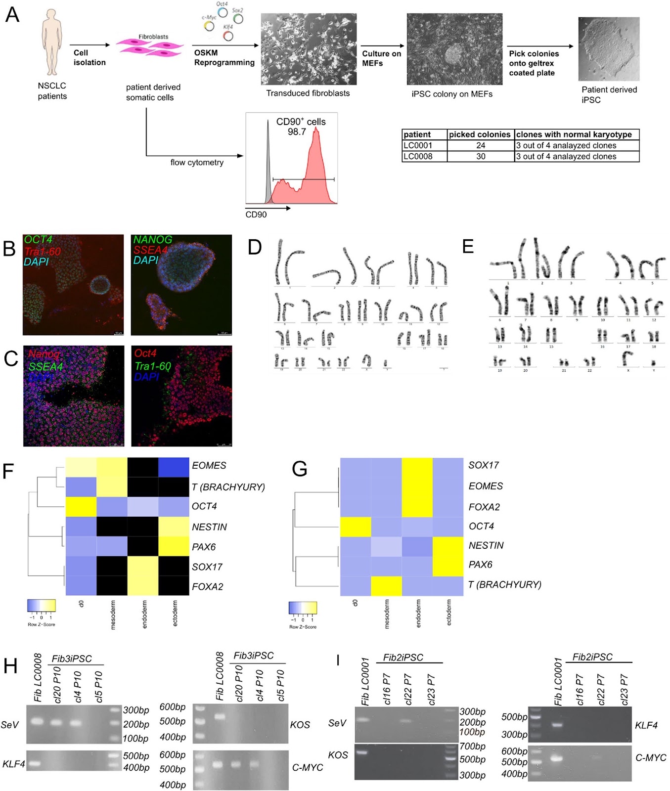
Transplantation of human endometrial perivascular stem cells with hydroxy saffron yellow A promotes uterine repair in rats
Graphical Abstract
Original ResearchVol. 15, None • pp. 358DOI: 10.1186/s13287-024-03984-x

Correction: Radiochemotherapy-induced DNA repair promotes the biogenesis of gastric cancer stem cells

Authors: Yu Lu, Xiaobo Zhang

This correction article addresses an inadvertent error in the original publication. In Fig. 5M of the original article, the image of the fourth lane (LEF-1) of the second picture (Doxorubicin-induced GCSCs) was inadvertently replaced with an incorrect version during the upload process. The authors wish to note a correction to the aforementioned picture via the corrected picture ahead in this Correction article. The authors deeply regret that this error occurred and sincerely apologize for any inconvenience.

Correction: Radiochemotherapy-induced DNA repair promotes the biogenesis of gastric cancer stem cells
Graphical Abstract
Original ResearchVol. 15, Issue 1 • pp. 22DOI: 10.1186/s13287-023-03572-5

Dysregulated lncRNAs regulate human umbilical cord mesenchymal stem cell differentiation into insulin-producing cells by forming a regulatory network with mRNAs

Authors: Tianqin Xie, Qiming Huang, Qiulan Huang, Yanting Huang, Shuang Liu, Haixia Zeng, Jianping Liu

Objective In recent years, cell therapy has emerged as a new research direction in the treatment of diabetes. However, the underlying molecular mechanisms of mesenchymal stem cell (MSC) differentiation necessary to form such treatment have not been clarified. Methods In this study, human umbilical cord mesenchymal stem cells (HUC-MSCs) isolated from newborns were progressively induced into insulin-producing cells (IPCs) using small molecules. HUC-MSC (S0) and four induced stage (S1–S4) samples were prepared. We then performed transcriptome sequencing experiments to obtain the dynamic expression profiles of both mRNAs and long noncoding RNAs (lncRNAs). Results We found that the number of differentially expressed lncRNAs and mRNAs trended downwards during differentiation. Gene Ontology (GO) analysis showed that the target genes of differentially expressed lncRNAs were associated with translation, cell adhesion, and cell connection. Kyoto Encyclopedia of Genes and Genomes (KEGG) analysis revealed that the NF-KB signalling pathway, MAPK signalling pathway, HIPPO signalling pathway, PI3K–Akt signalling pathway, and p53 signalling pathway were enriched in these differentially expressed lncRNA-targeting genes. We also found that the coexpression of the lncRNA CTBP1-AS2 with PROX1 and the lncRNAs AC009014.3 and GS1-72M22.1 with JARID2 mRNA was related to the development of pancreatic beta cells. Moreover, the coexpression of the lncRNAs: XLOC_ 050969, LINC00883, XLOC_050981, XLOC_050925, MAP3K14- AS1, RP11-148K1.12, and CTD2020K17.3 with p53, regulated insulin secretion by pancreatic beta cells. Conclusion In this study, HUC-MSCs combined with small molecule compounds were successfully induced into IPCs. Differentially expressed lncRNAs may regulate the insulin secretion of pancreatic beta cells by regulating multiple signalling pathways. The lncRNAs AC009014.3, Gs1-72m21.1, and CTBP1-AS2 may be involved in the development of pancreatic beta cells, and the lncRNAs: XLOC_050969, LINC00883, XLOC_050981, XLOC_050925, MAP3K14-AS1, RP11-148K1.12, and CTD2020K17.3 may be involved in regulating the insulin secretion of pancreatic beta cells, thus providing a lncRNA catalogue for future research regarding the mechanism of the transdifferentiation of HUC-MSCs into IPCs. It also provides a new theoretical basis for the transplantation of insulin-producing cells into diabetic patients in the future.

Dysregulated lncRNAs regulate human umbilical cord mesenchymal stem cell differentiation into insulin-producing cells by forming a regulatory network with mRNAs
Graphical Abstract
Original ResearchVol. 15, Issue 1 • pp. 322DOI: 10.1186/s13287-024-03931-w

Dissecting human adipose tissue heterogeneity using single-cell omics technologies

Authors: Giuliana Di Rocco, Angelo Trivisonno, Giovanni Trivisonno, Gabriele Toietta

Single-cell omics technologies that profile genes (genomic and epigenomic) and determine the abundance of mRNA (transcriptomic), protein (proteomic and secretomic), lipids (lipidomic), and extracellular matrix (matrisomic) support the dissection of adipose tissue heterogeneity at unprecedented resolution in a temporally and spatially defined manner. In particular, cell omics technologies may provide innovative biomarkers for the identification of rare specific progenitor cell subpopulations, assess transcriptional and proteomic changes affecting cell proliferation and immunomodulatory potential, and accurately define the lineage hierarchy and differentiation status of progenitor cells. Unraveling adipose tissue complexity may also provide for the precise assessment of a dysfunctional state, which has been associated with cancer, as cancer-associated adipocytes play an important role in shaping the tumor microenvironment supporting tumor progression and metastasis, obesity, metabolic syndrome, and type 2 diabetes mellitus. The information collected by single-cell omics has relevant implications for regenerative medicine because adipose tissue is an accessible source of multipotent cells; alternative cell-free approaches, including the use of adipose tissue stromal cell-conditioned medium, extracellular vesicles, or decellularized extracellular matrix, are clinically valid options. Subcutaneous white adipose tissue, which is generally harvested via liposuction, is highly heterogeneous because of intrinsic biological variability and extrinsic inconsistencies in the harvesting and processing procedures. The current limited understanding of adipose tissue heterogeneity impinges on the definition of quality standards appropriate for clinical translation, which requires consistency and uniformity of the administered product. We review the methods used for dissecting adipose tissue heterogeneity and provide an overview of advances in omics technology that may contribute to the exploration of heterogeneity and dynamics of adipose tissue at the single-cell level.

Dissecting human adipose tissue heterogeneity using single-cell omics technologies
Graphical Abstract
Original ResearchVol. 15, None • pp. 320DOI: 10.1186/s13287-024-03943-6

Mesenchymal stem cell therapy for liver transplantation: clinical progress and immunomodulatory properties

Authors: Fuli Wen, Guokai Yang, Saihua Yu, Haiyan Liu, Naishun Liao, Zhengfang Liu

Although liver transplantation (LT) is an effective strategy for end-stage liver diseases, the shortage of donor organs and the immune rejection hinder its widespread implementation in clinical practice. Mesenchymal stem cells (MSCs) transplantation offers a promising approach for patients undergoing liver transplantation due to their immune regulatory capabilities, hepatic protection properties, and multidirectional differentiation potential. In this review, we summarize the potential applications of MSCs transplantation in various LT scenarios. MSCs transplantation has demonstrated effectiveness in alleviating hepatic ischemia-reperfusion injury, enhancing the viability of liver grafts, preventing acute graft-versus-host disease, and promoting liver regeneration in split LT therapy. We also discuss the clinical progress, and explore the immunomodulatory functions of MSCs in response to both adaptive and innate immune responses. Furthermore, we emphasize the interactions between MSCs and different immune cells, including T cells, B cells, plasma cells, natural killer cells, dendritic cells, Kupffer cells, and neutrophils, to provide new insights into the immunomodulatory properties of MSCs in adoptive cell therapy.

Mesenchymal stem cell therapy for liver transplantation: clinical progress and immunomodulatory properties
Graphical Abstract
Original ResearchVol. 15, None • pp. 232DOI: 10.1186/s13287-024-03819-9

Robust bioprocess design and evaluation of commercial media for the serial expansion of human induced pluripotent stem cell aggregate cultures in vertical-wheel bioreactors

Authors: Breanna S. Borys, Tiffany Dang, Hannah Worden, Leila Larijani, Jessica M. Corpuz, Brett D. Abraham, Emilie J. Gysel, Julia Malinovska, Roman Krawetz, Tamas Revay, Bob Argiropoulos, Derrick E. Rancourt, Michael S. Kallos, Sunghoon Jung

Background: While pluripotent stem cell (PSC) therapies move toward clinical and commercial applications at a rapid rate, manufacturing reproducibility and robustness are notable bottlenecks in regulatory approval. Therapeutic applications of PSCs require large cell quantities to be generated under highly robust, well-defined, and economically viable conditions. Small-scale and short-term process optimization, however, is often performed in a linear fashion that does not account for time needed to verify the bioprocess protocols and analysis methods used. Design of a reproducible and robust bioprocess should be dynamic and include a continuous effort to understand how the process will respond over time and to different stresses before transitioning into large-scale production where stresses will be amplified. Methods: This study utilizes a baseline protocol, developed for the short-term culture of PSC aggregates in Vertical-Wheel® bioreactors, to evaluate key process attributes through long-term (serial passage) suspension culture. This was done to access overall process robustness when performed with various commercially available media and cell lines. Process output variables including growth kinetics, aggregate morphology, harvest efficiency, genomic stability, and functional pluripotency were assessed through short and long-term culture. Results: The robust nature of the expansion protocol was demonstrated over a six-day culture period where spherical aggregate formation and expansion were observed with high-fold expansions for all five commercial media tested. Profound differences in cell growth and quality were revealed only through long-term serial expansion and in-vessel dissociation operations. Some commercial media formulations tested demonstrated maintenance of cell growth

Robust bioprocess design and evaluation of commercial media for the serial expansion of human induced pluripotent stem cell aggregate cultures in vertical-wheel bioreactors
Graphical Abstract
Original ResearchVol. 15, None • pp. 209DOI: 10.1186/s13287-024-03835-9

Single-cell sequencing of facial adipose tissue unveils FKBP5 as a therapeutic target for facial infiltrating lipomatosis

Authors: Hongrui Chen, Bin Sun, Shih-Jen Chang, Zhang Yu, Yajing Qiu, Chen Hua, Xiaoxi Lin

Background Facial infiltrating lipomatosis is characterized by excessive growth of adipose tissue. Its etiology is associated with somatic phosphatidylinositol 3-kinase catalytic subunit alpha (PIK3CA) variants, but the specific mechanisms are not yet fully understood. Methods We collected facial adipose tissue from both FIL patients and non-FIL individuals, isolated the stromal vascular fraction (SVF) and performed single-cell transcriptome sequencing on these samples. Results We mapped out the cellular landscape within the SVF, with a specific focus on a deeper analysis of fibro-adipogenic precursor cells (FAPs). Our analysis revealed that FAPs from FIL patients (FIL-FAPs) significantly overexpressed FK506 binding protein 51 (FKBP5) compared to FAPs from individuals without FIL. Further experiments indicated that FKBP5 is regulated by the PI3K-AKT signaling pathway. The overactivation of this pathway led to an increase in FKBP5 expression. In vitro experiments demonstrated that FKBP5 promoted adipogenic differentiation of FAPs, a process that could be hindered by FKBP5 knockdown or inhibition. Additionally, in vivo assessments confirmed FKBP5’s role in adipogenesis. Conclusions These insights into the pathogenesis of FIL underscore FKBP5 as a promising target for developing non-surgical interventions to manage the excessive adipose tissue growth in FIL.
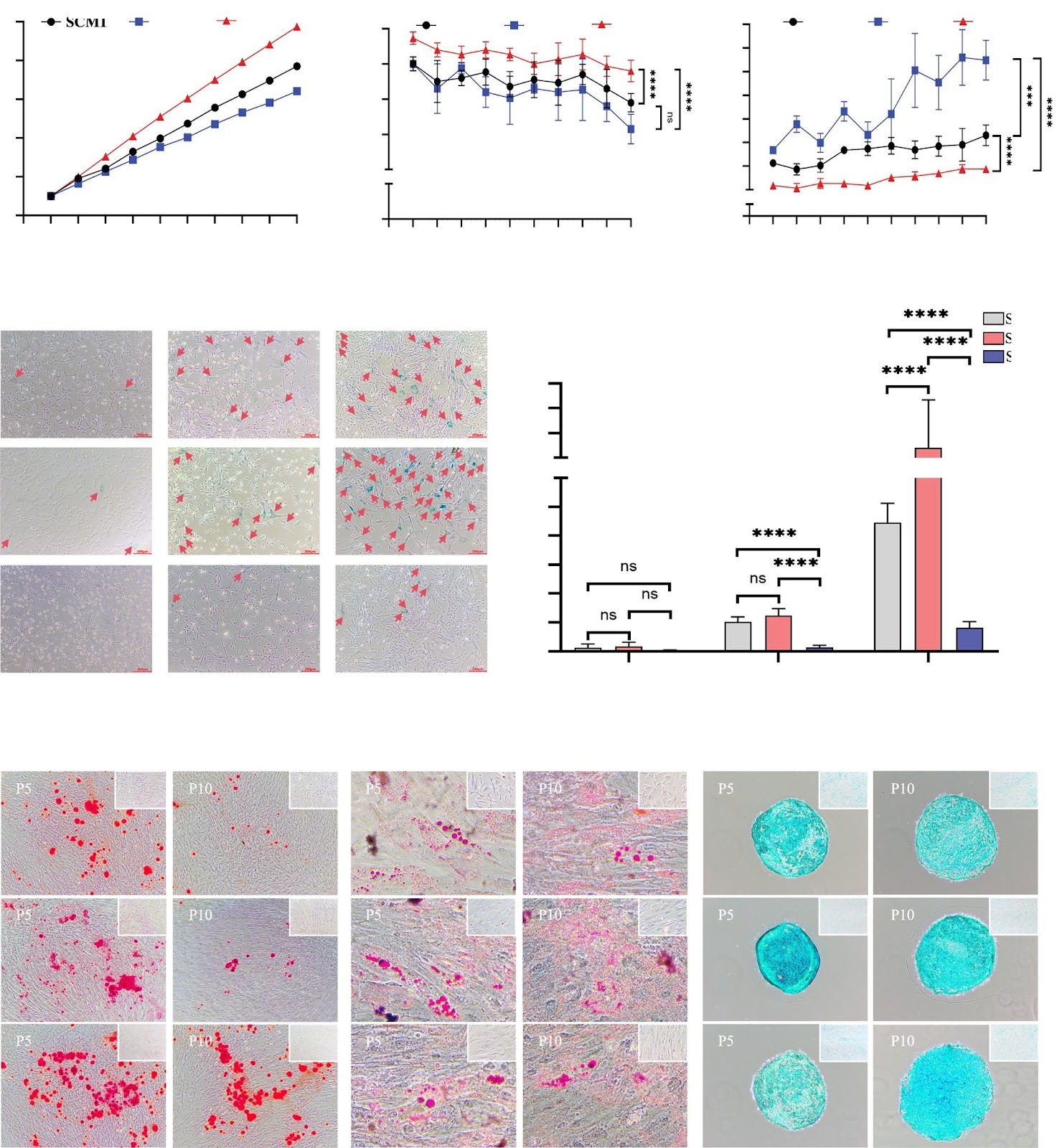
Single-cell sequencing of facial adipose tissue unveils FKBP5 as a therapeutic target for facial infiltrating lipomatosis
Graphical Abstract
Original ResearchVol. 15, Issue 1 • pp. 68DOI: 10.1186/s13287-023-03624-w

Mesenchymal stem/stromal cells from human pluripotent stem cell-derived brain organoid enhance the ex vivo expansion and maintenance of hematopoietic stem/progenitor cells

Authors: Ya Zhou, Xinping Cai, Xiuxiu Zhang, Yong Dong, Xu Pan, Mowen Lai, Yimeng Zhang, Yijin Chen, Xiaohong Li, Xia Li, Jiaxin Liu, Yonggang Zhang, Feng Ma

Background Mesenchymal stem/stromal cells (MSCs) are of great therapeutic value due to their role in maintaining the function of hematopoietic stem/progenitor cells (HSPCs). MSCs derived from human pluripotent stem cells represent an ideal alternative because of their unlimited supply. However, the role of MSCs with neural crest origin derived from HPSCs on the maintenance of HSPCs has not been reported. Methods Flow cytometric analysis, RNA sequencing and differentiation ability were applied to detect the characteristics of stromal cells from 3D human brain organoids. Human umbilical cord blood CD34+ (UCB-CD34+) cells were cultured in different coculture conditions composed of stromal cells and umbilical cord MSCs (UC-MSCs) with or without a cytokine cocktail. The hematopoietic stroma capacity of stromal cells was tested in vitro with the LTC-IC assay and in vivo by cotransplantation of cord blood nucleated cells and stroma cells into immunodeficient mice. RNA and proteomic sequencing were used to detect the role of MSCs on HSPCs. Results The stromal cells, derived from both H1-hESCs and human induced pluripotent stem cells forebrain organoids, were capable of differentiating into the classical mesenchymal-derived cells (osteoblasts, chondrocytes, and adipocytes). These cells expressed MSC markers, thus named pluripotent stem cell-derived MSCs (pMSCs). The pMSCs showed neural crest origin with CD271 expression in the early stage. When human UCB-CD34+ HSPCs were cocultured on UC-MSCs or pMSCs, the latter resulted in robust expansion of UCB-CD34+ HSPCs in long-term culture and efficient maintenance of their transplantability. Comparison by RNA sequencing indicated that coculture of human UCB-CD34+ HSPCs with pMSCs provided an improved microenvironment for HSC maintenance. The pMSCs highly expressed the Wnt signaling inhibitors SFRP1 and SFRP2, indicating that they may help to modulate the cell cycle to promote the maintenance of UCB-CD34+ HSPCs by antagonizing Wnt activation.

Mesenchymal stem/stromal cells from human pluripotent stem cell-derived brain organoid enhance the ex vivo expansion and maintenance of hematopoietic stem/progenitor cells
Graphical Abstract
Original ResearchVol. 15, None • pp. 253DOI: 10.1186/s13287-024-03864-4

Examining the potentials of stem cell therapy in reducing the burden of selected non-communicable diseases in Africa

Authors: Faith Ayobami Atewologun, Olalekan John Okesanya, Inibehe Ime Okon, Hassan Hakeem Kayode, Bonaventure Michael Ukoaka, Noah Olabode Olaleke, Jerico Bautista Ogaya, Lawal Azeez Okikiola, Emery Manirambona, Don Eliseo Lucero-Prisno III

Stem cell therapy (SCT) is a promising solution for addressing health challenges in Africa, particularly non-communicable diseases (NCDs). With their regenerative potential, stem cells have the inherent capacity to differentiate into numerous cell types for tissue repair. Despite infrastructural, ethical, and legal challenges, SCT holds immense promise for managing chronic illnesses and deep-seated tissue injuries. The rising prevalence of NCDs in Africa highlights the need for innovative strategies and treatment options. SCT offers hope in combating conditions like burns, osteoarthritis, diabetes, Alzheimer’s disease, stroke, heart failure and cancer, potentially reducing the burden of NCDs on the continent. Despite SCT’s opportunities in Africa, there are significant obstacles. However, published research on SCT in Africa is scarce, but recent initiatives such as the Basic School on Neural Stem Cells (NSC) express interest in developing NSC research in Africa. SCT research in African regions, notably on neurogenesis, demonstrates a concentration on studying neurological processes in indigenous settings. While progress has been made in South Africa and Nigeria, issues such as brain drain and impediments to innovation remain. Clinical trials have investigated the efficacy of stem cell treatments, emphasising both potential benefits and limitations in implementing these therapies efficiently. Financing research, developing regulatory frameworks, and resolving affordability concerns are critical steps toward realizing the potential of stem cell treatment in Africa.

Examining the potentials of stem cell therapy in reducing the burden of selected non-communicable diseases in Africa
Graphical Abstract
Original ResearchVol. 15, Issue 1 • pp. 423DOI: 10.1186/s13287-024-04032-4

N-CADHERIN+/CD168− subpopulation determines therapeutic variations of UC-MSCs for cardiac repair after myocardial infarction

Authors: Yukang Wu, Jianguo Li, Ke Feng, Ailing Tan, Yingying Gao, Wen Chen, Wenwen Jia, Xudong Guo, Jiuhong Kang

Background The efficiency of mesenchymal stem cells (MSCs) in treating myocardial infarction (MI) remains inconsistent, which limits their therapeutic applications. Therefore, exploring the mechanism for the inconsistent efficacy of MSCs and identification the criteria for screening MSCs are important for improving the efficiency of MSCs. Methods Mouse model after MI was utilized to test the role of MSCs from different donors and the functional subpopulation in improving cardiac function. Heterogeneity of MSCs was identified using single-cell RNA sequencing (scRNA-seq) of MSC-GY. GSEA and Scissor analyses were used to find the functional subpopulations of MSCs that promote angiogenesis. The role of functional subpopulations in promoting angiogenesis was verified by detecting the secretory proteins, the ratio of N-CADHERIN+/CD168− subpopulations in MSCs, and the tube formation, migration, and proliferation of HUVECs after treatment with conditional medium (CM) derived from different MSCs. Results We found that umbilical cord-derived MSCs (UC-MSCs) from different donors have varied therapeutic efficacy in MI mice and UC-MSCs with higher therapeutic effectiveness exhibited the most potent pro-angiogenic effects by secreting elevated levels of angiogenesis-related proteins, such as MYDGF, VEGFA, and FGF2. ScRNA-seq of 10,463 UC-MSCs revealed that the N-CADHERIN+/CD168− subpopulation was closely associated with pro-angiogenic effects, and the ratio of this cell subpopulation was positively correlated with the angiogenic potential of MSCs. We also found that the N-CADHERIN+/CD168− subpopulation was the functional subpopulation of MSCs in improving cardiac function of MI mice. Conclusions Our study identified that the N-CADHERIN+/CD168− subpopulation was the functional subpopulation of MSCs in treating MI, which was essential for the development and utilization of MSCs in MI treatment.
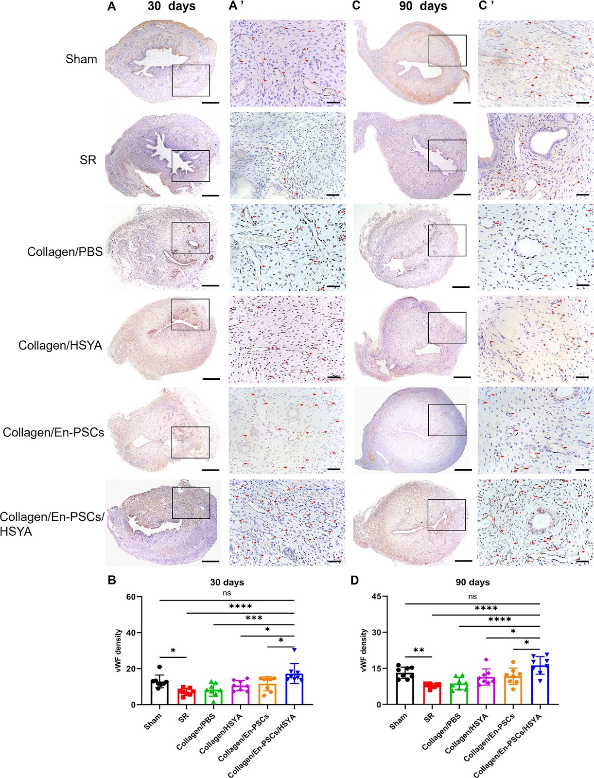
N-CADHERIN+/CD168− subpopulation determines therapeutic variations of UC-MSCs for cardiac repair after myocardial infarction
Graphical Abstract
Original ResearchVol. 15, None • pp. 296DOI: 10.1186/s13287-024-03917-8

Human intermediate prostate cancer stem cells contribute to the initiation and development of prostate adenocarcinoma

Authors: Jie Mu, Ruizhi Li, Yu Zheng, Yi Lu, Lei Ma, Lin Yin, Miao Zhang, Wenyu Ma, Mengjia Chang, Aihua Liu, Jing Li, Hai Zhu, Dong Wang

Background Intermediate cells are present in the early stages of human prostate development and adenocarcinoma. While primary cells isolated from benign human prostate tissues or tumors exhibit an intermediate phenotype in vitro, they cannot form tumors in vivo unless genetically modified. It is unclear about the stem cell properties and tumorigenicity of intermediate cells. Methods We developed a customized medium to culture primary human intermediate prostate cells, which were transplanted into male immunodeficient NCG mice to examine tumorigenicity in vivo. We treated the cells with different concentrations of dihydrotestosterone (DHT) and enzalutamide in vitro and surgically castrated the mice after cell transplantation in vivo. Immunostaining, qRT-PCR, RNA sequencing, and western blotting were performed to characterize the cells in tissues and 2D and 3D cultures. Results We found intermediate cells expressing AR+PSA+CK8+CK5+ in the luminal compartment of human prostate adenocarcinoma by immunostaining. We cultured the primary intermediate cells in vitro, which expressed luminal (AR+PSA+CK8+CK18+), basal (CK5+P63+), intermediate (IVL+), and stem cell (CK4+CK13+PSCA+SOX2+) markers. These cells resisted castration in vitro by upregulating the expression of AR, PSA, and proliferation markers KI67 and PCNA. The intermediate cells had high tumorigenicity in vivo, forming tumors in immunodeficient NCG mice in a month without any genetic modification or co-transplantation with embryonic urogenital sinus mesenchyme (UGSM) cells. We named these cells human castration-resistant intermediate prostate cancer stem cells or CriPCSCs and defined the xenograft model as patient primary cell-derived xenograft (PrDX). Human CriPCSCs resisted castration in vitro and

Human intermediate prostate cancer stem cells contribute to the initiation and development of prostate adenocarcinoma
Graphical Abstract
Original ResearchVol. 15, Issue 1 • pp. 288DOI: 10.1186/s13287-024-03903-0

Urine-derived stem cells serve as a robust platform for generating native or engineered extracellular vesicles

Authors: Anders Toftegaard Boysen, Bradley Whitehead, Anne Louise S. Revenfeld, Dhanu Gupta, Thor Petersen, Peter Nejsum

Background Mesenchymal stromal cell (MSC) therapy holds great potential yet efficacy and safety concerns with cell therapy persist. The beneficial effects of MSCs are often attributed to their secretome that includes extracellular vesicles (EVs). EVs carry biologically active molecules, protected by a lipid bilayer. However, several barriers hinder large-scale MSC EV production. A serum-free culturing approach is preferred for producing clinical-grade MSC-derived EVs but this can affect both yield and purity. Consequently, new strategies have been explored, including genetically engineering MSCs to alter EV compositions to enhance potency, increase circulation time or mediate targeting. However, efficient transfection of MSCs is challenging. Typical sources of MSC include adipose tissue and bone marrow, which both require invasive extraction procedures. Here, we investigate the use of urine-derived stem cells (USCs) as a non-invasive and inexhaustible source of MSCs for EV production. Methods We isolated, expanded, and characterized urine-derived stem cells (USCs) harvested from eight healthy donors at three different time points during the day. We evaluated the number of clones per urination, proliferation capacity and conducted flow cytometry to establish expression of surface markers. EVs were produced in chemically defined media and characterized. PEI/DNA transfection was used to genetically engineer USCs using transposon technology. Results There were no differences between time points for clone number, doubling time or viability. USCs showed immunophenotypic characteristics of MSCs, such as expression of CD73, CD90 and CD105, with no difference at the assessed time points, however, male donors had reduced CD73+ cells. Expanded USCs were incubated without growth factors or serum for 72 h without a loss in viability and EVs were isolated. USCs were transfected with high efficiency and after 10 days of selection, pure engineered cell cultures were established. Conclusions Isolation and expansion of MSCs from urine is non-invasive, robust, and without apparent sex-related differences. The sampling time point did not affect any measured markers or USC isolation potential. USCs offer an attractive production platform for EVs, both native and engineered.

Urine-derived stem cells serve as a robust platform for generating native or engineered extracellular vesicles
Graphical Abstract
Original ResearchVol. 15, None • pp. 231DOI: 10.1186/s13287-024-03855-5

Donor and recipient hematopoietic stem and progenitor cells mobilization in liver transplantation patients

Authors: Yao Zhi, Wei Qiu, Guangyao Tian, Shifei Song, Wenchao Zhao, Xiaodong Du, Xiaodong Sun, Yuguo Chen, Heyu Huang, Jing Li, Ying Yu, Mingqian Li, Guoyue Lv

Background Hematopoietic stem and progenitor cells (HSPCs) mobilize from bone marrow to peripheral blood in response to stress. The impact of alloresponse-induced stress on HSPCs mobilization in human liver transplantation (LTx) recipients remains under-investigated. Methods Peripheral blood mononuclear cell (PBMC) samples were longitudinally collected from pre- to post-LTx for one year from 36 recipients with acute rejection (AR), 74 recipients without rejection (NR), and 5 recipients with graft-versus-host disease (GVHD). 28 PBMC samples from age-matched healthy donors were collected as healthy control (HC). Multi-color flow cytometry (MCFC) was used to immunophenotype HSPCs and their subpopulations. Donor recipient-distinguishable major histocompatibility complex (MHC) antibodies determined cell origin. Results Before LTx, patients who developed AR after transplant contained more HSPCs in PBMC samples than HC, while the NR group patients contained fewer HSPCs than HC. After LTx, the HSPC ratio in the AR group sharply decreased and became less than HC within six months, and dropped to a comparable NR level afterward. During the one-year follow-up period, myeloid progenitors (MPs) biased differentiation was observed in all LTx recipients who were under tacrolimus-based immunosuppressive treatment. During both AR and GVHD episodes, the recipient-derived and donor-derived HSPCs mobilized into the recipient’s blood-circulation and migrated to the target tissue, respectively. The HSPCs percentage in blood reduced after the disease was cured. Conclusions A preoperative high HSPC ratio in blood characterizes recipients who developed AR after LTx. Recipients exhibited a decline in blood-circulating HSPCs after transplant, the cells mobilized into the blood and migrated to target tissue during alloresponse.

Donor and recipient hematopoietic stem and progenitor cells mobilization in liver transplantation patients
Graphical Abstract
Original ResearchVol. 15, Issue 1 • pp. 39DOI: 10.1186/s13287-024-03665-9

Correction: Primary explants of the postnatal thymus allow the expansion of clonogenic thymic epithelial cells that constitute thymospheres

Authors: Juan M. Ocampo-Godinez, Jose L. Gonzalez-Quiroz, Hector Cote-Palafox, Elizabeth George, Jael A. Vergara-Lope Nuñez, Guillermo Villagomez-Olea, Febe C. Vazquez-Vazquez, Edgar O. Lopez-Villegas, Gloria Leon-Avila, Maria L. Dominguez-Lopez, Marco A. Alvarez-Perez

The original article contains an erroneous attribution of co-author, Gloria Leon-Avila to affiliation #3. Dr Leon-Avila should instead be affiliated to affiliation #2 as shown in this correction article.

Correction: Primary explants of the postnatal thymus allow the expansion of clonogenic thymic epithelial cells that constitute thymospheres
Graphical Abstract
Original ResearchVol. 15, None • pp. 270DOI: 10.1186/s13287-024-03882-2

Xenogenous implanted dental follicle stem cells promote periodontal regeneration through inducing the N2 phenotype of neutrophils

Authors: Li Liu, Yuqi Wen, Liangrui Chen, Maoxue Li, Jialu Yu, Weidong Tian, Yafei Wu, Shujuan Guo

Background Periodontal tissue loss is the main reason for tooth mobility and loss caused by periodontal disease. Dental follicle stem cells (DFSCs) have significant therapeutic potential in periodontal regeneration, which maybe mainly depends on their potent immunomodulatory capacity. Consequently, this study aims to elucidate the impact of implanted xenogenous DFSCs on innate immune responses during early and late stages in the periodontal defect repair period. Methods To trace and investigate the immunomodulation mechanisms of DFSCs in vivo, DFSCs were engineered (E-DFSCs) using lentiviral vectors expressing CD63-enhanced green fluorescent protein (CD63-EGFP) and β-Actin-mCherry protein (ACTB-mCherry) to exhibit green and red fluorescence. The biological characteristics and functions of E-DFSCs were verified by proliferation, differentiation, and co-culture experiments in vitro. In vivo, the periodontal regeneration capacity of E-DFSCs was detected by implantation of murine periodontal defect model, and the response of innate immune cells was detected at the 1st, 3rd, and 5th days (early stage) and 4th week (late stage) after implantation. Results In vitro assessments showed that E-DFSCs retain similar properties to their non-engineered counterparts but exhibit enhanced macrophage immunomodulation capability. In mice models, four-week micro-CT and histological evaluations indicated that E-DFSCs have equivalent efficiency to DFSCs in periodontal defect regeneration. At the early stage of repair in mice periodontal defect, fluorescence tracking showed that implanted E-DFSCs might primarily activate endogenous cells through direct contact and indirect actions, and most of these cells are myeloperoxidase-positive neutrophils. Additionally, compared with the control group, the neutrophilic infiltration and conversion of N2-type were significantly increased in the E-DFSC group. At the late stage of defect regeneration, more M2-type

Xenogenous implanted dental follicle stem cells promote periodontal regeneration through inducing the N2 phenotype of neutrophils
Graphical Abstract
Original ResearchVol. 15, Issue 1 • pp. 48DOI: 10.1186/s13287-024-03656-w

Kupffer cells abrogate homing and repopulation of allogeneic hepatic progenitors in injured liver site

Authors: Nasir Abbas, Kai You, Anteneh Getachew, Feima Wu, Muzammal Hussain, Xinping Huang, Yan Chen, Tingcai Pan, Yinxiong Li

Background Allogeneic hepatocyte transplantation is an emerging approach to treat acute liver defects. However, durable engraftment of the transplanted cells remains a daunting task, as they are actively cleared by the recipient’s immune system. Therefore, a detailed understanding of the innate or adaptive immune cells-derived responses against allogeneic transplanted hepatic cells is the key to rationalize cell-based therapies. Methods Here, we induced an acute inflammatory regenerative niche (3–96 h) on the surface of the liver by the application of cryo-injury (CI) to systematically evaluate the innate immune response against transplanted allogeneic hepatic progenitors in a sustained micro-inflammatory environment. Results The resulting data highlighted that the injured site was significantly repopulated by alternating numbers of innate immune cells, including neutrophils, monocytes and Kupffer cells (KCs), from 3 to 96 h. The transplanted allo-HPs, engrafted 6 h post-injury, were collectively eliminated by the innate immune response within 24 h of transplantation. Selective depletion of the KCs demonstrated a delayed recruitment of monocytes from day 2 to day 6. In addition, the intrasplenic engraftment of the hepatic progenitors 54 h post-transplantation was dismantled by KCs, while a time-dependent better survival and translocation of the transplanted cells into the injured site could be observed in samples devoid of KCs. Conclusion Overall, this study provides evidence that KCs ablation enables a better survival and integration of allo-HPs in a sustained liver inflammatory environment, having implications for rationalizing the cell-based therapeutic interventions against liver defects.

Kupffer cells abrogate homing and repopulation of allogeneic hepatic progenitors in injured liver site
Graphical Abstract
Original ResearchVol. 15, Issue 1 • pp. 158DOI: 10.1186/s13287-024-03753-w

Harnessing three-dimensional porous chitosan microsphere embedded with adipose-derived stem cells to promote nerve regeneration

Authors: Yaqiong Zhu, Dan Yi, Jing Wang, Yongyi Zhang, Molin Li, Jun Ma, Yongjiao Ji, Jiang Peng, Yuexiang Wang, Yukun Luo

Background Nerve guide conduits are a promising strategy for reconstructing peripheral nerve defects. Improving the survival rate of seed cells in nerve conduits is still a challenge and microcarriers are an excellent three-dimensional (3D) culture scaffold. Here, we investigate the effect of the 3D culture of microcarriers on the biological characteristics of adipose mesenchymal stem cells (ADSCs) and to evaluate the efficacy of chitosan nerve conduits filled with microcarriers loaded with ADSCs in repairing nerve defects. Methods In vitro, we prepared porous chitosan microspheres by a modified emulsion cross-linking method for loading ADSCs and evaluated the growth status and function of ADSCs. In vivo, ADSCs-loaded microcarriers were injected into chitosan nerve conduits to repair a 12 mm sciatic nerve defect in rats. Results Compared to the conventional two-dimensional (2D) culture, the prepared microcarriers were more conducive to the proliferation, migration, and secretion of trophic factors of ADSCs. In addition, gait analysis, neuro-electrophysiology, and histological evaluation of nerves and muscles showed that the ADSC microcarrier-loaded nerve conduits were more effective in improving nerve regeneration. Conclusions The ADSCs-loaded chitosan porous microcarrier prepared in this study has a high cell engraftment rate and good potential for peripheral nerve repair.

Harnessing three-dimensional porous chitosan microsphere embedded with adipose-derived stem cells to promote nerve regeneration
Graphical Abstract
Original ResearchVol. 15, Issue 1 • pp. 11DOI: 10.1186/s13287-023-03626-8

Good manufacturing practice production of human corneal limbus-derived stromal stem cells and in vitro quality screening for therapeutic inhibition of corneal scarring

Authors: Mithun Santra, Moira L. Geary, Elizabeth Rubin, Michael Y. S. Hsu, Martha L. Funderburgh, Christine Chandran, Yiqin Du, Deepinder K. Dhaliwal, Vishal Jhanji, Gary Hin-Fai Yam

Background Mesenchymal stem cells in the adult corneal stroma (named corneal stromal stem cells, CSSCs) inhibit corneal inflammation and scarring and restore corneal clarity in pre-clinical corneal injury models. This cell therapy could alleviate the heavy reliance on donor materials for corneal transplantation to treat corneal opacities. Herein, we established Good Manufacturing Practice (GMP) protocols for CSSC isolation, propagation, and cryostorage, and developed in vitro quality control (QC) metric for in vivo anti-scarring potency of CSSCs in treating corneal opacities. Methods A total of 24 donor corneal rims with informed consent were used—18 were processed for the GMP optimization of CSSC culture and QC assay development, while CSSCs from the remaining 6 were raised under GMP-optimized conditions and used for QC validation. The cell viability, growth, substrate adhesion, stem cell phenotypes, and differentiation into stromal keratocytes were assayed by monitoring the electric impedance changes using xCELLigence real-time cell analyzer, quantitative PCR, and immunofluorescence. CSSC’s conditioned media were tested for the anti-inflammatory activity using an osteoclastogenesis assay with mouse macrophage RAW264.7 cells. In vivo scar inhibitory outcomes were verified using a mouse model of anterior stromal injury caused by mechanical ablation using an Algerbrush burring. Results By comparatively assessing various GMP-compliant reagents with the corresponding non-GMP research-grade chemicals used in the laboratory-based protocols, we finalized GMP protocols covering donor limbal stromal tissue processing, enzymatic digestion, primary CSSC culture, and cryopreservation. In establishing the in vitro QC metric, two parameters—stemness stability of ABCG2 and nestin and anti-inflammatory ability (rate of inflammation)—were factored into a novel formula to calculate a Scarring Index (SI) for each CSSC batch. Correlating with the in vivo scar inhibitory outcomes, the CSSC batches with SI < 10 had a predicted 50% scar reduction potency, whereas cells with SI > 10 were ineffective to inhibit scarring. Conclusions We established a full GMP-compliant protocol for donor CSSC cultivation, which is essential toward clinical-grade cell manufacturing. A novel in vitro QC–in vivo potency correlation was developed to predict the anti-scarring efficacy of donor CSSCs in treating corneal opacities. This method is applicable to other cell-based therapies and pharmacological treatments.

Good manufacturing practice production of human corneal limbus-derived stromal stem cells and in vitro quality screening for therapeutic inhibition of corneal scarring
Graphical Abstract
Original ResearchVol. 15, None • pp. 176DOI: 10.1186/s13287-024-03800-6

Regenerative medicine in the treatment of specific dermatologic disorders: a systematic review of randomized controlled clinical trials

Authors: Alireza Jafarzadeh, Arash Pour Mohammad, Haniyeh Keramati, Roya Zeinali, Mina Khosravi, Azadeh Goodarzi

Aims and objectives: The aim of this study is to systematically review randomized controlled clinical trials (RCTs) studying various types of regenerative medicine methods (such as platelet-rich plasma, stromal vascular fraction, cell therapy, conditioned media, etc.) in treating specific dermatologic diseases. Rejuvenation, scarring, wound healing, and other secondary conditions of skin damage were not investigated in this study. Method: Major databases, including PubMed, Scopus, and Web of Science, were meticulously searched for RCTs up to January 2024, focusing on regenerative medicine interventions for specific dermatologic disorders (such as androgenetic alopecia, vitiligo, alopecia areata, etc.). Key data extracted encompassed participant characteristics and sample sizes, types of regenerative therapy, treatment efficacy, and adverse events. Results: In this systematic review, 64 studies involving a total of 2888 patients were examined. Women constituted 44.8% of the study population, while men made up 55.2% of the participants, with an average age of 27.64 years. The most frequently studied skin diseases were androgenetic alopecia (AGA) (45.3%) and vitiligo (31.2%). The most common regenerative methods investigated for these diseases were PRP and the transplantation of autologous epidermal melanocyte/keratinocyte cells, respectively. Studies reported up to 68.4% improvement in AGA and up to 71% improvement in vitiligo. Other diseases included in the review were alopecia areata, melasma, lichen sclerosus et atrophicus (LSA), inflammatory acne vulgaris, chronic telogen effluvium, erosive oral lichen planus, and dystrophic epidermolysis bullosa. Regenerative medicine was found to be an effective treatment option in all of these studies, along with other methods. The regenerative medicine techniques investigated in this study comprised the transplantation of autologous epidermal melanocyte/keratinocyte cells, isolated melanocyte transplantation, cell transplantation from hair follicle origins, melanocyte–keratinocyte suspension in PRP, conditioned media injection, a combination of PRP and basic fibroblast growth factor, intravenous injection of mesenchymal stem cells, concentrated growth factor, stromal vascular fraction (SVF), a combination of PRP and SVF, and preserving hair grafts in PRP. Conclusion: Regenerative medicine holds promise as a treatment for specific dermatologic disorders. To validate our findings, it is recommended to conduct numerous clinical trials focusing on various skin conditions. In our study, we did not explore secondary skin lesions like scars or ulcers. Therefore, assessing the effectiveness of this treatment method for addressing these conditions would necessitate a separate study.

Regenerative medicine in the treatment of specific dermatologic disorders: a systematic review of randomized controlled clinical trials
Graphical Abstract
Original ResearchVol. 15, Issue 1 • pp. 197DOI: 10.1186/s13287-024-03787-0

Essential role of p21Waf1/Cip1 in the modulation of post-traumatic hippocampal Neural Stem Cells response

Authors: Francesco Chiani, Valentina Mastrorilli, Nicole Marchetti, Andrea Macioce, Chiara Nappi, Georgios Strimpakos, Miriam Pasquini, Alessia Gambadoro, Jonathan Isacco Battistini, Debora Cutuli, Laura Petrosini, Sara Marinelli, Raffaella Scardigli, Stefano Farioli Vecchioli

Background Traumatic Brain Injury (TBI) represents one of the main causes of brain damage in young people and the elderly population with a very high rate of psycho-physical disability and death. TBI is characterized by extensive cell death, tissue damage and neuro-inflammation with a symptomatology that varies depending on the severity of the trauma from memory loss to a state of irreversible coma and death. Recently, preclinical studies on mouse models have demonstrated that the post-traumatic adult Neural Stem/Progenitor cells response could represent an excellent model to shed light on the neuro-reparative role of adult neurogenesis following damage. The cyclin-dependent kinase inhibitor p21Waf1/Cip1 plays a pivotal role in modulating the quiescence/activation balance of adult Neural Stem Cells (aNSCs) and in restraining the proliferation progression of progenitor cells. Based on these considerations, the aim of this work is to evaluate how the conditional ablation of p21Waf1/Cip1 in the aNSCS can alter the adult hippocampal neurogenesis in physiological and post-traumatic conditions. Methods We designed a novel conditional p21Waf1/Cip1 knock-out mouse model, in which the deletion of p21Waf1/Cip1 (referred as p21) is temporally controlled and occurs in Nestin-positive aNSCs, following administration of Tamoxifen. This mouse model (referred as p21 cKO mice) was subjected to Controlled Cortical Impact to analyze how the deletion of p21 could influence the post-traumatic neurogenic response within the hippocampal niche. Results The data demonstrates that the conditional deletion of p21 in the aNSCs induces a strong increase in activation of aNSCs as well as proliferation and differentiation of neural progenitors in the adult dentate gyrus of the hippocampus, resulting in an enhancement of neurogenesis and the hippocampal-dependent working memory. However, following traumatic brain injury, the increased neurogenic response of aNSCs in p21 cKO mice leads to a fast depletion of the aNSCs pool, followed by declined neurogenesis and impaired hippocampal functionality. Conclusions These data demonstrate for the first time a fundamental role of p21 in modulating the post-traumatic hippocampal neurogenic response, by the regulation of the proliferative and differentiative steps of aNSCs/progenitor populations after brain damage.

Essential role of p21Waf1/Cip1 in the modulation of post-traumatic hippocampal Neural Stem Cells response
Graphical Abstract
Original ResearchVol. 15, Issue 1 • pp. 424DOI: 10.1186/s13287-024-04022-6

Transcriptional landscape of the interaction of human Mesenchymal Stem Cells with Glioblastoma in bioprinted co-cultures

Authors: Lisa Oliver, Yuna Landais, Catherine Gratas, Pierre-François Cartron, François Paris, Dominique Heymann, François M. Vallette, Aurelien Serandour

Background The interaction between mesenchymal stem cells (MSC) and Glioblastoma (GBM), although potentially of the highest importance, is ill-understood. This is due, in part, to the lack of relevant experimental models. The similarity between the in vitro situations and the in vivo situation can be improved by 3D co-culture as it reproduces key cell–cell interactions between the tumor microenvironment (TME) and cancer cells. Methods MSC Can acquired characteristics of cancer associated fibroblasts (CAF) by being cultured with conditioned medium from GBM cultures and thus are called MSCCAF. We co Cultured MSCCAF with patient derived GBM in a scaffold 3D bioprinted model. We studied the response to current GBM therapy (e.g. Temozolomide +/Radiation) on the co cultures by bulk transcriptomic (RNA Seq) and epigenetic (ATAC Seq) analyses Results The transcriptomic modifications induced by standard GBM treatment in bioprinted scaffolds of mono- or co-cultures of GBM ± MSC can be analyzed. We found that mitochondrial encoded OXPHOS genes are overexpressed under these conditions and are modified by both co-culture and treatment (chemotherapy ± radiation). We have identified two new markers of MSC/GBM interactions, one epigenetically regulated (i.e. TREM-1) associated with an increased overall survival in GBM patients and another implicated in post-transcriptional regulation (i.e. the long non-coding RNA, miR3681HG), which is associated with a reduced overall survival in GBM patients.

Transcriptional landscape of the interaction of human Mesenchymal Stem Cells with Glioblastoma in bioprinted co-cultures
Graphical Abstract
Original ResearchVol. 15, None • pp. 418DOI: 10.1186/s13287-024-04029-z

Phenotypic and transcriptomic profiling of induced pluripotent stem cell (iPSC)-derived NK cells and their cytotoxicity against cancers

Authors: Nontaphat Thongsin, Siriwal Suwanpitak, Punn Augsornworawat, Jakkrapatra Srisantitham, Kritayaporn Saiprayong, Piroon Jenjaroenpun, Methichit Wattanapanitch

Background: Adoptive immunotherapy using natural killer (NK) cells has attracted considerable interest in numerous clinical trials targeting both hematological and solid tumors. Traditionally, NK cells are primarily derived from either peripheral blood (PB) or umbilical cord blood (UCB). However, these methods can lead to variability and heterogeneity within the NK cell population. In contrast, induced pluripotent stem cell (iPSC)-derived NK (iNK) cells provide a more controlled and uniform cellular population, suitable for large-scale clinical applications. This makes iNK cells a promising option for developing “off-the-shelf” immunotherapeutic products. Nevertheless, current NK cell differentiation protocols, which rely on embryoid body (EB) cultures, are labor-intensive and susceptible to unwanted heterogeneity during differentiation. Here, we developed a more efficient approach for generating iNK cells by employing a monolayer and feeder-free differentiation protocol, alongside optimized culture media. Methods: The iNK cells were generated using a two-step in vitro monolayer feeder-free system following NK cell development. To evaluate their maturity, phenotypic analysis was performed using flow cytometry, comparing with PB-NK cells and the NK-92 cell line. Additionally, single-cell RNA sequencing was performed to examine their transcriptomic profiles. The cytotoxic activity of the iNK cells was evaluated by co-culturing with cholangiocarcinoma (CCA) and breast cancer (BCA) cell lines in both monolayer (2D) and tumor spheroid (3D) co-culture systems. Results: We successfully differentiated iPSCs into mesoderm (ME), hematopoietic stem/progenitor cells (HSPCs), and NK cells. The resulting iNK cells exhibited typical NK cell markers such as CD45, CD56, and CD16, and expressed key functional proteins, including both activating and inhibitory receptors. Single-cell RNA sequencing confirmed that the transcriptomic profile of our iNK cells closely resembles that of PB-NK cells. Importantly, our iNK cells demonstrated strong cytotoxic abilities against various CCA and BCA cell lines, surpassing the NK-92 cell line in both monolayer cultures and tumor spheroid cultures. Conclusion: This study highlights the potential of iPSCs as an effective alternative cell source for generating NK cells. Using a two-step in vitro monolayer feeder-free system, we successfully generated iNK cells that not only expressed key NK cell markers and their receptors but also displayed a transcriptomic profile closely resembling PB-NK cells. Furthermore, iNK cells exhibited cytotoxicity against CCA and BCA cell lines comparable to that of PB-NK cells. This approach could pave the way for off-the-shelf NK cell products, potentially enhancing the effectiveness of adoptive NK cell therapy.

Phenotypic and transcriptomic profiling of induced pluripotent stem cell (iPSC)-derived NK cells and their cytotoxicity against cancers
Graphical Abstract
Original ResearchVol. 15, None • pp. 388DOI: 10.1186/s13287-024-03994-9

Identification of CD141+ vasculogenic precursor cells from human bone marrow and their endothelial engagement in the arteriogenesis by co-transplantation with mesenchymal stem cells

Authors: Gabee Park, Dae Yeon Hwang, Do Young Kim, Ji Young Han, Euiseon Lee, Hwakyung Hwang, Jeong Seop Park, Dae Wook Kim, Seonmin Hong, Sung Vin Yim, Hyun Sook Hong, Youngsook Son

Background Critical limb ischemia (CLI) is a condition characterized by insufficient blood flow to the lower limbs, resulting in severe ischemia and potentially leading to amputation. This study aims to identify novel vasculogenic precursor cells (VPCs) in human bone marrow and evaluate their efficacy in combination with bone marrow-derived mesenchymal stem cells (BM-MSCs) for the treatment of CLI. Methods Ex vivo cultured VPCs and BM-MSCs from bone marrow were characterized and their effects on neovascularization and long-term tissue regeneration were tested in a mouse CLI model. Results VPCs, expressing high levels of hepatocyte growth factor and c-MET, were identified from human bone marrow aspirates. These cells exhibited strong vasculogenic capacity in vitro but possessed a cellular phenotype distinct from those of previously reported endothelial precursor cells in circulation or cord blood. They also expressed most surface markers of BM-MSCs and demonstrated multipotent differentiation ability. Screening of 376 surface markers revealed that VPCs uniquely display CD141 (thrombomodulin). CD141+VPCs are present in BM aspirates as a rare population and can be expanded ex vivo with a population doubling time of approximately 20 h, generating an elaborate vascular network even under angiogenic factor-deficient conditions and recruiting BM-MSCs to the network as pericyte-like cells. Intramuscular transplantation of a combination of human CD141+VPCs and BM-MSCs at a ratio of 2:1 resulted in limb salvage, blood flow recovery, and regeneration of large vessels in the femoral artery-removed CLI model, with an efficacy superior to that of singular transplantation. Importantly, large arteries and arterioles in dual cell transplantation expressed human CD31 in the intima and human α-smooth muscle actin in media layer at 4 weeks post-transplantation, indicating direct engagement of transplanted cells in arteriogenesis. Conclusions CD141+VPCs represent a novel bone marrow-derived vasculogenic precursor cell population with potent therapeutic potential for CLI when combined with BM-MSCs, offering a promising cell-based strategy for vascular regeneration.
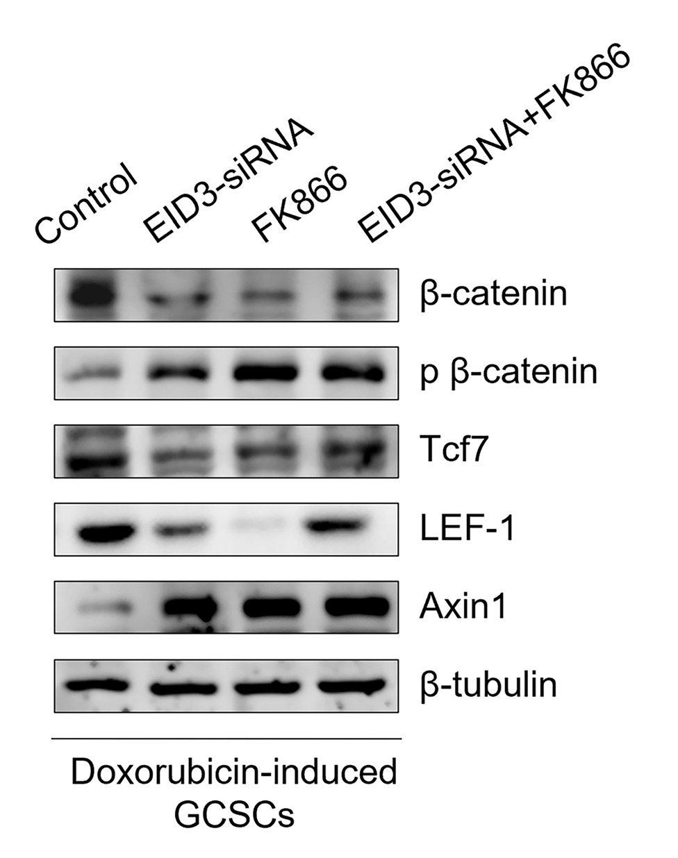
Identification of CD141+ vasculogenic precursor cells from human bone marrow and their endothelial engagement in the arteriogenesis by co-transplantation with mesenchymal stem cells
Graphical Abstract
Original ResearchVol. 15, None • pp. 321DOI: 10.1186/s13287-024-03938-3

The effect of exogenous mitochondria in enhancing the survival and volume retention of transplanted fat tissue in a nude mice model

Authors: Haoran Li, Zhengyao Li, Xiaoyu Zhang, Yan Lin, Tongtong Zhang, Leijuan Gan, Dali Mu

Background: Despite the pivotal role of fat grafting in plastic, reconstructive, and aesthetic surgery, inconsistent survival rates of transplanted adipose tissue, primarily due to early ischemic and hypoxic insults, remain a significant challenge. The infusion of healthy mitochondria has emerged as a promising intervention to support tissue recovery from ischemic, hypoxic, and other types of damages across various organ systems. Objectives: This study aims to evaluate the impact of supplementing human adipose tissue grafts with healthy exogenous mitochondria on their volume and mass retention rates when transplanted into the subcutaneous layers of nude mice. This approach seeks to improve and optimize fat grafting techniques. Methods: Human adipose tissues were preconditioned with exogenous mitochondria (10 µg/mL), a combination of exogenous mitochondria and the inhibitor Dyngo-4a, Dyngo-4a alone, or PBS, and then transplanted into the subcutaneous tissue of 24 nude mice. Samples were harvested at 1 and 3 months post-transplantation for analysis of mass and volume retention. The structural morphology and integrity of the adipose tissues were assessed using Hematoxylin and Eosin (H&E) staining. Results: Mitochondrial preconditioning significantly enhanced the retention of mass and volume in fat grafts, demonstrating superior structural morphology and integrity compared to the control group. Conclusions: This study highlights the potential of exogenous mitochondrial augmentation in fat transplantation to significantly improve fat graft survival, thereby optimizing the success of fat grafting procedures.
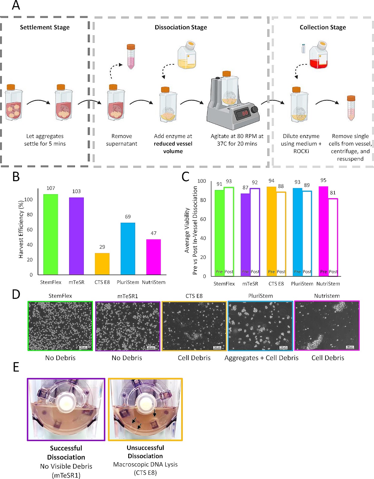
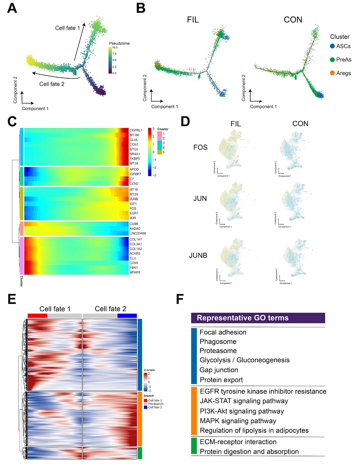
The effect of exogenous mitochondria in enhancing the survival and volume retention of transplanted fat tissue in a nude mice model
Graphical Abstract
Original ResearchVol. 15, None • pp. 116DOI: 10.1186/s13287-024-03727-y

Development and application of haploid embryonic stem cells

Authors: Hai-Song Wang, Xin-Rui Ma, Yi-Hong Guo

Haploid cells are a kind of cells with only one set of chromosomes. Compared with traditional diploid cells, haploid cells have unique advantages in gene screening and drug-targeted therapy, due to their phenotype being equal to the genotype. Embryonic stem cells are a kind of cells with strong differentiation potential that can differentiate into various types of cells under specific conditions in vitro. Therefore, haploid embryonic stem cells have the characteristics of both haploid cells and embryonic stem cells, which makes them have significant advantages in many aspects, such as reproductive developmental mechanism research, genetic screening, and drug-targeted therapy. Consequently, establishing haploid embryonic stem cell lines is of great significance. This paper reviews the progress of haploid embryonic stem cell research and briefly discusses the applications of haploid embryonic stem cells.

Development and application of haploid embryonic stem cells
Graphical Abstract