🧬 SinoBioData Academic Portal
🏛️ Indexed Academic JournalOriginal: 生物化学与生物物理学报

Acta Biochimica et Biophysica Sinica

Premier Chinese Biomedical Journal indexed in SinoBioData: Acta Biochimica et Biophysica Sinica (生物化学与生物物理学报).

Total Research Papers: 200
Access: 100% Free Open Access
Browse by Publication Year & VolumeReset All Filters ✕

Published Research PapersFiltered: Year 2025 • 57

Showing 73 of 200 peer-reviewed papers with full Graphical Abstracts.

Original ResearchVol. 57, Issue 7 • pp. 1164-1174DOI: 10.3724/abbs.2024240

miR-199a-3p suppresses Vldlr expression to promote cardiomyocyte proliferation

Authors: Rui Jiang, Lijuan Pei, Hongjie Zhang, Fenglian He, Yuhan Min, Xinhang Li, Ke Wei

The proliferative capacity of cardiomyocytes is limited in adult mammals, and replacing lost tissue following acute ischemic injury is challenging. Previous studies have demonstrated that miR-199a-3p can promote cardiomyocyte proliferation, but the exact mechanism by which this occurs remains unclear, although multiple targets of miR-199a-3p have been identified. We recently showed that very-low-density-lipoprotein receptor (Vldlr) inhibits cardiomyocyte proliferation, and in this study we aim to test whether Vldlr is a functional target gene of miR-199a-3p. 3′UTR reporter assays demonstrate that miR-199a-3p directly binds to the 3′UTR of Vldlr and inhibits its translation. Overexpressing Vldlr blunts the pro-proliferative effect of miR-199a-3p on cardiomyocytes, suggesting that Vldlr is indeed a functional target of miR-199a-3p. Mechanistically, Vldlr reduces S807/811 phosphorylation of RB1, and inhibiting CDK4/6 to prevent RB1 phosphorylation can block the pro-proliferative effect of both Vldlr knockdown and miR-199a-3p, suggesting that RB1 phosphorylation is required for the cardiomyocyte proliferation induced by miR-199a-3p and Vldlr knockdown. The findings of this study reveal Vldlr as a novel functional target of miR-199a-3p in cardiomyocytes and identify RB1 as a downstream effector of cardiomyocyte proliferation. The identification of the role of the miR-199a-3p-Vldlr-RB1 axis in cardiomyocyte proliferation may provide potential therapeutic targets for cardiac regenerative medicine.

miR-199a-3p suppresses Vldlr expression to promote cardiomyocyte proliferation
Graphical Abstract
Original ResearchVol. 57, Issue 8 • pp. 1222-1233DOI: 10.3724/abbs.2025041

A positive feedback loop between FOSB and miR-133b controls colon cancer cell proliferation

Authors: Wanwan Li, Qionggui Hu, Changwei Lin, Xiaorong Li, Yang Bai, Min Ma

FOSB, a member of the FOS gene family, forms heterodimers with JUN family proteins to engage in diverse cellular processes. Its biological impacts vary among different types of tumors, yet its specific function in colon cancer (CC) remains ambiguous. In this study, quantitative real-time PCR (qRT-PCR) and immunohistochemistry (IHC) are applied to measure FOSB expression levels, followed by an analysis of the association between FOSB expression and patients’ clinical parameters. In vitro experiments are performed to assess cell proliferation, including growth rate, cell cycle distribution, and apoptosis. A subcutaneous xenograft model in nude mice is utilized to monitor tumor growth in vivo. Additionally, chromatin immunoprecipitation (ChIP) and luciferase reporter assays are conducted to dissect the interactions among FOSB, miR-133b, and POU2F1. The results indicate that FOSB expression is downregulated in CC tissues relative to normal controls. Overexpression of FOSB suppresses proliferation and promotes apoptosis in CC cells. Mechanistically, FOSB binds to the promoter region of miR-133b, enhancing its transcription and subsequently repressing POU2F1 expression. Notably, decreased POU2F1 expression also alleviates the transcriptional repression of the FOSB promoter region, establishing a FOSB-miR-133b-POU2F1 feedback loop that inhibits CC proliferation. In summary, our findings suggest that FOSB acts as a tumor suppressor gene in CC and may exert its inhibitory effects on CC growth via the FOSB-miR-133b-POU2F1 feedback loop.

A positive feedback loop between FOSB and miR-133b controls colon cancer cell proliferation
Graphical Abstract
Original ResearchVol. 57, Issue 9 • pp. 1507-1516DOI: 10.3724/abbs.2025023

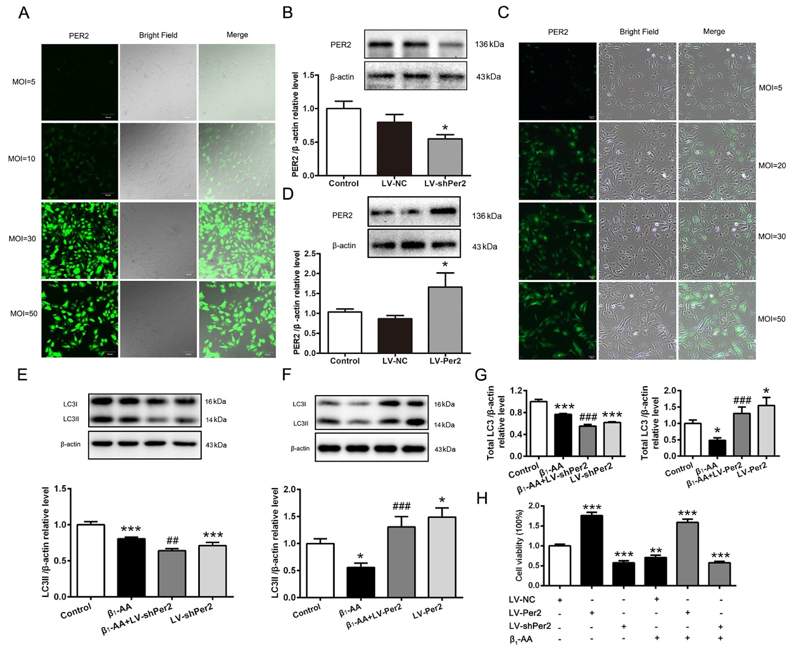
Reduced expression of the PER2 protein contributes to β1-AA-induced cardiac autophagy rhythm disorders

Authors: Pengjia Li, Jiayan Feng, Jiao Guo, Jin Xue, Yang Li, Shiyuan Wen, Xiaohui Wang, Huirong Liu, Li Wang

Heart failure may be linked to fluctuations in the rhythm of autophagy in cardiomyocytes throughout the day. Circadian rhythms depend on the regulation of core biological clock proteins, with PER2 playing a crucial role. Our previous research confirmed that the presence of β1-adrenergic receptor autoantibodies (β1-AAs) could inhibit myocardial autophagy, leading to cell death and heart failure. However, it remains unclear whether β1-AA induces cardiac autophagy rhythm disorders by affecting PER2 expression. In this study, we find that β1-AA disrupts the autophagy rhythm in cardiomyocytes, which is primarily indicated by decreased expression of the autophagy marker protein LC3. β1-AA disrupts the rhythmic expression of the PER2 protein in myocardial cells, which is manifested mainly by a decrease in PER2 protein expression. Metoprolol is used to verify that the β1-adrenergic receptor contributes to the reduction in the Per2 protein caused by β1-AA. Knockdown of Per2 with lentivirus reduces the inhibition of LC3 expression caused by β1-AA, whereas overexpression of Per2 in cardiomyocytes using lentivirus significantly restores the β1-AA-induced decrease in LC3 expression. Moreover, mTORC1 activation is found to participate in β1-AA-induced autophagy inhibition in cardiomyocytes after pretreatment with the mTORC1 inhibitor rapamycin. Furthermore, the decreased expression of the PER2 protein caused by β1-AA disrupts the myocardial autophagy rhythm by promoting mTORC1 activation through lentiviruses that knock down or overexpress the Per2 gene. This study provides an experimental basis for the precise treatment of cardiovascular diseases from the perspective of biological rhythm.

Reduced expression of the PER2 protein contributes to β1-AA-induced cardiac autophagy rhythm disorders
Graphical Abstract
Original ResearchVol. 57, Issue 12 • pp. 2048-2061DOI: 10.3724/abbs.2025077

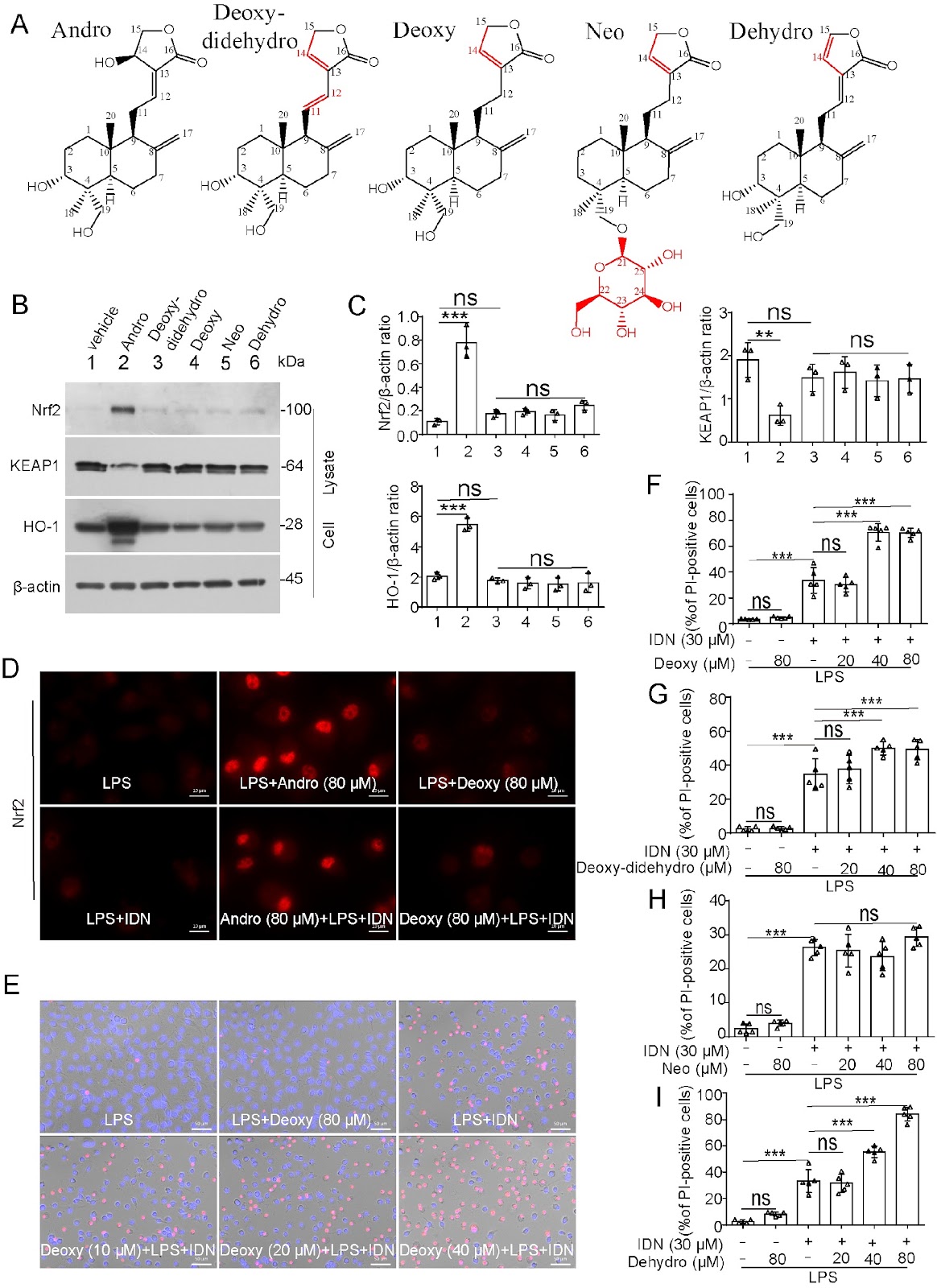
Andrographolide prevents necroptosis by suppressing the generation of reactive oxygen species

Authors: Na Lu, Qing Li, Linghan Duan, Rong Xu, Yaping Li, Fuli Shi, Zhiya Zhou, Yingqing Gan, Bo Hu, Jinhua Li, Xianhui He, Dongyun Ouyang, Qingbing Zha

Andrographolide (Andro), a natural product extracted from the Chinese traditional medicine herb Andrographis paniculata, has been applied for the treatment of diverse inflammatory diseases. However, its effects on necroptosis, a lytic form of cell death implicated in various inflammatory diseases, remain uncharacterized. In the present study, we investigate whether Andro and its derivatives can suppress necroptosis. Our results demonstrate that Andro notably inhibits necroptosis in the in vitro cellular models induced by either lipopolysaccharide (LPS) plus IDN-6556 or a combination of TNF-α, LCL-161 (Smac mimetic) and IDN-6556. In these cellular models, Andro inhibits the phosphorylation of receptor-interacting protein kinase 1 (RIPK1), RIPK3, and mixed lineage kinase domain-like pseudokinase (MLKL), as well as the formation of necrosomes. Specifically, Andro reduces the levels of intracellular reactive oxygen species (ROS) and mitochondrial superoxide (mtROS), preserves the mitochondrial membrane potential during necroptotic induction, and activates the antioxidant transcription factor nuclear factor E2-related factor 2 (Nrf2). Upon necroptotic stimulation, some mitochondrial proteins, such as Bcl-2 and Bak, oligomerize and co-localize with RIPK1, RIPK3, and phosphorylated MLKL (p-MLKL) in necrosomes. However, this process of necrosome formation can be prevented by Andro. In contrast, derivatives, including dehydroandrographolide, neoandrographolide, 14-deoxy-11,12-didehydroandrographolide, and 14-deoxyandrographolide, have no anti-necroptotic effects and fail to upregulate Nrf2. Collectively, our findings demonstrate that Andro specifically inhibits the RIPK1/RIPK3/MLKL signaling axis to suppress necroptosis, highlighting its therapeutic potential against necroptosis-related disorders.

Andrographolide prevents necroptosis by suppressing the generation of reactive oxygen species
Graphical Abstract
Original ResearchVol. 57, Issue 12 • pp. 2034-2047DOI: 10.3724/abbs.2025081

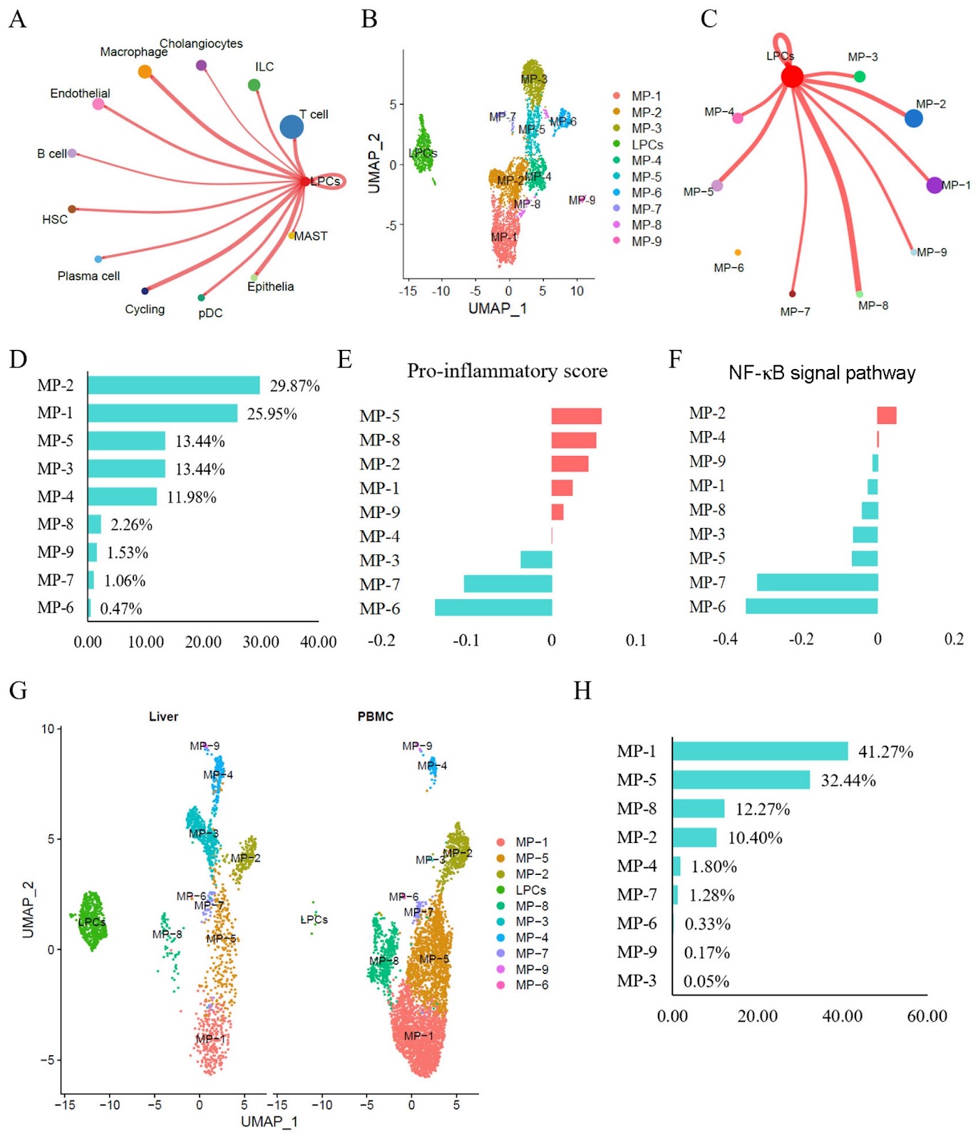
Resident CD24+LCN2+ LPCs aggravate fibrosis and inflammatory progression via the recruitment of TPPP3+COL10A1+ macrophages in NASH

Authors: Min Ding, Xiaoshu Qi, Weijian Huang, Yan Lin, Hexin Yan

Resident CD24+LCN2+ liver progenitor cells (LPCs) reportedly contribute to the expanding ductular reaction and macrophage-mediated inflammation associated with chronic liver damage. Both ductular reactions and macrophage-driven inflammation are associated with liver fibrosis and injury in various mouse liver disorders. This study aims to investigate the molecular phenotypes of LPCs and their regulatory mechanisms in humans with non-alcoholic steatohepatitis (NASH). Single-cell RNA sequencing (scRNA-seq) datasets are used to characterize the status and molecular phenotypes of LPCs in clinical NASH samples. To elucidate the regulatory mechanisms of LPCs, CellChat and NicheNet are employed to assess cell-cell communication between LPCs and other cell types. The findings are validated using RNA sequencing datasets associated with NASH progression, NASH mouse models (CDAHFD and HFD), and human NASH liver samples. Results show that resident CD24+LCN2+ LPCs are identified and found to be significantly enriched in NASH patients. Cell communication analyses predict strong interactions between LPCs and proinflammatory macrophage subtypes. Additionally, in NASH, the liver recruits peripheral blood mononuclear cell (PBMC)-derived macrophages and polarizes them into proinflammatory subtypes. The macrophage subtype MP-2 is identified as the primary recipient of LPC-derived signals, exhibiting marked hyperactivation of the NF-κB pathway and a strong association with liver fibrosis. Finally, the MP-2 markers COL10A1 and TPPP3 are characterized and validated. In summary, this study reveals that resident CD24+LCN2+ LPCs are activated in NASH and contribute to fibrosis progression by promoting the activation of the proinflammatory COL10A1+TPPP3+ macrophage subtype.

Resident CD24+LCN2+ LPCs aggravate fibrosis and inflammatory progression via the recruitment of TPPP3+COL10A1+ macrophages in NASH
Graphical Abstract
Original ResearchVol. 57, Issue 12 • pp. 2074-2093DOI: 10.3724/abbs.2025100

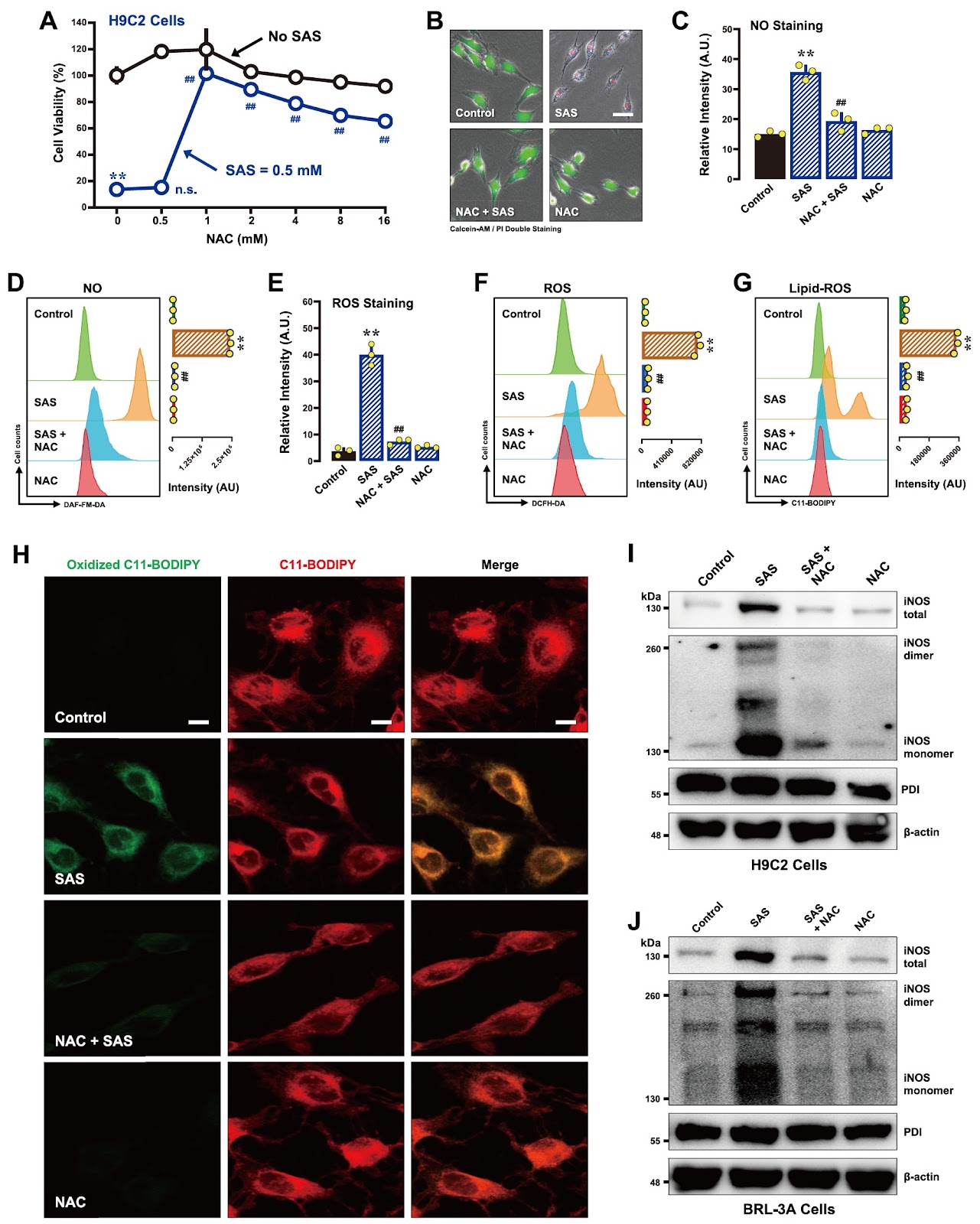
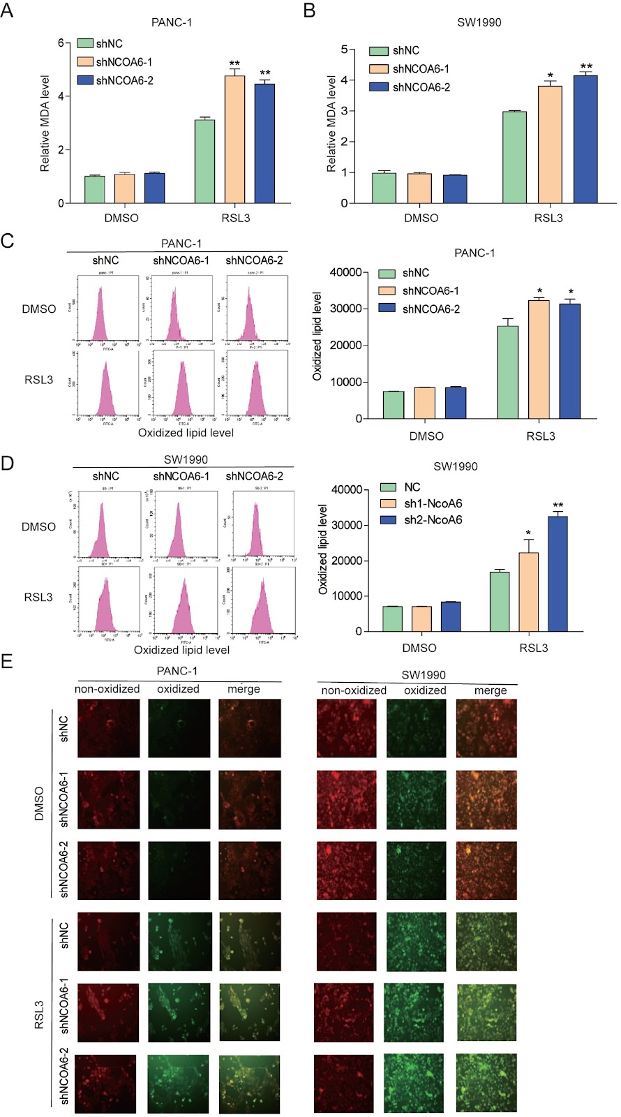
Characterization of the mechanisms underlying sulfasalazine-induced ferroptotic cell death: role of protein disulfide isomerase-mediated NOS activation and NO accumulation

Authors: Yi-Chen Jia, Jia-Ling Zhong, Xiangyu Hao, Bao Ting Zhu

Sulfasalazine (SAS), a clinically utilized anti-inflammatory drug, has been shown to induce ferroptosis by inhibiting system Xc− activity, thereby causing cellular glutathione depletion. Recently, protein disulfide isomerase (PDI) was shown to be an upstream mediator of the oxidative cell death (oxytosis/ferroptosis) induced by glutamate, erastin, RSL3 and SAS. The present study aims to further characterize the detailed biochemical and cellular mechanisms of SAS-induced ferroptosis in two cell lines, i.e., H9C2 rat cardiomyocytes and BRL-3A rat hepatocytes, with a focus on elucidating the critical role of PDI in mediating SAS-induced toxicity. We find that SAS can induce ferroptosis in H9C2 and BRL-3A cells, which is accompanied by a sequential increase in the buildup of cellular nitric oxide (NO), reactive oxygen species (ROS) and lipid-ROS. SAS activates PDI-mediated dimerization of inducible NO synthase (iNOS) and cellular accumulation of NO, and these effects are followed by ROS and lipid-ROS accumulation. Furthermore, SAS markedly upregulates the iNOS protein levels in these cells. Knockdown of PDI or pharmacological inhibition of PDI catalytic activity effectively suppresses SAS-induced iNOS dimerization, abrogates SAS-induced accumulation of NO, ROS and lipid-ROS, and prevents ferroptosis. On the other hand, PDI activation through the use of TrxR1 inhibitors sensitizes these cells to SAS-induced ferroptosis. These findings provide further experimental support for a pivotal role of PDI in SAS-induced cytotoxicity through the activation of the PDI-NOS-NO axis, which then leads to the accumulation of cellular ROS and lipid-ROS and ultimately the induction of oxidative cell death.

Characterization of the mechanisms underlying sulfasalazine-induced ferroptotic cell death: role of protein disulfide isomerase-mediated NOS activation and NO accumulation
Graphical Abstract
Original ResearchVol. 57, Issue 12 • pp. 1983-1998DOI: 10.3724/abbs.2025109

Rhamnose alleviates the proinflammatory response during endotoxemia via the CEACAM1/LGALS9-p38 axis

Authors: Rongjuan Wei, Tao Zhong, Ke Deng, Xianglong Zhang, Dongping Li, Meiling Chen, Ping Chang, Peng Wu, Zhanguo Liu

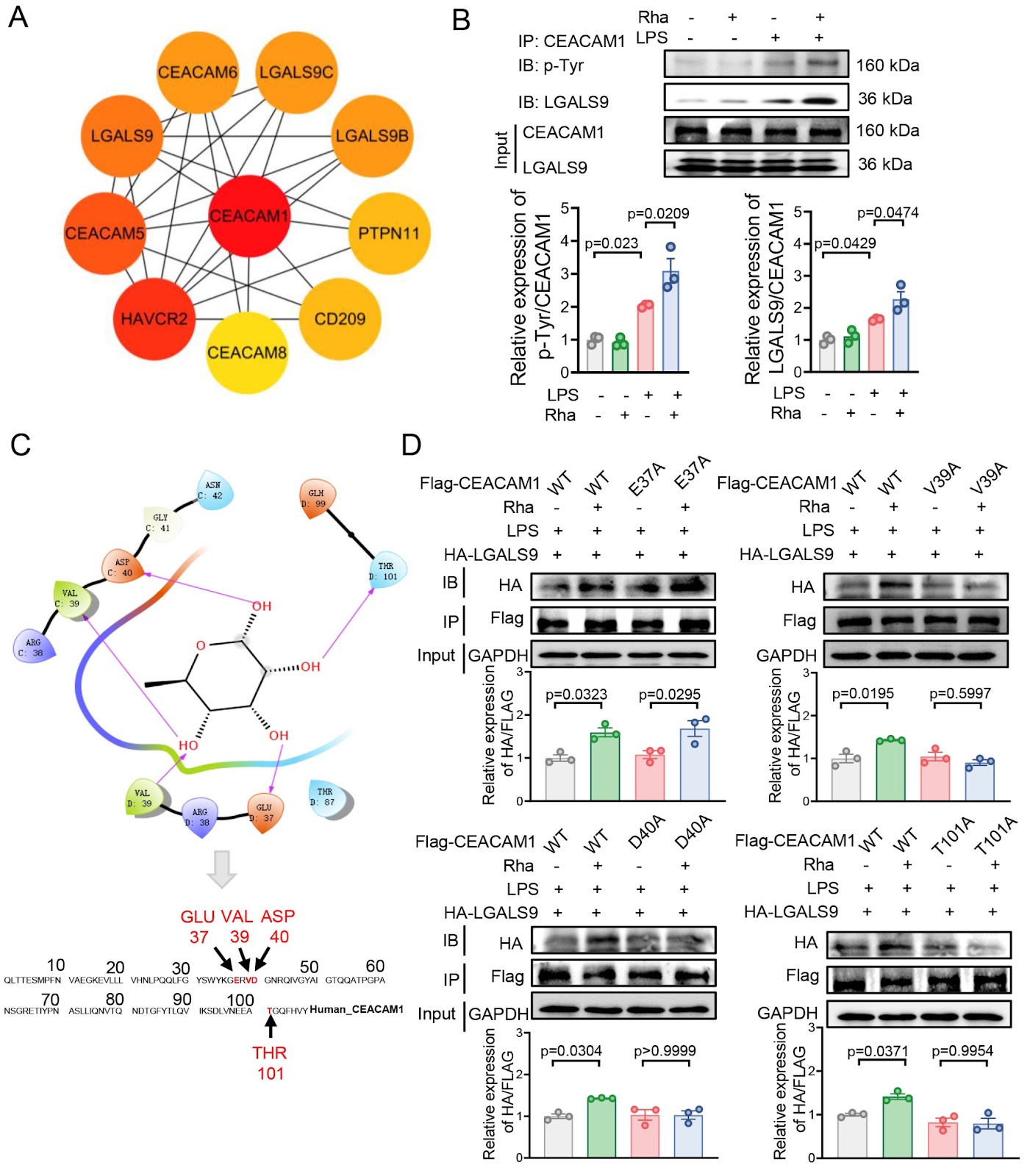
Gut microbiota plays an important role in orchestrating the host immune response. We previously reported that gut microbiota-derived rhamnose enhances the phagocytosis of macrophages, upon which we further asked whether rhamnose has modulatory effects on inflammation. Here, we show that, in an LPS-induced endotoxic mouse model, plasma rhamnose levels are increased. This bacteria-derived sugar alone does not impact inflammatory cytokine homeostasis or cause organ damage. In contrast, it is able to alleviate endotoxin-induced systemic inflammation and organ damage. Mechanistically, in macrophages in vitro, rhamnose binds to the V39, D40, and T101 sites of carcinoembryonic antigen-associated cell adhesion molecule 1 (CEACAM1), subsequently promoting the interaction between CEACAM1 and galectin 9 (LGALS9), which increases the protein level of dual-specificity protein phosphatase 1 (DUSP1). This inhibits p38 phosphorylation and thus attenuates the LPS-triggered expressions of proinflammatory factors. Collectively, our results suggest that rhamnose signals via the CEACAM1/LGALS9-p38 axis, which suppresses endotoxemia-associated inflammation, and that rhamnose is a candidate anti-inflammatory agent for the control of infection-induced organ damage.

Rhamnose alleviates the proinflammatory response during endotoxemia via the CEACAM1/LGALS9-p38 axis
Graphical Abstract
Original ResearchVol. 57, Issue 12 • pp. 1969-1982DOI: 10.3724/abbs.2025145

Angptl4 is upregulated by microenvironmental factors during the wound healing process and promotes epidermal stem cell proliferation via PRL8a6

Authors: Siyuan Yu, Pengxiang Ji, Ting Du, Zuohua Liu, Yuan Yang, Zhenkun Lv, Lei Xu, Qianheng Jin, Weijuan Gong, Yingying Le, Yi Fu, Ruixing Hou

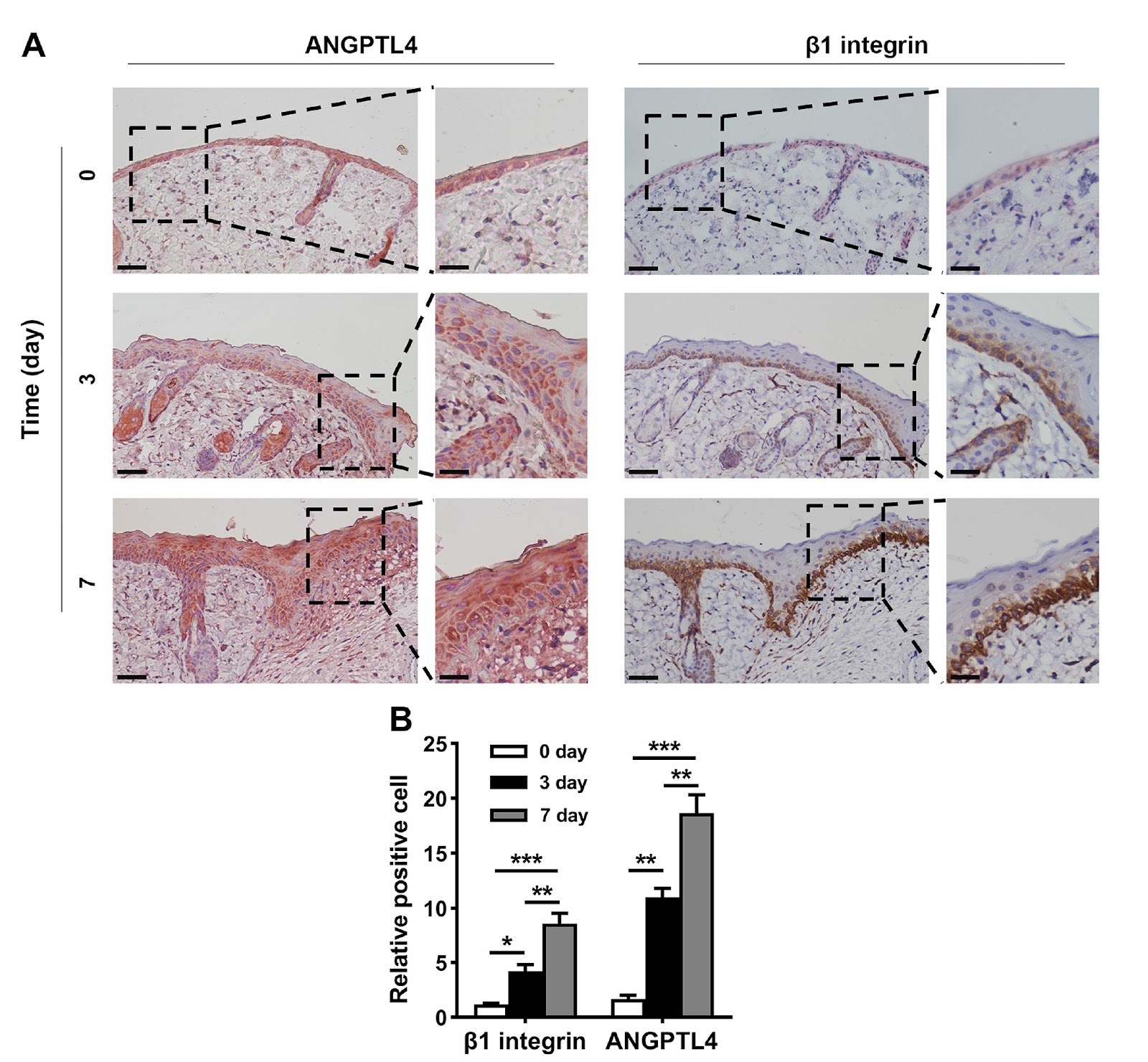
Angiopoietin-like 4 (ANGPTL4) expression is increased in wound tissue and contributes to wound healing. However, the underlying mechanisms are not fully understood. Here, we demonstrate that ANGPTL4 expression is significantly increased in epidermal stem cells (EpSCs) in the periwound epidermis during wound healing in mice. Increased Angptl4 expression is positively correlated with increased expressions of tumor growth factor-α, interleukin-1β, epidermal growth factor, nerve growth factor, fibroblast growth factor 7, and transforming growth factor-β1. Each of these molecules induces Angptl4 expression in mouse EpSCs. RNA sequencing of EpSCs derived from wild-type and Angptl4 knockout (Angptl4–/–) mice reveals altered expressions of genes involved in the cell cycle and cell proliferation in Angptl4–/– EpSCs, including a decrease in cyclin E2/A2/B1 and cyclin-dependent kinase 1 (Cdk1) expression; an increase in Cdk inhibitor 2a (Cdkn2a) and Cdkn2b expression; and a decrease in the prolactin (PRL) family members Prl2a1, Prl8a1, Prl8a9, and Prl8a6. Mechanistic studies reveal that ANGPTL4 stimulates EpSC proliferation via PRL8a6-mediated upregulation of cyclins A2/E2/B1 and Cdk1, downregulation of Cdkn2a, and acceleration of cell cycle progression from the G1 to the S and G2 phases. In vivo studies demonstrate that Prl8a6 mRNA is upregulated by ANGPTL4 in mouse periwound tissue during skin wound healing. Knockdown of Angptl4 or Prl8a6 in periwound skin tissue impairs EpSC proliferation and delays wound re-epithelialization. In conclusion, our study demonstrates that, after skin injury, elevated levels of proinflammatory cytokines and growth factors in periwound tissue stimulate Angptl4 expression in EpSCs and that ANGPTL4 promotes EpSC proliferation by increasing Prl8a6 expression, thereby accelerating wound re-epithelialization.

Angptl4 is upregulated by microenvironmental factors during the wound healing process and promotes epidermal stem cell proliferation via PRL8a6
Graphical Abstract
Original ResearchVol. 57, Issue 12 • pp. 2062-2073DOI: 10.3724/abbs.2025097

Integration of the hammerhead ribozyme into structured RNAs to measure ligand-binding events for riboswitch candidates and aptamers

Authors: Shenglan Zhang, Yinghong Lin, Ting Gao, Binfen Chen, Weibin Wu, Shanshan Fang, Kexin Fan, Yuqing Lai, Yezi Lin, Rongqin Ke, Sanshu Li

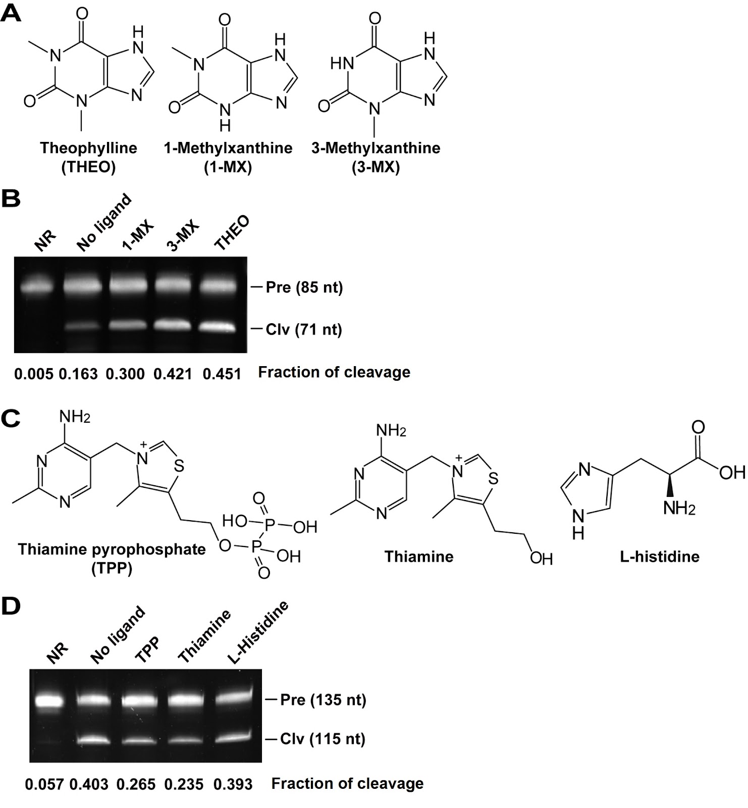
Some structured RNAs, such as riboswitches and aptamers, can bind to their cognate ligands and have been used in biosensors and gene expression control elements. However, current methods for detecting ligand binding to structured RNAs are either severely limited or inconvenient. In this study, we design a multibase pair bridge to integrate a hammerhead ribozyme into structured RNAs to detect ligand binding events. The experimental results demonstrate that the length of the bridge has a significant effect on the cleavage of the ribozyme; optimal cleavage can be achieved with three to six base pairs in the bridge. The dissociation constant (KD) values obtained through this method are in agreement with those determined by in-line probing techniques, and 1 pmol of allosteric ribozyme RNA is sufficient for measurement. We apply this method to evaluate the binding affinity of the riboswitch candidate Motif_9307. Our findings indicate that this motif has no binding affinity for S-adenosylmethionine or several other tested ligands, which is consistent with the results of the in-line probing experiments. Notably, our method reveals an increase in cleavage activity when yeast extract is added as a mixture of ligands, suggesting that the ligand of Motif_9307 is present in the extract. In conclusion, we develop an alternative approach for measuring ligand binding events associated with riboswitch candidates and aptamers.

Integration of the hammerhead ribozyme into structured RNAs to measure ligand-binding events for riboswitch candidates and aptamers
Graphical Abstract
Original ResearchVol. 57, Issue 12 • pp. 1953-1968DOI: 10.3724/abbs.2025089

LPS mediates cuproptosis and inflammation in THP-1 macrophages through HKDC1

Authors: Langlin Ou, Zitong Meng, Jian Mei, Hao Yuan, Xiangrui Zhu, Xiaoying Wang, Ao Shen, Zhaosi Wang, Lixin Zhang, Song Wang, Yingli Chen, Xiangming Pang, Yuxiang Liu, Yadong Xu, Cui Ma

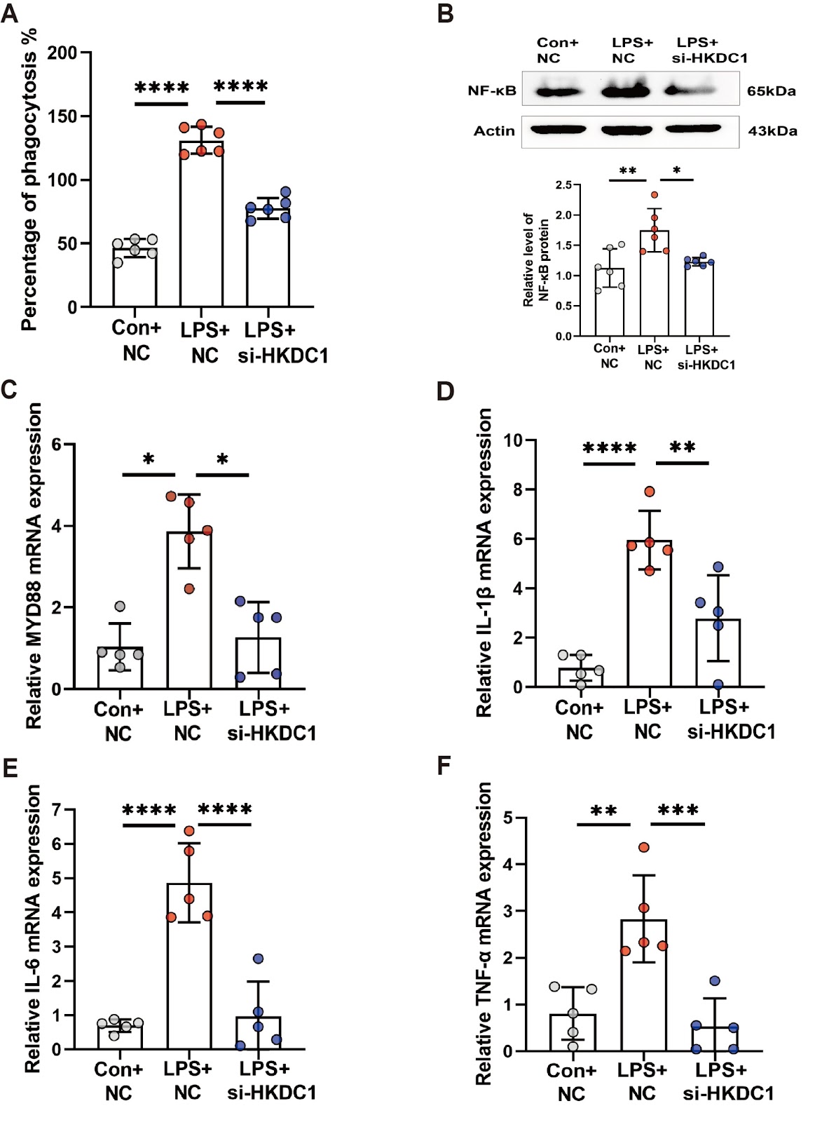
Cuproptosis is a recently identified form of copper-driven cell death characterized by the aggregation of acylated proteins and proteotoxic stress in the mitochondrial tricarboxylic acid cycle, which plays a role in inflammation. Recent studies suggest that hexokinase structural domain protein 1 (HKDC1), a fifth hexokinase, is involved in regulating mitochondrial function. However, the role of HKDC1 in cuproptosis and LPS-induced macrophage inflammation remains unclear. Here, we assess macrophage plasticity using CCK8 viability assays and phagocytosis activity experiments in an in vitro inflammatory model of THP-1 cells. We measure the levels of inflammatory factors and cuproptosis-related proteins using western blot analysis and RT-qPCR. Additionally, we examine the expression and localization of the HKDC1 protein using ChIP-qPCR and immunofluorescence staining. We find that LPS promotes the expressions of inflammatory factors and decreases cuproptosis levels in THP-1-derived macrophages while also activating glycolysis and inducing the expression of HKDC1 via the Toll-like receptor 4 (TLR4) receptor. We further demonstrate that HKDC1 knockdown inhibits glycolysis and induces cuproptosis. Mechanistically, we provide the first evidence that LPS promotes the binding of Yin Yang 1 (YY1) to the HKDC1 promoter, thereby regulating HKDC1 transcription. HKDC1 interacts with heat shock cognate B (HSCB) and ferredoxin 1 (FDX1), leading to increased intracellular copper levels and subsequent cuproptosis. HKDC1 knockdown in vivo alleviates acute sepsis by activating copper-dependent cell death pathways. Collectively, our findings suggest that LPS mitigates cuproptosis and promotes inflammation via HKDC1, suggesting a new cuproptosis-dependent anti-inflammatory strategy.

LPS mediates cuproptosis and inflammation in THP-1 macrophages through HKDC1
Graphical Abstract
Original ResearchVol. 57, Issue 7 • pp. 1139-1150DOI: 10.3724/abbs.2024215

High-resolution imaging atlas reveals the context-dependent role of pancreatic sympathetic innervation in diabetic mice

Authors: Qingqing Xu, Yuxin Chen, Xinyan Ni, Hanying Zhuang, Shenxi Cao, Liwei Zhao, Leying Wang, Jianhui Chen, Wen Z Yang, Wenwen Zeng, Xi Li, Hongbin Sun, Wei L Shen

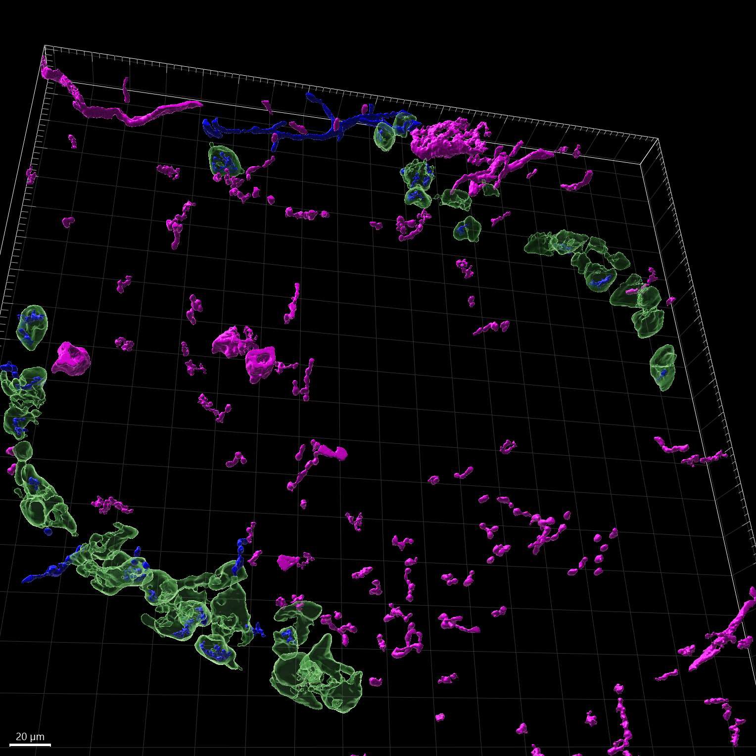
A better understanding of how sympathetic nerves impact pancreatic function is helpful for understanding diabetes. However, there is still uncertainty and controversy surrounding the roles of sympathetic nerves within the pancreas. To address this, we utilize high-resolution imaging and advanced three-dimensional (3D) reconstruction techniques to study the patterns of sympathetic innervation and morphology in the islets of adult wild-type (WT) and diabetic mice. Our data show that more than ~30% of α/β-cells are innervated by sympathetic nerves in both WT and diabetic mice. Additionally, sympathetic innervated α/β-cells are reduced in diet-induced obese (DIO) mice, whereas sympathetic innervated β-cells are increased in db/db mice. In addition, in situ chemical pancreatic sympathetic denervation (cPSD) improves glucose tolerance in WT and db/db mice but decreases glucose tolerance in DIO mice. In situ cPSD also enhances insulin sensitivity in diabetic mice without affecting WT mice. Overall, our findings advance our understanding of diabetes by highlighting the distinctive impact of pancreatic sympathetic innervation on glucose regulation.

High-resolution imaging atlas reveals the context-dependent role of pancreatic sympathetic innervation in diabetic mice
Graphical Abstract
Original ResearchVol. 57, Issue 12 • pp. 1939-1952DOI: 10.3724/abbs.2025171

Single-cell transcriptomics reveals apolipoprotein A4-mediated metabolic-immune reprogramming in lymphocytes during early obesity-related chronic kidney disease

Authors: Yang Wei, Ting Zhang, Yingying Jin, Xiaohuan Liu, Jinting Zhou, Na Huang, Yiying Wang

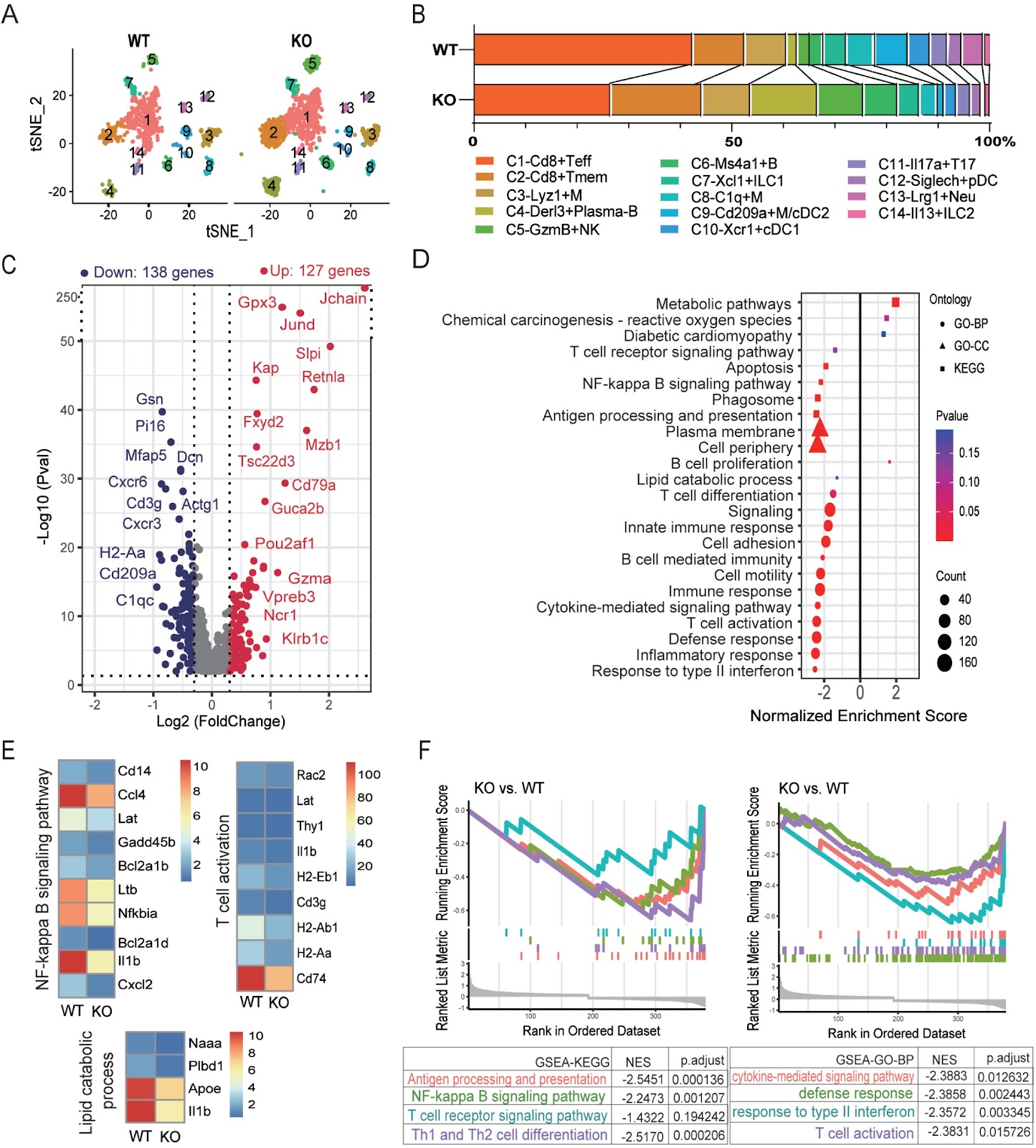
Obesity-induced metabolic inflammation is a key driver of chronic kidney disease (CKD), with immune dysregulation, particularly among lymphocytes, contributing to early disease pathology. To explore the role of apolipoprotein A4 (Apoa4) in regulating immune cell metabolism and function, we establish high-fat diet-induced obese (DIO) models using wild-type and Apoa4-knockout (KO) mice. KO mice exhibit exacerbated insulin resistance and renal lipid accumulation. Single-cell RNA sequencing reveals that Apoa4 deletion remodeled the renal immune-metabolic landscape. This remodeling broadly compromises the immune functions of T, NK, and B cells, even as it expands the proportions of cytotoxic Gzma+ NK cells and Derl3+ plasma cells. Mechanistically, Apoa4 deletion aggravates metabolic dysregulation and oxidative stress and downregulates the expression levels of key effector genes, including Ifng and Il1b. Furthermore, the regulatory network activities of key transcription factors, such as Lef1 and Runx3 in Cd8+ T cells; Irf8, T-bet, and Eomes in NK cells; and Tcf4, Lmo2, and Xbp1 in B cells, are perturbed. CellChat analysis predicts disruptions in pro-inflammatory (IFN-II and IL-1), immunoregulatory (FASLG), and metabolic regulatory (ENHO and ANGPTL) signaling, alongside enhanced IL-2-mediated suppression. These findings are corroborated by flow cytometry, immunofluorescence staining, and qPCR. Our results establish Apoa4 as a crucial regulator of lymphocyte metabolic and immune homeostasis in the early stages of obesity-associated CKD.

Single-cell transcriptomics reveals apolipoprotein A4-mediated metabolic-immune reprogramming in lymphocytes during early obesity-related chronic kidney disease
Graphical Abstract
Original ResearchVol. 57, Issue 12 • pp. 2124-2128DOI: 10.3724/abbs.2025055

Quantitative liquid chromatography-tandem mass spectrometric analysis of 11dH-TXB2 and creatinine in urine

Authors: Chunyan Li, Wuzheng Liu, Yana Xiao, Tenglong Dai, Yu Su, Yubin Wang, Ao Zhang, Ruichen Liu, Xianglong Zhao, Zhao Zhang, Shangqi Yin, Jun Wu

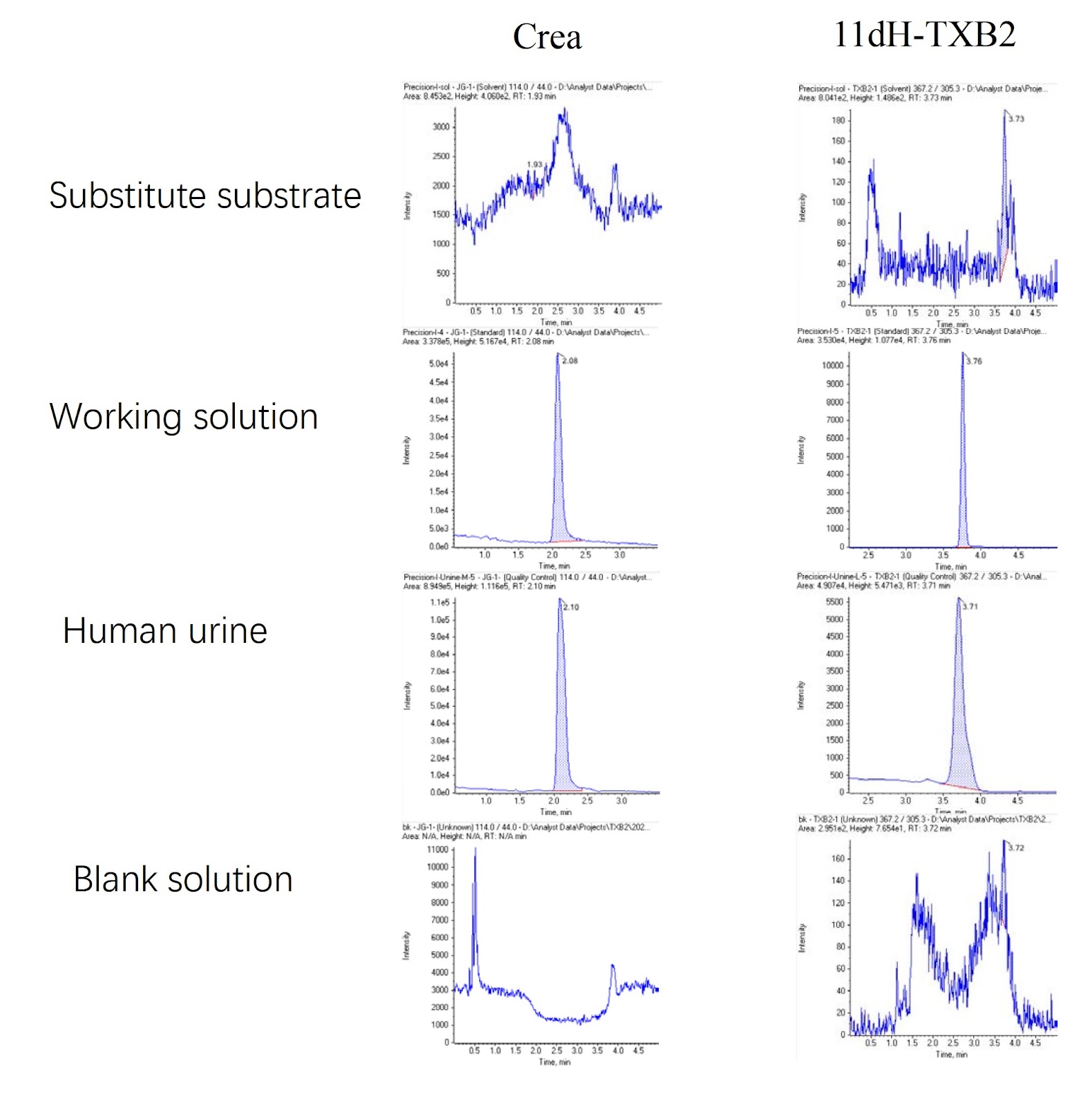
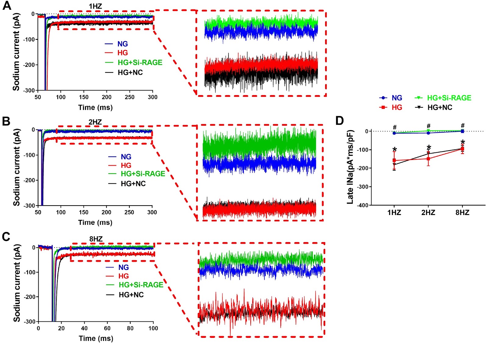
Platelets circulate in an inactive form in the body until they contact with defective areas of endothelial cells or encounter a clotting cascade [1]. Activated platelets release and express bioactive substances and acquire the ability to bind plasma fibrinogen. Abnormal activation of platelets is involved in atherosclerosis and thrombosis [2,3]. When platelets are stimulated and activated, phospholipase A2 is activated at the same time, which then cleaves membrane phospholipids and frees arachidonic acid (AA) [4]. The latter catalyzes thromboxane A2 (TXA2) via thromboxane synthetase [5], which is induced by the cyclooxygenase COX-1 to produce prostaglandins G2 and H2 [6]. TXA2 is highly unstable, with a half-life of only 30 s, and it is rapidly hydrolyzed to relatively stable thromboxane B2 (TXB2), which is then converted in the liver to 11-dehydrothromboxane B2 (11dH-TXB2), which has a longer half-life and is excreted in the urine [7]. Dehydrothromboxane B2 is the final stable metabolite of thromboxane A2, which is derived only from arachidonic acid metabolism and can represent thromboxane A2 level in the body [8]. Specifically, by inhibiting the action of COX-1, the most important enzyme in the process of arachidonic acid metabolism, aspirin inhibits the production of thromboxane A2, that is, the concentration of TXA2 affects the effect of aspirin on platelet aggregation. However, the half-life of TXA2 (including the metabolic intermediate TXB2) is too short to be accurately measured, so the detection of its metabolic end product 11dH-TXB2 can very accurately reflect the sensitivity of the body to aspirin [7]. The concentration of 11dH-TXB2 in the serum correlates well with the concentration of 11dH-TXB2 in the urine, so the determination of 11dH-TXB2 in the urine can more effectively reflect the production of TXA2 in vivo [8]. The 11dH-TXB2 concentration needs to be corrected with the urinary creatinine concentration to rule out the effects of the urine concentration and renal function, so random urine samples can be used for testing [6]. Aspirin can acetylate serine at the key site of cyclooxygenase and thus irreversibly inhibits the activity of COX-1, reduces the synthesis of TXA2, and blocks the production of TXA2 and its induced platelet aggregation. Low-dose aspirin (30–75 mg/day) can effectively inhibit 95% of COX-1 activity [7]. Since the production of TXA2 in serum is largely dependent on platelet COX-1 (a therapeutic target of aspirin), 11dH-TXB2 can be used as a monitor for aspirin-induced platelet inhibition [3]. Creatinine is a metabolic byproduct of muscle metabolism that is primarily excreted via glomerular filtration, and its level is indicative of renal function [9]. The 24-h creatinine clearance can also be used to determine the integrity of the sample or to correct the urine sample concentration with the creatinine ratio [10]. Currently, creatinine detection methods include the Jaffe method, enzymolysis spectrophotometry, HPLC, capillary electrophoresis, capillary zone electrophoresis, gas chromatography tandem mass spectrometry (GC-MS) and liquid chromatography tandem mass spectrometry (LC-MS/MS) [11]. At present, there is no method for the simultaneous detection of 11dH-TXB2 and creatinine. When both analytes are needed, separate tests must be performed, increasing the workload and sample volume requirements. Thus, developing a method that enables the concurrent quantification of 11dH-TXB2 and creatinine in a single assay remains a critical challenge. The aim of this study was to provide a method for the simultaneous detection of 11dH-TXB2 and creatinine and to alleviate the problem that 11dH-TXB2 and creatinine cannot be simultaneously detected. By developing a standardized quantitative approach for measuring 11dHTXB2 and creatinine in human urine, this study aims to provide reliable concentration data, thereby facilitating further clinical research and methodology optimization.

Quantitative liquid chromatography-tandem mass spectrometric analysis of 11dH-TXB2 and creatinine in urine
Graphical Abstract
Original ResearchVol. 57, Issue 11 • pp. 1875-1887DOI: 10.3724/abbs.2025091

Unique gene patterns lead to distinct functional phenotypes and chemosensitivity profiles among subclones obtained from a single glioblastoma cell line

Authors: Daxing Xu, Yingdi Jiang, Jie Li, Lingli Gong, Zhenkun Yang, Bo Zhang, Koukou Li, Jian Zou

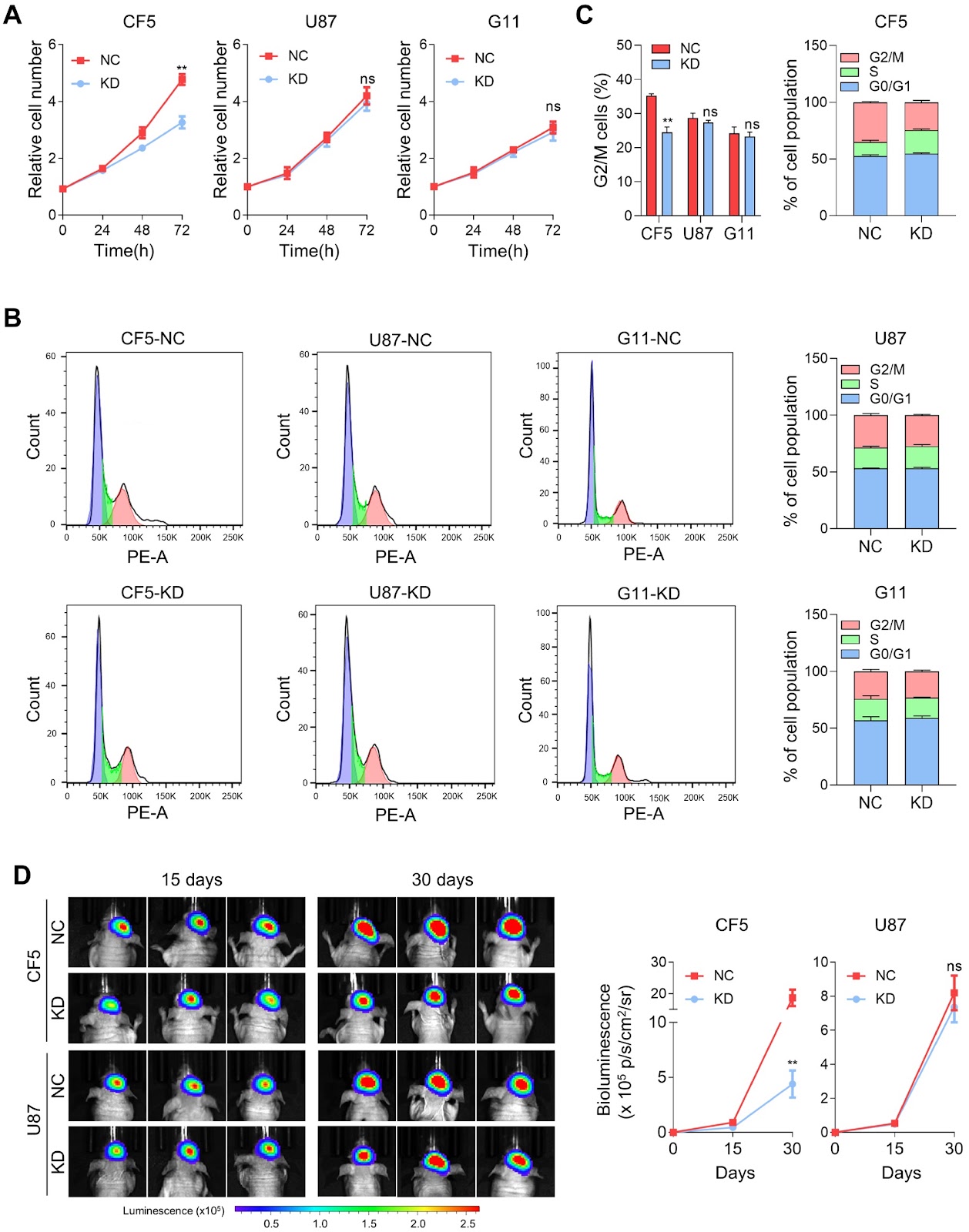
One of the characteristics of malignant tumors is heterogeneity, which refers to the molecular or genetic differences among progeny cells during tumor growth. This heterogeneity contributes to variations in the tumor growth rate, invasive ability, drug sensitivity, and prognosis. To gain a deeper understanding of the molecular background underlying tumor heterogeneity, we construct monoclonal cell lines derived from the glioblastoma (GBM) cell line U87-MG by limiting dilution assays. The selected CF5 and G11 subclones exhibit completely different cell morphologies and, more importantly, distinct functional phenotypes. CF5 exhibits stronger proliferative properties and chemoresistance, whereas G11 shows greater motility and invasion. Transcriptomic sequencing reveals great differences in gene expression among the CF5, G11, and U87 cell lines, and downregulated genes in individual clones are significantly enriched in gene sets related to extracellular matrix function. ITGA11 and ITGA6, as research subjects, are demonstrated to exclusively regulate functional phenotypes and chemotherapy sensitivity in CF5 or G11 cells. In U87 cells, combined knockdown of these two genes significantly inhibits tumor growth and increases chemotherapy sensitivity, but knockdown of either gene alone does not. In summary, these data reveal that even under uniform growth conditions, the heterogeneity of tumor cells and their diverse genetic backgrounds remain significant and persistent. This finding is crucial for accurately identifying tumor-related genes and their functional phenotypes, and a thorough understanding of the genetic and molecular background underlying tumor heterogeneity is essential for comprehensive cancer treatment.

Unique gene patterns lead to distinct functional phenotypes and chemosensitivity profiles among subclones obtained from a single glioblastoma cell line
Graphical Abstract
Original ResearchVol. 57, Issue 11 • pp. 1743-1757DOI: 10.3724/abbs.2025028

ISGylation: is our genome yearning for such a modification?

Authors: Zheng Chen, Zheng Li, Ying Wang, Zaure Dushimova, Kapanova Gulnara, Shunichi Takeda, Zhongjun Zhou, Xingzhi Xu

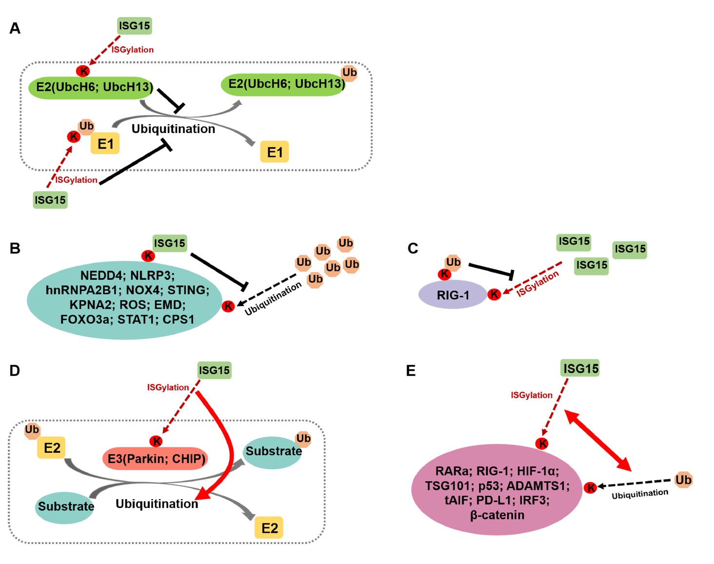
ISGylation is the post-translational modification of protein substrates covalently conjugated with the ubiquitin-like protein, interferon-stimulated gene 15 (ISG15). Although initially linked to antiviral immunity, recent evidence highlights important roles for ISGylation in various biological processes, such as maintaining genomic stability, promoting tumourigenesis, and being involved in other pathological conditions. In this review, we examine the molecular mechanisms underlying ISGylation, its interplay with other post-translational modifications, and its involvement in diverse biological and pathological processes. We propose future research directions to advance the field and discuss how ISGylation might be harnessed to ensure human health, particularly genome instability-associated diseases.

ISGylation: is our genome yearning for such a modification?
Graphical Abstract
Original ResearchVol. 57, Issue 12 • pp. 2133-2136DOI: 10.3724/abbs.2025048

Withaferin A combined with ricolinostat: a potent synergistic therapy for cervical cancer through regulating p53 ubiquitination and acetylation

Authors: Tian Chen, Yiting Xu, Kunming Yang, Yutong Du, Zhuan Zhu, Lingling Xu, Xinrong Wang, Yi Yin, Yu Hu, Chengcheng Wang, Ronggui Hu, Chuanyin Li

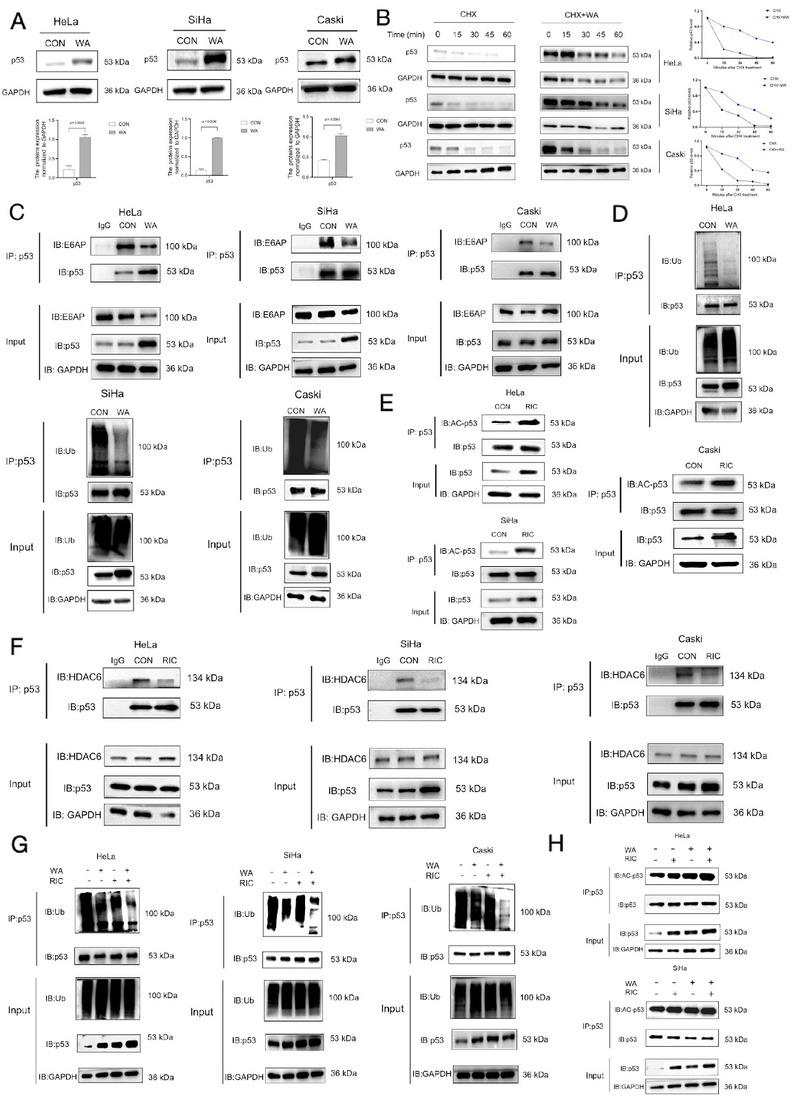
As a classic tumor suppressor gene, p53 has been extensively studied since its discovery in the mid-1980s. Research findings have revealed that p53 protein expression is suppressed in various cancers [1]. For example, in cervical cancer, p53 predominantly exists in a wild-type form to maintain its biological function [2]. Nevertheless, its tumor-suppressive activity is significantly impaired because of rapid protein degradation, short half-life, and low levels. Post-translational modifications (PTMs) of p53, such as ubiquitination, acetylation, phosphorylation and methylation, are critical regulators of its stability, activity, conformation, localization, and interactions with cofactors [3]. Among these, ubiquitination and acetylation play central roles in controlling p53 protein stability and activity [4]. Therefore, targeting p53 PTMs to modulate its ubiquitination and acetylation levels represents an effective strategy to increase its stability and tumor-suppressive function, offering a promising avenue for cervical cancer drug development. In 99% of cervical cancers (high-risk human papillomavirus-positive), E3 ubiquitin ligase E6-associated protein (E6AP) mediates the ubiquitination degradation of p53 [5], whereas histone deacetylase 6 (HDAC6) deacetylates p53. In this study, we explored the possibility of combining the natural product withferin A (WA) with the HDAC6 inhibitor ricolinostat (RIC) to treat cervical cancer cells, with a focus on the ubiquitination and acetylation of p53 and the consequences for its stability. These results suggested that the combination of WA and RIC is more effective than either treatment alone in inhibiting the degradation and increasing the stability of p53, thereby synergistically slowing the onset and progression of cervical cancer.

Withaferin A combined with ricolinostat: a potent synergistic therapy for cervical cancer through regulating p53 ubiquitination and acetylation
Graphical Abstract
Original ResearchVol. 57, Issue 11 • pp. 1834-1846DOI: 10.3724/abbs.2025202

VSIG2 hinders gastric cancer progression by suppressing ANXA2-mediated NF-κB pathway activation

Authors: Qingfeng Ni, Yang Wang, Xinyue Bian, Qiuchan Qu, Boyuan Shen, Yuanjie Niu, Jiawei Yu, Jianwei Zhu

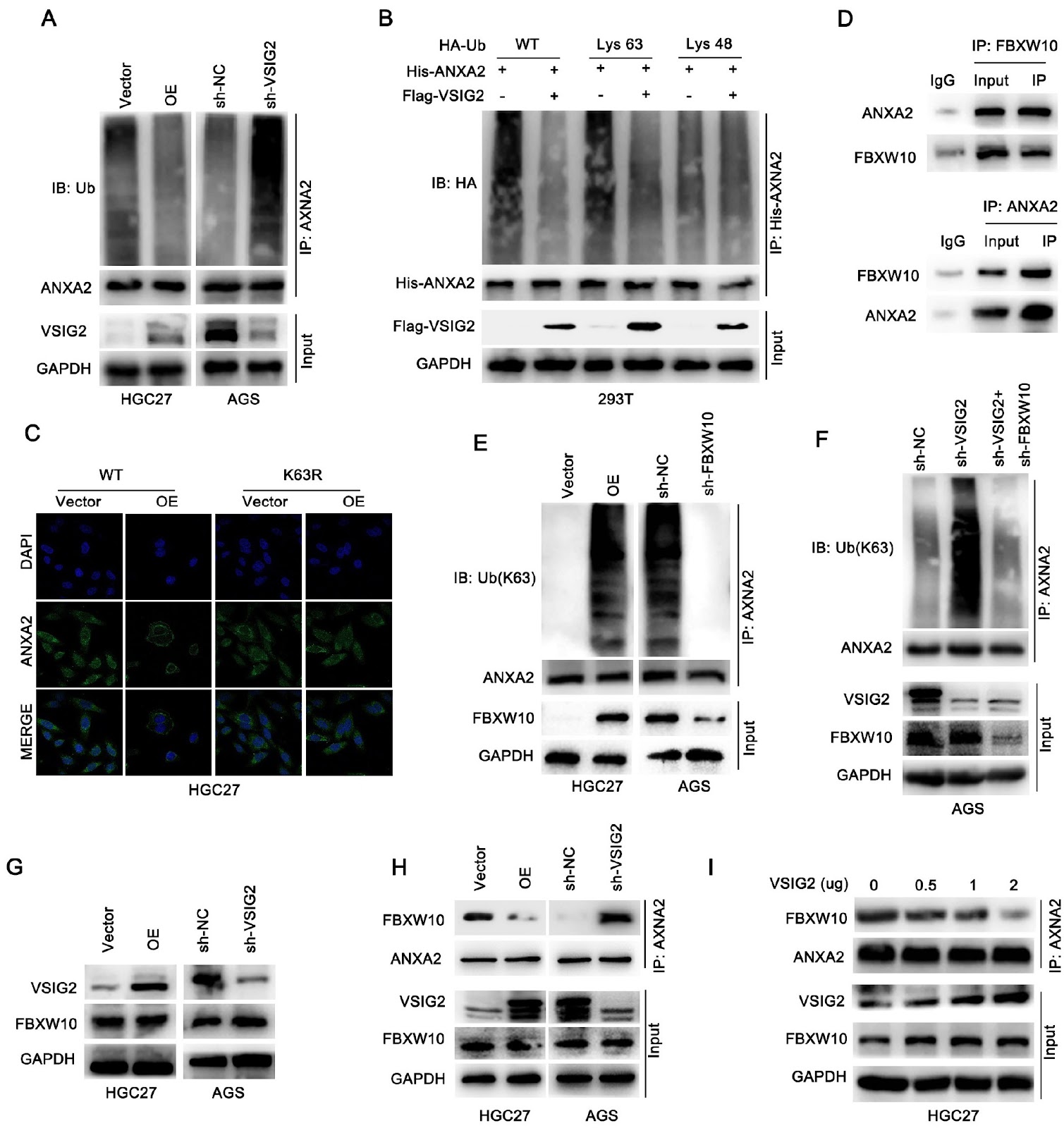
As the fifth most common cancer and the third leading cause of cancer death worldwide, gastric cancer (GC) has long been a serious global health challenge. The purpose of this study was to explore the expression of V-set and immunoglobulin domain containing 2 (VSIG2) in GC and to elucidate its role in GC progression and related mechanisms. Western blot analysis, qRT-PCR and immunohistochemical (IHC) staining are used to detect the expression of VSIG2 in GC cells and tissues. Kaplan-Meier survival curve analysis is performed. The effects of VSIG2 on biological effects related to GC progression in vitro are detected by CCK-8, EdU, Transwell and wound healing assays and in vivo by a nude mouse subcutaneous tumor model and a liver metastasis model. Mechanistically, co-immunoprecipitation, immunofluorescence and ubiquitination experiments are used to explore the regulatory effect of VSIG2 on ANXA2 and the regulatory effect between FBXW10 and ANXA2. VSIG2 is abnormally expressed at low levels in patients with GC and is associated with patient prognosis. Low VSIG2 expression is closely related to tumor size, lymph node metastasis, TNM stage and vascular invasion in GC patients. Functionally, in vitro and in vivo experiments reveal that VSIG2 could inhibit the growth, proliferation and metastasis of GC. Mechanistically, VSIG2 and ANXA2 interact directly in GCs and co-localize at the cell membrane. Further exploration reveals that highly expressed VSIG2 competes with FBXW10 for binding to ANXA2 and relies on FBXW10-mediated K63 polyubiquitination of ANXA2 to induce membrane localization of ANXA2 and further inactivate NF-κB, thereby suppressing GC progression. In summary, VSIG2 is expressed at abnormally low levels in patients with GC, and its low expression is associated with poor patient prognosis. VSIG2 can inhibit the proliferation and migration of GC via the ANXA2/NF-κB pathway. This study elucidates a new mechanism by which VSIG2 inhibits GC progression, which may provide a new perspective for the diagnosis and treatment of GC patients.

VSIG2 hinders gastric cancer progression by suppressing ANXA2-mediated NF-κB pathway activation
Graphical Abstract
Original ResearchVol. 57, Issue 12 • pp. 2137-2138DOI: 10.3724/abbs.2025233

Caveolin-1-deficient fibroblasts promote migration, invasion, and stemness by activating the TGF-β/Smad signaling pathway in breast cancer cells

Authors: Qingyun Huang, Longyuan Wu, Yi Wang, Xinyu Kong, Xinhua Xiao, Qiyuan Huang, Miao Li, Yujia Zhai, Fuxiu Shi, Ruichen Zhao, Junpei Zhong, and Lixia Xiong

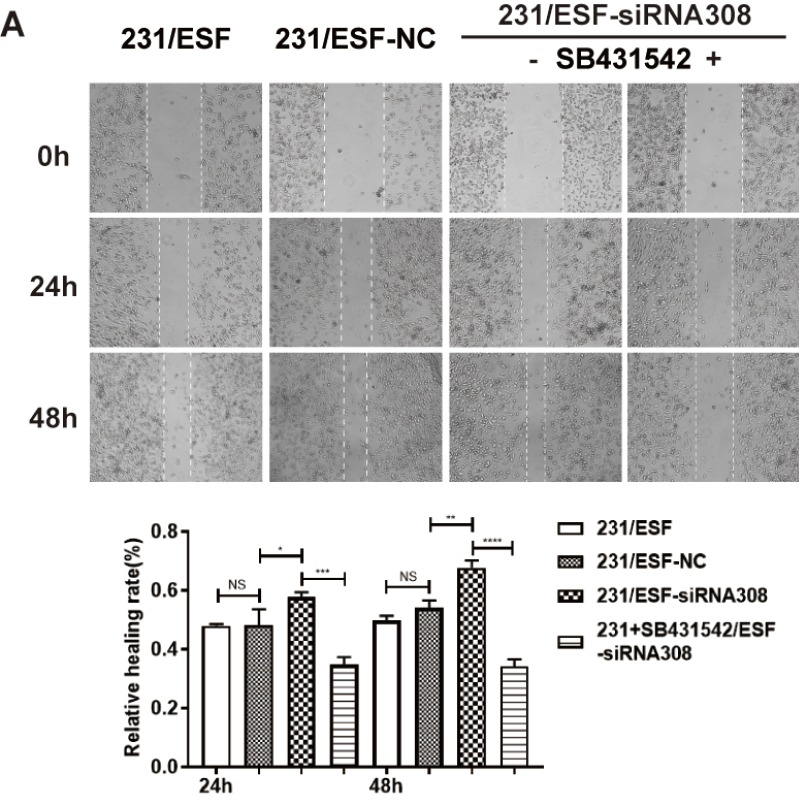
This is a corrigendum to the article 'Caveolin-1-deficient fibroblasts promote migration, invasion, and stemness by activating the TGF-β/Smad signaling pathway in breast cancer cells' published in Acta Biochim Biophys Sin 54: 1587–1598. The authors identified inaccuracies in the preparation of several figures (Figure 2D, 4A, and 5A) and have replaced them with corrected versions. The errors are strictly confined to figure presentation and do not impact the underlying data, statistical analysis, or main conclusions. The authors apologize for the oversight.

Caveolin-1-deficient fibroblasts promote migration, invasion, and stemness by activating the TGF-β/Smad signaling pathway in breast cancer cells
Graphical Abstract
Original ResearchVol. 57, Issue 11 • pp. 1814-1823DOI: 10.3724/abbs.2025057

New feature of hMEIOB and hSPATA22 binding to ssDNA from a single-molecule perspective

Authors: Yating Xu, Wei Qu, Erchi Zhou, Qi Sun, Weihao Gong, Lei Xu, Yaoke Lei, Zhangying Jia, Hanqing Shi, Xinghua Zhang, Mengcheng Luo

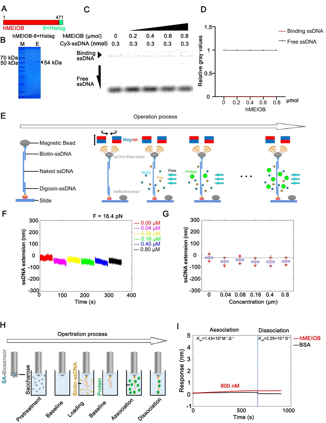
MEIOB and SPATA22 are gonad-specific proteins that function in meiosis recombination. Mutations in these two proteins cause oligospermia or azoospermia in human males. It has been reported that the heterodimer composed of MEIOB and SPATA22 recognizes and binds to the single-strand DNA (ssDNA) protected by the replication protein A (RPA) complex to promote DNA damage repair during homologous recombination. However, the amino acid sequences of the two proteins are inconsistent in humans and rodents, which leads to functional differences in meiosis. In this study, human-derived MEIOB (hMEIOB) and SPATA22 (hSPATA22) are expressed and purified for electrophoretic mobility shift assay (EMSA), magnetic tweezer (MT) assay and bio-layer interferometry (BLI) assay to analyze the ssDNA binding patterns. The results show that hMEIOB has low ssDNA-binding affinity and stability alone, but hSPATA22 binds to ssDNA faster and more stably and promotes ssDNA condensation. Strong binding affinity and stability to ssDNA are present when the hMEIOB-hSPATA22 heterodimer is formed. Moreover, we find that multiple hMEIOB-hSPATA22 heterodimers spontaneously aggregate in vitro. hRPA complex weakens the binding affinity of hMEIOB, hSPATA22 and hMEIOB-hSPATA22 heterodimer to ssDNA, and it can also bind to hSPATA22 and hMEIOB-hSPATA22 heterodimer in vitro, which might be related to the proven function of RPA complex to protect ssDNA and recruit proteins related to DNA damage repair during meiosis. Overall, this study is the first time to elucidate the binding patterns of the hMEIOB and hSPATA22 to ssDNA in vitro, and to verify the relationship between the RPA complex and meiosis-related proteins, MEIOB and SPATA22, from single-molecule perspective.

New feature of hMEIOB and hSPATA22 binding to ssDNA from a single-molecule perspective
Graphical Abstract
Original ResearchVol. 57, Issue 12 • pp. 1923-1938DOI: 10.3724/abbs.2025099

The dual role of RNA-binding proteins: promotion of tumorigenesis, drug resistance, and emerging therapeutic targets

Authors: Zemei Xu, Yukun Cui

Cancer is a complex and multifaceted disease characterized by a multitude of molecular factors. RNA-binding proteins (RBPs) have emerged as pivotal regulators of tumor development, progression, and chemoresistance through their interactions with target transcripts. These interactions regulate a multitude of processes, including alternative splicing, cleavage and polyadenylation, RNA localization, translation, N6-methyladenosine (m6A) RNA modification, and DNA double-strand break repair. The RBP family comprises over 2000 proteins and plays a critical role in oncogene expression, invasion, metastasis, and inhibition of apoptosis. However, the mechanisms by which RBPs selectively recognize RNAs remain an active area of research. In this review, we examine recent advancements in understanding RNA-binding domains and the RNA processes regulated by RBPs in tumorigenesis, summarize and highlight the roles of RNA-binding domains in cancers and the molecular mechanisms of RBPs in chemotherapy resistance, discuss the potential of targeting RBPs for cancer therapy and review RBPs that are dysregulated in cancers. Additionally, we highlight recently developed tools for predicting RBP-RNA binding activities to provide valuable support for ongoing research efforts.

The dual role of RNA-binding proteins: promotion of tumorigenesis, drug resistance, and emerging therapeutic targets
Graphical Abstract
Original ResearchVol. 57, Issue 11 • pp. 1888-1896DOI: 10.3724/abbs.2025125

Construction of an ASFV proteome library via multiple optimization strategies for high-throughput analysis

Authors: Songxin Guo, Li Ouyang, Hui Zhang, Ming Li, Wei Zhou, Ao Liang, Lu Wang, Rui Gong, Dianbing Wang, Chenli Liu, Zhuojun Dai, Shengce Tao, Jiaoyu Deng, Guimin Zhang, Xian-En Zhang, Feng Li

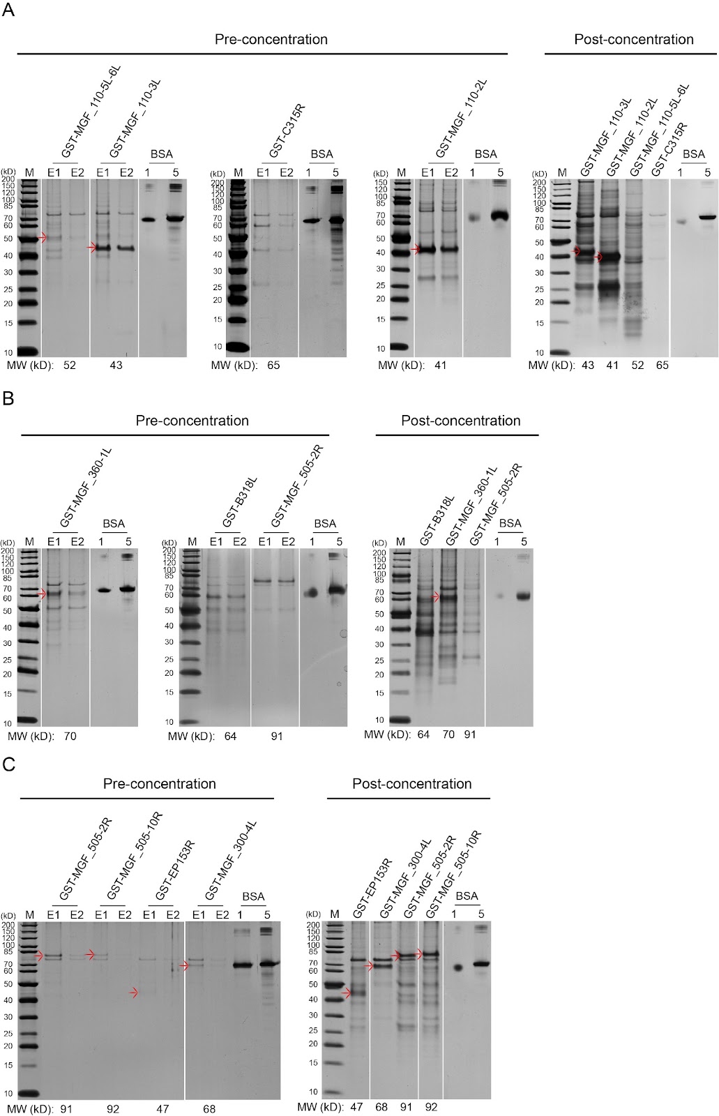
African swine fever virus (ASFV) is a large and structurally complex DNA virus encoding more than 160 proteins, including more than 68 structural proteins. A protein library covering recombinant ASFV proteins is fundamentally important for studies on protein function, antigenicity, vaccine development, and virus-host interactions. Here, to construct an ASFV protein library, we add a glutathione S-transferase (GST) tag at the N-terminus of each ASFV protein to facilitate solubilization and purification and express the recombinant proteins in the yeast host. By optimizing codons, expression vectors and strains and conditions of expression and purification, we achieve satisfactory protein yields for analytical applications and maximized access to the whole proteome of ASFV, with coverage of ca. 95%. Using the library, a protein chip is constructed and used to screen for interactions between ASFV and swine proteins (e.g., IRF3, p65, and IκBα). The ASFV protein library lays the groundwork for understanding and combatting ASFV. The methods for constructing the library are instructive for generating other protein libraries for high-throughput applications.

Construction of an ASFV proteome library via multiple optimization strategies for high-throughput analysis
Graphical Abstract
Original ResearchVol. 57, Issue 11 • pp. 1901-1903DOI: 10.3724/abbs.2025060

Osteocalcin carboxylation/undercarboxylation levels and gene variants associated with type 2 diabetes mellitus in the Chinese Han population

Authors: Luyue Qi, Hong Wu, Xiangqi Li, Yang Xu, Liangyong Liu

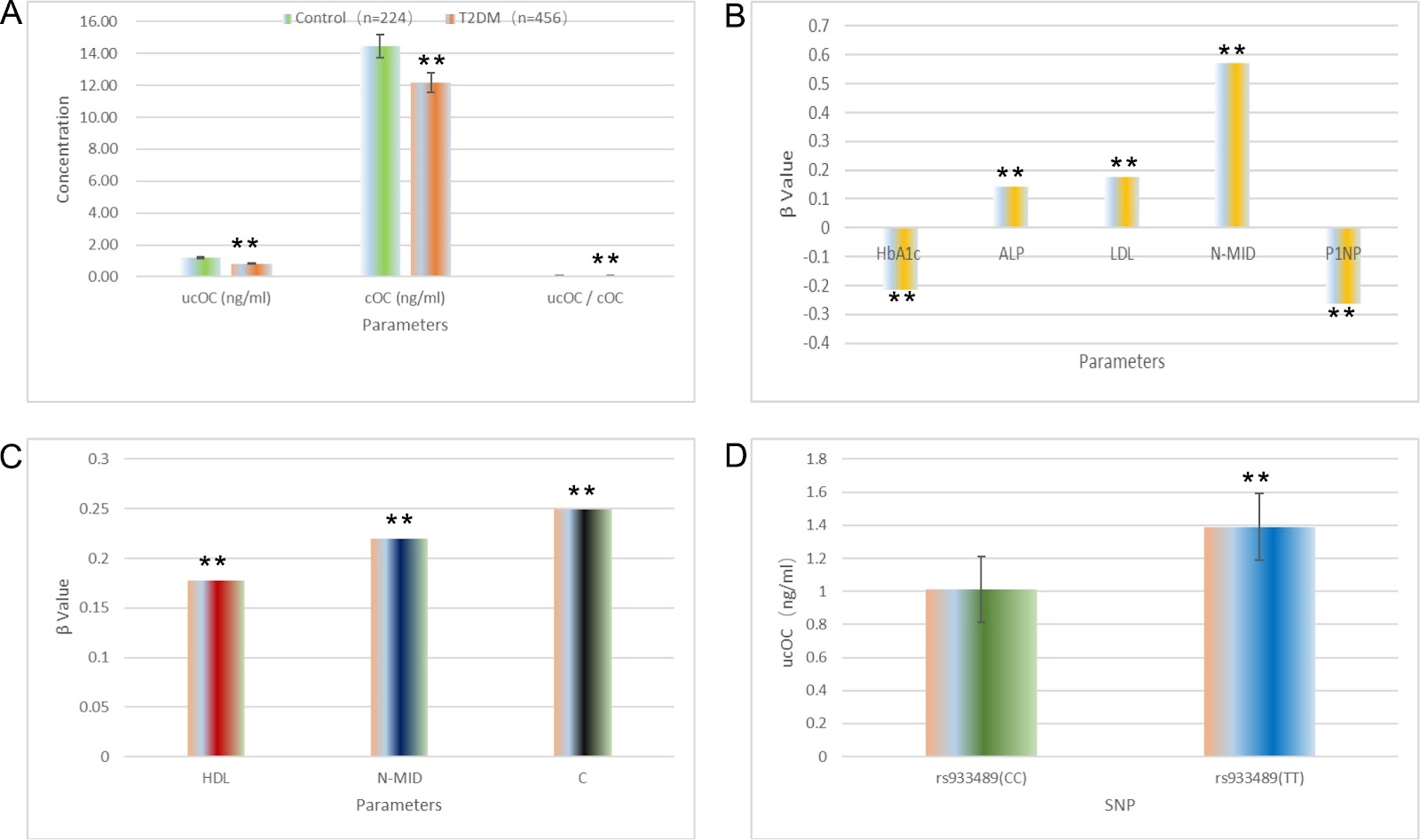
Type 2 diabetes mellitus (T2DM) is an endocrine metabolic disorder characterized by insulin secretion dysfunction and/or insulin resistance. Osteocalcin (OC), or bone γ-carboxyglutamic acid protein (BGP), is a bone matrix protein predominantly produced by osteoblasts. Vitamin K-dependent carboxylation converts OC into gamma-carboxyglutamic acid (Gla)-rich carboxylated osteocalcin (cOC), which binds to hydroxyapatite and can be decarboxylated to undercarboxylated osteocalcin (ucOC) under acidic conditions. While cOC influences bone formation and mineralization, ucOC regulates energy metabolism. This study aimed to explore the associations between T2DM, serum OC levels (including cOC and ucOC), and OC gene polymorphisms in the Chinese Han population. T2DM patients and a healthy cohort, all of Han ethnicity, were categorized into a T2DM group (n = 456) and a control group (n = 224). Serum levels of cOC and ucOC were determined via ELISA. Insulin sensitivity was assessed via HOMA-IR, and pancreatic β-cell function via HOMA-β. Nine SNPs in the OC gene were genotyped via SNaPshot. Compared with controls, T2DM patients presented significantly lower levels of ucOC, cOC, and ucOC/cOC ratio. Additionally, T2DM subjects had elevated BMI, HbA1c, HOMA-IR, ALP, TG, HDL, and LDL levels, with decreased HOMA-β, ALT, AST, hsCRP, and FFA levels. In terms of bone metabolism, T2DM patients presented increased blood phosphorus, ICTP, P1NP, and 25(OH)D levels and decreased blood calcium, N-MID, PTH, and β-CTX levels. Associations between serum cOC and ucOC and various factors were analyzed. In the T2DM group, cOC was inversely correlated with HbA1c and P1NP, and positively correlated with ALP, LDL, and N-MID. ucOC was positively associated with N-MID. In controls, cOC was positively correlated with HDL, N-MID, and PINP, while ucOC correlated with PINP. The study also examined SNPs in the OC gene and their relationships with serum cOC and ucOC.

Osteocalcin carboxylation/undercarboxylation levels and gene variants associated with type 2 diabetes mellitus in the Chinese Han population
Graphical Abstract
Original ResearchVol. 57, Issue 10 • pp. 1660-1669DOI: 10.3724/abbs.2025036

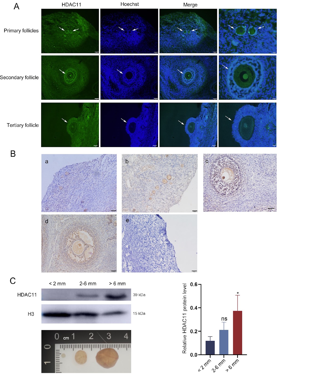
HDAC11 in ovarian granulosa cells coordinates LH in the maturation of oocytes in Tan sheep

Authors: Jiaqi Shi, Donghuan Lv, Yaxiu Xu, Xiangyan Wang, Zhipeng Qi, Yujie Yan, Jinghua Wang, Hongyuan Song, Hui Yang, Luguo Jin, Zhengyi Yang, Xiaoning Yang, Xiumei Kang, Xinfeng Liu, Zhuming Zhang, Chao Wang

Oocyte maturation plays an important role in supporting mammalian reproduction. Histone deacetylase 11 (HDAC11), the only member of the class IV histone deacetylase family and the smallest histone deacetylases (HDACs), has been shown to regulate oocyte maturation in mice and pigs. However, the epigenetic effects of HDACs in follicular granulosa cells in response to LH induction remain elusive in sheep. In this study, the effects of follicular somatic cell-derived HDAC11 on oocyte maturation in Tan sheep are evaluated. The expression changes of HDAC11 and related proteins are detected by means of immunofluorescence, immunohistochemistry, western blot analysis and enzyme-linked immunosorbent assay. Our results indicate that the level of HDAC11 in follicular granulosa cells as well as oocytes in Tan sheep increases with the growth and maturation of the follicles. Specific inhibition of HDAC11 by SIS17 remarkably reduces the oocyte maturation rate under LH supplementation in vitro. Accordingly, the acetylation level of H3K9 in granulosa cells is increased, while the EGF-like growth factor AREG is remarkably decreased. Furthermore, inhibition of HDAC11 markedly decreases the level of YAP1, which is a negative regulator of AREG in granulosa cells. Conclusively, HDAC11 in the granulosa cells of Tan sheep contributes to the LH induced production of AREG during oocyte in vitro maturation by decreasing the level of H3K9 acetylation and increasing the level of YAP1.

HDAC11 in ovarian granulosa cells coordinates LH in the maturation of oocytes in Tan sheep
Graphical Abstract
Original ResearchVol. 57, Issue 11 • pp. 1824-1833DOI: 10.3724/abbs.2025047

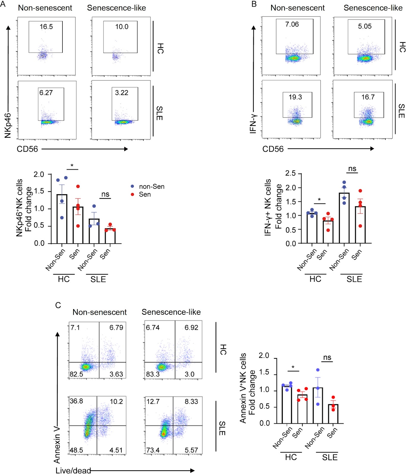
Increased neutrophil senescence is associated with impaired immunosuppressive activity in systemic lupus erythematosus

Authors: Lei Han, Fengling Huang, Qingchen Zhu, Huan Wang, Tianlin Lu, Chunyuan Xiao, Jing Xu, Xiaoyan Zhang, Yichuan Xiao, Xinfang Huang

Systemic lupus erythematosus (SLE) is an autoimmune disease characterized by a complex pathogenesis that was previously thought to involve primarily adaptive immunity. Emerging evidence underscores the role of neutrophils in shaping immune dysregulation and inducing organ damage in lupus. This study aims to investigate the dynamics of neutrophil senescence and its relationship with lupus, an area that remains poorly understood. Here, we identify a significantly elevated proportion of CXCR4hiCD62Llo senescence-like neutrophils in the peripheral blood of SLE patients compare to that in the healthy donors. Increased numbers of senescence-like neutrophils are positively correlated with SLE disease activity and autoantibody production in SLE patients. In addition, senescence-like neutrophils derived from SLE patients exhibit an impaired ability to suppress the proinflammatory activity of natural killer (NK) cells and CD4+ T cells. Further mechanistic exploration suggests that these senescence-like neutrophils might exert their immunosuppressive effects via reactive oxygen species (ROS) production under physiological conditions. Our results demonstrate that senescence-like neutrophils could serve as biomarkers for assessing the disease activity of SLE. The compromised immunosuppressive function of senescence-like neutrophils provides a new perspective on SLE pathophysiology and may pave the way for the development of novel therapies.

Increased neutrophil senescence is associated with impaired immunosuppressive activity in systemic lupus erythematosus
Graphical Abstract
Original ResearchVol. 57, Issue 11 • pp. 1897-1900DOI: 10.3724/abbs.2025094

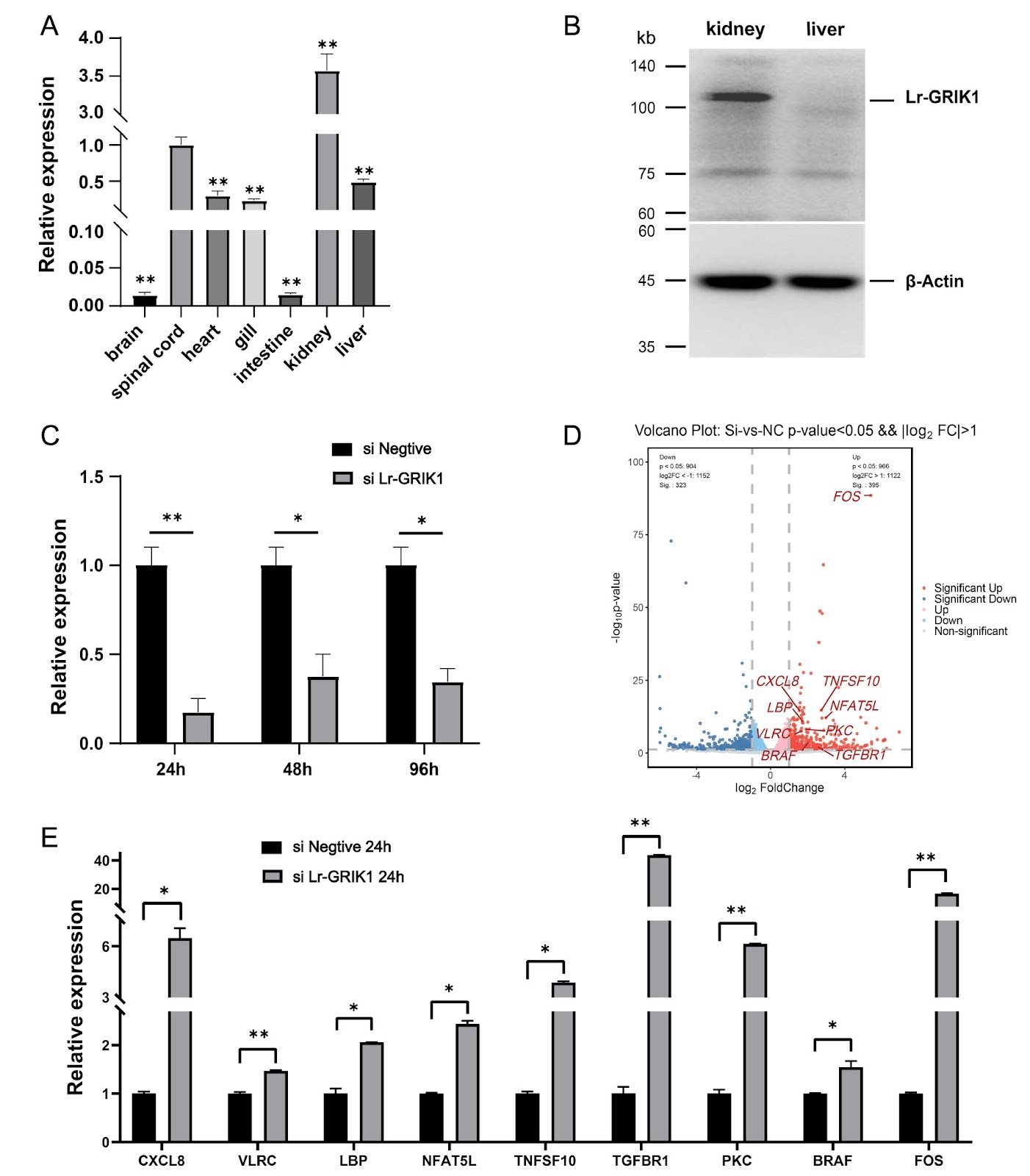
KARs negatively regulate the immune response in lamprey

Authors: Ruyu Zhuang, Zihao Yan, Shuyuan Zhang, Meixuan Li, Feng Sun, Ya Pang, Ding Li, Liang Zhao, Yinglun Han

Kainate receptors (KARs) are one of the ionotropic glutamate receptor (iGluR) families, and their antagonists are being investigated for the treatment of several neurological disorders, including Alzheimer’s disease, a neurodegenerative condition, etc. As early as 1990, Bettler et al. [1] first cloned the GRIK1 subunit of KARs, marking a pivotal advancement in understanding these receptors. Members of the iGluR family have been identified in other jawed vertebrates and exhibit conserved structural features. However, research into iGluRs in jawless vertebrates has been limited. Owing to the unique evolutionary position of lampreys, their iGluRs might also present functions distinct from those of jawed vertebrates; therefore, it is particularly important to study iGluRs in lampreys. In this study, we identified four homologous subunits of iGluRs in lampreys, including Lr-GRIA2, Lr-GRIA4, Lr-GRIK1 and Lr-GRIN2B. Lampreys occupy a unique evolutionary position, making phylogenetic analysis of iGluR subunits between lampreys and other species essential for understanding iGluR evolution. Given the distinctive functional characteristics of iGluR family members, particularly KAR subtypes, we focused on the functional validation of Lr-GRIK1. First, we confirmed the expression of Lr-GRIK1 in lampreys and examined its expression profiles across various tissues via qPCR and western blotting. To elucidate the functional role of Lr-GRIK1 in lampreys, we used an siRNA to silence Lr-GRIK1. We subsequently conducted transcriptome sequencing of both the silenced and control groups to construct and analyze their expression profiles. Our analysis revealed differential expression of genes enriched in pathways related to signal transduction and the immune system, highlighting potential roles of Lr-GRIK1 beyond traditional neurotransmission functions. Unlike in jawed vertebrates, transcriptome enrichment provides a new direction for understanding the function of Lr-GRIK1. Therefore, we monitored the changes in Lr-GRIK1 expression in the kidney tissue of lampreys after stimulation. In addition, we confirmed that Lr-GRIK1 affects the expression levels of immune-related molecules during the immune response process. These findings provide insights into the broader functional significance of Lr-GRIK1 in the biology of lampreys.

KARs negatively regulate the immune response in lamprey
Graphical Abstract
Original ResearchVol. 57, Issue 11 • pp. 1767-1779DOI: 10.3724/abbs.2025113

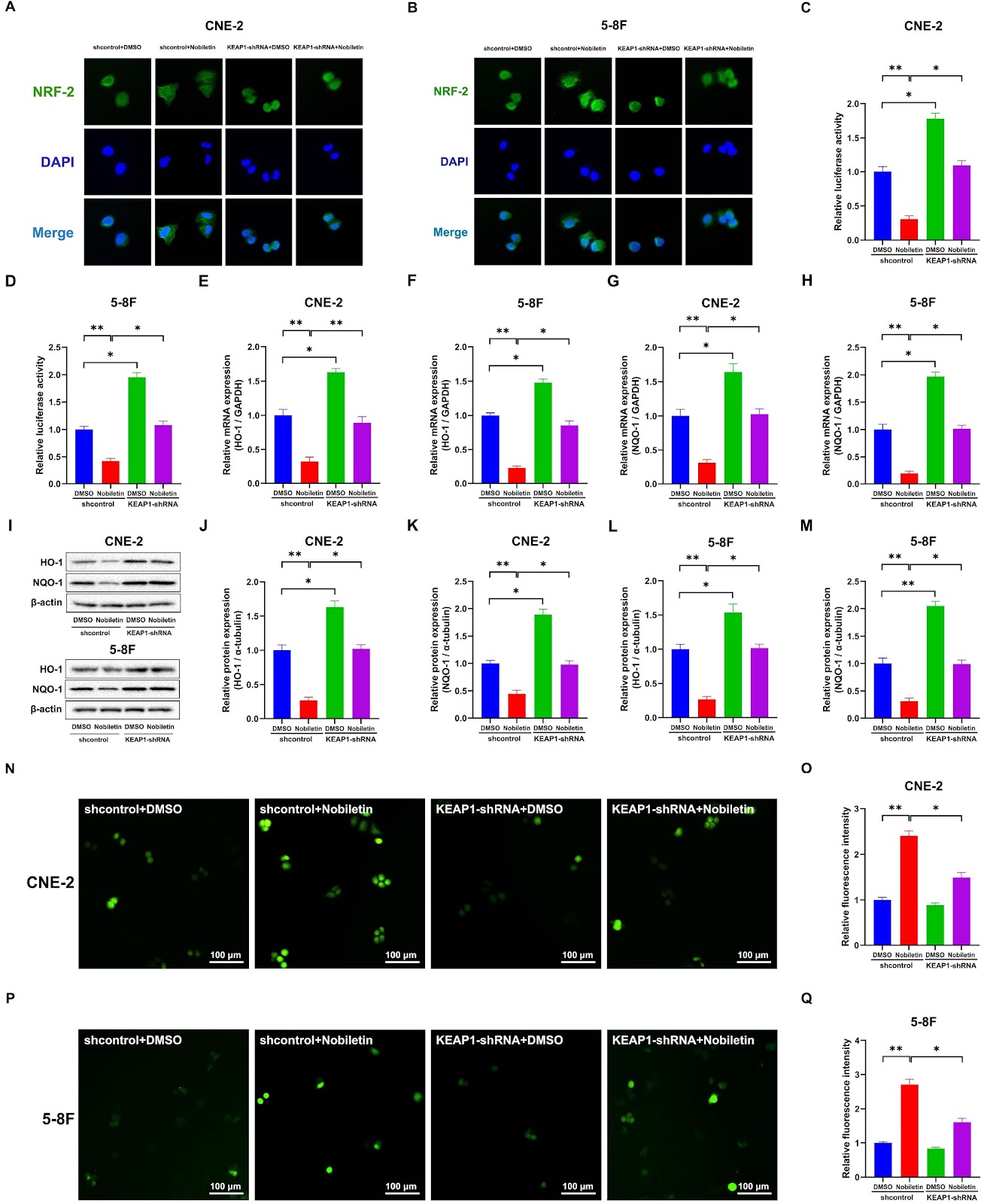
Nobiletin suppresses nasopharyngeal carcinoma by regulating the KEAP1/NRF2/ARE pathway

Authors: Yiyao Liang, Minyan Wei, Yunan Yao, Baizhong Chen, Jinji Deng, Shiqi Xu, Liming Li, Wen Liu, Yi Cai, Guodong Zheng

Nasopharyngeal carcinoma (NPC) ranks among the most prevalent malignancies, particularly in East Asia and Southeast Asia. Nobiletin (NOB), an exclusive polymethoxyflavonoid derived from citrus peel, exhibits diverse physiological properties, notably its potent anticancer activity. Kelch-like ECH-associated protein 1 (KEAP1), the repressor protein regulating the nuclear factor erythroid 2-related factor 2 (NRF2) transcription factor, has emerged as a promising strategy for addressing oxidative stress in various diseases. The KEAP1/NRF2/ARE signal is a fundamental pathway within the cellular homeostatic defense system. This study robustly demonstrates the chemopreventive potential of NOB through comprehensive in vitro and in vivo assessments using subcutaneous tumor mouse models. Furthermore, our groundbreaking findings reveal that NOB effectively hinders the migration and invasion capacities of CNE-2 and 5-8F (NPC) cells in a dose- and time-dependent manner. Mechanistically, NOB, a potent KEAP1 activator, significantly disrupts the NRF2/ARE signaling pathway by accelerating the proteasomal degradation of NRF2 and suppressing its nuclear translocation. Consequently, this cascade reduces the expressions of ARE-driven genes and antioxidant enzymes, thereby increasing intracellular reactive oxygen species (ROS) levels and increasing antitumor immunity. Moreover, the sensitivity induced by NOB is markedly diminished in CNE-2 cells following the gene silencing of KEAP1. These findings underscore the pivotal role of NOB in activating KEAP1. Overall, KEAP1 has emerged as a compelling target for potential malignancy treatment in nasopharyngeal carcinoma cell lines. Our results suggest the promising application of NOB as a natural sensitizer in chemotherapy, opening avenues for promising therapeutic interventions.

Nobiletin suppresses nasopharyngeal carcinoma by regulating the KEAP1/NRF2/ARE pathway
Graphical Abstract
Original ResearchVol. 57, Issue 10 • pp. 1611-1624DOI: 10.3724/abbs.2025056

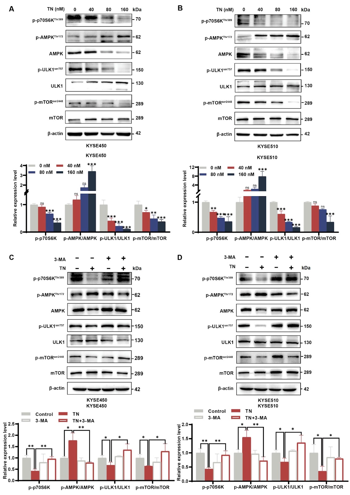
Triptonide facilitates autophagy-mediated apoptosis in esophageal squamous cell carcinoma by targeting the AMPK-mTOR-ULK1 axis

Authors: Jiujun Ju, Nuo Xu, Bohan Li, Dan Shi, Jiahui Cai, Qiusheng Zheng, Lei Ye, Shaosen Zhang, Caixia Wang

Triptonide (TN) is a small-molecule compound initially derived from Tripterygium wilfordii Hook. f used in traditional Chinese medicine. However, its potential antitumor mechanisms are still far from adequately understood. The purpose of this research is to elucidate the antitumor and pharmacological effects of TN on esophageal squamous cell carcinoma (ESCC). Functional assays, such as CCK-8 and colony formation assays, are used to evaluate the effects of TN on KYSE450 and KYSE510 cells. Subsequently, western blot analysis, Hoechst 33258 staining, flow cytometric analysis, autophagic flux detection, and transmission electron microscopy (TEM) are used to determine the effects of TN on apoptosis and autophagy in ESCC cells. Additionally, the autophagy inhibitor 3-methyladenine (3-MA) and the AMPK inhibitor dorsomorphin (Compound C, CC) are administered to explore the molecular mechanisms and crucial pathways in ESCC cells. Our findings provide strong evidence that TN induces autophagy-dependent apoptosis by targeting the AMPK-mTOR-ULK1 axis in ESCC cells. Collectively, this study sheds light on the anticancer mechanisms of TN in esophageal squamous cell carcinoma and suggests that TN is a promising candidate for the antitumor phytomedicine.

Triptonide facilitates autophagy-mediated apoptosis in esophageal squamous cell carcinoma by targeting the AMPK-mTOR-ULK1 axis
Graphical Abstract
Original ResearchVol. 57, Issue 10 • pp. 1589-1600DOI: 10.3724/abbs.2025034

Melatonin attenuates kidney injury by alleviating lysosomal damage in diabetic kidney disease

Authors: Jiaqi Chen, Shuting Zhang, Xiaoquan Xue, Xiaoqin Ma, Aomiao Chen, Yichuan Wu, Geningyue Wang, Qian Zhang, Yaoming Xue, Yijie Jia, Zongji Zheng

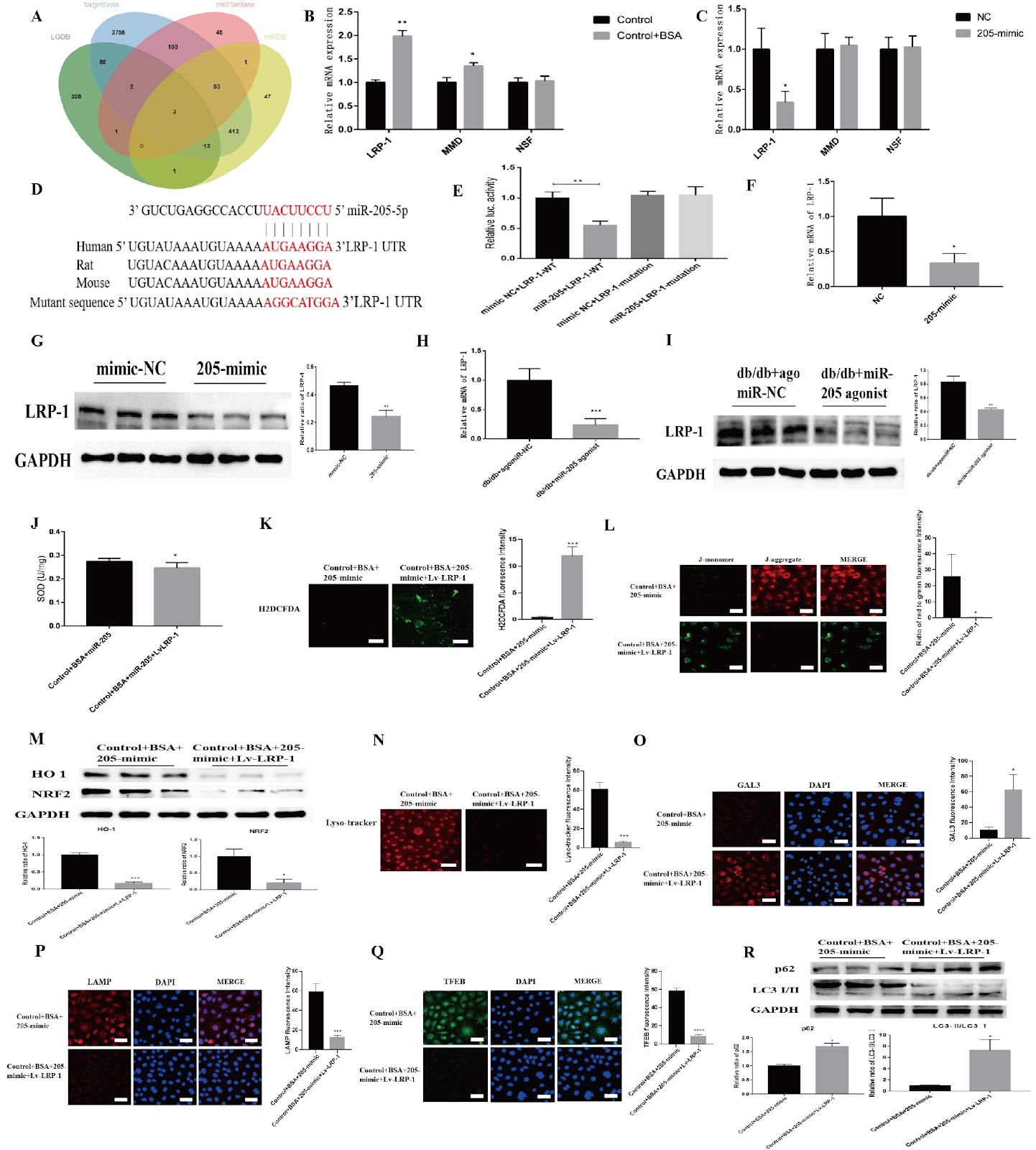
Proteinuria-induced damage to renal tubular epithelial cells is one of the main causes of diabetic kidney disease (DKD), and the clearance of overloaded albumin by lysosomes is crucial for maintaining the homeostasis of renal tubular epithelial cells. Therefore, lysosomal damage is closely related to the pathogenesis of DKD, but effective prevention and treatment measures are still lacking. Melatonin (MLT) is secreted by the pineal gland and can not only regulate circadian rhythms but also maintain lysosomal homeostasis. In this study, we demonstrate the presence of significant lysosomal damage in the renal tubules of DKD patients, which causes autophagy impairment and a concomitant oxidative stress imbalance; however, MLT can upregulate transcription factor EB (TFEB) to improve lysosomal damage and restore the biosynthesis of this organelle. Mechanistically, MLT may protect lysosomes via the upregulation of TFEB and the miR-205-5p-LRP-1 pathway in renal tubules, thus improving autophagy dysfunction and oxidative imbalance in DKD.

Melatonin attenuates kidney injury by alleviating lysosomal damage in diabetic kidney disease
Graphical Abstract
Original ResearchVol. 57, Issue 10 • pp. 1574-1588DOI: 10.3724/abbs.2025102

Nanchangmycin suppresses influenza A virus infection by blocking endosomal acidification

Authors: Hao Zhang, Changhai Liu, Shuo Cai, Yuting Wu, Lu Shang, Fayu Yang, Jing Liu, Nan Wei, Yingchun Liu, Mi Wang, Fei Gao, Qinfang Liu, Hongjun Chen, Guangzhi Tong, Yin Chen, Feng Gu

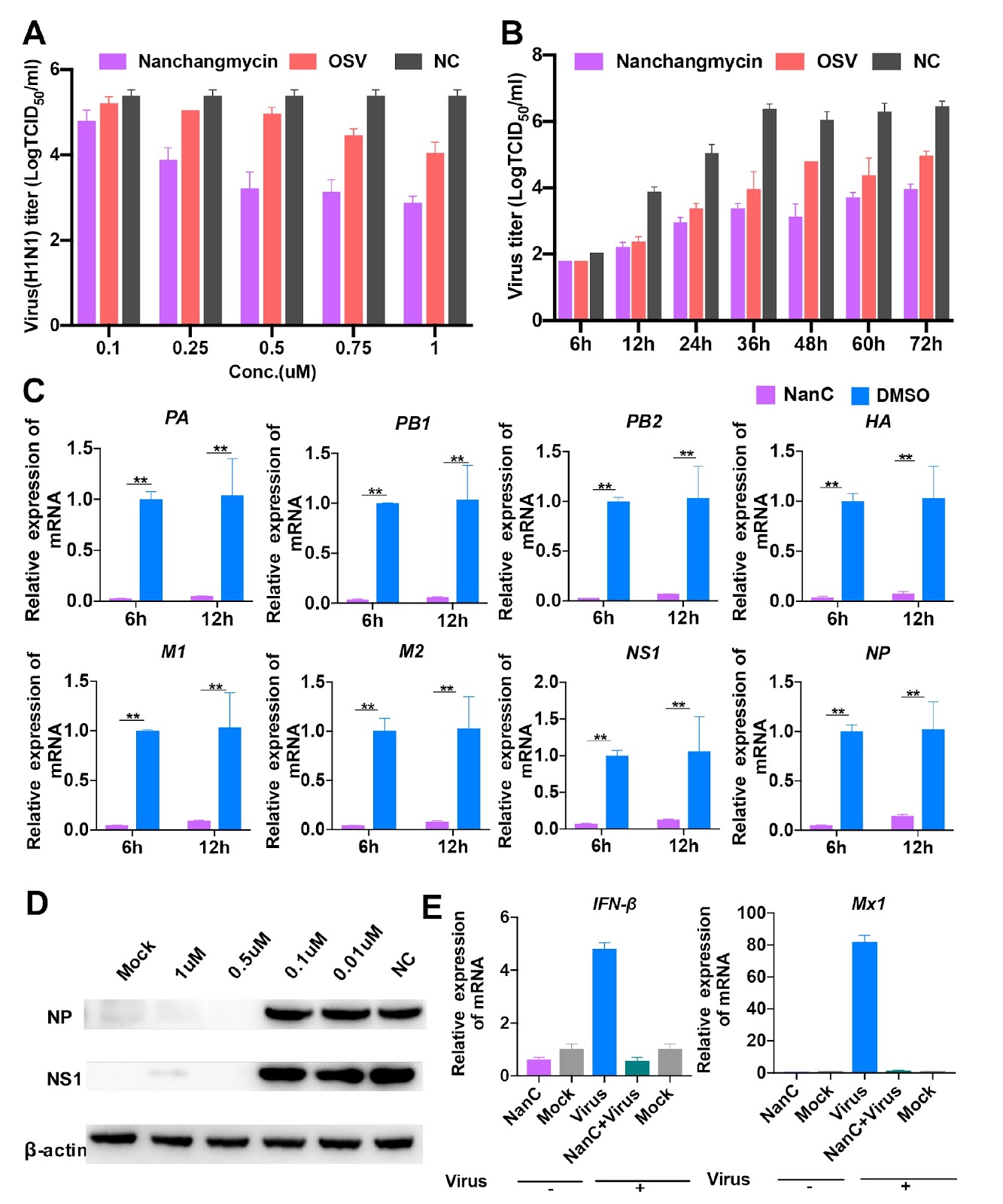
Influenza A viruses (IAVs) constitute a major threat to human and animal health. Currently, M2 ion-channel inhibitors, neuraminidase (NA) inhibitors, RNA polymerase inhibitors, and cap-dependent endonuclease inhibitors have been applied clinically as therapeutics against IAVs. However, IAVs possess adaptive mutations to these inhibitors, especially M2 ion channel and NA inhibitors. Thus, novel antiviral agents should be developed. In the present study, we screen approximately 5500 compounds and identify an IAV inhibitor, nanchangmycin, which possesses a robust antiviral activity both in vitro and in vivo. In addition, it exhibits broad-spectrum antiviral activity for additional virus infections, including pseudorabies virus, herpes virus, porcine epidemic diarrhea virus, porcine reproductive and respiratory syndrome virus. Most importantly, it has antiviral activity against oseltamivir-resistant strains in sub-μM ranges and promotes the survival of MDCK cells infected with the oseltamivir-resistant influenza A virus strain. Further studies reveal that it blocks the nuclei migration of viral nuclear proteins (NPs), resulting in NP accumulation in the cytoplasm, particularly within perinuclear endosomes. Also, it inhibits IAVs by blocking endosomal acidification. Overall, nanchangmycin has the potential to be developed as an anti-influenza agent.

Nanchangmycin suppresses influenza A virus infection by blocking endosomal acidification
Graphical Abstract
Original ResearchVol. 57, Issue 10 • pp. 1625-1635DOI: 10.3724/abbs.2025042

PDGFC secreted by cancer-associated fibroblasts promotes epithelial-mesenchymal transition and immunosuppression in lung adenocarcinoma

Authors: Meimei Cui, Xiaodi Ding, Yu Jiang, Liying Zhang, Wangkai Cao, Yongming Wang, Zhimei Sheng, Wei Sun, Ai Guo, Lihui Gu, Xiurong Zhang, Wanli Duan, Lihong Shi, Baogang Zhang

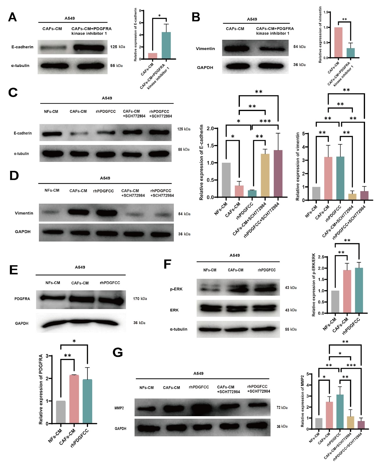
This study elucidates the mechanisms by which cancer-associated fibroblast (CAF)-derived platelet-derived growth factor C (PDGFC) promotes the progression of lung adenocarcinoma (LUAD) and explores the impact of PDGFC on immune regulation within the tumor microenvironment (TME). Our results show that there is higher expression of PDGFC in CAFs than in nontumor tissue fibroblasts (NFs) and that higher expression of PDGFC is correlated with poor prognosis in LUAD patients. Furthermore, CAF-derived PDGFC promotes epithelial-mesenchymal transition (EMT) in cancer cells as well as matrix metalloproteinase 2 (MMP2) expression through the PDGF receptor A (PDGFRA)-mitogen-activated protein kinase/extracellular signal-regulated kinase (MAPK/ERK) pathway. Moreover, our study demonstrates that CAF-derived PDGFC is essential for the activation and infiltration of fibroblasts in the TME, as well as the inflammatory infiltration of different immune cell types and the immunosuppressive conditions within the TME. In particular, PDGFC induces increased PDGFRA expression in both tumor cells and fibroblasts, which can lead to reciprocally positive feedback to accelerate malignant tumor progression. This discovery provides a novel TME-targeted strategy for LUAD treatment.

PDGFC secreted by cancer-associated fibroblasts promotes epithelial-mesenchymal transition and immunosuppression in lung adenocarcinoma
Graphical Abstract
Original ResearchVol. 57, Issue 10 • pp. 1684-1695DOI: 10.3724/abbs.2025045

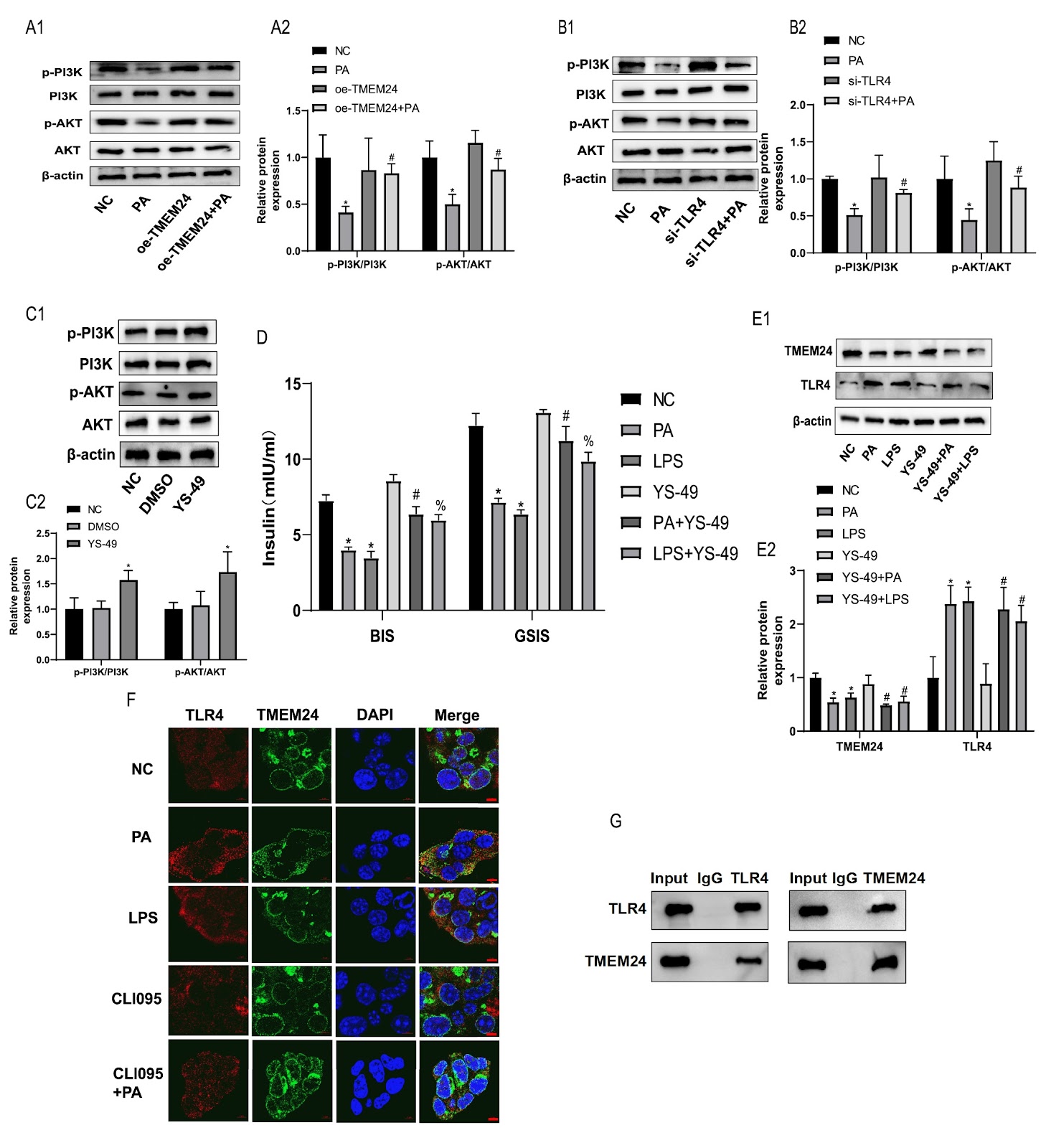
TLR4 mediates lipotoxic β-cell dysfunction by inhibiting the TMEM24/PI3K/AKT pathway

Authors: Chao Lan, Yan Li, Zhiyan Weng, Wei Pan, Wanxin Lin, Zhen Jiang, Liyong Yang, Ximei Shen

Immune imbalance is the core pathophysiological mechanism of the deterioration of β-cell function driven by lipid metabolism disorders. Toll-like receptor 4 (TLR4) inflammatory signaling is a key pathway that mediates lipotoxic injury in β-cells, but the underlying mechanism needs to be further elucidated. Transmembrane protein 24 (TMEM24) is a key transporter that regulates pulsatile insulin secretion, but its pathophysiology in lipotoxicity remains unclear. In this study, we investigate whether TLR4-mediated lipotoxicity is affected by the inhibition of TMEM24 expression. The PPI network shows that TLR4 is associated with both insulin secretion and ER stress proteins in islets from obese rats. Using in vitro lipotoxic β-cell models, we found that TMEM24 is the target signal of palmitic acid (PA)-induced insulin secretion impairment in islet β-cells, and TLR4 plays a mediating role in this process. Mechanistically, TLR4 mediates lipotoxicity by binding to TMEM24 and downregulating its protein expression to suppress PI3K/AKT signaling, leading to β-cell dysfunction. TLR4 knockout ameliorates islet function impairment through TMEM24/PI3K/AKT signaling in HFD-induced obese rats. Taken together, our results show that TLR4 mediates lipotoxicity in islet β-cells by inhibiting the TMEM24/PI3K/AKT pathway, and the mechanism of TLR4-mediated lipotoxicity is elucidated from the perspective of insulin vesicular secretion.

TLR4 mediates lipotoxic β-cell dysfunction by inhibiting the TMEM24/PI3K/AKT pathway
Graphical Abstract
Original ResearchVol. 57, Issue 10 • pp. 1557-1573DOI: 10.3724/abbs.2025071

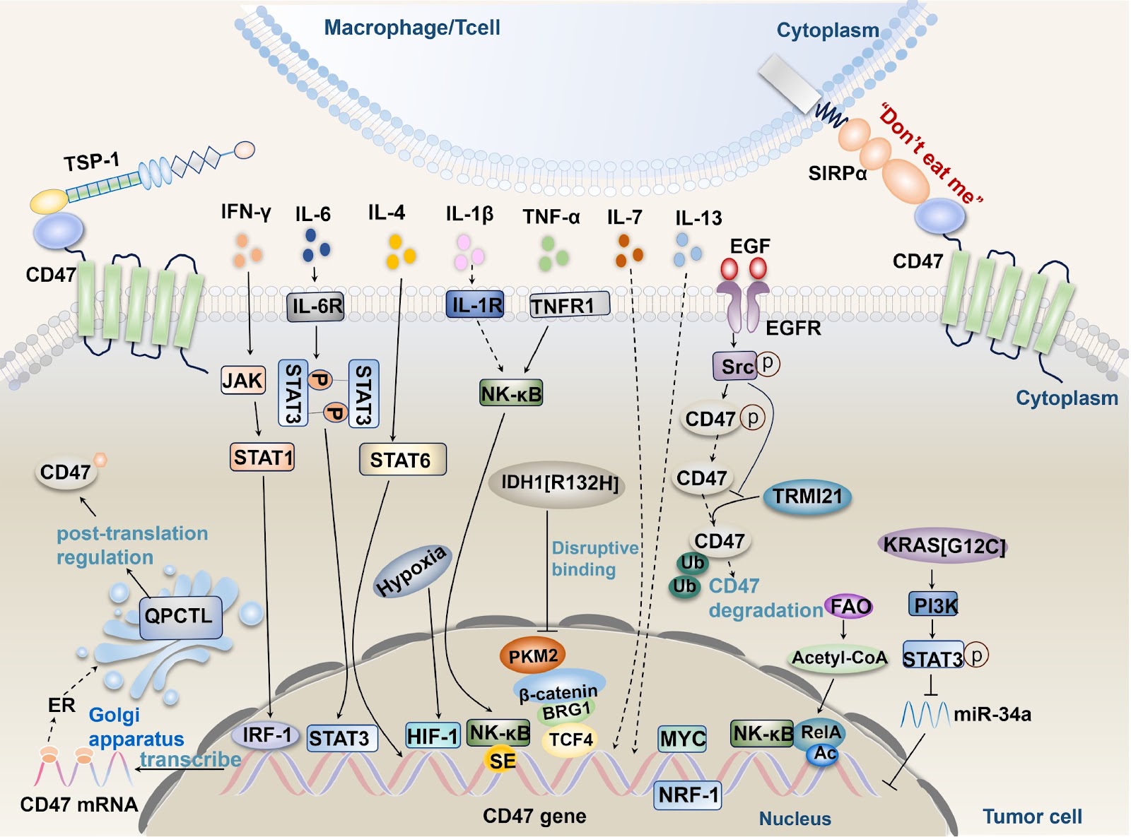
CD47-mediated tumor microenvironment remodeling: a central mechanism in immune evasion

Authors: Hemei Yuan, Lingling Zhu, Longhuan Yang, Yong Yi, Tao Lv

Immune evasion is a crucial strategy for tumor growth and survival, with the tumor microenvironment facilitating tumor immune evasion and cancer progression. CD47, a transmembrane protein highly expressed in various cancer cell types, interacts with its ligands SIRPα and TSP-1 to induce immune tolerance, enabling tumor cells to evade immune surveillance and phagocytosis by immune cells. Understanding the pathways driving CD47 signaling and related activation factors is essential. In this review, we discuss the interactions between CD47 and its ligands SIRPα and TSP-1; their roles in inhibiting the functions of immune cells (macrophages, dendritic cells (DCs), glial cells, T cells, NK cells, etc.); and the mechanisms involved. Furthermore, we also explore the influence of factors within the tumor microenvironment, including TNF-α, IFN-γ, ILs, HIF-1, oncogenes, isocitrate dehydrogenase 1, metabolic enzymes, and exosomes, on CD47-mediated immune evasion. Recent monoclonal antibody drugs targeting CD47 for cancer treatment have shown side effects and cause economic losses. Researchers can explore alternative approaches, such as designing targeted drugs with minimal side effects or investigating other related molecules or pathways. Combination therapy and further research into the molecular mechanisms of CD47 could offer new directions for antitumor drug development.

CD47-mediated tumor microenvironment remodeling: a central mechanism in immune evasion
Graphical Abstract
Original ResearchVol. 57, Issue 10 • pp. 1601-1610DOI: 10.3724/abbs.2025063

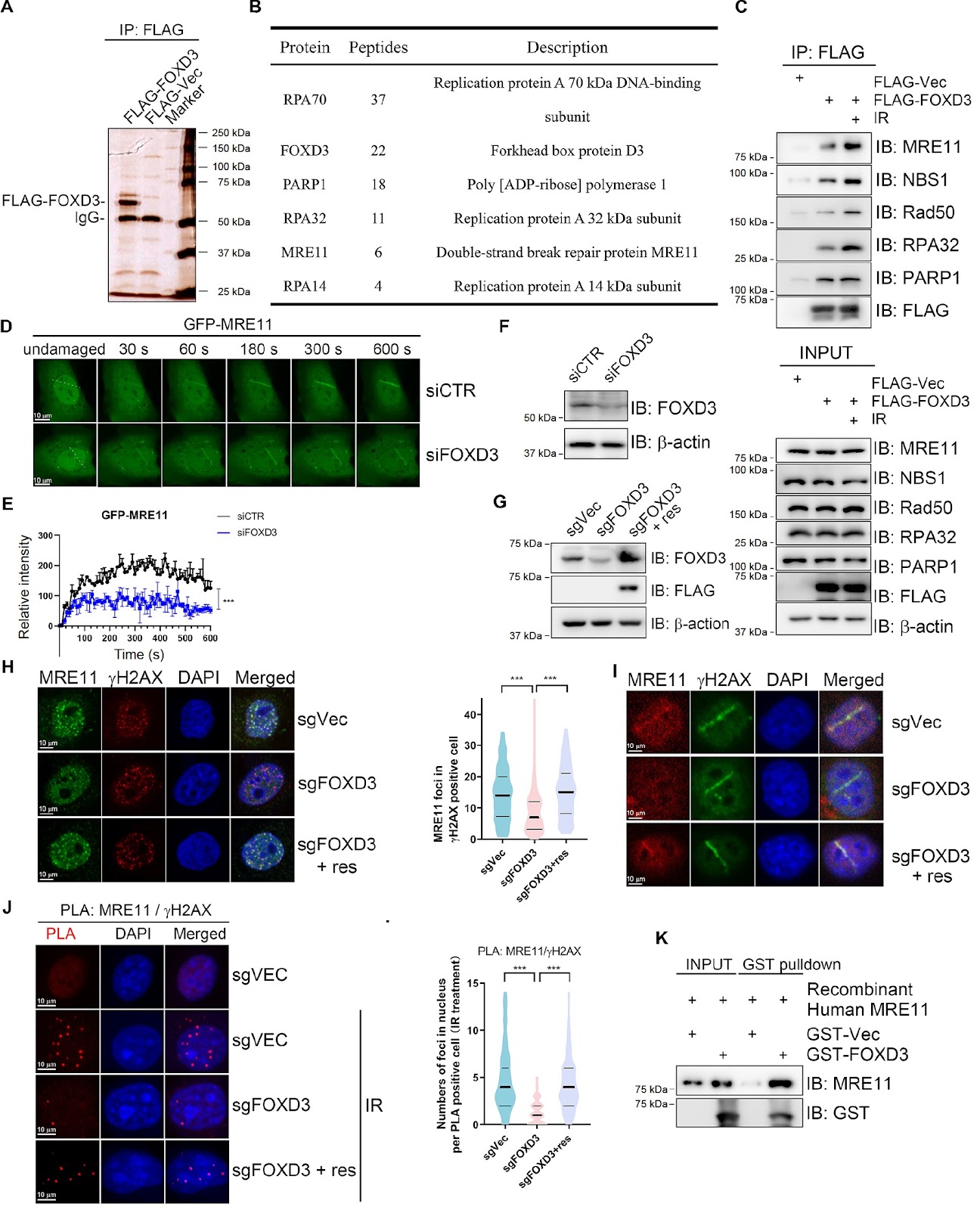
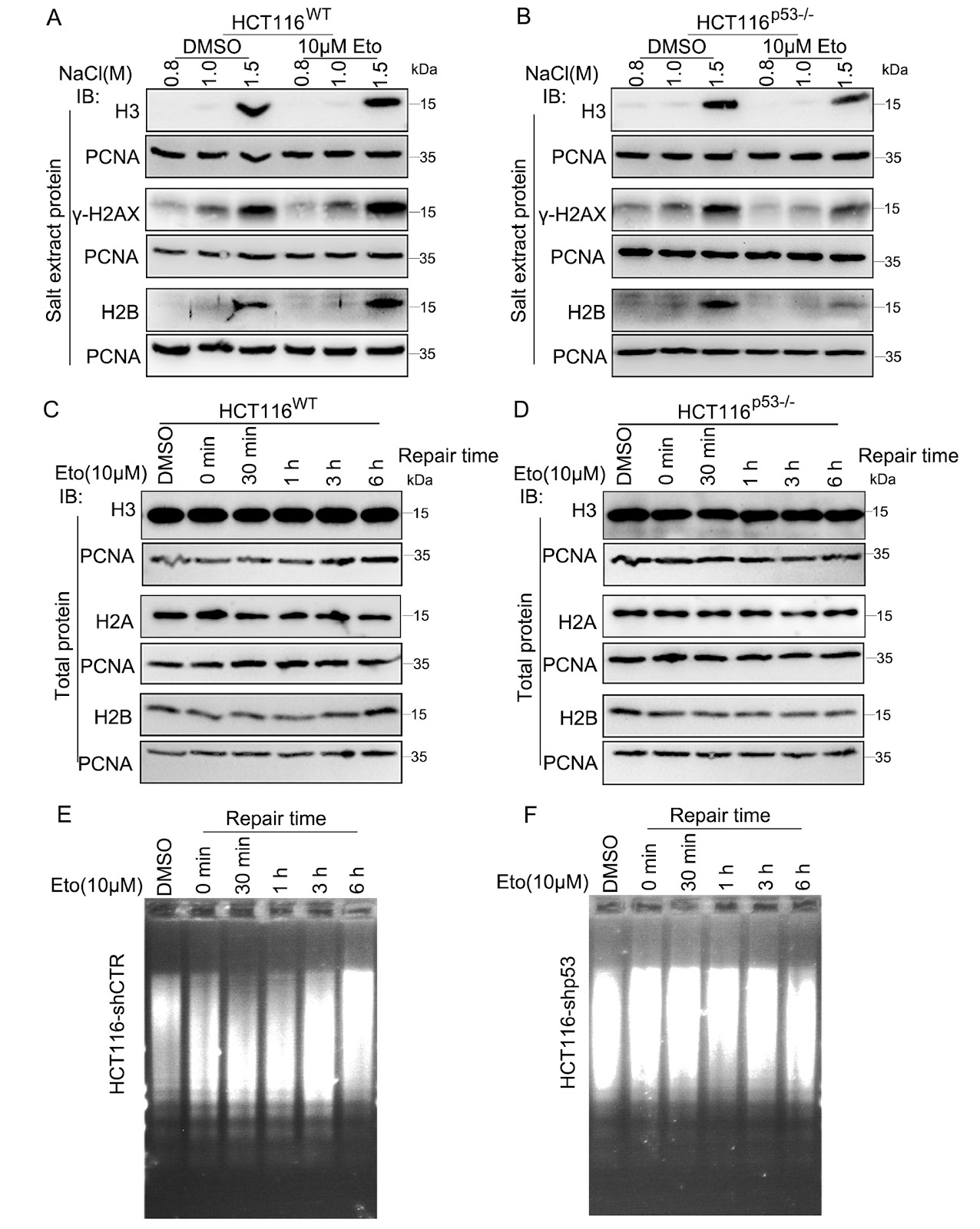
FOXD3 promotes homologous recombination repair and genomic stability by facilitating MRE11-mediated DNA end resection

Authors: Shibin Xu, Jingyu Zhang, Congwen Gao, Ziyi Xiong, Yamin Gong, Bao Chai, Hongxiang Chen, Xingzhi Xu

Homologous recombination (HR) is crucial for the high-fidelity repair of DNA double-strand breaks (DSBs), ensuring the maintenance of genome stability. In this study, we show that FOXD3 interacts with poly (ADP-ribose) polymerase 1 (PARP1) and is recruited to DSBs in a PARP1-dependent manner. FOXD3 directly binds to the DSB repair protein MRE11 and promotes its recruitment to DSB sites, ensuring proper end resection. Inhibition of FOXD3 expression compromises HR-mediated DSB repair and chromosome stability and sensitizes cancer cells to ionizing radiation. Collectively, our findings demonstrate that FOXD3 promotes HR-mediated DSB repair and genome stability.

FOXD3 promotes homologous recombination repair and genomic stability by facilitating MRE11-mediated DNA end resection
Graphical Abstract
Original ResearchVol. 57, Issue 10 • pp. 1709-1717DOI: 10.3724/abbs.2025005

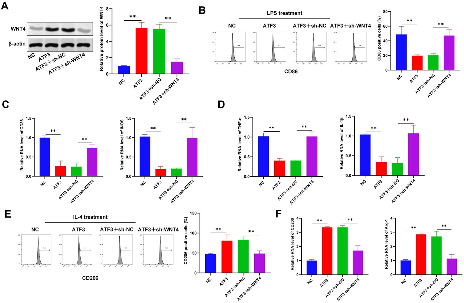
ATF3 triggers M2 macrophage polarization to protect against pulp inflammation through WNT4 regulation

Authors: Liu Liu, Jie Wang, Jie Yu, Jing Wang, Jinhua Yu

Pulpitis is a common inflammatory oral disease that can lead to pulp necrosis. The aim of this study is to investigate the expression and regulatory mechanisms of ATF3, a potential therapeutic marker, in pulpitis. A mouse pulpitis model with different degrees of inflammation is established, and the expression of ATF3 in pulpitis is explored. The histological features of healthy pulp and pulpitis are analyzed by HE staining, and classical inflammatory factors are detected by immunohistochemistry (IHC). In an in vitro study, we investigate the role of ATF3 in the regulation of WNT4 transcription and explore the effects of the ATF3/WNT4 axis on the polarization of RAW264.7 macrophages, the inflammatory response and the osteogenic differentiation of human dental pulp stem/stromal cells (hDPSCs). Our results show that ATF3 is expressed at low levels in inflamed pulp tissues; overexpression of ATF3 reduces the area of pulp necrosis, decreases the level of pro-inflammatory factors, and promotes macrophage polarization toward the M2 type. Furthermore, we reveal that ATF3 binds to the WNT4 promoter region and positively regulates the expression of WNT4 and that ATF3 downregulates M1 markers and increases the expression of M2 markers by regulating WNT4 expression. In addition, ATF3 promotes the osteogenic differentiation of dental pulp stem cells. In summary, this study reveals that ATF3 promotes M2 macrophage polarization by regulating WNT4, which in turn inhibits pulpal inflammatory responses and promotes the osteogenic differentiation of dental pulp stem cells. These findings suggest that ATF3 may be a potential target for pulpitis treatment.

ATF3 triggers M2 macrophage polarization to protect against pulp inflammation through WNT4 regulation
Graphical Abstract
Original ResearchVol. 57, Issue 10 • pp. 1647-1659DOI: 10.3724/abbs.2025070

Schisandrin A ameliorates the diabetes-associated memory impairment by alleviating inflammation and ferroptosis

Authors: Guandi Ma, Min Lei, Shuang Guo, Yuqing Zhang, Yixuan Sun, Huimin Ji, Changhan Ouyang, Xiaosong Yang, Youzhi Zhang, Xiufen Liu, Baoqing Zhao, Xiying Guo

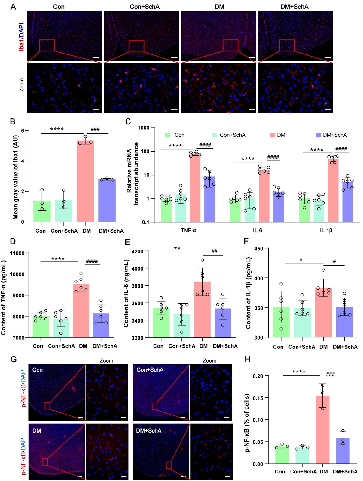
Schisandrin A (SchA), a bioactive lignan that was isolated from the dried fruit of Schisandra chinensis, has attracted much attention because of its diverse spectrum of pharmacological effects. The aim of this study is to clarify the function of SchA in diabetes-related fear memory impairment and its molecular mechanisms. Rats are randomly assigned to 4 groups: the control group (Con group), the DM group, the DM + SchA group, and the Con + SchA group. The results demonstrate that SchA treatment improves insulin sensitivity, reduces blood glucose, and significantly reduces memory impairment. SchA treatment also prevents histological damage, enhances synaptic protein production, and significantly decreases Aβ42 formation in the diabetic prefrontal cortex. Further research reveals that SchA therapy decreases microglial activation and the expression levels of variables linked to inflammation while increasing the phosphorylation of proteins implicated in the insulin resistance signaling pathway. Furthermore, in the prefrontal cortex of diabetic rats, SchA decreases ferroptosis by increasing the protein expressions of GPX4, SLC7A11, Nrf2, HO-1, and SIRT1. Overall, our findings suggest that SchA may lessen diabetes-associated fear memory impairment symptoms by, most likely, lowering ferroptosis and inflammatory responses in the prefrontal brain of diabetic rats. SchA may be a useful therapy for diabetes, including memory impairment.

Schisandrin A ameliorates the diabetes-associated memory impairment by alleviating inflammation and ferroptosis
Graphical Abstract
Original ResearchVol. 57, Issue 10 • pp. 1740-1742DOI: 10.3724/abbs.2025107

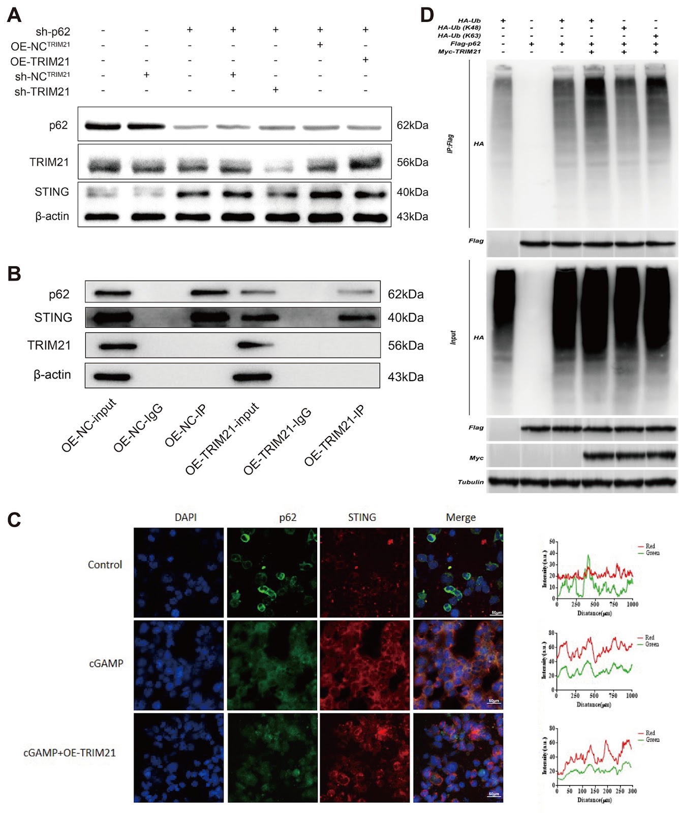
ArfGAP2 deficiency ameliorates autoinflammation by regulating STING signaling and proton channel activity

Authors: Min Zhang, Yufei Wang, Zhenwang Zhao, Xiaobo Hu

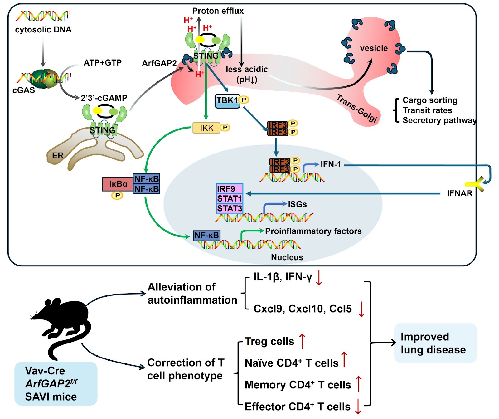
The cGAS-STING pathway is a critical regulator of innate immunity. When cyclic GMP-AMP synthase (cGAS) detects aberrant cytosolic DNA, it synthesizes the second messenger 2′3′-cGAMP, which binds and activates stimulator of interferon genes (STING) on the endoplasmic reticulum (ER). Activated STING then translocates to the Golgi apparatus, where it recruits and mutually phosphorylates TANK-binding kinase 1 (TBK1) and interferon regulatory factor 3 (IRF3). Phosphorylated IRF3 dimerizes and translocates to the nucleus, where it drives the production of type 1 interferons (IFN-1). In addition to being activated by IFN-1, STING also engages the nuclear factor kappa-B (NF-κB) pathway to induce the production of proinflammatory cytokines and chemokines. Moreover, IFN-1 signaling upregulates the expressions of interferon-stimulated genes (ISGs) through the IFN-α receptor (IFNAR). STING-associated vasculopathy with onset in infancy (SAVI) is a severe pediatric autoimmune disease caused by heterozygous gain-of-function mutations in STING, such as the N154S variant in humans and N153S in mice. Initially, classified as an interferonopathy due to constitutive activation of the STING pathway, SAVI is characterized by systemic inflammation, vasculopathy, interstitial lung disease, T-cell cytopenia, skin ulcerations, and premature death. However, recent studies challenge this paradigm, demonstrating that SAVI pathology develops independently of IFNAR-1 and IRFs (IRF3 and IRF7) [1,2], suggesting that alternative mechanisms drive disease progression. Emerging evidence indicates that STING restricts microbial infection through noncanonical autophagy and cell death pathways [3,4]. These functions may depend on its recently identified role as a proton channel in the Golgi apparatus [4,5]. Xun et al. [4] demonstrated that ligand-bound STING forms an ion channel in its transmembrane domain, facilitating proton efflux from post-Golgi vesicles and inducing Golgi deacidification. This raises a critical question: could STING-mediated Golgi deacidification be a potential mechanism underlying SAVI pathogenesis? A recent study by Poddar et al. [6] identified ADP ribosylation factor GTPase-activating protein 2 (ArfGAP2), which is involved in coatomer protein-1 (COP-1) coating in Golgi vesicles, as a key regulator of both STING signaling and proton channel activity, offering novel therapeutic insights for SAVI. First, to elucidate the role of STING in SAVI pathogenesis, they conducted a genome-wide CRISPR-Cas9 screen in T cells resistant to chronic STING activation and identified ArfGAP2 as a critical STING modulator among multiple Golgi-related proteins. Further investigation revealed that genetic ablation of ArfGAP2 in Jurkat T cells significantly attenuated STING-mediated ISG induction. Compelling evidence indicates that STING activation is associated with its subcellular location [7]. While ArfGAP family proteins typically regulate Golgi membrane trafficking, vesicle transport, and cargo sorting [8], Poddar et al. [6] surprisingly reported that ArfGAP2 enhances STING-mediated ISG induction and promotes LC3 lipidation without altering STING palmitoylation or its Golgi localization. Further experiments revealed that ArfGAP2 enhances STING signaling and IFN-β secretion in mouse bone marrow-derived macrophages (BMDMs). In addition to promoting IFN-1 induction, ArfGAP2 promotes the secretion of NF-κB-dependent proinflammatory cytokines activated by STING in THP-1 monocytes [6]. In addition to inducing ISGs and NF-κB signaling, activated STING acts as a proton channel triggering Golgi deacidification [4]. Given the well-established importance of the Golgi pH in regulating enzyme activity, protein modification, and membrane trafficking [9], researchers have further explored how STING and ArfGAP2 modulate cargo transport and secretion. They reported that the loss of ArfGAP2 impairs STING-mediated proton channel activity in the Golgi, leading to a lower luminal pH. Moreover, ArfGAP2-deficient cells presented significant alterations in the cell surface proteome upon STING activation, accompanied by altered sorting, secretion and trafficking rates of specific protein cargos in the Golgi [6].

ArfGAP2 deficiency ameliorates autoinflammation by regulating STING signaling and proton channel activity
Graphical Abstract
Original ResearchVol. 57, Issue 9 • pp. 1539-1547DOI: 10.3724/abbs.2025068

Intracellular acetyl phosphate modulates Escherichia coli pyruvate metabolism

Authors: Ling Zhang, Hongmei Shi, Zixiang Liu, Jing Gu, Jiaoyu Deng

Lysine acetylation has been shown to be an abundant and vital post-translational modification (PTM) that utilizes acetyl phosphate (AcP) as one of the acetyl group donors in bacteria. The pyruvate dehydrogenase (PDH) complex catalyzes the conversion from pyruvate to acetyl coenzyme A (acetyl-CoA). Thus far, the connection between lysine acetylation and pyruvate metabolism has not been thoroughly investigated. In this study, we show that AcP could acetylate Escherichia coli pyruvate dehydrogenase (AceE) in vitro and in vivo, which could be reversed by protein lysine deacetylase (CobB). In vitro treatment of AceE with AcP also causes increased phosphorylation of the protein, whereas deleting ackA does not affect the phosphorylation of the protein. As a result, in vitro treatment of AceE by AcP leads to decreased enzymatic activity. In contrast, deleting ackA leads to increased acetylation and enzymatic activity of AceE, and deleting pta results in the decreased acetylation and enzymatic activity of AceE. As expected, deleting pta in E. coli causes pyruvate accumulation. Although deleting ackA also causes pyruvate accumulation, decreased expression of the two genes involved in pyruvate metabolism (ldhA and poxB) is observed in the mutant, indicating that AcP could affect pyruvate metabolism by other routes in addition to modulating the AceE activity. Thus, our results demonstrate that intracellular AcP could modulate pyruvate metabolism in E. coli. For the first time, a linkage between AcP-mediated protein lysine acetylation, pyruvate dehydrogenase activity, and pyruvate metabolism is established.

Intracellular acetyl phosphate modulates Escherichia coli pyruvate metabolism
Graphical Abstract
Original ResearchVol. 57, Issue 9 • pp. 1433-1445DOI: 10.3724/abbs.2025051

Vesicle-mediated transport-related gene SEC23A promotes cell proliferation by regulating cell cycle leading to gastric cancer progression

Authors: Kang Wang, Shihao Rao, Xujin Wei, Wen Xie, Zhijun Hong, Jia Cheng, Xin Chen, Jingjing Hou, Huiqin Zhuo

Gastric cancer (GC) is a highly prevalent and lethal gastrointestinal cancer. Dysregulation of vesicle-mediated transport-related genes (VMTRGs) is closely associated with tumorigenesis and disease progression. However, the prognostic value of VMTRGs in GC remains unclear. In this study, on the basis of our proteomics data and public databases, we identify differentially expressed VMTRGs in infiltrative-type GC with more metastases and recurrences identified by Ming’s classification. Least absolute shrinkage and selection operator (LASSO) regression identifies 3 VMTRGs (SEC23A, RAB31, and GABARAPL2) from 41 infiltrative-associated VMTRGs, based on which a risk model Vesicle-Infiltrative Lasso System (VILS) is constructed, and its effectiveness and potential importance are validated by immune microenvironment analysis and functional enrichment analysis. As an independent prognostic factor for GC, VILS, combined with other clinically independent prognostic factors to form a nomogram, is effective in predicting GC prognosis. The VILS high-risk group has higher M2 macrophage and cancer-associated fibroblast infiltration, and lower infiltration of Th1 cells and natural killer cells. SEC23A is highly expressed in GC tissues and cells. The importance of SEC23A in GC cells is evaluated by in vitro assays including colony formation assay and CCK-8 assay, and by in vivo assay using a subcutaneous xenograft mouse model. The results show that SEC23A promotes GC cell proliferation and tumor growth through regulation of the cell cycle in vitro and in vivo. VILS provides excellent prognostic prediction for GC patients and is correlated with antitumor immune cell infiltration. SEC23A, the dominant gene of VILS, is highly expressed in GC and promotes GC growth and malignant progression through various molecular mechanisms. Our study reveals the effect of SEC23A on the proliferation of gastric cancer cells for the first time. Therefore, SEC23A has the potential to be a new therapeutic target for the diagnosis and treatment of GC.

Vesicle-mediated transport-related gene SEC23A promotes cell proliferation by regulating cell cycle leading to gastric cancer progression
Graphical Abstract
Original ResearchVol. 57, Issue 9 • pp. 1420-1432DOI: 10.3724/abbs.2024232

Inhibition of HMOX1 alleviates diabetic cardiomyopathy by targeting ferroptosis

Authors: Huiping Yang, Gongyi Xiao, Dinghui Wang, Tianhua Xiong, Jing Wang, Xiaodong Jing, Bingquan Xiong, Junmei Xie, Bin Liu, Qiang She

Diabetic cardiomyopathy (DCM) is an important complication of chronic diabetes mellitus. However, its pathologic process and pathogenesis have not been fully elucidated. This study aims to investigate the role of ferroptosis in DCM and clarify the effect of heme oxygenase-1 (HMOX1) on DCM by targeting ferroptosis. In vivo, an animal model of DCM is established by subjecting mice to a high-fat diet (HFD) combined with low-dose streptozotocin (STZ) injection. We induce an in vitro DCM model by exposing H9C2 cells to high glucose and palmitic acid. Transcriptome sequencing reveals that the differentially expressed genes (DEGs) are enriched primarily in fatty acid metabolism and mitochondrial fatty acid β-oxidation, which are closely related to ferroptosis. The experimental results show that the diabetic microenvironment induces ferroptosis both in vivo and in vitro. Western blot analysis reveals the decreased expressions of the antioxidant proteins GPX4, SLC7A11 and ferritin in the DCM group. However, qPCR demonstrates the elevated expressions of the ferroptosis markers PTGS2 and ACSL4. Biochemical indicators further support the occurrence of ferroptosis, with increased levels of malondialdehyde (MDA) and lactate dehydrogenase (LDH), along with decreased level of glutathione (GSH). In vitro, intervention with high glucose and palmitic acid in H9C2 cells results in ferroptosis, which is reversed by ferrostatin-1 (Fer-1). Results show the elevated expression of HMOX1 in DCM. Moreover, knockdown of HMOX1 ameliorates ferroptosis, thereby alleviating diabetic cardiomyopathy by reducing cardiac fibrosis and improving cardiac function. Our study elucidates the role of HMXO1 in DCM pathogenesis and provides a potential therapeutic strategy for clinical treatment.

Inhibition of HMOX1 alleviates diabetic cardiomyopathy by targeting ferroptosis
Graphical Abstract
Original ResearchVol. 57, Issue 9 • pp. 1457-1468DOI: 10.3724/abbs.2024239

MYB represses ζ-globin expression through upregulating ETO2

Authors: Zejun Dong, Yuhua Ye, Wei Zhang, Hualei Luo, Jialong Li, Qianqian Zhang, Xinhua Zhang, Xiang Guo, Xiangmin Xu

Reactivating the embryonic ζ-globin gene represents a potential therapeutic approach to ameliorate the severe clinical phenotype of α-thalassemia and sickle cell disease. The transcription factor MYB has been extensively proven to be a master regulator of the γ-globin gene, but its role in the regulation of ζ-globin remains incompletely understood. Here, we report a mechanistic study on the derepression of ζ-globin both in vivo and in vitro. We show that MYB depletion in mouse models and human hematopoietic stem cells leads to consistent and remarkable reactivation of ζ-globin. Furthermore, multiomics analysis and functional validation of MYB-knockout and wild-type cell lines reveal that ETO2 functions as a novel repressor of ζ-globin through coordination with NuRD nucleosome remodeling and the deacetylation complex to modulate histone deacetylation of ζ-globin. Additionally, we evaluate the clinical significance of these findings by knocking out ETO2 in primary CD34+ cells from nondeletional hemoglobin H patients, which results in a significant increase in ζ-globin expression. The RNA-seq data reveal that key erythroid genes are more co-regulated by Myb and Eto2 than by Myb and Klf1, highlighting a distinctly enhanced erythroid-specific transcriptional impact within the MYB-ETO2 regulatory axis. Compared with ETO2 knockout alone, codepletion of ETO2 and BCL11A did not significantly activate ζ-globin, suggesting that the MYB-ETO2 pathway primarily silences ζ-globin. Our study reveals a linear MYB-ETO2 signaling pathway crucial for ζ-globin repression and offers new targets for treating α-thalassemia and sickle cell disease.

MYB represses ζ-globin expression through upregulating ETO2
Graphical Abstract
Original ResearchVol. 57, Issue 8 • pp. 1270-1280DOI: 10.3724/abbs.2025043

D-mannose suppresses the angiogenesis and progression of colorectal cancer

Authors: Yu Du, Xinchao Zhang, Yixin Xu, Yuefan Zhou, Yanping Xu

Angiogenesis is an important factor influencing the development of solid tumors, and vascular endothelial growth factor receptor-2 (VEGFR2) is a central regulator of angiogenesis. Antibodies and inhibitors against VEGFR2 have been widely used in various malignancies. However, the regulatory mechanism of VEGFR2 has not been fully clarified. Here, we show that D-mannose can significantly inhibit angiogenesis and tumor growth by degrading VEGFR2. Specifically, D-mannose inactivates GSK3β by promoting the phosphorylation of GSK3β at Ser9, enhances the nuclear translocation of TFE3, and promotes lysosomal biogenesis, thereby increasing the lysosome-mediated degradation of VEGFR2. Thus, D-mannose significantly inhibits the proliferation, migration, and capillary formation of human umbilical vein endothelial cells (HUVECs) in vitro. Oral administration of D-mannose dramatically inhibits angiogenesis and tumor growth in mice. Our findings reveal a previously unrecognized anti-tumor mechanism of D-mannose by destabilizing VEGFR2 and provide a new strategy for the clinical treatment of colorectal cancer (CRC).

D-mannose suppresses the angiogenesis and progression of colorectal cancer
Graphical Abstract
Original ResearchVol. 57, Issue 8 • pp. 1260-1269DOI: 10.3724/abbs.2024221

NCOA6 knockdown enhances RSL3-induced ferroptosis in pancreatic cancer cells and increases the sensitivity to gemcitabine

Authors: Yuming Jia, Zeng Ye, Xin Wang, Yanli Deng, Chao Wang, Zhilei Zhang, Guixiong Fan, Wuhan Yang, Xiaowu Xu, Yi Qin, Li Peng

Ferroptosis is a type of programmed death characterized by iron-dependent lipid peroxidation, and targeting ferroptosis has been shown to efficiently kill highly aggressive cancer cells. Previously, we confirmed that nuclear receptors regulate ferroptosis in pancreatic cancer. However, whether nuclear receptor co-activators regulate ferroptosis is unclear. Here, we show that knocking down the nuclear receptor co-activator, NCOA6, enhances the sensitivity of pancreatic cancer cells to ferroptosis. Mechanistically, NCOA6 knockdown promotes the expression of ACSL4 while inhibiting the expression of SCD1, resulting in changes in lipid metabolism, sensitivity to RSL3-induced ferroptosis, and sensitivity to gemcitabine in pancreatic cancer. The relationships between NCOA6 and ACSL4 or SCD1 are further explored in clinical specimens. This study reveals that targeting NCOA6 might alleviate gemcitabine resistance in pancreatic cancer.

NCOA6 knockdown enhances RSL3-induced ferroptosis in pancreatic cancer cells and increases the sensitivity to gemcitabine
Graphical Abstract
Original ResearchVol. 57, Issue 9 • pp. 1556DOI: 10.3724/abbs.2025129

Corrigendum to: Magnolol promotes the autophagy of esophageal carcinoma cells by upregulating HACE1 gene expression

Authors: Kenan Huang, Biao Zhang, Yu Feng, Haitao Ma

This is a corrigendum to the article 'Magnolol promotes the autophagy of esophageal carcinoma cells by upregulating HACE1 gene expression' published in Acta Biochimica et Biophysica Sinica 2024, 56(7): 1044–1054. In the original publication, the corresponding author's email address was personal. To comply with the institution's publishing policy, it has been changed from '[email protected]' to the institutional address '[email protected]'. The authors apologize for any confusion it may have caused.

Corrigendum to: Magnolol promotes the autophagy of esophageal carcinoma cells by upregulating HACE1 gene expression
Graphical Abstract
Original ResearchVol. 57, Issue 8 • pp. 1281-1291DOI: 10.3724/abbs.2025013

GWAS study of myelosuppression among NSCLC patients receiving platinum-based combination chemotherapy

Authors: Hanxue Huang, Junyan Liu, Qi Xiao, Chenxue Mao, Lei She, Lulu Yu, Bing Yu, Mengrong Lei, Ying Gao, Baimei He, Pinhua Pan, Xi Li, Jiye Yin, Zhaoqian Liu

Platinum-based chemotherapy remains the mainstay for non-small cell lung cancer (NSCLC), but it frequently causes dose-limiting myelosuppression, with significant individual variability in susceptibility. However, the genetic basis of myelosuppression side effects remains elusive, greatly hindering personalized therapeutic approaches. In this study, we perform a comprehensive genome-wide association analysis on 491 NSCLC patients receiving platinum-based chemotherapy, examining 4,690,998 single-nucleotide polymorphisms (SNPs) to identify relevant genetic variants. LDBlockShow, FUMA, and MAGMA are utilized to explore linkage disequilibrium, expression quantitative trait loci (eQTLs), chromatin interaction, and conduct gene-based and gene set-based analysis of candidate SNPs. The GWAS results reveal that rs6856089 and its linked SNPs are significantly associated with platinum-based chemotherapy-induced myelosuppression. Specifically, patients with the A allele of rs6856089 have a significantly lower risk of myelosuppression [odds ratio (OR) = 0.1300, P = 7.59 × 10⁻⁸]. Furthermore, gene-based analysis reveals that EMCN (P = 2.47 × 10⁻⁵), which encodes endomucin, a marker for hematopoietic stem cells, might mediate myelosuppression. This study provides a scientific basis for the individual differences in platinum-based chemotherapy-induced myelosuppression.

GWAS study of myelosuppression among NSCLC patients receiving platinum-based combination chemotherapy
Graphical Abstract
Original ResearchVol. 57, Issue 8 • pp. 1350-1362DOI: 10.3724/abbs.2025014

Knockdown of lncRNA XR_877193.1 suppresses ferroptosis and promotes osteogenic differentiation via the PI3K/AKT signaling pathway in SONFH

Authors: Huixia Yang, Ning Ding, Shi Qing, Yinju Hao, Cilin Zhao, Kai Wu, Guizhong Li, Huiping Zhang, Shengchao Ma, Zhigang Bai, Yideng Jiang

Ferroptosis is a novel form of regulated cell death characterized by the iron-dependent accumulation of lipid peroxides. Recent research has suggested that ferroptosis in osteoblasts contributes to steroid-induced osteonecrosis of the femoral head (SONFH). However, the relationship between ferroptosis and SONFH remains unclear. In this study, in vitro experiments show that dexamethasone (Dex) treatment reduces the expressions of key ferroptosis regulators, SLC7A11 and GPX4, in MC3T3-E1 cells. This reduction leads to a decrease in intracellular glutathione (GSH) levels, accompanied by elevated levels of total iron, malondialdehyde (MDA), and reactive oxygen species (ROS). Importantly, the ferroptosis inhibitor ferrostatin-1 (Fer-1) effectively reverses Dex-induced ferroptosis in MC3T3-E1 cells. Furthermore, RNA-seq analysis reveals that the long noncoding RNA (lncRNA) XR_877193.1 is significantly upregulated in Dex-treated MC3T3-E1 cells. Functional studies demonstrate that the knockdown of lncRNA XR_877193.1 promotes osteogenic differentiation by inhibiting Dex-induced ferroptosis in MC3T3-E1 cells, whereas its overexpression exacerbates cell death via ferroptosis. Kyoto Encyclopedia of Genes and Genomes (KEGG) enrichment analysis reveals that the differentially expressed lncRNA XR_877193.1 is enriched in ferroptosis-related pathways, including the PI3K/AKT signaling pathway. Moreover, PI3K/AKT inhibitors reverse ferroptosis in MC3T3-E1 cells inhibited by lncRNA XR_877193.1 knockdown. Collectively, our findings indicate that lncRNA XR_877193.1 knockdown exerts anti-ferroptosis effects by stimulating the PI3K/AKT signaling pathway, suggesting a promising therapeutic strategy for attenuating SONFH.

Knockdown of lncRNA XR_877193.1 suppresses ferroptosis and promotes osteogenic differentiation via the PI3K/AKT signaling pathway in SONFH
Graphical Abstract
Original ResearchVol. 57, Issue 8 • pp. 1371-1375DOI: 10.3724/abbs.2025016

CRATS: a one-pot CRISPR-Cas12b and RPA combined assay with a temperature switch for highly sensitive detection of monkeypox virus

Authors: Guangxi Yu, Yue Wang, Yukang Chen, Jiangyuan Liu, Hongtao Kang, Xiaodong Luan, Song Gao, Pei Wang

CRISPR-Cas nucleases have been extensively used in molecular detections, especially highly sensitive nucleic acid detections. In these detections, Cas nucleases are programmed by the guide RNA to respond to the detection targets and cleave the chemically labelled molecular beacons by the trans-cleavage activity to produce the detection signal. To improve sensitivity, nucleic acid amplification technologies are usually introduced to give a pre-amplification of the nucleic acid targets, increasing the detection sensitivity extraordinarily. Polymerase chain reaction (PCR) technology has been used for pre-amplification in laboratories, and isothermal amplification technologies are applied to meet point-of-care testing (POCT) needs because they avoid the use of sophisticated thermal cycling devices. The recombinase polymerase amplification (RPA) technology that amplifies nucleic acid targets isothermally at 37–42°C has been combined with CRISPR-Cas nucleases to establish advantageous nucleic acid detection assays, e.g., the SHERLOCK, which combines with Cas13a, and the DETECTR, which combines with Cas12a. It has been challenging to integrate Cas nucleases and RPA in a one-pot reaction system because the cleavage activity of Cas stimulated by even small amounts of the targets can interfere with amplification by digesting the primers or the newly amplified fragments. Thus, many assays based on Cas nucleases and RPA are in a two-step setting, with pre-amplification and Cas cleavage being isolated as two independent procedures. The two-step setting ensures that amplification and cleavage occur under favorable conditions but sacrifices operational convenience and introduces the risk of cross-contamination. In efforts to establish one-pot RPA-Cas assays, many strategies have been applied, including the use of photocontrolled guide RNA to activate the Cas nuclease at a preferred timepoint, the use of a suboptimal protospacer adjacent motif (PAM) to suppress Cas activity, the generation of dynamic aqueous multiphase with sucrose or glycerol to partially separate the two reactions, and extensive optimization of the RPA-CRISPR reaction system to achieve a subtle balance between the two reactions. In this study, a one-pot CRISPR-Cas12b and RPA combined assay with a temperature switch (CRATS) was established utilizing the reaction temperature difference between RPA and CRISPR-Cas12b cleavage. The Cas12b used in this study, AaCas12b, is a type V-B CRISPR‒Cas nuclease from Alicyclobacillus acidiphilus. It has a bi-lobed architecture consisting of an α-helical recognition lobe containing the REC domains and a nuclease lobe containing the WED, RuvC and Nuc domains. As a dual-RNA-guided DNA endonuclease, Cas12b can be guided by a chimeric single-guide (sg) RNA, and its trans-cleavage activity is specifically activated by the DNA target and results in nonspecific cleavage of single-stranded (ss) DNA molecules, which can be used to produce detection signals if the ssDNA is appropriately labelled as the molecular beacon. As the temperature for RPA is 37°C and the trans-cleavage of Cas12b is active at 60°C, CRATS uses temperature switching to adjust the major on-going reaction in the one-pot system and realizes sequential amplification of the target and cleavage reactions for signal detection. The detection target of this study, monkeypox virus, is an infectious pathogen that has caused the announcement of the Public Health Emergency of International Concern (PHEIC) by the World Health Organization (WHO) twice in recent years. In this one-pot CRATS assay, the reaction reagents of CRISPR-Cas12b and RPA are mixed in a single tube. After the addition of the sample containing the detection target, the reaction was carried out at 37°C for 20 min for amplification, followed by 60°C for 20 min for Cas12b cleavage. The fluorescently labelled molecular beacon is cleaved by Cas12b to release the FAM fluorophore from quenching, producing a fluorescence signal that is visualized under blue light. CRATS shows a high sensitivity of 100 copies of the target DNA per reaction and good specificity, providing a novel strategy of temperature switching to integrate CRISPR-Cas and RPA in a one-pot reaction system. Moreover, it provides a POCT-friendly tool for the detection of the important infectious pathogen monkeypox virus.

CRATS: a one-pot CRISPR-Cas12b and RPA combined assay with a temperature switch for highly sensitive detection of monkeypox virus
Graphical Abstract
Original ResearchVol. 57, Issue 8 • pp. 1207-1221DOI: 10.3724/abbs.2025020

Essential role of the metabolite α-ketoglutarate in bone tissue and bone-related diseases

Authors: Zuping Wu, Yuzhe Guan, Qian Chen, Ruifeng Song, Jing Xie, Xin Zhang, Yan Wang, Qianming Chen, Xiaoyan Chen

Bone metabolism in bone tissue is constantly maintained in a state of dynamic equilibrium. The mass of bone and joint tissues is determined by both bone formation and bone resorption. It is hypothesized that disrupted metabolic balance leads to osteoporosis, osteoarthritis, rheumatoid arthritis, and bone tumors. Such disruptions often manifest as either a reduction or abnormality in bone mass and are frequently accompanied by pathological changes such as inflammation, fractures, and pain. α-Ketoglutarate (α-KG) serves as a pivotal intermediate in various metabolic pathways in mammals, significantly contributing to cellular energy metabolism, amino acid metabolism, and other physiological processes. α-KG may be a therapeutic target for a variety of bone-related diseases, such as osteoporosis, osteoarthritis, and rheumatoid arthritis, because of its role in maintaining the metabolic balance of bone. After the application of α-KG, bone loss and inflammation in bone tissue are alleviated. This review focuses on the regulatory effects of α-KG on various cells in bone and joint tissues. Owing to the regulatory effect of α-KG on the balance of bone metabolism, the application of α-KG in the treatment of osteoporosis, osteoarthritis, rheumatoid arthritis, bone tumors, and other bone tissue diseases has been clarified.

Essential role of the metabolite α-ketoglutarate in bone tissue and bone-related diseases
Graphical Abstract
Original ResearchVol. 57, Issue 8 • pp. 1304-1311DOI: 10.3724/abbs.2025039

Puerarin prevents cadmium-induced endoplasmic reticulum stress via SIRT1-dependent PERK-CHOP pathway in HepG2 cells

Authors: Di Huang, Mengqi Qiu, Kuanhong Luo, Yanzhe Zhu, Siyu Zhang, Zhen He, Xiaobo Hu, Zhaohui Cao

Cadmium (Cd) is a high-risk heavy metal that induces oxidative stress, endoplasmic reticulum (ER) stress and inflammation, damaging organs such as the liver. Puerarin (PUE) has been shown to treat liver injury and especially prevent Cd-induced hepatic damage via its antioxidant activity. Sirtuin 1 (SIRT1), a histone deacetylase, is a key protector against various stress insults. However, its role in the protection of PUE against Cd-induced liver damage has not been clarified. Thus, this study is designed to elucidate the molecular mechanism in the human hepatoma cell line HepG2. The results first reveal that Cd-induced apoptosis is significantly restored by PUE pretreatment, as confirmed by the CCK-8, flow cytometric, Hoechst 33258 and TUNEL assays. Mechanistically, PUE significantly decreases ROS production and increases SOD levels in Cd-treated HepG2 cells. Moreover, PUE pretreatment alleviates ER stress by inhibiting the PERK-eIF2α-ATF4-CHOP axis and subsequently partially restores ER function as revealed by decreased Ca2+ release from the ER. In addition, further study demonstrates that PUE upregulates SIRT1 expression, which suppresses the PERK signaling cascade and reduces CHOP levels. Collectively, our results first demonstrate that PUE protects HepG2 cells from Cd-induced apoptosis at least partially by inhibiting the PERK-eIF2α-ATF4-CHOP pathway in a SIRT1 expression-dependent manner. Puerarin appears to have great potential as a hepatoprotective agent.

Puerarin prevents cadmium-induced endoplasmic reticulum stress via SIRT1-dependent PERK-CHOP pathway in HepG2 cells
Graphical Abstract
Original ResearchVol. 57, Issue 8 • pp. 1234-1243DOI: 10.3724/abbs.2024217

Ageing-associated gut dysbiosis deteriorates mouse cognition

Authors: Huihui Ju, Yile Zhou, Wanting Wei, Yan Hu, Hongwei Fang, Zhouyi Chen, Xia Sun, Yi Shi, Hao Fang

Ageing is an independent factor for cognitive dysfunction. Ageing-associated alterations in the gut microbiota also affect cognition. The present study is designed to investigate changes in the gut microbiota and their participation in ageing-associated cognitive impairment. Both 10-week-old and 18-month-old mice are used. Mouse cognition is examined by novel object recognition and T-maze tests. Mouse feces are collected for sequencing and transplantation. Protein expression in the mouse intestine and hippocampus is studied using immunohistochemistry and immunofluorescence staining. Senescent neurons are induced by hydrogen peroxide in vitro. The cell lysates are used for western blot analysis and adenosine triphosphate (ATP) measurement. Our results show that 18-month-old mice exhibit cognitive dysfunction compared with young mice. In aged mice, transplanting the microbiota of young mice increases the protein presence of synaptophysin in the hippocampus and partially restores cognition. The protein expressions of mucin-2 and E-cadherin in the intestine are reduced in aged mice but are increased by transplantation. Gut microbiota analyses reveal that the reduced abundance of the microbe Bacilli-Lactobacillales-Lactobacillaceae-Lactobacillus in aged mice is restored by transplantation. Fecal microbiota transplantation in young mice increases the serum level of acetic acid in aged mice. Hydrogen peroxide stimulation induces senescence and reduces the protein expression levels of synaptophysin and acetyl-coenzyme A synthetase member 2 (ACSS2) in primary neurons. Incubation with acetic acid upregulates the protein expressions of ACSS2 and synaptophysin and further increases ATP production in senescent neurons. In summary, gut microbiota transplantation increases the abundance of Lactobacillales, elevates serum acetic acid level, and improves cognitive function in aged mice. Gut microbiota transplantation has therapeutic importance for ageing-associated cognitive decline.

Ageing-associated gut dysbiosis deteriorates mouse cognition
Graphical Abstract
Original ResearchVol. 57, Issue 7 • pp. 1037-1046DOI: 10.3724/abbs.2025025

The role of cryptochrome (CRY) in cancer: molecular mechanisms and clock-based therapeutic strategies

Authors: Shuzhao Zhang, Xue Chen, Jiayi Li, An’an Xu, Ann M. Bode, Xiangjian Luo

The circadian rhythm is a phenomenon in which physiological, behavioral, and biochemical processes within an organism naturally fluctuate over a period of approximately 24 hours. This phenomenon is ubiquitous in living organisms. Disruption of circadian rhythms in mammals leads to different diseases, such as cancer, and neurodegenerative and metabolic disorders. In specific tissues, numerous genes have been found to have circadian oscillations, suggesting a broad role for rhythm genes in the regulation of gene expression. This review systematically summarizes the role of cryptochromes (CRYs) in the initiation and progression of different types of cancer and discusses the relationships between clock genes and the tumor microenvironment (TME), as well as clock-based therapeutic strategies.

The role of cryptochrome (CRY) in cancer: molecular mechanisms and clock-based therapeutic strategies
Graphical Abstract
Original ResearchVol. 57, Issue 6 • pp. 955-967DOI: 10.3724/abbs.2024235

Gastrodin inhibits reactive astrocyte-mediated inflammation in hypoxic-ischemic brain damage through S100B/RAGE-Smad3 signaling

Authors: Pengxiang Wang, Hanjun Zuo, Haolong Shi, Zhao Wang, Xueqi Ren, Jinsha Shi, Tao Guo, Xianfeng Kuang, Min Zhao, Jinghui Li, Juanjuan Li

Activated astrocytes and their associated inflammatory responses play critical roles in the pathogenesis of hypoxic-ischemic brain damage (HIBD). Gastrodin (GAS), an anti-inflammatory herbal agent, is known to suppress microglial activation. Here, we investigate whether it exerts a similar effect on activated astrocytes and whether it acts through S100B/RAGE-Smad3 signaling. The expression changes of S100B/RAGE-Smad3 signaling pathway-related proteins, inflammatory factors and A1/A2 astrocyte markers were detected by ELISA, western blot analysis, immunofluorescence and immunohistochemistry. The results show that GAS decreases the expression of sRAGE in the brain tissue and S100B in the serum and brain tissue of HIBD mice. However, it promotes the expression of sRAGE in the serum of HIBD mice. Moreover, GAS inhibits the expressions of RAGE, p-Smad3, TNF-α, and C3 (A1 astrocyte marker), and promotes the expressions of S100A10 (A2 astrocyte marker) and BDNF in HIBD model mice, as well as in oxygen glucose deprivation (OGD)-treated TNC-1 astrocytes. The immunofluorescence and immunohistochemical results of RAGE and p-Smad3, as well as the immunofluorescence results of C3 and S100A10, reveal the same trend. Interestingly, FPS-ZM1 (a specific inhibitor of RAGE) inhibits the expressions of p-Smad3, TNF-α, C3, and S100A10, but promotes that of BDNF compared with those in the OGD group. The combination of GAS and FPS-ZM1 further decreases the expression of C3. These results indicate that GAS can inhibit the activation of Smad3 through S100B/RAGE signaling and regulate the expression of A1/A2-type astrocytes.

Gastrodin inhibits reactive astrocyte-mediated inflammation in hypoxic-ischemic brain damage through S100B/RAGE-Smad3 signaling
Graphical Abstract
Original ResearchVol. 57, Issue 8 • pp. 1244-1251DOI: 10.3724/abbs.2024208

D-CAPS: an efficient CRISPR-Cas9-based phage defense system for E. coli

Authors: Mingjun Sun, Jie Gao, Hongjie Tang, Hengyi Wang, Liyan Zhou, Chuan Song, Yongqiang Tian, Qi Li

Escherichia coli is widely used in industrial chemical synthesis but faces significant challenges due to bacteriophage contamination, which reduces product quality and yield. Therefore, developing an efficient antiphage system is essential. In this study, we develop a CRISPR-Cas9-based antiphage system (CAPS) targeting essential genes of the T7 phage (gene 5 and gene 19) with single gRNAs transformed into MG1655 strains expressing Cas9. While CAPS provides limited resistance, with plating efficiencies ranging from 10–5 to 10–1, further optimization is needed. To enhance efficacy, we design a double-site-targeting CRISPR-Cas9-based antiphage system (D-CAPS). D-CAPS demonstrates complete resistance, with no plaques observed even at a high multiplicity of infection (MOI of 2), and growth curve analysis reveals that antiphage E. coli strains grow normally, similar to the wild-type strain, even at a high multiplicity of infection. Furthermore, D-CAPS is effective against BL21(DE3) strains, showing strong resistance and demonstrating its versatility across different E. coli strains. Protein expression analysis via green fluorescent protein confirms that E. coli carrying D-CAPS could maintain normal protein expression levels even in the presence of phages, comparable to wild-type strains. Overall, D-CAPS offers a robust and versatile approach to enhancing E. coli resistance to phages, providing a practical solution for protecting industrial E. coli strains and improving fermentation processes.

D-CAPS: an efficient CRISPR-Cas9-based phage defense system for E. coli
Graphical Abstract
Original ResearchVol. 57, Issue 7 • pp. 1068-1080DOI: 10.3724/abbs.2025026

The protective effect of naringenin on ulcerative colitis in mice through increasing Nrf2 pathway activity

Authors: Jiaxiang Li, Li Hua, Meichun Hu, Ni Zhu, Sijin Dong, Xiaoli Jing, Zihuan Zhu, Yifei Liu, Yanhong Zhou

Ulcerative colitis (UC) is a chronic inflammatory disease with an increasing prevalence worldwide. Naringenin (NAR) has been proven effective in preventing UC, but its mechanism has not been fully elucidated. In this study, network pharmacology and bioinformatics methods are used to screen the genes associated with NAR and UC. A mouse model of dextran sulfate sodium (DSS)-induced UC is established. After treatment with NAR, the disease activity index (DAI) is scored, and colonic histopathology is observed via hematoxylin-eosin (HE) staining. The expressions of the nuclear factor erythroid 2-related factor 2 (Nrf2) signaling pathway and inflammation-related factors in the colons of UC mice are examined via western blot analysis and immunohistochemistry (IHC). The results of the animal experiments reveal that the model group of UC mice present the most severe weight loss and the highest DAI scores. After the administration of NAR, weight loss is alleviated, and DAI scores are reduced (P < 0.05). NAR improves pathological manifestations in the mouse colon, such as reducing inflammatory cell infiltration and restoring goblet cell loss (P < 0.05). NAR significantly increases the protein expression levels of Nrf2, heme oxygenase 1 (HO-1), and NAD(P)H dehydrogenase [quinone] 1 (NQO1) in the colon (P < 0.05) but decreases the protein expression levels of nuclear factor kappa-B (NF-κB), tumor necrosis factor-α (TNF-α), and interleukin-1β (IL-1β) (P < 0.05), thus alleviating the inflammatory response in UC model mice.

The protective effect of naringenin on ulcerative colitis in mice through increasing Nrf2 pathway activity
Graphical Abstract
Original ResearchVol. 57, Issue 7 • pp. 1081-1092DOI: 10.3724/abbs.2025003

Regulation of immune responses by a tumor necrosis factor in pearl oysters: insights from PmTNF gene expression and function

Authors: Yifan Wu, Bidan Liang, Haiying Liang

Tumor necrosis factor (TNF) is a multifunctional cytokine that regulates cellular processes such as inflammation, apoptosis, differentiation, and proliferation and activates various functions of the immune system. This article reports the discovery and characterization of a novel tumor necrosis factor gene in the pearl oyster Pinctada fucata martensii, which is named PmTNF. The deduced PmTNF protein sequence displays the typical structural characteristics of a TNF domain, and phylogenetic analysis of the sequences of PmTNF and its putative orthologs shows that they conform to the current taxonomy. Analysis of PmTNF mRNA expression via real-time PCR reveals its constitutive expression in all the examined tissues, with the highest expression in the gills. Furthermore, PmTNF expression in the gills varies upon exposure to pathogen-derived stimuli, with modest upregulation in response to lipopolysaccharides, but with significant downregulation in response to polyinosinic:polycytidylic acid. Nucleus insertion surgery induces an increase in PmTNF mRNA level in the gills at 12 h postoperation. Knocking down PmTNF through RNA interference significantly inhibits the expressions of immune-related genes in the NF-κB signaling pathway in the gills by 24 h (P < 0.05). The function of PmTNF is further characterized by studying the activity of an engineered recombinant PmTNF protein (rPmTNF) in vivo. Upon nuclear insertion, treatment with rPmTNF for 6 h upregulates several genes in the NF-κB pathway. Similarly, rPmTNF increases the activities of the antioxidant enzymes, including superoxide dismutase, glutathione and peroxidase, which reflect the total antioxidant capacity. Collectively, these results indicate that PmTNF participates in pearl oyster immunity by modulating the NF-κB pathway and activating the antioxidant defense system.

Regulation of immune responses by a tumor necrosis factor in pearl oysters: insights from PmTNF gene expression and function
Graphical Abstract
Original ResearchVol. 57, Issue 7 • pp. 1115-1124DOI: 10.3724/abbs.2025018

Atrial APD prolongation caused by the upregulation of RAGE and subsequent INaL increase in diabetic patients

Authors: Yingchun Luo, Wenbo Ma, Qi Kang, Han Pan, Ling Shi, Jiudong Ma, Jiahui Song, Dongmei Gong, Kai Kang, Xuexin Jin

Diabetes mellitus (DM) is a risk factor for the development of atrial fibrillation (AF). The action potential duration (APD) has been demonstrated to be prolonged in the atrium of diabetic mice. In contrast, the APD is generally shortened in AF patients. It is unclear what change occurs in the atrial APD of diabetic patients. In this study, we explore the APD change of atrial myocytes from diabetic patients and the underlying molecular mechanisms. The whole-cell patch-clamp technique is used to detect single-cell electrical activity in diabetic and nondiabetic human samples. The results show that both APD50 and APD90, the APD at 50% and 90% repolarization, are increased in diabetic patients compared with those in nondiabetic controls. The density of late sodium current (INaL) in the atrial myocytes of diabetic patients is greater than that in the myocytes of nondiabetic patients. The expression of receptor for advanced glycation end products (RAGE) is increased in the atria of diabetic patients. In cultured HL-1 cells, high glucose (HG) treatment increases INaL, and the expression of RAGE prolongs APD. The siRNA-mediated knockdown of RAGE reduces the INaL and shortens the APD. The APD is prolonged in the atria of diabetic patients because of the upregulation of RAGE and the subsequent increase in INaL. Our findings provide novel insights into atrial electrical remodeling in diabetic patients.

Atrial APD prolongation caused by the upregulation of RAGE and subsequent INaL increase in diabetic patients
Graphical Abstract
Original ResearchVol. 57, Issue 7 • pp. 1151-1163DOI: 10.3724/abbs.2024230

AKR1C3 protects cardiomyocytes against hypoxia-induced cell apoptosis through the Nrf-2/NF-κB pathway

Authors: Wenlu Zhang, Wei Tian, Xin Xia, Hua Tian, Ting Sun

Hypoxia-induced apoptosis plays a critical role in the progression of various cardiac diseases, such as heart failure and acute myocardial infarction (AMI). Aldosterone reductase 1C3 (AKR1C3), a member of the aldo-keto reductase superfamily, participates in the metabolism of steroid hormones and redox reactions in vivo. Imbalances in prostaglandin levels have been linked to coronary events. However, the function and molecular mechanism by which AKR1C3 influences AMI are not yet fully understood. This study aims to investigate the role of AKR1C3 in hypoxia-induced myocardial cell damage and elucidate its mechanism. Our findings reveal that a hypoxic microenvironment triggers cardiomyocyte apoptosis and elevates AKR1C3 expression in H9C2 and AC16 cells, as well as in cardiac tissue from rats and mice with AMI. The overexpression of AKR1C3 promotes cardiomyocyte proliferation and cell vitality, whereas the silencing of AKR1C3 exerts the opposite effects in vitro. AKR1C3 protects cardiomyocytes against hypoxia-induced cell apoptosis by reducing ROS levels, preventing mitochondrial damage, and maintaining the oxygen consumption rate (OCR) and ATP production; conversely, AKR1C3 knockdown leads to adverse outcomes. Moreover, the application of a ROS inhibitor (MitoQ10) mitigates the increase in mitochondrial ROS in cardiomyocytes induced by AKR1C3 knockdown under hypoxic conditions. Mechanically, AKR1C3 increases Nrf-2 expression through the ubiquitin-proteasome pathway in cardiomyocytes and subsequently inhibits the NF-κB signaling pathway, thereby inhibiting Bax/caspase-3 signaling. Collectively, these results suggest that AKR1C3 prevents hypoxia-induced cardiomyocyte injury by modulating the Nrf-2/NF-κB axis, suggesting new insights into the mechanisms underlying myocardial protection.

AKR1C3 protects cardiomyocytes against hypoxia-induced cell apoptosis through the Nrf-2/NF-κB pathway
Graphical Abstract
Original ResearchVol. 57, Issue 7 • pp. 1205-1206DOI: 10.3724/abbs.2025088

Corrigendum to: Vitamin B6 prevents Isocarbophos-induced posterior cerebral artery injury in offspring rats through up-regulating S1P receptor expression

Authors: Yanhua Liu, Kunli Yang, Ling Wang, Jinfang Yang, Yang Wang, Hu Luo, Peng Li, Yaling Yin

This corrigendum corrects errors in the original article 'Vitamin B6 prevents Isocarbophos-induced posterior cerebral artery injury in offspring rats through up-regulating S1P receptor expression' published in Acta Biochim Biophys Sin 2021, 53(12): 1691–1701. The errors were found in Figure 2B (Vit B6 + Fingolimod), Figure 5 (Saline), and Figure 7 (Isocarbophos/Control). The correct figures are shown. The authors apologize for the error. The corrigendum does not affect the interpretation of data and conclusions.

Corrigendum to: Vitamin B6 prevents Isocarbophos-induced posterior cerebral artery injury in offspring rats through up-regulating S1P receptor expression
Graphical Abstract
Original ResearchVol. 57, Issue 6 • pp. 1029-1032DOI: 10.3724/abbs.2025040

Histone acetylases are required for iron homeostasis in yeast

Authors: Jian Zhang, Yong Xue, Xinya Zhang, Renjie Qi, Yaqi Zhang, Chen Lu, Zhidan Luo

Iron, an ancient and essential transition metal, is involved in various biological functions, including oxygen transport, DNA synthesis, heme production, and iron-sulfur clusters, which participate in electron transport, DNA repair, and other cellular processes. However, excessive iron can lead to oxidative stress, lipid peroxidation, and cell damage. Thus, maintaining the iron content within an appropriate safe range and maintaining the balance of iron metabolism play crucial roles in both cellular function and human health [1]. An important aspect of maintaining the balance of iron homeostasis is the regulation of the iron uptake system. In Saccharomyces cerevisiae, cells can either obtain iron from the external environment via the non-reducing siderophore transport system or transport iron from the extracellular space to the intracellular space via the reducing iron transport system [2]. Iron uptake system-related genes are regulated mainly by the transcription factor Aft1p. During iron deficiency, Aft1p translocates into the nucleus, binds to genes involved in iron metabolism, and regulates the expressions of genes involved in iron uptake systems [3]. In addition, when there is a problem in the synthesis of iron-sulfur clusters in the mitochondria, such as the lack of the iron chaperone Yfh1p, which promotes the synthesis of iron-sulfur clusters, the transcription and nuclear entry of the transcription factor Aft1p are also activated, thereby regulating the expressions of iron metabolism-related genes [4]. In addition to transcription factors, gene expression is also regulated by histones and their modifications at the epigenetic level. For example, histone H3K4 methylation is related to gene activation, H3K36 methylation plays an important role in the elongation of transcription, and histone acetylation results in the loss of nucleosome structure and facilitates gene expression [5]. Therefore, histone modifications should also play important roles in the regulation of iron homeostasis. The relationship between histone modifications and iron homeostasis has been reported in the literature. For example, the DNA methylation-binding protein MBD5 can change histone acetylation in the promoter region of the ferritin gene by recruiting the histone acetylase KAT2A protein [6]. Histone acetylation has also been reported to be reduced in iron-deficient environments [7,8], and direct effects of histone acetylation on iron homeostasis gene loci have also been reported both in C. albicans [9] and mammals [10]. More recently, the histone H3-H4 tetramer was found to be a copper reductase enzyme, and H3-mediated Cu+ toxicity is a major determinant of the cellular functional pool of iron-sulfur (Fe-S) clusters [11,12]. However, information on the role of histone modifications in the regulation of iron homeostasis is limited. The mechanism by which and how histone modifications are involved in the transcriptional regulation of iron uptake-related genes or the iron deficiency response require further investigation. To determine whether histone acetylation and methylation are involved in the iron deficiency response, we first deleted the histone acetylase genes, including GCN5 (histones H2B and H3 N-terminal lysine acetylase, partial deletion of the ADA2 interaction sequence [13]), RTT109 (H3 lysine 9 and 56 acetylase), SAS2 (H4 lysine 16 acetylase), and YNG2 (subunit of the histone acetyltransferase complex NuA4 for acetylation of histone H4 or histone H2A) in the wild-type (WT) strain, as well as the histone methyltransferases SET1 (H3 lysine 4 methyltransferase), SET2 (H3 lysine 36 methyltransferase) and DOT1 (H3 lysine 79 methyltransferase). The genomic deletions were confirmed by colony PCR and genomic coverage analysis, as depicted in Supplementary Figures S1 and S2. The sensitivity of single histone modification enzyme mutants to iron deficiency induced by the iron chelator bathophenanthroline disulfonate (BPS) was tested. As shown in Supplementary Figure S3, the histone acetylation-related mutants gcn51–316, rtt109Δ, sas2Δ, and yng2Δ did not exhibit significant growth defects compared with the WT on the YPD + BPS plate. None of the histone methyltransferase knockout strains presented significant growth defects. As a positive control, the iron-responsive transcription factor gene AFT1 knockout strain grew slowly on YPD + BPS plates. It is possible that histone modifications do not have a strong effect on the equilibrium status of iron deficiency but still regulate transcription induction during the iron deficiency response. To investigate the role of histone modifications during the induction of the iron deficiency response, the expressions of iron response genes in the wild-type and mutant strains before and 4 h after BPS treatment were examined.
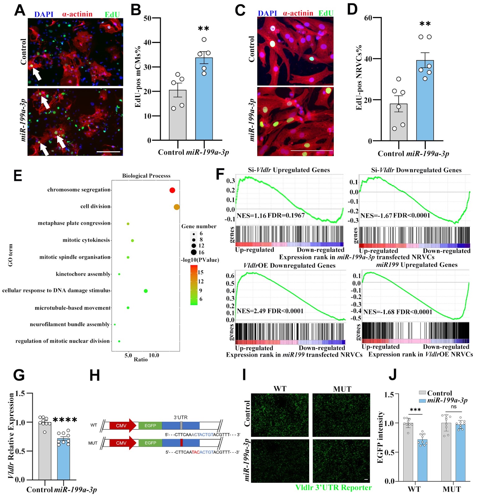
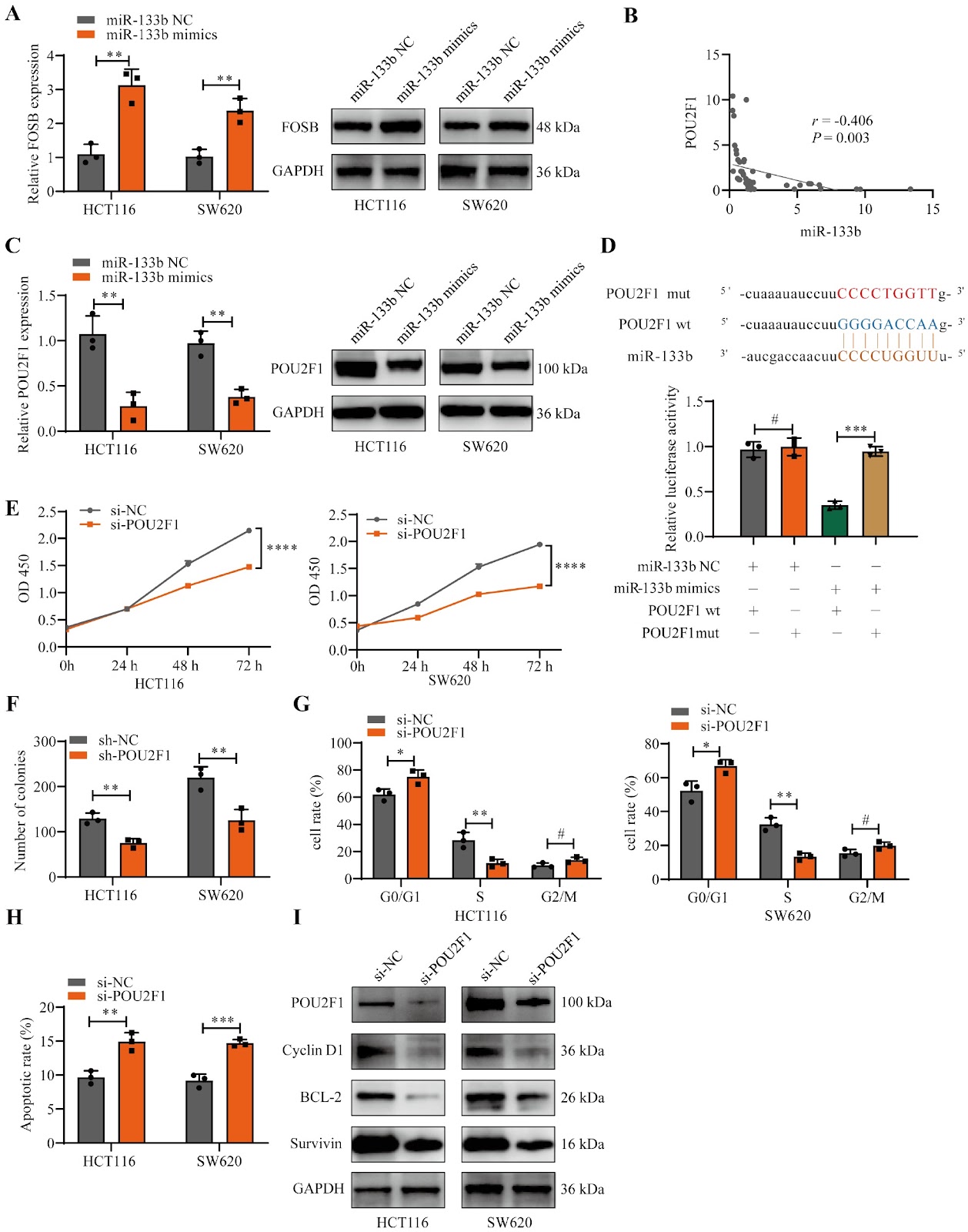
Histone acetylases are required for iron homeostasis in yeast
Graphical Abstract
Original ResearchVol. 57, Issue 1 • pp. 1036DOI: 10.3724/abbs.2025079

Corrigendum to: Stattic sensitizes osteosarcoma cells to epidermal growth factor receptor inhibitors via blocking the interleukin 6-induced STAT3 pathway

Authors: Shenglin Wang, Yunqing Wang, Zhen Huang, Hongxiang Wei, Xinwen Wang, Rongkai Shen, Wenbin Lan, Guangxian Zhong, Jianhua Lin

This is a corrigendum to the original article published in Acta Biochim Biophys Sin (Shanghai) 2021, 53(12): 1670–1680. In the original version, errors were found in Figure 2 and Figure 6. The correct figures are shown in this corrigendum. The authors apologize for the error.

Corrigendum to: Stattic sensitizes osteosarcoma cells to epidermal growth factor receptor inhibitors via blocking the interleukin 6-induced STAT3 pathway
Graphical Abstract
Original ResearchVol. 57, Issue 6 • pp. 890-900DOI: 10.3724/abbs.2024202

Similarities and differences in the response and molecular characteristics of peripheral sensory neurons associated with pain and itch

Authors: Li Liu, Jiemin Yin, Youqiang Meng, Congrui Ye, Junhui Chen, Sa Wang, Wen Yin, Po Gao, Yingfu Jiao, Weifeng Yu, Yinghui Fan

Dorsal root ganglion (DRG) neurons are responsible for the primary detection and transmission of peripheral noxious stimuli, mainly pain and itch. However, as two distinct noxious sensations, how DRG neurons respond differently to and code pain and itch is still an attractive topic. Here, we investigate the response and activation spectrum of DRG neurons under peripheral pain and itch stimuli using in vivo two-photon calcium imaging and find differences in the response intensity to pain and itch between multisensory neurons (both pain and itch) and single-sensory neurons (either pain or itch). In addition, single-cell RNA sequencing (scRNA-seq) is used to reveal the heterogeneity of distinct subpopulations on the basis of their expressions of pain- or itch-related marker genes and to determine the similarities and differences in their transcriptomic changes under chronic pain and itch. Our results show that primary sensory neurons with different sensory patterns respond differently to the same nociceptive stimuli. Additionally, distinct clusters of neurons exhibit unique transcriptomic changes in the development of chronic pain and itch, which may offer new insights for treating these conditions.

Similarities and differences in the response and molecular characteristics of peripheral sensory neurons associated with pain and itch
Graphical Abstract
Original ResearchVol. 57, Issue 5 • pp. 758-769DOI: 10.3724/abbs.2024183

Cannabidiol alleviates the inflammatory response in rats with traumatic brain injury through the PGE2-EP2-cAMP-PKA signaling pathway

Authors: Yan Cao, Hengxi Li, Jiali Li, Tenghan Ling, Aiping Yin, Xinyuan Luo, Ying Zhou, Jinghui Li, Hongyan Jiang, Huawei Wang, Li Yang, Haiying Wu, Ping Li

Traumatic brain injury (TBI) is a recognized global public health problem. However, there are still limitations in the available therapeutic approaches and a lack of clinically effective drugs. Therefore, an in-depth exploration of the secondary pathological mechanism of TBI and the identification of new effective drugs are urgently needed. Cannabidiol (CBD), a component derived from the cannabis plant, has potential therapeutic effects on neurological diseases and has received increasing attention. However, few reports on CBD intervention in TBI patients exist. Here, we use the Feeney free-fall method to establish a rat TBI model. CBD significantly improves neurological deficit scores, neuronal damage and blood-brain barrier permeability in rats and significantly inhibits the expressions of the brain injury markers S-100β and NSE. Mechanistically, CBD attenuates TBI-induced astrocyte activation, reduces inflammation, and attenuates the expressions of inflammatory prostaglandin system indicators. The use of TG6-10-1 (EP2 inhibitor) and H-89 (PKA inhibitor) indicates that CBD attenuates TBI-induced neurological damage via the PGE2-EP2-cAMP-PKA signaling pathway. Overall, this research provides a novel drug candidate for the treatment of clinical brain trauma.
Cannabidiol alleviates the inflammatory response in rats with traumatic brain injury through the PGE2-EP2-cAMP-PKA signaling pathway
Graphical Abstract
Original ResearchVol. 57, Issue 6 • pp. 871-878DOI: 10.3724/abbs.2025011

Melanoma-derived versican reactivates tumor-associated macrophages by upregulating pyruvate carboxylase through TLR2-MyD88-RelB axis under normoxia

Authors: Yuxin Shu, Linmin Zhou, Jinqin Qian, Wei-Guo Zhu

Relieving hypoxia in the tumor microenvironment (TME) promotes innate and adaptive immunity. Our previous research demonstrated that reoxygenation of the TME promotes the phagocytosis and tumor-killing functions of tumor-associated macrophages (TAMs) by upregulating pyruvate carboxylase (PCB). However, the mechanism remains obscure. In the present study, we find that versican derived from melanoma cells binds to TLR2 and activates the downstream transcription factor RelB, which transcribes PCB under normoxia. Blocking the versican-TLR2-MyD88-RelB axis not only reverses the upregulation of PCB in TAMs but also hinders the clearance of tumor cells by TAMs. Our work suggests a pathway that modulates the functions of TAMs under normoxia, which could be harnessed for strengthening anti-tumor immunity.

Melanoma-derived versican reactivates tumor-associated macrophages by upregulating pyruvate carboxylase through TLR2-MyD88-RelB axis under normoxia
Graphical Abstract
Original ResearchVol. 57, Issue 5 • pp. 834-846DOI: 10.3724/abbs.2025046

TRIM21 promotes type I interferon by inhibiting the autophagic degradation of STING via p62/SQSTM1 ubiquitination in systemic lupus erythematosus

Authors: Chen Li, Ang Ma, Yu Bai, Zitao Liu, Linghan Tian, Ziyuan Wang, Huaishun Ma, Zhengpu Chen, Zhengheng Gao, Shijie Feng, Ping Fu

The cGAS-STING signaling pathway serves as a pivotal surveillance mechanism for cytosolic double-stranded DNA (dsDNA) detection in mammalian systems. While STING-mediated type I interferon production is crucial for host defense, sustained activation of this pathway contributes to autoimmune pathologies, including systemic lupus erythematosus (SLE). Maintaining immune homeostasis requires precise regulation of STING activity to prevent hyperactivation. Our study identifies TRIM21 as a novel positive regulator of cGAS-STING signaling in SLE pathogenesis. Our results demonstrate that TRIM21 overexpression stabilizes STING by suppressing autophagic degradation, whereas TRIM21 depletion accelerates this clearance process. Mechanistically, TRIM21 catalyzes the K63-linked polyubiquitylation of the selective autophagy receptor p62/SQSTM1, disrupting its interaction with STING. This post-translational modification prevents the sequestration of STING into autophagosomes, thereby stabilizing the adaptor protein and amplifying downstream type I interferon responses. Our findings reveal a previously unrecognized regulatory circuit in which TRIM21 orchestrates cross-talk between ubiquitin signaling and autophagy to control STING turnover. The TRIM21-p62 axis represents a potential therapeutic target for attenuating pathological interferon production in STING-dependent autoimmune disorders. This work advances our understanding of immune regulation by demonstrating how E3 ligase-mediated ubiquitin modifications modulate cargo recognition in selective autophagy pathways. The identified mechanism provides new insights into the molecular interplay between protein ubiquitylation and autophagic degradation in maintaining the innate immune balance, offering novel perspectives for developing targeted therapies against interferonopathies associated with cGAS-STING hyperactivity.

TRIM21 promotes type I interferon by inhibiting the autophagic degradation of STING via p62/SQSTM1 ubiquitination in systemic lupus erythematosus
Graphical Abstract
Original ResearchVol. 57, Issue 5 • pp. 701-711DOI: 10.3724/abbs.2025008

p53-dependent chromatin relaxation is required for DNA double-strand break repair

Authors: Hongyu Chen, Jin Shan, Wenjing Qi, Lili Chen, Xianlu Zeng

The tumor suppressor p53, an indispensable nuclear transcription factor, plays a central role in orchestrating cellular responses when DNA damage occurs. In this study, we demonstrate that in the initial phases of DNA double-strand break (DSB) repair, p53 is rapidly recruited to sites of damage and the surrounding chromatin, where it enhances DSB repair efficiency. This enhancement occurs through the modulation of chromatin dynamics and the promotion of a more relaxed chromatin configuration, a process influenced by p53 in response to DSB-inducing factors such as etoposide, ultraviolet radiation, and nucleases. These results underscore the pivotal function of p53 as a rapid responder to DSBs, delineating a significant departure from its traditionally recognized role as a downstream transcriptional regulator in DNA damage repair processes. This study emphasizes that the direct engagement of p53 in DNA repair through chromatin structure regulation extends beyond its established involvement in UV irradiation-induced nucleotide excision repair (NER), demonstrating analogous mechanistic attributes in the context of DSB repair. This newly illuminated perspective enhances our understanding of the multifaceted roles of p53 in genome stability and integrity.

p53-dependent chromatin relaxation is required for DNA double-strand break repair
Graphical Abstract
Original ResearchVol. 57, Issue 5 • pp. 749-757DOI: 10.3724/abbs.2024182

CD98hc, a novel of galectin-8 receptor, binds to galectin-8 in an N-glycosylation-dependent manner

Authors: Yunlong Si, Jiahui Zhu, Hend Sayed, Kevin H. Mayo, Yifa Zhou, Guihua Tai, Jiyong Su

Glycan-mediated recognition plays a critical role in facilitating cell-cell and cell-matrix interactions. Galectin-8 (Gal-8), classified as a ‘tandem-repeat’ type of galectin, binds to cell surface glycans to modulate various cellular functions, including cell adhesion, migration, apoptosis, pathogen recognition, autophagy, and immunomodulation. Despite the known function of Gal-8 in binding to various glycosylated proteins, only a few interactions have been reported to date. In this study, mass spectrometry is used to identify CD98hc as a novel binding partner for Gal-8. Both the N-terminal and C-terminal carbohydrate recognition domains (CRDs) of Gal-8 (Gal-8N and Gal-8C) bind to CD98hc, an interaction that is specifically inhibited by lactose but not sucrose, as confirmed by pull-down assays. The binding affinity between CD98hc and Gal-8 measured by microscale thermophoresis (MST) is 1.51 ± 0.17 μM. In addition, Gal-8N and Gal-8C have the binding affinities of 0.22 ± 0.03 μM and 10.68 ± 1.69 μM, respectively. Gal-8N and Gal-8C are both involved in the recognition and binding process of CD98hc. Furthermore, both full-length Gal-8 and its individual CRDs bind specifically to N-glycosylated glycans on CD98hc, as demonstrated by the use of tunicamycin to inhibit N-glycosylation in cells. In addition, Gal-8 and its individual CRDs can pull down glycosylated CD98hc-ED but not free CD98hc-ED in vitro, indicating that the binding of Gal-8 to glycosylated CD98hc-ED is N-glycosylation-dependent. Overall, our findings establish CD98hc as a novel binding partner for Gal-8 and provide insights for further exploration of the diverse biological functions of Gal-8.

CD98hc, a novel of galectin-8 receptor, binds to galectin-8 in an N-glycosylation-dependent manner
Graphical Abstract
Original ResearchVol. 57, Issue 4 • pp. 507-520DOI: 10.3724/abbs.2024228

Annexins: central regulators of plant growth and stress signaling

Authors: Chen Xie, Mingyue Zhu, Ruirui Shi, Liu Yang, Xiaoya An, Chao Wang

Annexins are a family of multifunctional calcium-dependent and phospholipid-binding proteins that are widely distributed in the plant kingdom. They have a highly conserved evolutionary history that dates back to single-celled protists. Plant annexins, as soluble proteins, can flexibly bind to endomembranes and plasma membranes, exhibiting unique calcium-dependent and calcium-independent characteristics. Members of the annexin family have diverse functions, including binding to F-actin, participating in ATP and GTP hydrolysis, and even serving as peroxidases or cation channels. Annexins play pivotal roles in plant growth and stress signaling. They can respond sensitively to environmental, metabolic, and developmental signals, thereby affecting cytoskeleton remodeling and exocytosis mechanisms. Plant annexin gene families have been successfully identified in multiple species, and their expression and intracellular localization are precisely regulated by developmental processes and environmental factors. Although research on plant annexins has aroused great interest, their depth and breadth still need further expansion compared with those of animal annexins. This article provides a comprehensive and in-depth review of the characteristics and functions of plant annexin families, revealing their core roles in plant growth and adaptation, and yielding valuable references and insights for future research.

Annexins: central regulators of plant growth and stress signaling
Graphical Abstract
Original ResearchVol. 57, Issue 3 • pp. 473-485DOI: 10.3724/abbs.2025010

Immunopathological characteristics and therapeutic effects of UC-MSCs in a pigeon breeder’s lung mouse model

Authors: Jingran Xu, Li Li, Yaping Zhou, Zulipikaer Abudureheman, Lexin Xue, Chao Wu, Xiaoguang Zou

Hypersensitivity pneumonitis (HP), including pigeon breeder’s lung (PBL), often progresses from acute inflammation to fibrosis, impairing lung function and limiting targeted therapeutic strategies. Mechanistic studies on PBL progression are limited by the lack of preclinical animal models and a predominant focus on patient data. This study explores the immunopathological characteristics of all stages of PBL in mice and evaluates the therapeutic potential of human umbilical cord-derived mesenchymal stem cells (UC-MSCs) during the non-fibrotic stage. PBL models are created in A/J mice through tracheal instillation of pigeon dropping extract (PDE) protein powder. Different doses (0.4 × 10⁶, 0.8 × 10⁶, and 1.6 × 10⁶ cells per animal) and frequencies (1–2 times) are administered to the model. The immunopathological characteristics of PBL and the therapeutic effects of UC-MSCs are assessed using micro-CT, pulmonary function, histopathology, cell counts in BALF, HYP levels, inflammatory factor levels, immunohistochemistry, and fibrosis marker expression in lung tissues. The results show that PDE exposure consistently impairs pulmonary function and increases the levels of inflammation and fibrosis markers as the disease progresses. Model mice experience non-fibrotic stages (acute inflammation) from days 0–36, mild fibrosis from days 37–77, and severe fibrosis from day 78 onwards. UC-MSCs, particularly at the highest dose (1.6 × 10⁶ cells), effectively treat non-fibrotic PBL by improving pulmonary function (lung ventilation area recovers) and reducing inflammation and fibrosis. This study successfully establishes PBL mouse models reflecting both the acute (inflammatory) and chronic (fibrotic) stages, and UC-MSCs have the potential to delay fibrosis, providing new therapeutic options for PBL and other inflammation-induced lung fibrotic diseases.

Immunopathological characteristics and therapeutic effects of UC-MSCs in a pigeon breeder’s lung mouse model
Graphical Abstract
Original ResearchVol. 57, Issue 4 • pp. 646-655DOI: 10.3724/abbs.2025027

Iron overload mediates cytarabine resistance in AML by inhibiting the TP53 signaling pathway

Authors: Yan Jia, Ling Li, Ying Li, Xunxun Zhu, Haiyan Wang, Bin Xu, Qiuping Li, Hao Zhang

Currently, chemotherapy remains the primary treatment for acute myeloid leukemia (AML). Drug resistance in AML cells is a critical factor contributing to the failure of chemotherapy remission and subsequent relapse. Iron overload frequently occurs in AML patients because of hematopoietic suppression or supportive blood transfusion therapy. Previous studies have indicated that iron overload may promote the progression of AML; however, the underlying mechanisms remain unclear. Our results demonstrate that, compared with TP53-wild-type AML cells, TP53-mutant AML cells exhibit increased resistance to cytarabine-induced cytotoxicity. Moreover, reducing TP53 expression in wild-type AML cells diminishes their sensitivity to cytarabine. The TP53 signaling pathway is essential for mediating cytarabine-induced apoptosis in AML cells. In this study, an iron overload model in AML cells via the use of ferric citrate is constructed. Our data indicate that iron overload can suppress the TP53/BCL2/BAX signaling pathway, counteracting cytarabine-induced apoptosis. In TP53 wild-type AML cells, TFR1 participates in iron-mediated resistance to cytarabine by regulating the entry of iron into the cells. These findings provide a foundation for further exploration of the molecular mechanisms involved in AML resistance to cytarabine.
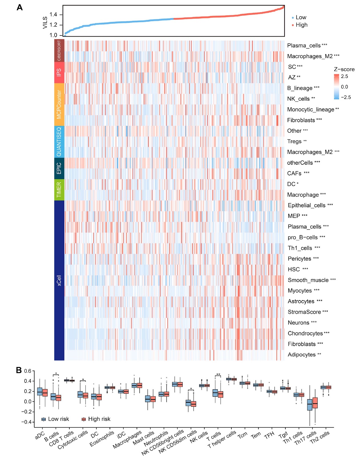
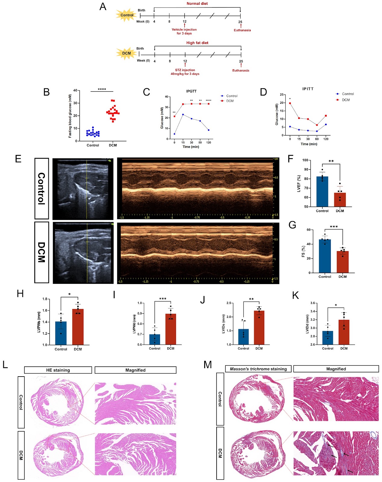
Iron overload mediates cytarabine resistance in AML by inhibiting the TP53 signaling pathway
Graphical Abstract
Original ResearchVol. 57, Issue 4 • pp. 676-678DOI: 10.3724/abbs.2025009

Head-to-head: IL-21 triumphs over IL-15 in NK cell therapy for glioblastoma

Authors: Jake C. Miller, Bihui Cao, Jia Shen

Glioblastoma (GBM) is the most aggressive primary brain tumor. Despite current treatment options, including surgery, radiotherapy, and temozolomide chemotherapy, patient outcomes remain poor, with a median survival of less than 15 months. This dire prognosis highlights an urgent need to develop more effective therapies. Natural Killer (NK) cells, a key component of the innate immune system, are being actively investigated as a potential treatment for GBM. NK cells continually surveil their environment for abnormal cells, including GBM stem cells (GSCs), which are central to GBM progression and recurrence. While NK cells exhibit some ability to target GSCs independently, their activity can be significantly amplified by inflammatory cytokines. One such cytokine, interleukin-15 (IL-15), is critical for NK cell survival and function, making it a focal point of research in GBM immunotherapy. However, IL-15 is not without complications; it has been associated with toxicity, and its overexpression has been shown to induce leukemia in mouse models, potentially due to heightened inflammatory responses. These issues make IL-15 overexpression a less-than-ideal strategy for enhancing NK cell anti-tumor activity. To address these limitations, Shanley and colleagues recently identified interleukin-21 (IL-21) as a promising alternative to IL-15 in their study published in Cancer Cell. Their findings revealed that IL-21 overexpression provides prolonged NK cell activity, even under repeated exposure to GSCs, and demonstrates efficacy both in vitro and in vivo. Importantly, IL-21-expressing NK cells showed no significant toxicity when injected into mouse brains. These results suggest that IL-21 could represent a safer and more effective cytokine for boosting NK cell-mediated GBM therapy.

Head-to-head: IL-21 triumphs over IL-15 in NK cell therapy for glioblastoma
Graphical Abstract
Original ResearchVol. 57, Issue 2 • pp. 325DOI: 10.3724/abbs.2024200

Corrigendum to: Downregulating integrin subunit alpha 7 (ITGA7) promotes proliferation, invasion, and migration of papillary thyroid carcinoma cells through regulating epithelial-to-mesenchymal transition

Authors: Yaoyao Guan, Adheesh Bhandari, Erjie Xia, Lingguo Kong, Xiaohua Zhang, Ouchen Wang

This is a corrigendum to the original article published in Acta Biochim Biophys Sin 2020, 52(2): 116–124. The authors correct an error in Figure 4 of the original manuscript. The correct figure is provided, and the authors apologize for the error.

Corrigendum to: Downregulating integrin subunit alpha 7 (ITGA7) promotes proliferation, invasion, and migration of papillary thyroid carcinoma cells through regulating epithelial-to-mesenchymal transition
Graphical Abstract
Original ResearchVol. 57, Issue 1 • pp. 1-2DOI: 10.3724/abbs.2024234

Safari in the RNA world: a special issue focused on RNA biogenesis, functions, and technologies

Authors: Ya-Nan Chang, Hong Cheng

RNA is one of the most essential biopolymers in cells. According to the central dogma, messenger RNAs (mRNAs) transmit genetic information from DNA to proteins through a complex process, facilitated by key non-coding RNAs (ncRNAs) such as ribosomal RNAs (rRNAs) and transfer RNAs (tRNAs). In addition to these essential RNAs, a wide variety of ncRNAs have been discovered, each playing key roles in gene regulation and contributing to the complexity of the RNA landscape. All RNAs undergo intricate processing and modifications before maturation and transport to their respective cellular compartments, where they perform their functions. Regulation of these processes often results in the generation of multiple isoforms from a single gene, further diversifying the RNA landscape. Understanding the mechanisms of RNA biogenesis and the functional roles of RNAs in both physiological and pathological contexts is essential for unraveling how cells respond to developmental and environmental cues, with profound implications for biomedicine. This special issue features 12 expert reviews in RNA research, each offering a comprehensive summary of the latest advances in RNA biology from their respective perspectives.

Safari in the RNA world: a special issue focused on RNA biogenesis, functions, and technologies
Graphical Abstract
Original ResearchVol. 57, Issue 8 • pp. 1252-1259DOI: 10.3724/abbs.2024229

CD40 ligation-induced ERK activation leads to enhanced radiosensitivity in cervical carcinoma cells via promoting autophagy

Authors: Baocai Liu, Yadong Zhang, Quan Wang, Qian Wang, Zhixin Wang, Li Feng

CD40, a member of the tumor necrosis factor (TNF) receptor superfamily, plays an important role not only in the immune system but also in tumor progression. CD40 ligation reportedly promotes autophagy in immune cells. However, the effects of CD40 ligation on autophagy and its mechanism in solid tumor cells are still unclear. In this study, we find that CD40 ligation promotes autophagosome formation and consequently promotes autophagic flux in cervical cancer cells. Mechanistically, this effect relies on ERK contributing to CD40 ligation-induced ATG13 upregulation by p53. Furthermore, we demonstrate that CD40 ligation-induced autophagy increases the radiosensitivity of cervical cancer cells. Taken together, our results provide new evidence for the involvement of the CD40 pathway in autophagy and radiotherapy in cervical cancer cells.

CD40 ligation-induced ERK activation leads to enhanced radiosensitivity in cervical carcinoma cells via promoting autophagy
Graphical Abstract
Original ResearchVol. 57, Issue 12 • pp. 1999-2010DOI: 10.3724/abbs.2025066

Crystal structures of Kif2A complexed with WDR5 reveal the structural plasticity of WIN-S7 sites

Authors: Yang Yang, Shuting Zhang, Zhangyu Wu, Wenwen Li, Xuefang Sun, Yumi Xuan, Tianrong Hang, Li Xu, Xuemin Chen

Chromosome congression and spindle assembly are essential for genomic stability and proper cell division, with deficiencies in these processes linked to tumorigenesis. WD repeat-containing protein 5 (WDR5), a core component of the mixed lineage leukemia (MLL) methyltransferase complex, directly binds to kinesin family member 2A (Kif2A) to regulate these mitotic events. Despite the importance of this interaction, its structural basis for Kif2A recognition by WDR5 remains unclear. Here, we determine the crystal structure of WDR5 in complex with a Kif2A-derived peptide (residues 114–122) at a resolution of 1.85 Å. Structural analysis reveals that Kif2A engages both the WIN and S7 sites of WDR5 via Arg117 and Ser121, with Ser121 forming hydrogen bonds with WDR5 Tyr191 and Lys259, driving Tyr191 rotation and opening the S7 pocket. Additional structures of WDR5 complexed with truncated or mutated Kif2A peptides and a WDR5 Y191F variant highlight the dynamic nature of Tyr191. Notably, anti-WDR5 compounds exhibit a similar binding mode at the WDR5 WIN-S7 site. The results of mutagenesis combined with isothermal titration calorimetry (ITC) assays underscore the critical roles of Arg117 and Ser121 in mediating the binding of Kif2A to WDR5. In summary, our findings provide atomic-level insights into the molecular mechanisms underlying the non-canonical mitotic function of the MLL/WDR5 complex and highlight WIN-S7 sites as promising therapeutic targets for diseases associated with chromosomal instability, such as cancers.

Crystal structures of Kif2A complexed with WDR5 reveal the structural plasticity of WIN-S7 sites
Graphical Abstract