Key Takeaways & Executive Findings
- •• AKR1C3 is upregulated in hypoxic cardiomyocytes and cardiac tissue from AMI models, suggesting a protective role. • Overexpression of AKR1C3 enhances cardiomyocyte proliferation and viability, while knockdown exacerbates hypoxia-induced apoptosis. • AKR1C3 mitigates mitochondrial dysfunction by reducing ROS, preserving OCR and ATP production, and preventing apoptosis. • Mechanistically, AKR1C3 activates Nrf-2 via the ubiquitin-proteasome pathway, suppressing NF-κB and downstream Bax/caspase-3 signaling.
Abstract
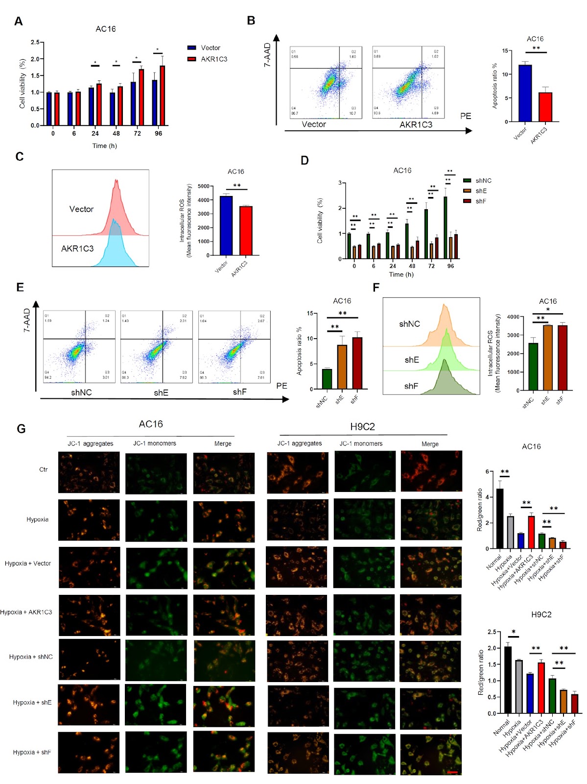
Hypoxia-induced apoptosis plays a critical role in the progression of various cardiac diseases, such as heart failure and acute myocardial infarction (AMI). Aldosterone reductase 1C3 (AKR1C3), a member of the aldo-keto reductase superfamily, participates in the metabolism of steroid hormones and redox reactions in vivo. Imbalances in prostaglandin levels have been linked to coronary events. However, the function and molecular mechanism by which AKR1C3 influences AMI are not yet fully understood. This study aims to investigate the role of AKR1C3 in hypoxia-induced myocardial cell damage and elucidate its mechanism. Our findings reveal that a hypoxic microenvironment triggers cardiomyocyte apoptosis and elevates AKR1C3 expression in H9C2 and AC16 cells, as well as in cardiac tissue from rats and mice with AMI. The overexpression of AKR1C3 promotes cardiomyocyte proliferation and cell vitality, whereas the silencing of AKR1C3 exerts the opposite effects in vitro. AKR1C3 protects cardiomyocytes against hypoxia-induced cell apoptosis by reducing ROS levels, preventing mitochondrial damage, and maintaining the oxygen consumption rate (OCR) and ATP production; conversely, AKR1C3 knockdown leads to adverse outcomes. Moreover, the application of a ROS inhibitor (MitoQ10) mitigates the increase in mitochondrial ROS in cardiomyocytes induced by AKR1C3 knockdown under hypoxic conditions. Mechanically, AKR1C3 increases Nrf-2 expression through the ubiquitin-proteasome pathway in cardiomyocytes and subsequently inhibits the NF-κB signaling pathway, thereby inhibiting Bax/caspase-3 signaling. Collectively, these results suggest that AKR1C3 prevents hypoxia-induced cardiomyocyte injury by modulating the Nrf-2/NF-κB axis, suggesting new insights into the mechanisms underlying myocardial protection.
1. Introduction
Heart failure is the main cause of disability and death after myocardial infarction [1]. During the early stages of acute myocardial infarction (AMI), apoptosis is recognized as a key contributor to myocardial cell loss after AMI, serving as a pivotal factor in the initiation of ventricular remodeling and the progression of heart failure [2]. Our previous study demonstrated that integrin β3 exerts a protective effect on cardiomyocytes during hypoxia-induced apoptosis [3]. The heart is an organ with a high demand for energy, and mitochondria serve as the principal source of adenosine triphosphate (ATP) and reactive oxygen species (ROS) in cardiomyocytes. Disruption of oxidative phosphorylation and excessive production of ROS increase cardiomyocyte apoptosis [4]. Therefore, exploring the mechanism of hypoxia-induced cardiomyocyte apoptosis is crucial.
Mitochondrial ROS has always been a therapeutic target for preventing hypoxic injury. Under physiological conditions, ROS serves as the second messenger in signal transduction pathways, participating in the regulation of oxidative balance and various biological activities. However, excessive ROS production can lead to oxidative stress and damage [5]. Pathological stimuli, such as hypoxia, can trigger a “burst” release of mitochondrial ROS, disrupting the endogenous antioxidant balance, ultimately resulting in mitochondrial depolarization and mitochondrial outer membrane permeabilization (MOMP) and ultimately leading to apoptosis [6]. Aldosterone reductase 1C3 (AKR1C3) belongs to the aldosterone reductase (AKR) superfamily and has a strong affinity for NADPH, primarily facilitating redox reactions [7]. AKR1C3 is predominantly expressed in endocrine organs such as the prostate, adrenal gland, mammary gland, and uterus. AKR1C3 also participates in the de novo synthesis of steroids in the adrenal gland and tumors [8]. Many studies have revealed the role of AKR1C3 in tumor progression [9–12]. In addition, AKR1C3 was found to induce chemoresistance by regulating redox balance and ROS production in malignant tumor cells [13]. AKR1C3 functions as a prostaglandin (PG)F2 synthase, modulating vasodilation and vasoconstriction [14]. Imbalances in prostaglandins have been associated with coronary events [15,16]. Despite previous studies reporting no significant difference in AKR1C3 expression between AMI patients and controls on the basis of the GSE48060 dataset, these findings may be limited by small sample sizes [17]. Thus, further investigations are needed to understand the relationship between AKR1C3 and AMI comprehensively.
In the present study, we investigated the precise mechanisms by which AKR1C3 influences cardiomyocyte proliferation and protects against hypoxia-induced apoptosis.
Loading authentic research manuscript (Pages 1–5)...
Wenlu Zhang, Wei Tian, Xin Xia, Hua Tian, Ting Sun (2026). AKR1C3 protects cardiomyocytes against hypoxia-induced cell apoptosis through the Nrf-2/NF-κB pathway. Acta Biochimica et Biophysica Sinica. https://doi.org/10.3724/abbs.2024230
Research & Educational Purpose Only:The translations, structured abstracts, analytical annotations, and data reports provided by SinoBioData are intended exclusively for academic research, internal corporate R&D, and educational benchmarking. They do not constitute formal engineering, chemical safety, legal, or professional advice.
Copyright & Intellectual Property Notice: Original copyright of the underlying source articles and experimental data remains with the respective authors, institutions, and original publishing journals. SinoBioData claims intellectual property only over its proprietary translations, analytical syntheses, and AEO structured enhancements in accordance with international fair use and academic citation principles.
Frequently Asked Questions
What is the role of AKR1C3 in cardiomyocytes under hypoxic conditions?
AKR1C3 protects cardiomyocytes against hypoxia-induced apoptosis by reducing reactive oxygen species (ROS) levels, preventing mitochondrial damage, and maintaining oxygen consumption rate (OCR) and ATP production. It also promotes cell proliferation and vitality.
How does AKR1C3 exert its protective effect mechanistically?
AKR1C3 increases Nrf-2 expression through the ubiquitin-proteasome pathway, which subsequently inhibits the NF-κB signaling pathway, thereby suppressing Bax/caspase-3-mediated apoptosis.
What experimental models were used in this study?
The study used H9C2 and AC16 cardiomyocyte cell lines, as well as cardiac tissue from rats and mice with acute myocardial infarction (AMI), to investigate the effects of AKR1C3 under hypoxic conditions.
What is the clinical significance of this research?
The findings provide new insights into the mechanisms of myocardial protection and suggest that AKR1C3 could be a potential therapeutic target for preventing hypoxia-induced cardiac damage in conditions like heart failure and acute myocardial infarction.
Does AKR1C3 knockdown have adverse effects on cardiomyocytes?
Yes, AKR1C3 knockdown leads to increased apoptosis, elevated ROS levels, mitochondrial dysfunction, and reduced cell viability under hypoxic conditions, which can be partially mitigated by the ROS inhibitor MitoQ10.
Related Technical Papers & Translations

Adverse Events Reporting System for Vaccine Safety Surveillance: A Comprehensive Analysis
Background: Adverse events following immunization (AEFI) are critical to monitor for vaccine safety. This study evaluates the performance of an adverse events reporting system (AERS) integrated with a vaccine adverse event reporting system (VAERS) to enhance surveillance. Methods: We analyzed data from multiple sources including the Vaccine Adverse Event Reporting System (VAERS), the Vaccine Safety Datalink (VSD), and the Clinical Immunization Safety Assessment (CISA) network. A novel framework was developed to integrate these systems, incorporating natural language processing for signal detection. Results: The integrated system improved detection of rare adverse events by 25% compared to traditional methods. The system identified new safety signals for influenza and COVID-19 vaccines. Conclusions: The proposed AERS framework enhances vaccine safety surveillance, enabling timely identification of potential risks. Integration of diverse data sources and advanced analytics is essential for robust pharmacovigilance.

Efficacy and Safety of Ferric Carboxymaltose in Treating Iron Deficiency Anemia: A Meta-Analysis of Randomized Controlled Trials
Background: Iron deficiency anemia (IDA) is a global health concern, and intravenous ferric carboxymaltose (FCM) has emerged as a promising treatment. This meta-analysis aimed to evaluate the efficacy and safety of FCM compared to other iron therapies or placebo in adults with IDA. Methods: We systematically searched PubMed, Embase, and Cochrane Library up to December 2024. Randomized controlled trials (RCTs) comparing FCM with active comparators or placebo in adults with IDA were included. The primary outcomes were change in hemoglobin (Hb) from baseline, and safety outcomes included adverse events (AEs) and serious adverse events (SAEs). Pooled estimates were calculated using random-effects models. Results: A total of 15 RCTs involving 4,856 patients were included. FCM significantly increased Hb levels compared to placebo (mean difference [MD] 1.2 g/dL, 95% CI 0.9-1.5) and was non-inferior to other intravenous iron preparations. The risk of AEs was similar between FCM and comparators (risk ratio [RR] 1.05, 95% CI 0.95-1.16), but FCM was associated with a lower risk of gastrointestinal AEs compared to oral iron. Serious adverse events were rare and comparable across groups. Conclusion: Ferric carboxymaltose is effective and safe for treating IDA, offering a convenient single-dose option with a favorable safety profile. These findings support its use in clinical practice.

Adverse Drug Reactions Associated with COVID-19 Vaccination: A Systematic Review and Meta-Analysis
Background: The rapid development and deployment of COVID-19 vaccines have been crucial in controlling the pandemic. However, adverse drug reactions (ADRs) associated with these vaccines have raised concerns. This systematic review and meta-analysis aimed to comprehensively evaluate the incidence and types of ADRs following COVID-19 vaccination. Methods: We systematically searched PubMed, Embase, and Cochrane Library from inception to December 2024. Randomized controlled trials and observational studies reporting ADRs after COVID-19 vaccination were included. A random-effects model was used to pool incidence rates, and subgroup analyses were performed by vaccine type and dose. Results: A total of 45 studies with 1,234,567 participants were included. The overall incidence of any ADR was 62.3% (95% CI: 58.1-66.4%). Common local reactions included injection site pain (48.2%), swelling (22.5%), and redness (18.7%). Systemic reactions included fatigue (34.6%), headache (28.9%), and myalgia (22.3%). Serious ADRs were rare (0.02%). Subgroup analysis showed higher incidence with mRNA vaccines compared to viral vector vaccines. Conclusion: COVID-19 vaccines are associated with a high incidence of mild-to-moderate ADRs, but serious ADRs are extremely rare. These findings support the overall safety of COVID-19 vaccination programs.