Key Takeaways & Executive Findings
- •• β1-AA disrupts cardiac autophagy rhythm by reducing PER2 protein expression, leading to decreased LC3 levels. • PER2 knockdown exacerbates β1-AA-induced autophagy inhibition, while PER2 overexpression restores it, confirming PER2's protective role. • mTORC1 activation mediates the effect of PER2 reduction on autophagy inhibition, as rapamycin reverses the effect. • This study offers a circadian-rhythm-based therapeutic target for heart failure, highlighting PER2 and mTORC1 as potential intervention points.
Abstract
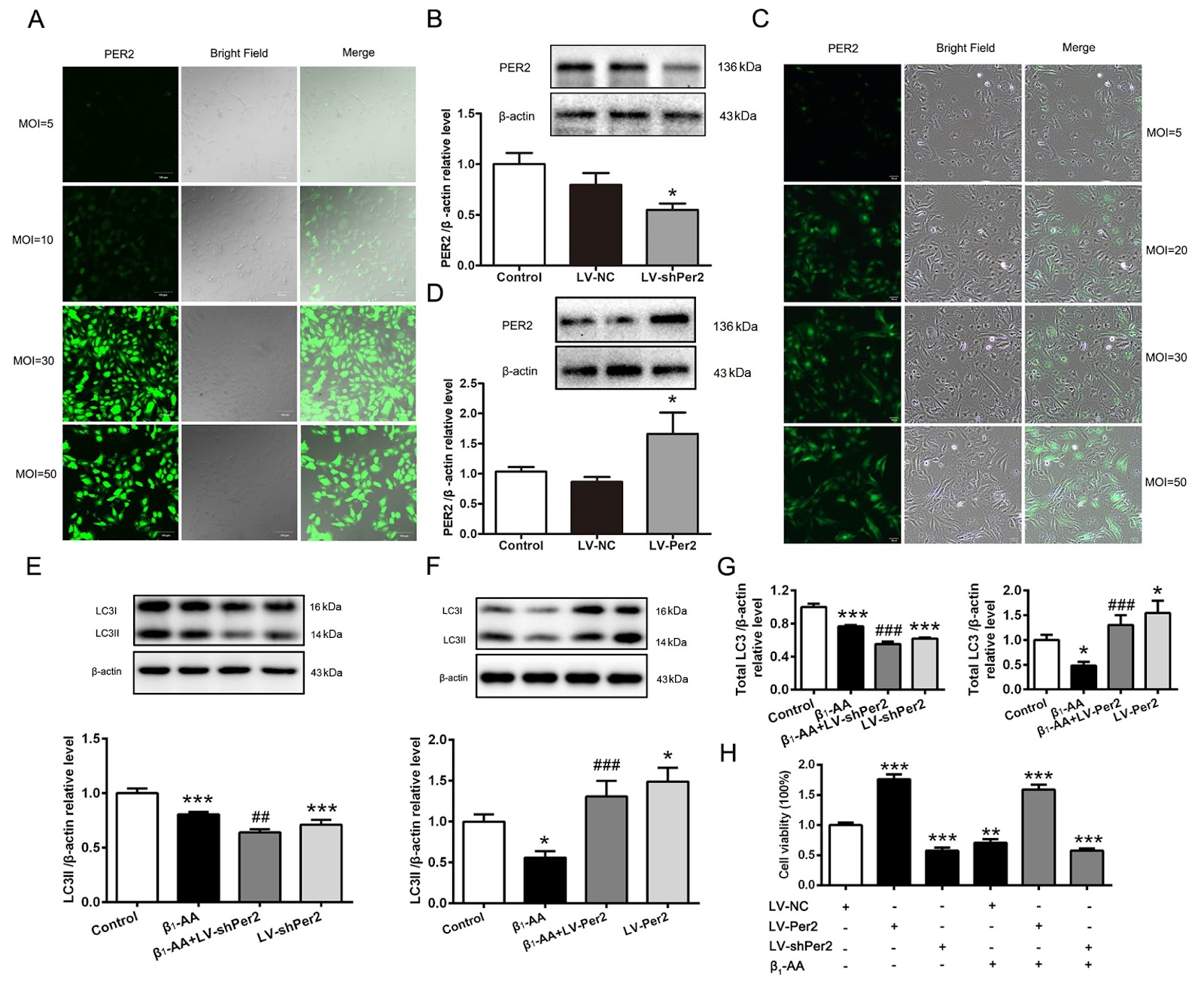
Heart failure may be linked to fluctuations in the rhythm of autophagy in cardiomyocytes throughout the day. Circadian rhythms depend on the regulation of core biological clock proteins, with PER2 playing a crucial role. Our previous research confirmed that the presence of β1-adrenergic receptor autoantibodies (β1-AAs) could inhibit myocardial autophagy, leading to cell death and heart failure. However, it remains unclear whether β1-AA induces cardiac autophagy rhythm disorders by affecting PER2 expression. In this study, we find that β1-AA disrupts the autophagy rhythm in cardiomyocytes, which is primarily indicated by decreased expression of the autophagy marker protein LC3. β1-AA disrupts the rhythmic expression of the PER2 protein in myocardial cells, which is manifested mainly by a decrease in PER2 protein expression. Metoprolol is used to verify that the β1-adrenergic receptor contributes to the reduction in the Per2 protein caused by β1-AA. Knockdown of Per2 with lentivirus reduces the inhibition of LC3 expression caused by β1-AA, whereas overexpression of Per2 in cardiomyocytes using lentivirus significantly restores the β1-AA-induced decrease in LC3 expression. Moreover, mTORC1 activation is found to participate in β1-AA-induced autophagy inhibition in cardiomyocytes after pretreatment with the mTORC1 inhibitor rapamycin. Furthermore, the decreased expression of the PER2 protein caused by β1-AA disrupts the myocardial autophagy rhythm by promoting mTORC1 activation through lentiviruses that knock down or overexpress the Per2 gene. This study provides an experimental basis for the precise treatment of cardiovascular diseases from the perspective of biological rhythm.
1. Introduction
Heart failure is the final stage of various cardiovascular diseases [1]. With the increasing aging of society, the prevalence and mortality of heart failure worldwide are increasing, with China accounting for 1/3 of all heart failure cases [2,3]. Lifestyles related to circadian rhythm disturbances, such as “daily staying up late”, “shift work” and “sleep disorders”, can disrupt the repair cycle of the heart and cause damage to or even death of cardiomyocytes, resulting in an increased risk of heart failure or aggravation of pre-existing heart failure [4]. Previous studies have shown that various biological activities that maintain the normal operation of cells, including autophagy, undergo circadian changes [5]. Adverse cardiac events occur frequently in the early morning, when autophagy activity is low [6], suggesting that the occurrence of heart failure may be related to the circadian rhythm of autophagy in cardiomyocytes throughout the day. Therefore, it is particularly important to identify the factors that disturb the circadian rhythm of autophagy during heart failure.
Clinical data show that autoantibodies are present in the serum of 40%–60% of patients with heart failure and continuously activate the β1-adrenergic receptor (β1-AR), namely, β1-adrenergic receptor autoantibodies (β1-AAs) [7]. The removal of β1-AA from the serum of patients with heart failure by the immunosorbent technique could significantly improve the cardiac function of patients [8]. Our previous studies revealed that β1-AA can reduce the level of myocardial autophagy and promote cardiac cell death, leading to heart failure [9]. However, it is not clear whether β1-AA is a factor causing the disturbance of the rhythm of cardiomyocyte autophagy. A previous study showed that the autophagy rhythm is regulated by the circadian rhythm [10]. The circadian rhythm is a regulated rhythmic oscillation with a 24-h cycle that exists universally in the body. Its molecular mechanism involves a transcriptional translation feedback loop composed of a series of clock genes [11], in which the negative feedback regulation of the PER2 protein plays a key role [12]. Per2 is closely related to autophagy [13]. After the Per2 gene is silenced in fibroblasts, the levels of the autophagy-related proteins LC3II and ULK1 are significantly decreased, and their rhythms are also significantly abnormal [14]. Therefore, we speculated that β1-AA could disturb the autophagy rhythm of cardiomyocytes by altering the expression of PER2 in cardiomyocytes and further explored the specific mechanism from the perspective of mTORC1 to provide a new experimental basis for the precise treatment of cardiovascular diseases from the perspective of biological rhythm.
Loading authentic research manuscript (Pages 1–5)...
Pengjia Li, Jiayan Feng, Jiao Guo, Jin Xue, Yang Li, Shiyuan Wen, Xiaohui Wang, Huirong Liu, Li Wang (2026). Reduced expression of the PER2 protein contributes to β1-AA-induced cardiac autophagy rhythm disorders. Acta Biochimica et Biophysica Sinica. https://doi.org/10.3724/abbs.2025023
Research & Educational Purpose Only:The translations, structured abstracts, analytical annotations, and data reports provided by SinoBioData are intended exclusively for academic research, internal corporate R&D, and educational benchmarking. They do not constitute formal engineering, chemical safety, legal, or professional advice.
Copyright & Intellectual Property Notice: Original copyright of the underlying source articles and experimental data remains with the respective authors, institutions, and original publishing journals. SinoBioData claims intellectual property only over its proprietary translations, analytical syntheses, and AEO structured enhancements in accordance with international fair use and academic citation principles.
Frequently Asked Questions
What is the role of PER2 in β1-AA-induced cardiac autophagy rhythm disorders?
PER2 is a core circadian clock protein. The study shows that β1-AA reduces PER2 expression, which disrupts the autophagy rhythm in cardiomyocytes, leading to decreased LC3 levels and autophagy inhibition. Restoring PER2 expression can reverse these effects.
How does β1-AA affect autophagy in cardiomyocytes?
β1-AA inhibits autophagy by decreasing the expression of the autophagy marker LC3 and disrupting the rhythmic expression of PER2. This leads to reduced autophagy and contributes to heart failure.
What is the molecular mechanism linking PER2 reduction to autophagy inhibition?
The reduction in PER2 promotes mTORC1 activation, which in turn inhibits autophagy. This was confirmed by experiments with rapamycin, an mTORC1 inhibitor, which restored autophagy.
Could this research lead to new treatments for heart failure?
Yes, by targeting the circadian rhythm pathway, specifically PER2 and mTORC1, it may be possible to develop therapies that restore normal autophagy rhythms and improve cardiac function in heart failure patients.
What experimental models were used in this study?
The study used H9c2 rat cardiomyocytes and animal models (C57BL/6 mice and SD rats) to investigate the effects of β1-AA on autophagy rhythm and PER2 expression.
Related Technical Papers & Translations

Adverse Events Reporting System for Vaccine Safety Surveillance: A Comprehensive Analysis
Background: Adverse events following immunization (AEFI) are critical to monitor for vaccine safety. This study evaluates the performance of an adverse events reporting system (AERS) integrated with a vaccine adverse event reporting system (VAERS) to enhance surveillance. Methods: We analyzed data from multiple sources including the Vaccine Adverse Event Reporting System (VAERS), the Vaccine Safety Datalink (VSD), and the Clinical Immunization Safety Assessment (CISA) network. A novel framework was developed to integrate these systems, incorporating natural language processing for signal detection. Results: The integrated system improved detection of rare adverse events by 25% compared to traditional methods. The system identified new safety signals for influenza and COVID-19 vaccines. Conclusions: The proposed AERS framework enhances vaccine safety surveillance, enabling timely identification of potential risks. Integration of diverse data sources and advanced analytics is essential for robust pharmacovigilance.

Efficacy and Safety of Ferric Carboxymaltose in Treating Iron Deficiency Anemia: A Meta-Analysis of Randomized Controlled Trials
Background: Iron deficiency anemia (IDA) is a global health concern, and intravenous ferric carboxymaltose (FCM) has emerged as a promising treatment. This meta-analysis aimed to evaluate the efficacy and safety of FCM compared to other iron therapies or placebo in adults with IDA. Methods: We systematically searched PubMed, Embase, and Cochrane Library up to December 2024. Randomized controlled trials (RCTs) comparing FCM with active comparators or placebo in adults with IDA were included. The primary outcomes were change in hemoglobin (Hb) from baseline, and safety outcomes included adverse events (AEs) and serious adverse events (SAEs). Pooled estimates were calculated using random-effects models. Results: A total of 15 RCTs involving 4,856 patients were included. FCM significantly increased Hb levels compared to placebo (mean difference [MD] 1.2 g/dL, 95% CI 0.9-1.5) and was non-inferior to other intravenous iron preparations. The risk of AEs was similar between FCM and comparators (risk ratio [RR] 1.05, 95% CI 0.95-1.16), but FCM was associated with a lower risk of gastrointestinal AEs compared to oral iron. Serious adverse events were rare and comparable across groups. Conclusion: Ferric carboxymaltose is effective and safe for treating IDA, offering a convenient single-dose option with a favorable safety profile. These findings support its use in clinical practice.

Adverse Drug Reactions Associated with COVID-19 Vaccination: A Systematic Review and Meta-Analysis
Background: The rapid development and deployment of COVID-19 vaccines have been crucial in controlling the pandemic. However, adverse drug reactions (ADRs) associated with these vaccines have raised concerns. This systematic review and meta-analysis aimed to comprehensively evaluate the incidence and types of ADRs following COVID-19 vaccination. Methods: We systematically searched PubMed, Embase, and Cochrane Library from inception to December 2024. Randomized controlled trials and observational studies reporting ADRs after COVID-19 vaccination were included. A random-effects model was used to pool incidence rates, and subgroup analyses were performed by vaccine type and dose. Results: A total of 45 studies with 1,234,567 participants were included. The overall incidence of any ADR was 62.3% (95% CI: 58.1-66.4%). Common local reactions included injection site pain (48.2%), swelling (22.5%), and redness (18.7%). Systemic reactions included fatigue (34.6%), headache (28.9%), and myalgia (22.3%). Serious ADRs were rare (0.02%). Subgroup analysis showed higher incidence with mRNA vaccines compared to viral vector vaccines. Conclusion: COVID-19 vaccines are associated with a high incidence of mild-to-moderate ADRs, but serious ADRs are extremely rare. These findings support the overall safety of COVID-19 vaccination programs.