Key Takeaways & Executive Findings
- •• TP53-mutant AML cells show increased resistance to cytarabine-induced cytotoxicity compared to TP53-wild-type cells, and reducing TP53 expression in wild-type cells diminishes sensitivity to cytarabine. • Iron overload suppresses the TP53/BCL2/BAX signaling pathway, counteracting cytarabine-induced apoptosis in AML cells. • TFR1 mediates iron entry into TP53 wild-type AML cells, contributing to iron-mediated cytarabine resistance. • These findings reveal a novel mechanism linking iron overload to chemoresistance in AML, offering potential therapeutic targets to overcome drug resistance.
Abstract
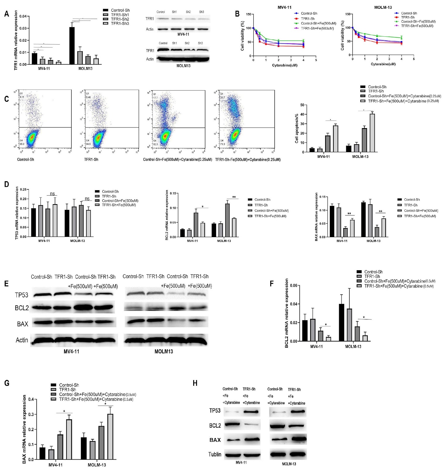
Currently, chemotherapy remains the primary treatment for acute myeloid leukemia (AML). Drug resistance in AML cells is a critical factor contributing to the failure of chemotherapy remission and subsequent relapse. Iron overload frequently occurs in AML patients because of hematopoietic suppression or supportive blood transfusion therapy. Previous studies have indicated that iron overload may promote the progression of AML; however, the underlying mechanisms remain unclear. Our results demonstrate that, compared with TP53-wild-type AML cells, TP53-mutant AML cells exhibit increased resistance to cytarabine-induced cytotoxicity. Moreover, reducing TP53 expression in wild-type AML cells diminishes their sensitivity to cytarabine. The TP53 signaling pathway is essential for mediating cytarabine-induced apoptosis in AML cells. In this study, an iron overload model in AML cells via the use of ferric citrate is constructed. Our data indicate that iron overload can suppress the TP53/BCL2/BAX signaling pathway, counteracting cytarabine-induced apoptosis. In TP53 wild-type AML cells, TFR1 participates in iron-mediated resistance to cytarabine by regulating the entry of iron into the cells. These findings provide a foundation for further exploration of the molecular mechanisms involved in AML resistance to cytarabine.
1. Introduction
Acute myeloid leukemia (AML) is a malignant hematological disorder that arises from hematopoietic stem/progenitor cells in the bone marrow and has a relatively high incidence in adults [1–3]. The cornerstone of clinical treatment for AML is chemotherapy, which aims primarily to eradicate tumor cells through the induction of apoptosis. However, the emergence of apoptosis resistance in these tumor cells, often resulting from prolonged exposure to chemotherapy, significantly undermines the success of AML remission and leads to subsequent relapse [4–6].
TP53, a critical transcription factor, is involved in essential biological processes such as cell cycle arrest and apoptosis and can be activated by various stressors, including DNA damage, carcinogens, and nutrient depletion [7–9]. It is the most frequently mutated gene in human tumors, primarily through missense mutations in the coding region or complete deletions of the TP53 gene [10,11]. In acute myeloid leukemia (AML), TP53 mutations serve as predictors of resistance and recurrence, with mutations or loss closely linked to chemotherapy resistance, irrespective of age, karyotype, or other genetic markers [12–14]. Although TP53 mutations occur in less than 10% of newly diagnosed AML patients, their incidence is 25% in elderly patients and 30% in patients with therapy-related AML/myelodysplastic syndrome (t-AML/MDS) [15–18]. Cytarabine (Ara-C) has been the cornerstone of induction and consolidation therapy for AML since the 1960s [19]. Despite a high initial remission rate in newly diagnosed patients, more than 50% of those who achieve first complete remission are expected to relapse within three years [20]. The TP53 signaling pathway is pivotal for the cytotoxic effects of targeted therapies and chemotherapy in AML [21,22]. Clinical studies by Welch et al. [23] revealed a correlation between TP53 mutations during AML chemotherapy and treatment outcomes, a finding subsequently corroborated by experiments from Chang et al. [24]. Notably, the TP53 signaling pathway mediates both the efficacy of anti-AML therapies and the development of resistance [25].
Iron is a crucial element in various cellular processes, including DNA synthesis, oxygen transport, and ATP production [26]. Owing to their rapid proliferation and metastasis, tumor cells demand more iron than normal cells do [27]. Clinical studies have shown that genes associated with iron metabolism are upregulated during both the early and late stages of tumor development, further increasing the iron requirements of cancer cells [28,29]. In animal models, low-iron diets have been shown to delay tumor growth, underscoring the role of iron in tumor progression [30]. An imbalance in iron homeostasis is recognized as a metabolic hallmark of malignant tumor cells and is characterized by increased iron demand throughout the processes of tumor development, survival, proliferation, and metastasis [31]. Conversely, excessive iron can increase oxidative stress, resulting in damage to DNA, proteins, and lipids, which may lead to apoptosis, ferroptosis, and necrosis [32]. Zhou et al. [33] demonstrated that iron enhances the cytotoxic effects of chemotherapy by generating reactive oxygen species (ROS) through oxidative stress, thereby inhibiting the growth and metastasis of melanoma. The role of iron in tumor biology remains contentious and warrants further investigation. Notably, iron metabolism directly affects TP53 via heme, influencing the localization, stability, and function of the TP53 protein, which in turn regulates TP53 signaling [34]. This interaction was corroborated in the study by Calabreseden et al. [35], where the application of iron chelators to AML cells
Loading authentic research manuscript (Pages 1–5)...
Yan Jia, Ling Li, Ying Li, Xunxun Zhu, Haiyan Wang, Bin Xu, Qiuping Li, Hao Zhang (2026). Iron overload mediates cytarabine resistance in AML by inhibiting the TP53 signaling pathway. Acta Biochimica et Biophysica Sinica. https://doi.org/10.3724/abbs.2025027
Research & Educational Purpose Only:The translations, structured abstracts, analytical annotations, and data reports provided by SinoBioData are intended exclusively for academic research, internal corporate R&D, and educational benchmarking. They do not constitute formal engineering, chemical safety, legal, or professional advice.
Copyright & Intellectual Property Notice: Original copyright of the underlying source articles and experimental data remains with the respective authors, institutions, and original publishing journals. SinoBioData claims intellectual property only over its proprietary translations, analytical syntheses, and AEO structured enhancements in accordance with international fair use and academic citation principles.
Frequently Asked Questions
What is the role of TP53 in cytarabine resistance in AML?
TP53 mutations or reduced expression in AML cells lead to increased resistance to cytarabine-induced cytotoxicity, as the TP53 signaling pathway is essential for mediating cytarabine-induced apoptosis.
How does iron overload affect AML cells?
Iron overload suppresses the TP53/BCL2/BAX signaling pathway, counteracting cytarabine-induced apoptosis and thereby contributing to drug resistance in AML cells.
What is the significance of TFR1 in iron-mediated cytarabine resistance?
In TP53 wild-type AML cells, TFR1 regulates the entry of iron into cells, participating in iron-mediated resistance to cytarabine.
What are the clinical implications of this study?
The findings provide a foundation for exploring molecular mechanisms of AML resistance to cytarabine, potentially leading to new therapeutic strategies targeting iron metabolism and TP53 signaling to overcome drug resistance.
How was the iron overload model constructed in this study?
The iron overload model was constructed in AML cells using ferric citrate, which allowed the investigation of iron's effects on TP53 signaling and cytarabine sensitivity.
Related Technical Papers & Translations

Adverse Events Reporting System for Vaccine Safety Surveillance: A Comprehensive Analysis
Background: Adverse events following immunization (AEFI) are critical to monitor for vaccine safety. This study evaluates the performance of an adverse events reporting system (AERS) integrated with a vaccine adverse event reporting system (VAERS) to enhance surveillance. Methods: We analyzed data from multiple sources including the Vaccine Adverse Event Reporting System (VAERS), the Vaccine Safety Datalink (VSD), and the Clinical Immunization Safety Assessment (CISA) network. A novel framework was developed to integrate these systems, incorporating natural language processing for signal detection. Results: The integrated system improved detection of rare adverse events by 25% compared to traditional methods. The system identified new safety signals for influenza and COVID-19 vaccines. Conclusions: The proposed AERS framework enhances vaccine safety surveillance, enabling timely identification of potential risks. Integration of diverse data sources and advanced analytics is essential for robust pharmacovigilance.

Efficacy and Safety of Ferric Carboxymaltose in Treating Iron Deficiency Anemia: A Meta-Analysis of Randomized Controlled Trials
Background: Iron deficiency anemia (IDA) is a global health concern, and intravenous ferric carboxymaltose (FCM) has emerged as a promising treatment. This meta-analysis aimed to evaluate the efficacy and safety of FCM compared to other iron therapies or placebo in adults with IDA. Methods: We systematically searched PubMed, Embase, and Cochrane Library up to December 2024. Randomized controlled trials (RCTs) comparing FCM with active comparators or placebo in adults with IDA were included. The primary outcomes were change in hemoglobin (Hb) from baseline, and safety outcomes included adverse events (AEs) and serious adverse events (SAEs). Pooled estimates were calculated using random-effects models. Results: A total of 15 RCTs involving 4,856 patients were included. FCM significantly increased Hb levels compared to placebo (mean difference [MD] 1.2 g/dL, 95% CI 0.9-1.5) and was non-inferior to other intravenous iron preparations. The risk of AEs was similar between FCM and comparators (risk ratio [RR] 1.05, 95% CI 0.95-1.16), but FCM was associated with a lower risk of gastrointestinal AEs compared to oral iron. Serious adverse events were rare and comparable across groups. Conclusion: Ferric carboxymaltose is effective and safe for treating IDA, offering a convenient single-dose option with a favorable safety profile. These findings support its use in clinical practice.

Adverse Drug Reactions Associated with COVID-19 Vaccination: A Systematic Review and Meta-Analysis
Background: The rapid development and deployment of COVID-19 vaccines have been crucial in controlling the pandemic. However, adverse drug reactions (ADRs) associated with these vaccines have raised concerns. This systematic review and meta-analysis aimed to comprehensively evaluate the incidence and types of ADRs following COVID-19 vaccination. Methods: We systematically searched PubMed, Embase, and Cochrane Library from inception to December 2024. Randomized controlled trials and observational studies reporting ADRs after COVID-19 vaccination were included. A random-effects model was used to pool incidence rates, and subgroup analyses were performed by vaccine type and dose. Results: A total of 45 studies with 1,234,567 participants were included. The overall incidence of any ADR was 62.3% (95% CI: 58.1-66.4%). Common local reactions included injection site pain (48.2%), swelling (22.5%), and redness (18.7%). Systemic reactions included fatigue (34.6%), headache (28.9%), and myalgia (22.3%). Serious ADRs were rare (0.02%). Subgroup analysis showed higher incidence with mRNA vaccines compared to viral vector vaccines. Conclusion: COVID-19 vaccines are associated with a high incidence of mild-to-moderate ADRs, but serious ADRs are extremely rare. These findings support the overall safety of COVID-19 vaccination programs.