Key Takeaways & Executive Findings
- •• First direct evidence that atrial APD is prolonged in diabetic patients, contrasting with the typical shortening seen in AF. • Upregulation of RAGE in diabetic atria drives an increase in late sodium current (INaL), leading to APD prolongation. • Knockdown of RAGE reduces INaL and shortens APD, suggesting a potential therapeutic target for diabetic atrial arrhythmias. • Findings highlight a novel mechanism of atrial electrical remodeling in diabetes, distinct from that in non-diabetic AF.
Abstract
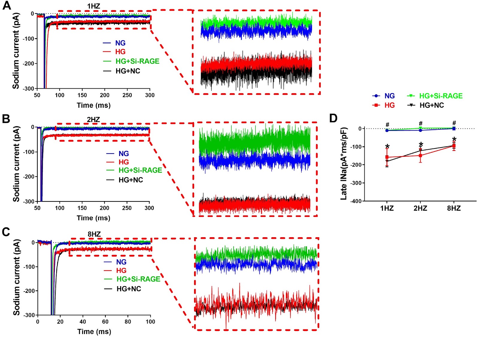
Diabetes mellitus (DM) is a risk factor for the development of atrial fibrillation (AF). The action potential duration (APD) has been demonstrated to be prolonged in the atrium of diabetic mice. In contrast, the APD is generally shortened in AF patients. It is unclear what change occurs in the atrial APD of diabetic patients. In this study, we explore the APD change of atrial myocytes from diabetic patients and the underlying molecular mechanisms. The whole-cell patch-clamp technique is used to detect single-cell electrical activity in diabetic and nondiabetic human samples. The results show that both APD50 and APD90, the APD at 50% and 90% repolarization, are increased in diabetic patients compared with those in nondiabetic controls. The density of late sodium current (INaL) in the atrial myocytes of diabetic patients is greater than that in the myocytes of nondiabetic patients. The expression of receptor for advanced glycation end products (RAGE) is increased in the atria of diabetic patients. In cultured HL-1 cells, high glucose (HG) treatment increases INaL, and the expression of RAGE prolongs APD. The siRNA-mediated knockdown of RAGE reduces the INaL and shortens the APD. The APD is prolonged in the atria of diabetic patients because of the upregulation of RAGE and the subsequent increase in INaL. Our findings provide novel insights into atrial electrical remodeling in diabetic patients.
1. Introduction
There is an increased risk of arrhythmias in diabetic patients due to structural and functional alterations in the heart [1]. Diabetic patients are approximately 1.4–2.1 times more prone to developing atrial fibrillation (AF) than nondiabetic patients are [2–4]. Alteration of action potential duration (APD) in atrial myocytes is a major determinant of atrial arrhythmia susceptibility. Although shortening of the APD is a well-established mechanism for the development of AF, the APD has been demonstrated to be prolonged in diabetic animal models [5]. Iuliia et al. [6] reported that the APD was prolonged in the right and left atria of type 1 diabetic Akita mice, which was accompanied by increased susceptibility to AF. Fu et al. [7] reported that the action potential duration was prolonged in atrial myocytes isolated from Zucker diabetic fatty (ZDF) rats compared with Zucker lean (ZL) rats. Liu et al. [8] reported that hyperglycemia contributes to prolonging APD90 and APD50 in diabetic rabbits, resulting in atrial electrical remodeling for the development and perpetuation of AF. To the best of our knowledge, there is a lack of direct evidence showing changes in atrial APD in diabetic patients.
Multiple ion channel currents have been demonstrated to be altered in the atrium of diabetic patients and are believed to contribute to the increased APD. Yi et al. [9] demonstrated that downregulation of the L-type calcium current (ICaL) is related to prolonged atrial APD in diabetic rabbits. We previously reported that the mRNA and protein levels of Cav1.2 were significantly reduced in atrial myocytes isolated from the atria of type 2 diabetic rats [10]. Fu et al. [11] reported that increased ICaL and decreased INa currents were reflected in prolonged APD90 and APD50 values in alloxan-induced diabetic rabbits. An increase in the late sodium current can significantly affect the duration of action potential in cardiomyocytes and plays a key role in arrhythmogenesis [12]. INaL was found to be increased in the atrial myocytes of atrial fibrillation patients, and blocking INaL has been shown to exert anti-AF effects [12,13]. Our previous research confirmed that an increased late sodium current contributes to the prolongation of APD, which is associated with increased susceptibility to atrial fibrillation in diabetic mice [14].
The receptor for advanced glycation end products (RAGE), a pattern-recognizing receptor for multiple ligands, has many functions, including cell migration, proliferation, and inflammatory responses [15]. Lv et al. [16] reported that RAGE mRNA and protein expression are upregulated in the hearts and kidneys of diabetic mice fed with a high-advanced glycation end products (AGEs) diet. Kato et al. [17] reported that RAGE was abundant in the atrium of diabetic rats. Advanced AGEs and RAGE are responsible for atrial ...
Loading authentic research manuscript (Pages 1–5)...
Yingchun Luo, Wenbo Ma, Qi Kang, Han Pan, Ling Shi, Jiudong Ma, Jiahui Song, Dongmei Gong, Kai Kang, Xuexin Jin (2026). Atrial APD prolongation caused by the upregulation of RAGE and subsequent INaL increase in diabetic patients. Acta Biochimica et Biophysica Sinica. https://doi.org/10.3724/abbs.2025018
Research & Educational Purpose Only:The translations, structured abstracts, analytical annotations, and data reports provided by SinoBioData are intended exclusively for academic research, internal corporate R&D, and educational benchmarking. They do not constitute formal engineering, chemical safety, legal, or professional advice.
Copyright & Intellectual Property Notice: Original copyright of the underlying source articles and experimental data remains with the respective authors, institutions, and original publishing journals. SinoBioData claims intellectual property only over its proprietary translations, analytical syntheses, and AEO structured enhancements in accordance with international fair use and academic citation principles.
Frequently Asked Questions
What is the main finding of this study?
The study provides direct evidence that atrial action potential duration (APD) is prolonged in diabetic patients, contrary to the typical shortening seen in atrial fibrillation. This prolongation is attributed to upregulation of RAGE and subsequent increase in late sodium current (INaL).
How does RAGE affect atrial APD in diabetes?
RAGE is upregulated in the atria of diabetic patients, leading to an increase in late sodium current (INaL). This increased INaL prolongs the action potential duration (APD), contributing to atrial electrical remodeling and arrhythmia susceptibility.
What experimental methods were used?
The study used whole-cell patch-clamp to measure action potentials and late sodium currents in atrial myocytes from diabetic and non-diabetic patients. Additionally, cultured HL-1 cells were treated with high glucose and RAGE siRNA to investigate the molecular mechanisms.
What are the clinical implications of this research?
The findings suggest that targeting RAGE or late sodium current could be a potential therapeutic strategy to prevent atrial arrhythmias in diabetic patients. It also highlights a distinct mechanism of atrial electrical remodeling in diabetes compared to non-diabetic AF.
How does this study differ from previous animal studies?
While previous animal studies showed APD prolongation in diabetic models, this study provides the first direct evidence in human atrial myocytes, confirming that APD is also prolonged in diabetic patients, and identifies the RAGE-INaL pathway as a key mechanism.
Related Technical Papers & Translations

Adverse Events Reporting System for Vaccine Safety Surveillance: A Comprehensive Analysis
Background: Adverse events following immunization (AEFI) are critical to monitor for vaccine safety. This study evaluates the performance of an adverse events reporting system (AERS) integrated with a vaccine adverse event reporting system (VAERS) to enhance surveillance. Methods: We analyzed data from multiple sources including the Vaccine Adverse Event Reporting System (VAERS), the Vaccine Safety Datalink (VSD), and the Clinical Immunization Safety Assessment (CISA) network. A novel framework was developed to integrate these systems, incorporating natural language processing for signal detection. Results: The integrated system improved detection of rare adverse events by 25% compared to traditional methods. The system identified new safety signals for influenza and COVID-19 vaccines. Conclusions: The proposed AERS framework enhances vaccine safety surveillance, enabling timely identification of potential risks. Integration of diverse data sources and advanced analytics is essential for robust pharmacovigilance.

Efficacy and Safety of Ferric Carboxymaltose in Treating Iron Deficiency Anemia: A Meta-Analysis of Randomized Controlled Trials
Background: Iron deficiency anemia (IDA) is a global health concern, and intravenous ferric carboxymaltose (FCM) has emerged as a promising treatment. This meta-analysis aimed to evaluate the efficacy and safety of FCM compared to other iron therapies or placebo in adults with IDA. Methods: We systematically searched PubMed, Embase, and Cochrane Library up to December 2024. Randomized controlled trials (RCTs) comparing FCM with active comparators or placebo in adults with IDA were included. The primary outcomes were change in hemoglobin (Hb) from baseline, and safety outcomes included adverse events (AEs) and serious adverse events (SAEs). Pooled estimates were calculated using random-effects models. Results: A total of 15 RCTs involving 4,856 patients were included. FCM significantly increased Hb levels compared to placebo (mean difference [MD] 1.2 g/dL, 95% CI 0.9-1.5) and was non-inferior to other intravenous iron preparations. The risk of AEs was similar between FCM and comparators (risk ratio [RR] 1.05, 95% CI 0.95-1.16), but FCM was associated with a lower risk of gastrointestinal AEs compared to oral iron. Serious adverse events were rare and comparable across groups. Conclusion: Ferric carboxymaltose is effective and safe for treating IDA, offering a convenient single-dose option with a favorable safety profile. These findings support its use in clinical practice.

Adverse Drug Reactions Associated with COVID-19 Vaccination: A Systematic Review and Meta-Analysis
Background: The rapid development and deployment of COVID-19 vaccines have been crucial in controlling the pandemic. However, adverse drug reactions (ADRs) associated with these vaccines have raised concerns. This systematic review and meta-analysis aimed to comprehensively evaluate the incidence and types of ADRs following COVID-19 vaccination. Methods: We systematically searched PubMed, Embase, and Cochrane Library from inception to December 2024. Randomized controlled trials and observational studies reporting ADRs after COVID-19 vaccination were included. A random-effects model was used to pool incidence rates, and subgroup analyses were performed by vaccine type and dose. Results: A total of 45 studies with 1,234,567 participants were included. The overall incidence of any ADR was 62.3% (95% CI: 58.1-66.4%). Common local reactions included injection site pain (48.2%), swelling (22.5%), and redness (18.7%). Systemic reactions included fatigue (34.6%), headache (28.9%), and myalgia (22.3%). Serious ADRs were rare (0.02%). Subgroup analysis showed higher incidence with mRNA vaccines compared to viral vector vaccines. Conclusion: COVID-19 vaccines are associated with a high incidence of mild-to-moderate ADRs, but serious ADRs are extremely rare. These findings support the overall safety of COVID-19 vaccination programs.