Key Takeaways & Executive Findings
- •• Sulfasalazine induces ferroptosis in H9C2 cardiomyocytes and BRL-3A hepatocytes via a sequential buildup of NO, ROS, and lipid-ROS. • PDI mediates SAS-induced iNOS dimerization and NO accumulation, which are upstream of oxidative damage. • Inhibition of PDI activity or knockdown of PDI suppresses SAS-induced ferroptosis, while PDI activation sensitizes cells. • The PDI-NOS-NO axis is a critical pathway for SAS cytotoxicity, offering potential therapeutic targets for modulating ferroptosis.
Abstract
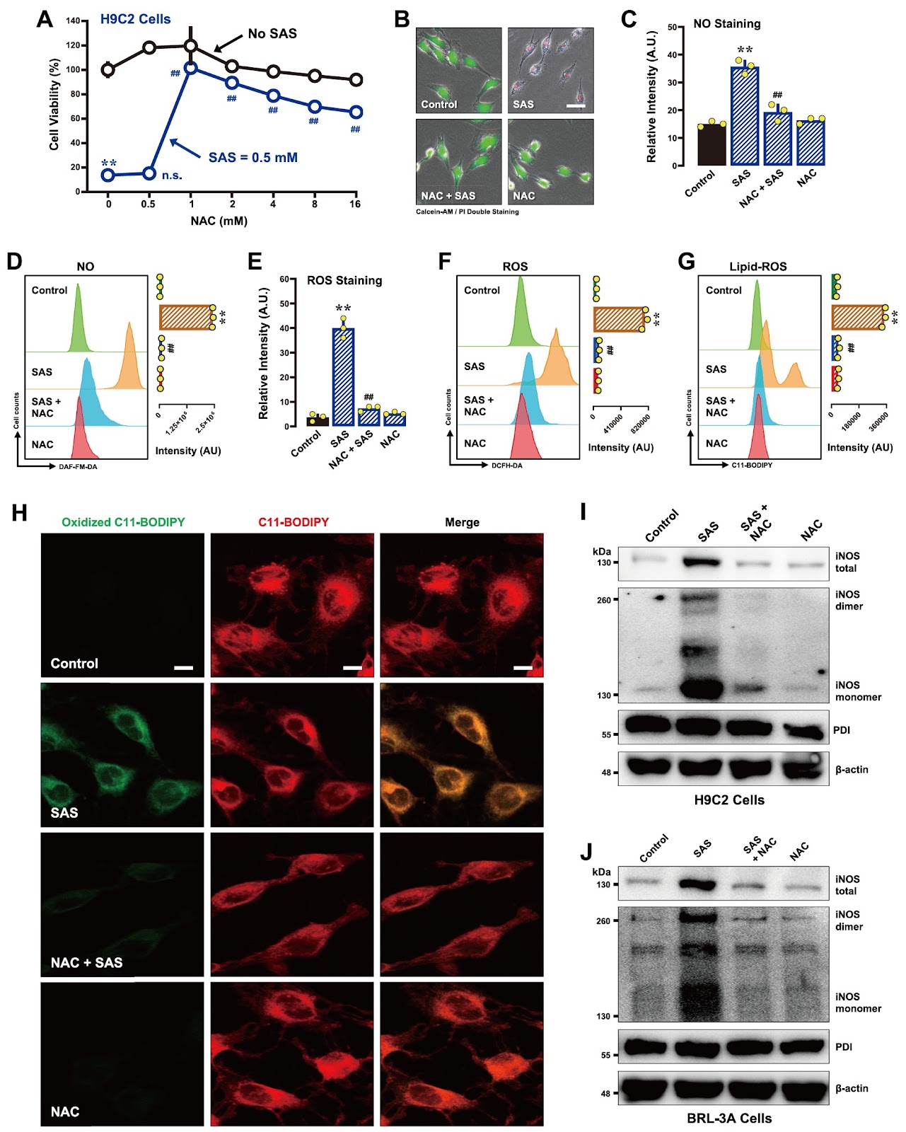
Sulfasalazine (SAS), a clinically utilized anti-inflammatory drug, has been shown to induce ferroptosis by inhibiting system Xc− activity, thereby causing cellular glutathione depletion. Recently, protein disulfide isomerase (PDI) was shown to be an upstream mediator of the oxidative cell death (oxytosis/ferroptosis) induced by glutamate, erastin, RSL3 and SAS. The present study aims to further characterize the detailed biochemical and cellular mechanisms of SAS-induced ferroptosis in two cell lines, i.e., H9C2 rat cardiomyocytes and BRL-3A rat hepatocytes, with a focus on elucidating the critical role of PDI in mediating SAS-induced toxicity. We find that SAS can induce ferroptosis in H9C2 and BRL-3A cells, which is accompanied by a sequential increase in the buildup of cellular nitric oxide (NO), reactive oxygen species (ROS) and lipid-ROS. SAS activates PDI-mediated dimerization of inducible NO synthase (iNOS) and cellular accumulation of NO, and these effects are followed by ROS and lipid-ROS accumulation. Furthermore, SAS markedly upregulates the iNOS protein levels in these cells. Knockdown of PDI or pharmacological inhibition of PDI catalytic activity effectively suppresses SAS-induced iNOS dimerization, abrogates SAS-induced accumulation of NO, ROS and lipid-ROS, and prevents ferroptosis. On the other hand, PDI activation through the use of TrxR1 inhibitors sensitizes these cells to SAS-induced ferroptosis. These findings provide further experimental support for a pivotal role of PDI in SAS-induced cytotoxicity through the activation of the PDI-NOS-NO axis, which then leads to the accumulation of cellular ROS and lipid-ROS and ultimately the induction of oxidative cell death.
1. Introduction
Sulfasalazine (SAS), a well-established drug for treating inflammatory disorders such as Crohn’s disease and ulcerative colitis [1–3], has recently been identified as a potent inducer of ferroptosis [4]. SAS inhibits the cystine/glutamate antiporter system Xc− [5,6], which is essential for cystine uptake and intracellular glutathione (GSH) synthesis [7]. By depleting GSH, SAS disrupts the cellular redox balance and triggers oxidative stress, culminating in ferroptotic cell death. Studies have shown that SAS can induce ferroptosis in several cell lines [8–15], and SAS can increase the efficacy of chemotherapeutic agents by increasing oxidative damage [5].
Ferroptosis is a form of regulated cell death often associated with the accumulation of reactive oxygen species (ROS) [16]. Unlike apoptosis or necrosis, ferroptosis has notable morphological and biochemical features, such as reduced mitochondrial size, increased membrane density, and GSH deficiency-associated oxidative damage to cellular lipids [17]. Central to this process is the depletion of cellular GSH and inactivation of glutathione peroxidase 4 (GPX4), and these changes disrupt a cell’s ability to neutralize reactive lipid peroxides (lipid-ROS), subsequently leading to cell death [18].
Protein disulfide isomerase (PDI or PDIA1) is a well-known member of the thioredoxin superfamily and is primarily localized in the endoplasmic reticulum [19]. Functionally, PDI serves as a dithiol/disulfide oxidoreductase and facilitates protein folding by catalyzing the isomerization of the intra- and intermolecular disulfide bonds [20]. Recent studies from our laboratory have shown that PDI is involved in mediating chemically-induced, glutathione (GSH) depletion-associated ferroptosis [21–23]. Specifically, GSH depletion leads to PDI oxidation and activates its catalytic activity for nitric oxide synthase (NOS) dimerization via disulfide bond formation, which subsequently results in the buildup of cellular nitric oxide (NO) and ROS/lipid-ROS and mitochondrial ROS and ultimately ferroptotic cell death [21–23]. Notably, the inhibition of PDI function has been shown to block NOS dimerization and NO accumulation and thus effectively rescues cells from chemically induced ferroptosis [21–23]. These findings position PDI as a pivotal upstream mediator of ferroptosis, linking GSH depletion to oxidative cell death pathways.
Loading authentic research manuscript (Pages 1–5)...
Yi-Chen Jia, Jia-Ling Zhong, Xiangyu Hao, Bao Ting Zhu (2026). Characterization of the mechanisms underlying sulfasalazine-induced ferroptotic cell death: role of protein disulfide isomerase-mediated NOS activation and NO accumulation. Acta Biochimica et Biophysica Sinica. https://doi.org/10.3724/abbs.2025100
Research & Educational Purpose Only:The translations, structured abstracts, analytical annotations, and data reports provided by SinoBioData are intended exclusively for academic research, internal corporate R&D, and educational benchmarking. They do not constitute formal engineering, chemical safety, legal, or professional advice.
Copyright & Intellectual Property Notice: Original copyright of the underlying source articles and experimental data remains with the respective authors, institutions, and original publishing journals. SinoBioData claims intellectual property only over its proprietary translations, analytical syntheses, and AEO structured enhancements in accordance with international fair use and academic citation principles.
Frequently Asked Questions
What is the main finding of this study?
The study reveals that sulfasalazine induces ferroptosis in H9C2 and BRL-3A cells through a PDI-mediated activation of NOS, leading to NO accumulation and subsequent ROS/lipid-ROS buildup, ultimately causing oxidative cell death.
How does sulfasalazine trigger ferroptosis?
Sulfasalazine inhibits system Xc−, depleting glutathione, which activates PDI. PDI then promotes iNOS dimerization, increasing NO production, which leads to ROS and lipid-ROS accumulation and ferroptosis.
What role does protein disulfide isomerase (PDI) play?
PDI acts as an upstream mediator: upon GSH depletion, it becomes oxidized and catalyzes NOS dimerization, which is essential for NO accumulation and subsequent oxidative damage.
Can inhibition of PDI prevent sulfasalazine-induced ferroptosis?
Yes, knockdown of PDI or pharmacological inhibition of its catalytic activity suppresses iNOS dimerization, NO accumulation, and ROS/lipid-ROS production, thereby preventing ferroptosis.
What are the potential therapeutic implications?
Targeting the PDI-NOS-NO axis could modulate ferroptosis, offering new strategies for diseases where ferroptosis is involved, such as cancer or ischemia-reperfusion injury.
Related Technical Papers & Translations

Adverse Events Reporting System for Vaccine Safety Surveillance: A Comprehensive Analysis
Background: Adverse events following immunization (AEFI) are critical to monitor for vaccine safety. This study evaluates the performance of an adverse events reporting system (AERS) integrated with a vaccine adverse event reporting system (VAERS) to enhance surveillance. Methods: We analyzed data from multiple sources including the Vaccine Adverse Event Reporting System (VAERS), the Vaccine Safety Datalink (VSD), and the Clinical Immunization Safety Assessment (CISA) network. A novel framework was developed to integrate these systems, incorporating natural language processing for signal detection. Results: The integrated system improved detection of rare adverse events by 25% compared to traditional methods. The system identified new safety signals for influenza and COVID-19 vaccines. Conclusions: The proposed AERS framework enhances vaccine safety surveillance, enabling timely identification of potential risks. Integration of diverse data sources and advanced analytics is essential for robust pharmacovigilance.

Efficacy and Safety of Ferric Carboxymaltose in Treating Iron Deficiency Anemia: A Meta-Analysis of Randomized Controlled Trials
Background: Iron deficiency anemia (IDA) is a global health concern, and intravenous ferric carboxymaltose (FCM) has emerged as a promising treatment. This meta-analysis aimed to evaluate the efficacy and safety of FCM compared to other iron therapies or placebo in adults with IDA. Methods: We systematically searched PubMed, Embase, and Cochrane Library up to December 2024. Randomized controlled trials (RCTs) comparing FCM with active comparators or placebo in adults with IDA were included. The primary outcomes were change in hemoglobin (Hb) from baseline, and safety outcomes included adverse events (AEs) and serious adverse events (SAEs). Pooled estimates were calculated using random-effects models. Results: A total of 15 RCTs involving 4,856 patients were included. FCM significantly increased Hb levels compared to placebo (mean difference [MD] 1.2 g/dL, 95% CI 0.9-1.5) and was non-inferior to other intravenous iron preparations. The risk of AEs was similar between FCM and comparators (risk ratio [RR] 1.05, 95% CI 0.95-1.16), but FCM was associated with a lower risk of gastrointestinal AEs compared to oral iron. Serious adverse events were rare and comparable across groups. Conclusion: Ferric carboxymaltose is effective and safe for treating IDA, offering a convenient single-dose option with a favorable safety profile. These findings support its use in clinical practice.

Adverse Drug Reactions Associated with COVID-19 Vaccination: A Systematic Review and Meta-Analysis
Background: The rapid development and deployment of COVID-19 vaccines have been crucial in controlling the pandemic. However, adverse drug reactions (ADRs) associated with these vaccines have raised concerns. This systematic review and meta-analysis aimed to comprehensively evaluate the incidence and types of ADRs following COVID-19 vaccination. Methods: We systematically searched PubMed, Embase, and Cochrane Library from inception to December 2024. Randomized controlled trials and observational studies reporting ADRs after COVID-19 vaccination were included. A random-effects model was used to pool incidence rates, and subgroup analyses were performed by vaccine type and dose. Results: A total of 45 studies with 1,234,567 participants were included. The overall incidence of any ADR was 62.3% (95% CI: 58.1-66.4%). Common local reactions included injection site pain (48.2%), swelling (22.5%), and redness (18.7%). Systemic reactions included fatigue (34.6%), headache (28.9%), and myalgia (22.3%). Serious ADRs were rare (0.02%). Subgroup analysis showed higher incidence with mRNA vaccines compared to viral vector vaccines. Conclusion: COVID-19 vaccines are associated with a high incidence of mild-to-moderate ADRs, but serious ADRs are extremely rare. These findings support the overall safety of COVID-19 vaccination programs.