Key Takeaways & Executive Findings
- •• Resident CD24+LCN2+ LPCs are significantly enriched in NASH patients and contribute to fibrosis progression. • LPCs interact with proinflammatory macrophage subtypes, particularly MP-2, which shows NF-κB pathway hyperactivation. • The study identifies COL10A1 and TPPP3 as novel markers of the proinflammatory macrophage subtype associated with fibrosis. • These findings provide potential therapeutic targets for NASH by modulating LPC-macrophage crosstalk.
Abstract
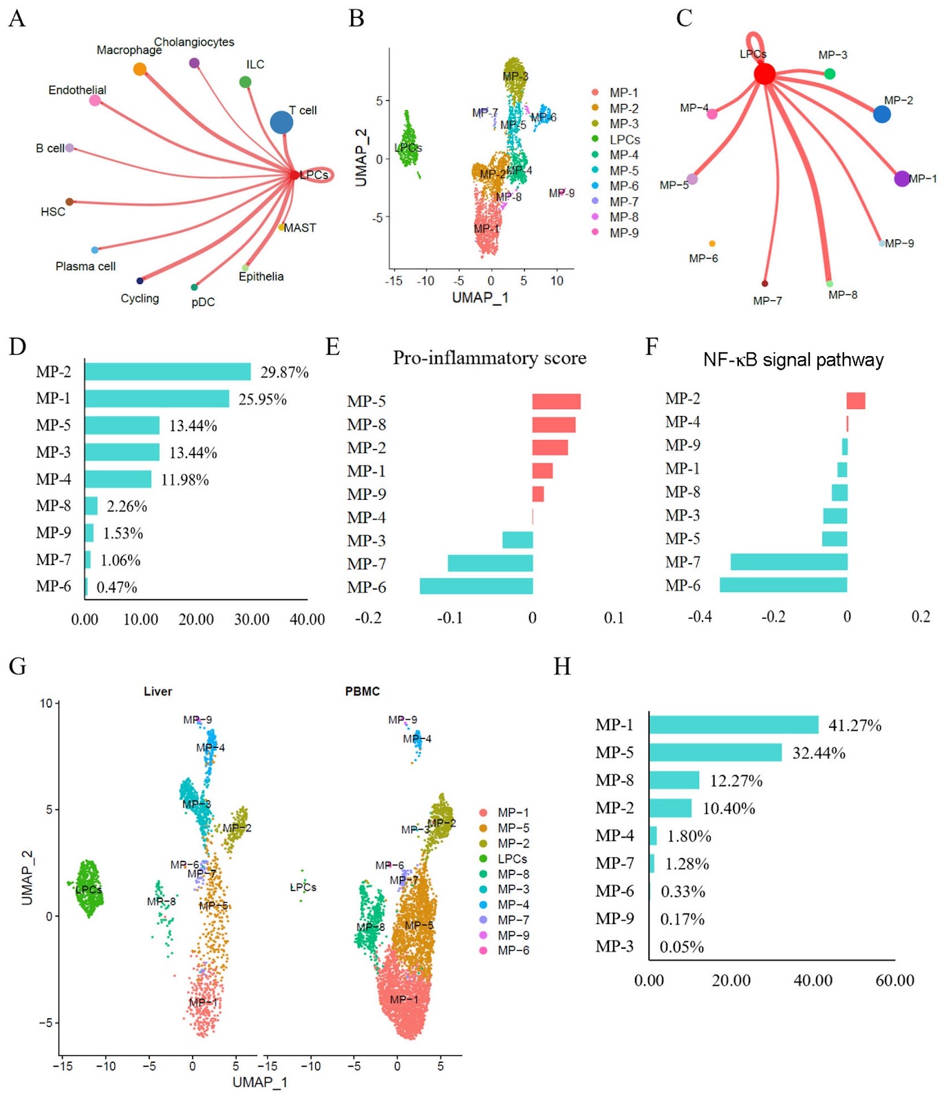
Resident CD24+LCN2+ liver progenitor cells (LPCs) reportedly contribute to the expanding ductular reaction and macrophage-mediated inflammation associated with chronic liver damage. Both ductular reactions and macrophage-driven inflammation are associated with liver fibrosis and injury in various mouse liver disorders. This study aims to investigate the molecular phenotypes of LPCs and their regulatory mechanisms in humans with non-alcoholic steatohepatitis (NASH). Single-cell RNA sequencing (scRNA-seq) datasets are used to characterize the status and molecular phenotypes of LPCs in clinical NASH samples. To elucidate the regulatory mechanisms of LPCs, CellChat and NicheNet are employed to assess cell-cell communication between LPCs and other cell types. The findings are validated using RNA sequencing datasets associated with NASH progression, NASH mouse models (CDAHFD and HFD), and human NASH liver samples. Results show that resident CD24+LCN2+ LPCs are identified and found to be significantly enriched in NASH patients. Cell communication analyses predict strong interactions between LPCs and proinflammatory macrophage subtypes. Additionally, in NASH, the liver recruits peripheral blood mononuclear cell (PBMC)-derived macrophages and polarizes them into proinflammatory subtypes. The macrophage subtype MP-2 is identified as the primary recipient of LPC-derived signals, exhibiting marked hyperactivation of the NF-κB pathway and a strong association with liver fibrosis. Finally, the MP-2 markers COL10A1 and TPPP3 are characterized and validated. In summary, this study reveals that resident CD24+LCN2+ LPCs are activated in NASH and contribute to fibrosis progression by promoting the activation of the proinflammatory COL10A1+TPPP3+ macrophage subtype.
1. Introduction
Metabolic dysfunction-associated steatotic liver disease (MASLD) is an increasingly recognized liver disease worldwide, with a reported incidence of approximately 20%–30% [1,2]. MASLD comprises a continuum of liver conditions, ranging from benign metabolic dysfunction-associated steatosis to its progressive form characterized by inflammation and fibrosis, known as non-alcoholic steatohepatitis (NASH). The prevalence of NASH is high, and it poses a significant risk of progression to cirrhosis and hepatocellular carcinoma (HCC) [3,4]. Current guidelines recommend lifestyle modifications and a limited number of MASLD-specific pharmacological treatments, such as vitamin E and pioglitazone, for managing the disease [4]. However, there is no effective strategy for reversing NASH or improving fibrosis. Therefore, further research is needed to elucidate the underlying mechanisms of NASH and explore potential innovative therapeutic approaches.
Recent studies have demonstrated that the ductular reaction is associated with liver fibrosis and damage in various liver disorders, including NASH. This process is often accompanied by the activation of liver progenitor cells (LPCs), which express markers related to biliary epithelial cells and exhibit proliferative and inflammatory responses [5,6]. Our previous studies identified CD24+LCN2+ LPCs as the predominant resident LPCs contributing to the expanding ductular reaction and macrophage-mediated inflammation in chronic liver injury [7]. Emerging evidence suggests that crosstalk between resident LPCs and infiltrating immune cells promotes fibrogenesis through macrophage-derived profibrotic mediators, including TNF-α, NF-κB, and TGF-β [8–12]. This fibrotic reprogramming represents a key driver of NASH pathogenesis, as fibrosis is the strongest histological predictor of adverse clinical outcomes, including cirrhosis progression, transplant-free survival, and liver-related mortality. However, the mechanism by which LPC-activated inflammatory responses promote the pathogenesis of NASH remains unclear. Therefore, understanding the interactions between liver intrinsic LPCs and inflammatory cells as well as their impact on fibrosis is critical and may help reveal the pathological mechanisms of NASH. This may further lead to the development of new therapeutic strategies.
Loading authentic research manuscript (Pages 1–5)...
Min Ding, Xiaoshu Qi, Weijian Huang, Yan Lin, Hexin Yan (2026). Resident CD24+LCN2+ LPCs aggravate fibrosis and inflammatory progression via the recruitment of TPPP3+COL10A1+ macrophages in NASH. Acta Biochimica et Biophysica Sinica. https://doi.org/10.3724/abbs.2025081
Research & Educational Purpose Only:The translations, structured abstracts, analytical annotations, and data reports provided by SinoBioData are intended exclusively for academic research, internal corporate R&D, and educational benchmarking. They do not constitute formal engineering, chemical safety, legal, or professional advice.
Copyright & Intellectual Property Notice: Original copyright of the underlying source articles and experimental data remains with the respective authors, institutions, and original publishing journals. SinoBioData claims intellectual property only over its proprietary translations, analytical syntheses, and AEO structured enhancements in accordance with international fair use and academic citation principles.
Frequently Asked Questions
What is the role of CD24+LCN2+ LPCs in NASH?
CD24+LCN2+ LPCs are resident liver progenitor cells that are activated in NASH and contribute to fibrosis progression by promoting the activation of proinflammatory macrophages.
How do LPCs interact with macrophages in NASH?
LPCs interact with proinflammatory macrophage subtypes, particularly MP-2, through cell-cell communication, leading to NF-κB pathway hyperactivation and fibrosis.
What are the novel markers identified in this study?
The study identifies COL10A1 and TPPP3 as markers of the proinflammatory macrophage subtype associated with fibrosis in NASH.
What methods were used in this study?
The study used single-cell RNA sequencing, CellChat, NicheNet, and validation with RNA sequencing datasets and mouse models.
What is the clinical significance of this research?
The findings provide potential therapeutic targets for NASH by modulating LPC-macrophage crosstalk, which could lead to new treatment strategies.
Related Technical Papers & Translations

Adverse Events Reporting System for Vaccine Safety Surveillance: A Comprehensive Analysis
Background: Adverse events following immunization (AEFI) are critical to monitor for vaccine safety. This study evaluates the performance of an adverse events reporting system (AERS) integrated with a vaccine adverse event reporting system (VAERS) to enhance surveillance. Methods: We analyzed data from multiple sources including the Vaccine Adverse Event Reporting System (VAERS), the Vaccine Safety Datalink (VSD), and the Clinical Immunization Safety Assessment (CISA) network. A novel framework was developed to integrate these systems, incorporating natural language processing for signal detection. Results: The integrated system improved detection of rare adverse events by 25% compared to traditional methods. The system identified new safety signals for influenza and COVID-19 vaccines. Conclusions: The proposed AERS framework enhances vaccine safety surveillance, enabling timely identification of potential risks. Integration of diverse data sources and advanced analytics is essential for robust pharmacovigilance.

Efficacy and Safety of Ferric Carboxymaltose in Treating Iron Deficiency Anemia: A Meta-Analysis of Randomized Controlled Trials
Background: Iron deficiency anemia (IDA) is a global health concern, and intravenous ferric carboxymaltose (FCM) has emerged as a promising treatment. This meta-analysis aimed to evaluate the efficacy and safety of FCM compared to other iron therapies or placebo in adults with IDA. Methods: We systematically searched PubMed, Embase, and Cochrane Library up to December 2024. Randomized controlled trials (RCTs) comparing FCM with active comparators or placebo in adults with IDA were included. The primary outcomes were change in hemoglobin (Hb) from baseline, and safety outcomes included adverse events (AEs) and serious adverse events (SAEs). Pooled estimates were calculated using random-effects models. Results: A total of 15 RCTs involving 4,856 patients were included. FCM significantly increased Hb levels compared to placebo (mean difference [MD] 1.2 g/dL, 95% CI 0.9-1.5) and was non-inferior to other intravenous iron preparations. The risk of AEs was similar between FCM and comparators (risk ratio [RR] 1.05, 95% CI 0.95-1.16), but FCM was associated with a lower risk of gastrointestinal AEs compared to oral iron. Serious adverse events were rare and comparable across groups. Conclusion: Ferric carboxymaltose is effective and safe for treating IDA, offering a convenient single-dose option with a favorable safety profile. These findings support its use in clinical practice.

Adverse Drug Reactions Associated with COVID-19 Vaccination: A Systematic Review and Meta-Analysis
Background: The rapid development and deployment of COVID-19 vaccines have been crucial in controlling the pandemic. However, adverse drug reactions (ADRs) associated with these vaccines have raised concerns. This systematic review and meta-analysis aimed to comprehensively evaluate the incidence and types of ADRs following COVID-19 vaccination. Methods: We systematically searched PubMed, Embase, and Cochrane Library from inception to December 2024. Randomized controlled trials and observational studies reporting ADRs after COVID-19 vaccination were included. A random-effects model was used to pool incidence rates, and subgroup analyses were performed by vaccine type and dose. Results: A total of 45 studies with 1,234,567 participants were included. The overall incidence of any ADR was 62.3% (95% CI: 58.1-66.4%). Common local reactions included injection site pain (48.2%), swelling (22.5%), and redness (18.7%). Systemic reactions included fatigue (34.6%), headache (28.9%), and myalgia (22.3%). Serious ADRs were rare (0.02%). Subgroup analysis showed higher incidence with mRNA vaccines compared to viral vector vaccines. Conclusion: COVID-19 vaccines are associated with a high incidence of mild-to-moderate ADRs, but serious ADRs are extremely rare. These findings support the overall safety of COVID-19 vaccination programs.