Key Takeaways & Executive Findings
- •• High-resolution 3D imaging reveals that over 30% of α/ and β-cells are sympathetically innervated in both wild-type and diabetic mice, providing a detailed anatomical atlas. • Sympathetic innervation patterns are context-dependent: reduced in diet-induced obese (DIO) mice but increased in db/db mice, suggesting differential roles in diabetes subtypes. • Chemical pancreatic sympathetic denervation (cPSD) improves glucose tolerance in wild-type and db/db mice but impairs it in DIO mice, highlighting a dual role in glucose regulation. • cPSD enhances insulin sensitivity in diabetic mice without affecting wild-type mice, offering potential therapeutic insights for diabetes management.
Abstract
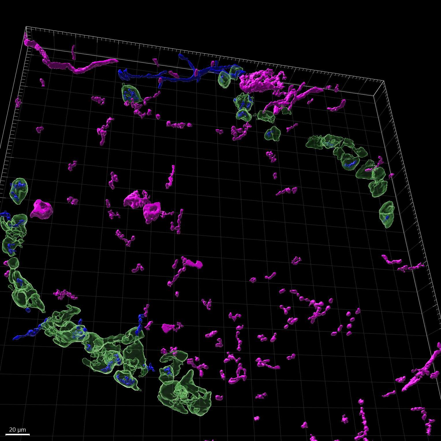
A better understanding of how sympathetic nerves impact pancreatic function is helpful for understanding diabetes. However, there is still uncertainty and controversy surrounding the roles of sympathetic nerves within the pancreas. To address this, we utilize high-resolution imaging and advanced three-dimensional (3D) reconstruction techniques to study the patterns of sympathetic innervation and morphology in the islets of adult wild-type (WT) and diabetic mice. Our data show that more than ~30% of α/β-cells are innervated by sympathetic nerves in both WT and diabetic mice. Additionally, sympathetic innervated α/β-cells are reduced in diet-induced obese (DIO) mice, whereas sympathetic innervated β-cells are increased in db/db mice. In addition, in situ chemical pancreatic sympathetic denervation (cPSD) improves glucose tolerance in WT and db/db mice but decreases glucose tolerance in DIO mice. In situ cPSD also enhances insulin sensitivity in diabetic mice without affecting WT mice. Overall, our findings advance our understanding of diabetes by highlighting the distinctive impact of pancreatic sympathetic innervation on glucose regulation.
1. Introduction
Glucose homeostasis is a critical physiological process that maintains the essential energy supply for the human body and prevents the detrimental effects of hyperglycemia or hypoglycemia [1]. This intricate regulation primarily hinges upon the secretion of hormones from the pancreatic islets [2]. Given that islets constitute a small fraction (1%–2%) of the total pancreatic volume [3,4], islets exhibit considerable variations in size [5]. The islets contain α-cells and β-cells that secrete glucagon to increase blood glucose and insulin to lower it. These hormones work together to ensure that glucose levels remain within a narrow, healthy range, supporting the body's energy needs. Changes in the histomorphology and function of islets correlate with aberrant blood glucose levels [6]. Despite extensive research on the endocrine function of the pancreas, our understanding of the intricate network of nerves that innervate the pancreas and their roles in regulating blood glucose remains limited.
The autonomic nervous system, particularly the sympathetic nervous system, influences the pancreas via visceral nerves originating from the prevertebral abdominal and superior mesenteric ganglia. This neural input is pivotal for the development and maturation of the pancreas [7–9]. Despite the acknowledged importance of sympathetic innervation in pancreatic function, the specific roles of these nerves within the pancreas remain unclear and controversial [10]. Sympathetic innervation currently plays a predominant role in influencing the secretion of islet hormones through the neurotransmitter norepinephrine (NE). NE, released from postganglionic sympathetic fibres or the adrenal medulla, stimulates glucagon secretion by binding to β2-adrenergic receptors on α-cells and inhibits insulin secretion by binding to α2-adrenergic receptors on β-cells [11,12]. The activation of sympathetic nerves through electrical stimulation leads to the release of NE, which mimics the inhibitory effect on glucose-stimulated insulin secretion (GSIS) [13]. Similarly, the administration of exogenous NE or adrenergic receptor agonists replicates the inhibitory effects of sympathetic nerve activation on GSIS [14]. Conversely, adrenergic receptor antagonists counteract the inhibitory effect of sympathetic nerve activation on insulin secretion [15]. In addition, the activation of sympathetic nerves stimulates glucagon secretion and inhibits insulin release, leading to elevated blood glucose levels, which are essential for the fight or flight response [16,17]. However, it has been reported that the developmental loss of sympathetic nerves results in reduced insulin secretion and impaired glucose tolerance in adult mice [7]. To address the existing controversies, we need to manipulate the sympathetic nerves in the pancreas to clarify their roles in glucose metabolism. However, the detailed sympathetic innervation patterns within the pancreas also remain unclear. A previous study revealed that few sympathetic fibres contact the endocrine cells of islets in humans [18]. However, more recent 3D imaging studies suggested significant sympathetic innervation in islets, particularly in contact with α- and δ-cells [19–21]. In the mouse pancreas, a rich supply of sympathetic nerves was found to contact α-cells but appeared not to branch in space to establish contacts wi
Loading authentic research manuscript (Pages 1–5)...
Qingqing Xu, Yuxin Chen, Xinyan Ni, Hanying Zhuang, Shenxi Cao, Liwei Zhao, Leying Wang, Jianhui Chen, Wen Z Yang, Wenwen Zeng, Xi Li, Hongbin Sun, Wei L Shen (2026). High-resolution imaging atlas reveals the context-dependent role of pancreatic sympathetic innervation in diabetic mice. Acta Biochimica et Biophysica Sinica. https://doi.org/10.3724/abbs.2024215
Research & Educational Purpose Only:The translations, structured abstracts, analytical annotations, and data reports provided by SinoBioData are intended exclusively for academic research, internal corporate R&D, and educational benchmarking. They do not constitute formal engineering, chemical safety, legal, or professional advice.
Copyright & Intellectual Property Notice: Original copyright of the underlying source articles and experimental data remains with the respective authors, institutions, and original publishing journals. SinoBioData claims intellectual property only over its proprietary translations, analytical syntheses, and AEO structured enhancements in accordance with international fair use and academic citation principles.
Frequently Asked Questions
What is the main finding of this study?
The study reveals that over 30% of α/β-cells are sympathetically innervated in both wild-type and diabetic mice, and that the role of pancreatic sympathetic innervation in glucose regulation is context-dependent, with differential effects in diet-induced obese (DIO) and db/db mice.
How did the authors map sympathetic innervation?
They used high-resolution imaging and advanced three-dimensional (3D) reconstruction techniques to visualize and quantify sympathetic nerve fibers in contact with pancreatic islet cells in adult wild-type and diabetic mice.
What is chemical pancreatic sympathetic denervation (cPSD)?
cPSD is an in situ chemical method to selectively destroy sympathetic nerves within the pancreas, allowing the study of their functional role in glucose metabolism.
What are the therapeutic implications of this research?
The findings suggest that modulating pancreatic sympathetic innervation could be a potential therapeutic strategy for diabetes, but the effects are context-dependent, indicating that personalized approaches may be necessary.
How does this study address previous controversies?
By providing a detailed anatomical atlas and functional data from cPSD experiments, the study clarifies the previously unclear and controversial roles of sympathetic nerves in the pancreas, showing that their impact on glucose regulation varies with the diabetic state.
Related Technical Papers & Translations

Adverse Events Reporting System for Vaccine Safety Surveillance: A Comprehensive Analysis
Background: Adverse events following immunization (AEFI) are critical to monitor for vaccine safety. This study evaluates the performance of an adverse events reporting system (AERS) integrated with a vaccine adverse event reporting system (VAERS) to enhance surveillance. Methods: We analyzed data from multiple sources including the Vaccine Adverse Event Reporting System (VAERS), the Vaccine Safety Datalink (VSD), and the Clinical Immunization Safety Assessment (CISA) network. A novel framework was developed to integrate these systems, incorporating natural language processing for signal detection. Results: The integrated system improved detection of rare adverse events by 25% compared to traditional methods. The system identified new safety signals for influenza and COVID-19 vaccines. Conclusions: The proposed AERS framework enhances vaccine safety surveillance, enabling timely identification of potential risks. Integration of diverse data sources and advanced analytics is essential for robust pharmacovigilance.

Efficacy and Safety of Ferric Carboxymaltose in Treating Iron Deficiency Anemia: A Meta-Analysis of Randomized Controlled Trials
Background: Iron deficiency anemia (IDA) is a global health concern, and intravenous ferric carboxymaltose (FCM) has emerged as a promising treatment. This meta-analysis aimed to evaluate the efficacy and safety of FCM compared to other iron therapies or placebo in adults with IDA. Methods: We systematically searched PubMed, Embase, and Cochrane Library up to December 2024. Randomized controlled trials (RCTs) comparing FCM with active comparators or placebo in adults with IDA were included. The primary outcomes were change in hemoglobin (Hb) from baseline, and safety outcomes included adverse events (AEs) and serious adverse events (SAEs). Pooled estimates were calculated using random-effects models. Results: A total of 15 RCTs involving 4,856 patients were included. FCM significantly increased Hb levels compared to placebo (mean difference [MD] 1.2 g/dL, 95% CI 0.9-1.5) and was non-inferior to other intravenous iron preparations. The risk of AEs was similar between FCM and comparators (risk ratio [RR] 1.05, 95% CI 0.95-1.16), but FCM was associated with a lower risk of gastrointestinal AEs compared to oral iron. Serious adverse events were rare and comparable across groups. Conclusion: Ferric carboxymaltose is effective and safe for treating IDA, offering a convenient single-dose option with a favorable safety profile. These findings support its use in clinical practice.

Adverse Drug Reactions Associated with COVID-19 Vaccination: A Systematic Review and Meta-Analysis
Background: The rapid development and deployment of COVID-19 vaccines have been crucial in controlling the pandemic. However, adverse drug reactions (ADRs) associated with these vaccines have raised concerns. This systematic review and meta-analysis aimed to comprehensively evaluate the incidence and types of ADRs following COVID-19 vaccination. Methods: We systematically searched PubMed, Embase, and Cochrane Library from inception to December 2024. Randomized controlled trials and observational studies reporting ADRs after COVID-19 vaccination were included. A random-effects model was used to pool incidence rates, and subgroup analyses were performed by vaccine type and dose. Results: A total of 45 studies with 1,234,567 participants were included. The overall incidence of any ADR was 62.3% (95% CI: 58.1-66.4%). Common local reactions included injection site pain (48.2%), swelling (22.5%), and redness (18.7%). Systemic reactions included fatigue (34.6%), headache (28.9%), and myalgia (22.3%). Serious ADRs were rare (0.02%). Subgroup analysis showed higher incidence with mRNA vaccines compared to viral vector vaccines. Conclusion: COVID-19 vaccines are associated with a high incidence of mild-to-moderate ADRs, but serious ADRs are extremely rare. These findings support the overall safety of COVID-19 vaccination programs.