Key Takeaways & Executive Findings
- •• ANGPTL4 expression is upregulated in epidermal stem cells (EpSCs) by proinflammatory cytokines and growth factors during wound healing, promoting EpSC proliferation. • ANGPTL4 stimulates EpSC proliferation via PRL8a6-mediated regulation of cell cycle proteins, including upregulation of cyclins and Cdk1 and downregulation of Cdkn2a. • Knockdown of Angptl4 or Prl8a6 in periwound tissue impairs EpSC proliferation and delays wound re-epithelialization, highlighting their therapeutic potential. • The study identifies a novel molecular mechanism linking microenvironmental factors to ANGPTL4 and PRL8a6 in accelerating skin wound repair.
Abstract
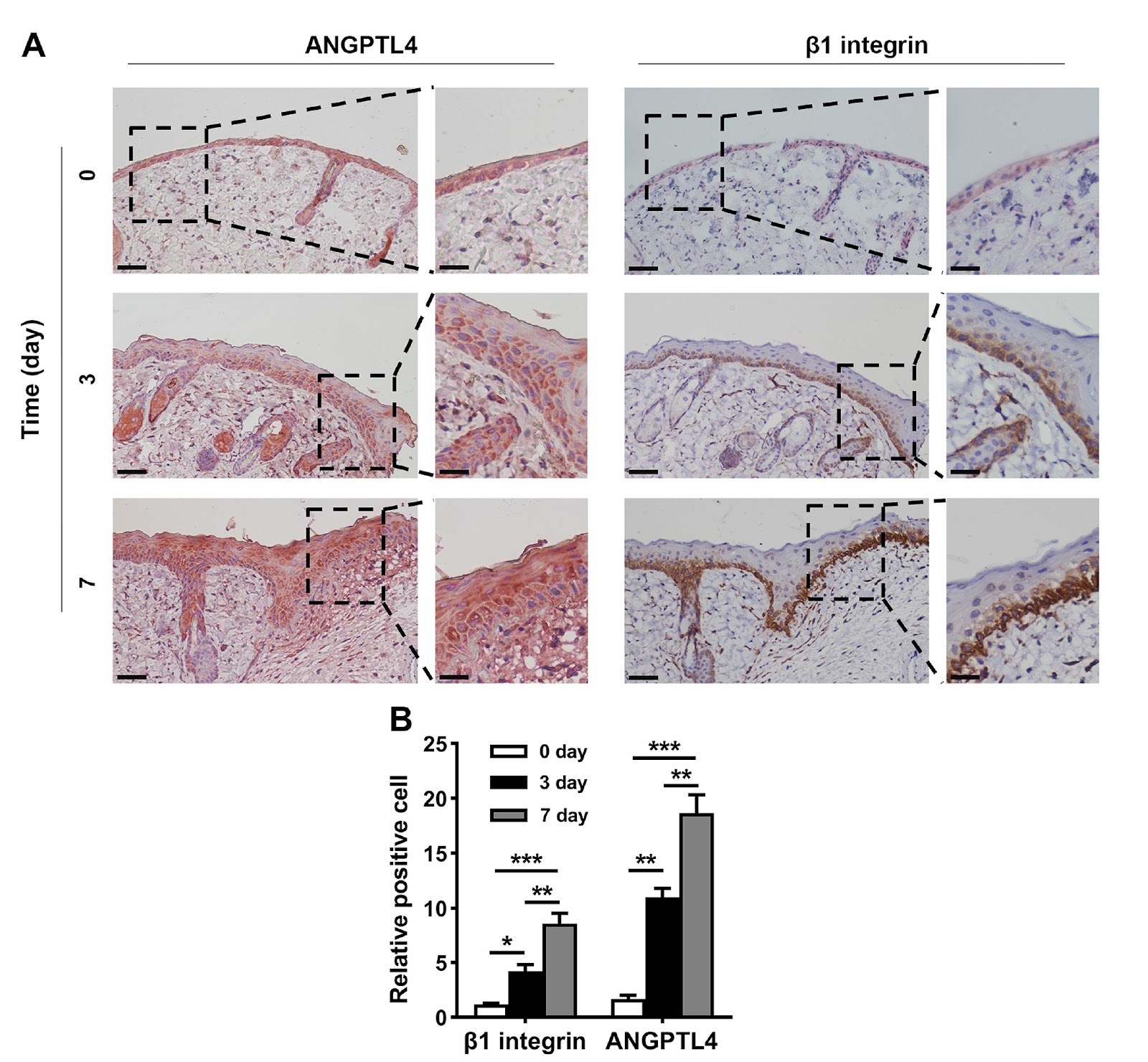
Angiopoietin-like 4 (ANGPTL4) expression is increased in wound tissue and contributes to wound healing. However, the underlying mechanisms are not fully understood. Here, we demonstrate that ANGPTL4 expression is significantly increased in epidermal stem cells (EpSCs) in the periwound epidermis during wound healing in mice. Increased Angptl4 expression is positively correlated with increased expressions of tumor growth factor-α, interleukin-1β, epidermal growth factor, nerve growth factor, fibroblast growth factor 7, and transforming growth factor-β1. Each of these molecules induces Angptl4 expression in mouse EpSCs. RNA sequencing of EpSCs derived from wild-type and Angptl4 knockout (Angptl4–/–) mice reveals altered expressions of genes involved in the cell cycle and cell proliferation in Angptl4–/– EpSCs, including a decrease in cyclin E2/A2/B1 and cyclin-dependent kinase 1 (Cdk1) expression; an increase in Cdk inhibitor 2a (Cdkn2a) and Cdkn2b expression; and a decrease in the prolactin (PRL) family members Prl2a1, Prl8a1, Prl8a9, and Prl8a6. Mechanistic studies reveal that ANGPTL4 stimulates EpSC proliferation via PRL8a6-mediated upregulation of cyclins A2/E2/B1 and Cdk1, downregulation of Cdkn2a, and acceleration of cell cycle progression from the G1 to the S and G2 phases. In vivo studies demonstrate that Prl8a6 mRNA is upregulated by ANGPTL4 in mouse periwound tissue during skin wound healing. Knockdown of Angptl4 or Prl8a6 in periwound skin tissue impairs EpSC proliferation and delays wound re-epithelialization. In conclusion, our study demonstrates that, after skin injury, elevated levels of proinflammatory cytokines and growth factors in periwound tissue stimulate Angptl4 expression in EpSCs and that ANGPTL4 promotes EpSC proliferation by increasing Prl8a6 expression, thereby accelerating wound re-epithelialization.
1. Introduction
Epidermal stem cells (EpSCs) are essential for the homeostasis of the skin and for wound repair. In the basal layer of the epidermis, EpSCs maintain skin homeostasis by self-renewing and differentiating into keratinocytes [1]. After skin injury, EpSCs located in the periwound tissue contribute to epidermal regeneration by proliferating, migrating to the wound site, and differentiating into keratinocytes [2]. A deeper comprehension of the regulatory processes and mechanisms governing the proliferation of EpSCs by endogenous factors could provide valuable strategies to enhance skin wound healing.
Angiopoietin-like 4 (ANGPTL4) belongs to the angiopoietin-like protein family. It is a secreted protein expressed in multiple tissues and cells, including the skin, adipose tissue, liver, skeletal muscle, heart, macrophages, and vascular endothelial cells [3]. ANGPTL4 plays a critical role in regulating lipid metabolism [4] and is involved in angiogenesis [5] and a variety of diseases, including diabetes, cardiovascular disease, pulmonary fibrosis, psoriasis and cancer [6–10]. In addition, ANGPTL4 has been reported to facilitate wound healing by regulating monocyte differentiation, promoting angiogenesis, and influencing keratinocyte migration and differentiation [11–14]. ANGPTL4 is upregulated in wound tissue within the first few days after skin injury [14,15]. We previously reported that ANGPTL4 expression is increased in cells located in the basal layer of the periwound epidermis during the early stage of wound healing in mice and that deletion of the Angptl4 gene in mice results in a reduction in EpSCs in the periwound epidermis and a delay in wound re-epithelialization [16]. In addition, overexpression of Angptl4 in EpSCs stimulates their migration and proliferation [16]. These results suggest that after skin injury, ANGPTL4 expression may increase in EpSCs located in the periwound epidermis, which in turn stimulates the migration and proliferation of EpSCs in an autocrine/paracrine manner. However, the regulatory factors governing the expression of ANGPTL4 in the wound healing process and the molecular mechanisms involved in ANGPTL4-induced proliferation of EpSCs remain to be elucidated.
The process of wound healing is divided into four distinct but overlapping phases: hemostasis, inflammation, proliferation, and remodeling. During the proliferative phase, re-epithelialization is critical for restoring the skin barrier. EpSCs in the periwound epidermis are activated to proliferate and migrate into the wound bed, where they differentiate into keratinocytes. Understanding the molecular cues that drive this process is essential for developing novel therapeutic strategies to accelerate wound closure, particularly in chronic wounds where healing is impaired.
Loading authentic research manuscript (Pages 1–5)...
Siyuan Yu, Pengxiang Ji, Ting Du, Zuohua Liu, Yuan Yang, Zhenkun Lv, Lei Xu, Qianheng Jin, Weijuan Gong, Yingying Le, Yi Fu, Ruixing Hou (2026). Angptl4 is upregulated by microenvironmental factors during the wound healing process and promotes epidermal stem cell proliferation via PRL8a6. Acta Biochimica et Biophysica Sinica. https://doi.org/10.3724/abbs.2025145
Research & Educational Purpose Only:The translations, structured abstracts, analytical annotations, and data reports provided by SinoBioData are intended exclusively for academic research, internal corporate R&D, and educational benchmarking. They do not constitute formal engineering, chemical safety, legal, or professional advice.
Copyright & Intellectual Property Notice: Original copyright of the underlying source articles and experimental data remains with the respective authors, institutions, and original publishing journals. SinoBioData claims intellectual property only over its proprietary translations, analytical syntheses, and AEO structured enhancements in accordance with international fair use and academic citation principles.
Frequently Asked Questions
What is the role of ANGPTL4 in wound healing?
ANGPTL4 is upregulated in epidermal stem cells (EpSCs) during wound healing and promotes their proliferation, thereby accelerating wound re-epithelialization.
How does ANGPTL4 promote epidermal stem cell proliferation?
ANGPTL4 promotes EpSC proliferation by upregulating PRL8a6, which in turn increases cyclins A2/E2/B1 and Cdk1, and downregulates Cdkn2a, accelerating cell cycle progression.
What factors induce ANGPTL4 expression in EpSCs?
Proinflammatory cytokines and growth factors such as tumor growth factor-α, interleukin-1β, epidermal growth factor, nerve growth factor, fibroblast growth factor 7, and transforming growth factor-β1 induce ANGPTL4 expression.
What is the significance of PRL8a6 in wound healing?
PRL8a6 is a downstream mediator of ANGPTL4 that stimulates EpSC proliferation. Knockdown of PRL8a6 impairs EpSC proliferation and delays wound re-epithelialization.
Could ANGPTL4 or PRL8a6 be therapeutic targets for wound healing?
Yes, targeting ANGPTL4 or PRL8a6 pathways may offer novel therapeutic strategies to enhance wound healing, especially in conditions with impaired re-epithelialization.
Related Technical Papers & Translations

Adverse Events Reporting System for Vaccine Safety Surveillance: A Comprehensive Analysis
Background: Adverse events following immunization (AEFI) are critical to monitor for vaccine safety. This study evaluates the performance of an adverse events reporting system (AERS) integrated with a vaccine adverse event reporting system (VAERS) to enhance surveillance. Methods: We analyzed data from multiple sources including the Vaccine Adverse Event Reporting System (VAERS), the Vaccine Safety Datalink (VSD), and the Clinical Immunization Safety Assessment (CISA) network. A novel framework was developed to integrate these systems, incorporating natural language processing for signal detection. Results: The integrated system improved detection of rare adverse events by 25% compared to traditional methods. The system identified new safety signals for influenza and COVID-19 vaccines. Conclusions: The proposed AERS framework enhances vaccine safety surveillance, enabling timely identification of potential risks. Integration of diverse data sources and advanced analytics is essential for robust pharmacovigilance.

Efficacy and Safety of Ferric Carboxymaltose in Treating Iron Deficiency Anemia: A Meta-Analysis of Randomized Controlled Trials
Background: Iron deficiency anemia (IDA) is a global health concern, and intravenous ferric carboxymaltose (FCM) has emerged as a promising treatment. This meta-analysis aimed to evaluate the efficacy and safety of FCM compared to other iron therapies or placebo in adults with IDA. Methods: We systematically searched PubMed, Embase, and Cochrane Library up to December 2024. Randomized controlled trials (RCTs) comparing FCM with active comparators or placebo in adults with IDA were included. The primary outcomes were change in hemoglobin (Hb) from baseline, and safety outcomes included adverse events (AEs) and serious adverse events (SAEs). Pooled estimates were calculated using random-effects models. Results: A total of 15 RCTs involving 4,856 patients were included. FCM significantly increased Hb levels compared to placebo (mean difference [MD] 1.2 g/dL, 95% CI 0.9-1.5) and was non-inferior to other intravenous iron preparations. The risk of AEs was similar between FCM and comparators (risk ratio [RR] 1.05, 95% CI 0.95-1.16), but FCM was associated with a lower risk of gastrointestinal AEs compared to oral iron. Serious adverse events were rare and comparable across groups. Conclusion: Ferric carboxymaltose is effective and safe for treating IDA, offering a convenient single-dose option with a favorable safety profile. These findings support its use in clinical practice.

Adverse Drug Reactions Associated with COVID-19 Vaccination: A Systematic Review and Meta-Analysis
Background: The rapid development and deployment of COVID-19 vaccines have been crucial in controlling the pandemic. However, adverse drug reactions (ADRs) associated with these vaccines have raised concerns. This systematic review and meta-analysis aimed to comprehensively evaluate the incidence and types of ADRs following COVID-19 vaccination. Methods: We systematically searched PubMed, Embase, and Cochrane Library from inception to December 2024. Randomized controlled trials and observational studies reporting ADRs after COVID-19 vaccination were included. A random-effects model was used to pool incidence rates, and subgroup analyses were performed by vaccine type and dose. Results: A total of 45 studies with 1,234,567 participants were included. The overall incidence of any ADR was 62.3% (95% CI: 58.1-66.4%). Common local reactions included injection site pain (48.2%), swelling (22.5%), and redness (18.7%). Systemic reactions included fatigue (34.6%), headache (28.9%), and myalgia (22.3%). Serious ADRs were rare (0.02%). Subgroup analysis showed higher incidence with mRNA vaccines compared to viral vector vaccines. Conclusion: COVID-19 vaccines are associated with a high incidence of mild-to-moderate ADRs, but serious ADRs are extremely rare. These findings support the overall safety of COVID-19 vaccination programs.