🧬 SinoBioData Academic Portal
📚 Peer-Reviewed Translated Literature

All Biomedical & Clinical Articles (Page 40)

Browse complete peer-reviewed translations from top Chinese biomedical, oncology, and genomics journals. Read verified previews and download full authentic clinical reports.

Published Research Papers

Showing 24 of 1542 peer-reviewed translated articles (Page 40 of 65)

The therapeutic efficacy comparison of MSCs derived different tissues unveilings anti-apoptosis more crucial than angiogenesis in treating acute myocardial infarctionGraphical AbstractVerified
Stem Cell Research & Therapy

The therapeutic efficacy comparison of MSCs derived different tissues unveilings anti-apoptosis more crucial than angiogenesis in treating acute myocardial infarction

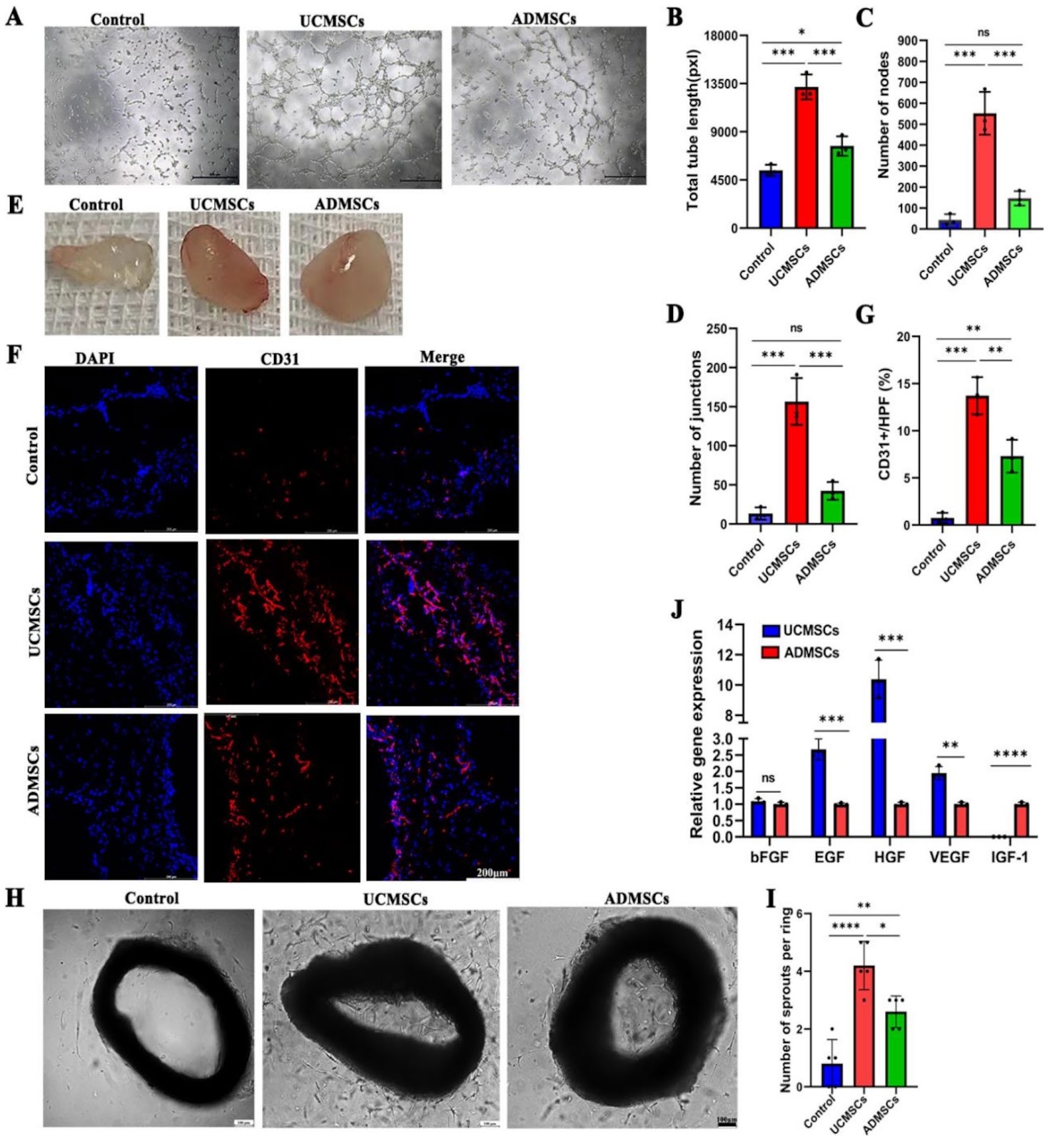
Background Myocardial infarction (MI) is a severe disease that often associated with impaired angiogenesis and increased myocardial apoptosis. Mesenchymal stromal cells (MSCs) have been a promising candidate for treating myocardial infarction. However, functional heterogeneity of MSCs leads to inconsistent therapeutic efficiency and the current MSCs-based therapy lacks the concept and implementation of precision medicine. In this study, we compared the cardioprotective effect of UCMSCs and ADMSCs targeting the angiogenesis in a mouse MI model and screened out optimum MSCs candidate for precise clinical application. Methods The gene expression profiles of UCMSCs and ADMSCs were investigated through RNA sequencing analysis. To compare their angiogenic potential, we performed tube formation assay, Matrigel plug assays, and aortic ring assay, and analyzed pro-angiogenic genes via qPCR. Subsequently, UCMSCs and ADMSCs were respectively injected into myocardium after MI surgery in mice. On day 28 post-MI, echocardiography was performed to assess cardiac function. Histological analysis was performed to assess MSCs retention, angiogenesis, and myocardial apoptosis. Additionally, the anti-apoptosis effects mediated by MSCs were further evaluated using flow cytometry in hypoxia H9C2 and HL-1 cells. Results The RNA sequencing analysis revealed differences in gene expression related to angiogenesis and apoptosis pathways between UCMSCs and ADMSCs. UCMSCs presented greater pro-angiogenesis activity than ADMSCs in vitro and in vivo. Both of UCMSCs and ADMSCs improved cardiac function, decreased infarction area and inhibited cardiomyocyte apoptosis while promoting angiogenesis post-MI in mice. Notably, ADMSCs exerted a better cardioprotective function than UCMSCs and stronger anti-apoptotic effect on residual cardiomyocytes.
Read Full Abstract10.1186/s13287-025-04378-3
E2 pretreatment alleviates aggregation of intravenously injected mesenchymal stem cells in TBI by regulating BRG1 to affect adhesionGraphical AbstractVerified
Stem Cell Research & Therapy

E2 pretreatment alleviates aggregation of intravenously injected mesenchymal stem cells in TBI by regulating BRG1 to affect adhesion

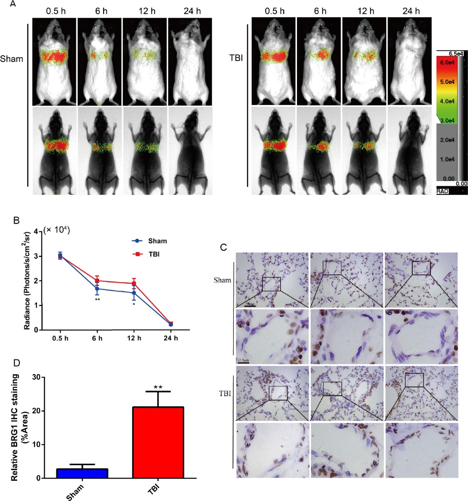
Background Human umbilical cord mesenchymal stem cells (hUMSCs) are considered an effective prospect for treating TBI, but they tend to accumulate in the lungs after intravenous injection, hindering further clinical translation. Brahma-related gene 1(BRG1) can be influenced by estrogen to regulate adhesion, and ourprevious studies have found that the expression of BRG1 in lungs increases after TBI. However, the relationship between BRG1, estrogen, TBI, and stem cell lung aggregation is not clear. Methods By regulating the expression levels of BRG1 in vascular endothelial cells and hUMSCs, Western Blot and immunohistochemistry were used to explore its changes in adhesion and possible mechanisms; used in vivo bioluminescenece imaging analysis, real-time tracking the distribution of stem cells after transplantation; and therapeutic drug E2 is introduced to observe the effect of changes in BRG1 expression on the aggregation of hUMSCs in the lungs of model animals, as well as the therapeutic effect of E2-pretreated hUMSCs on inflammation after TBI. Results After TBI, the retention of hUMSCs in the lungs was higher in the TBI groups than in the Sham groups, and the level of BRG1 in lung was higher in the TBI groups than in the Sham groups; the expression of BRG1 in HUVECs, HPAECs, and hUMSCs treated with TNF-α and LPS were higher than those in the control groups, showing dose- and time-dependent effects. E2 can inhibit the expression of BRG1 and adhesion proteins; after intervention with estrogen receptor inhibitor (ICI 182780) and NF-κ B inhibitor SC75741, BRG1 expression increased and adhesion protein decreased; E2-pretreated MSCs can reduce pulmonary retention, and has no adverse effects on the inflammatory response for TBI.

Read Full Abstract10.1186/s13287-025-04637-3
Development and evaluation of siRNA-mediated gene silencing strategies for ADO2 therapy utilizing iPSCs model and DMPC-SPIONs delivery systemGraphical AbstractVerified
Stem Cell Research & Therapy

Development and evaluation of siRNA-mediated gene silencing strategies for ADO2 therapy utilizing iPSCs model and DMPC-SPIONs delivery system

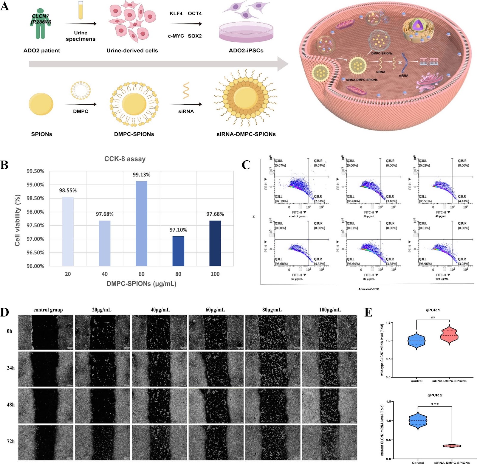
Background Autosomal dominant osteodystrophy type II (ADO2) is an inherited disease characterized by an abnormal increase in bone mineral density, and CLCN7 (R286W) is its most common causative mutation. The aim of this study was to explore the new idea of siRNA technology applied to the in vitro treatment of ADO2. Methods Urinary-derived cells from ADO2 patients were collected to establish induced pluripotent stem cells (iPSCs) model. The siRNA targeting CLCN7 (R286W) mutant mRNA was designed. the cytotoxicity of the delivery vector DMPC-SPIONs was comprehensively evaluated by CCK-8 assay, flow cytometry and scratch assay. Finally, qPCR was utilized to verify the post-transcriptional silencing effect of siRNAs. Results We found that DMPC-SPIONs had low cytotoxicity and were able to effectively deliver siRNAs into ADO2-iPSCs. qPCR confirmed that siRNA-DMPC-SPIONs were able to significantly reduce the expression level of mutant CLCN7 (66%), while there was no significant effect on the expression of wild-type CLCN7. Conclusions This study developed a gene silencing strategy based on siRNAs and DMPC-SPIONs, which provides a potential new approach for the treatment of ADO2 and demonstrates the potential application of siRNA technology in the treatment of autosomal dominant genetic diseases. Innovative statements In this study, we used the established ADO2-iPSCs using patient's urine-derived cells to explore the safety and efficacy of siRNA technology based on the principle of RNA interference for ADO2 treatment for the first time. In addition, we chose DMPC-SPIONs as the delivery vehicle for siRNA, which cleverly exploits the advantages of nanoparticles such as superparamagnetism, low cytotoxicity, and good bio-histocompatibility.

Read Full Abstract10.1186/s13287-025-04151-6
Osteoinductive micro-nano guided bone regeneration membrane for in situ bone defect repairGraphical AbstractVerified
Stem Cell Research & Therapy

Osteoinductive micro-nano guided bone regeneration membrane for in situ bone defect repair

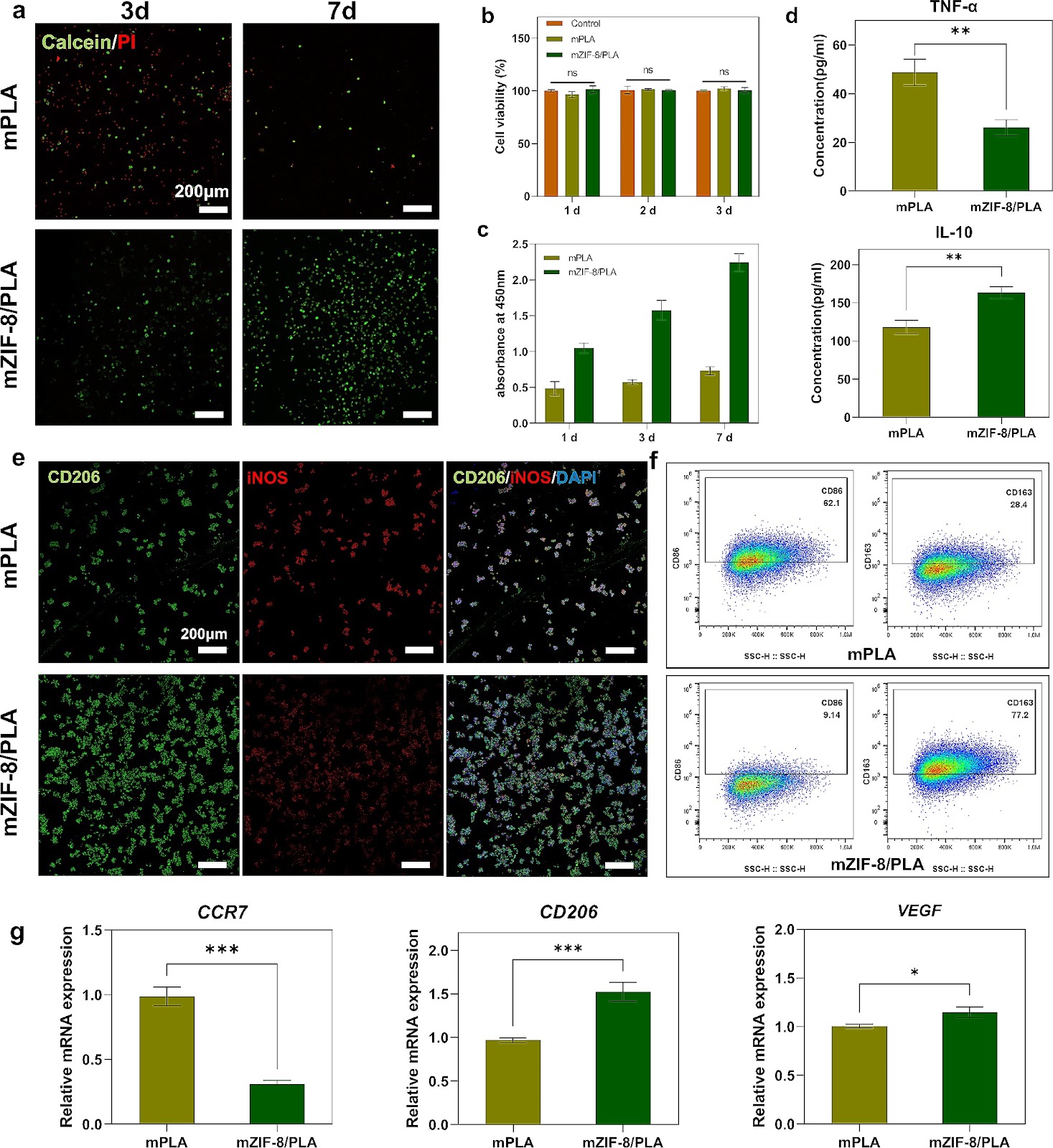
Background Biomaterials used in bone tissue engineering must fulfill the requirements of osteoconduction, osteoinduction, and osseointegration. However, biomaterials with good osteoconductive properties face several challenges, including inadequate vascularization, limited osteoinduction and barrier ability, as well as the potential to trigger immune and inflammatory responses. Therefore, there is an urgent need to develop guided bone regeneration membranes as a crucial component of tissue engineering strategies for repairing bone defects. Methods The mZIF-8/PLA membrane was prepared using electrospinning technology and simulated body fluid external mineralization method. Its ability to induce biomimetic mineralization was evaluated through TEM, EDS, XRD, FT-IR, zeta potential, and wettability techniques. The biocompatibility, osteoinduction properties, and osteo-immunomodulatory effects of the mZIF-8/PLA membrane were comprehensively evaluated by examining cell behaviors of surface-seeded BMSCs and macrophages, as well as the regulation of cellular genes and protein levels using PCR and WB. In vivo, the mZIF-8/PLA membrane's potential to promote bone regeneration and angiogenesis was assessed through Micro-CT and immunohistochemical staining. Results The mineralized deposition enhances hydrophilicity and cell compatibility of mZIF-8/PLA membrane. mZIF-8/PLA membrane promotes up-regulation of osteogenesis and angiogenesis related factors in BMSCs. Moreover, it induces the polarization of macrophages towards the M2 phenotype and modulates the local immune microenvironment. After 4-weeks of implantation, the mZIF-8/PLA membrane successfully bridges critical bone defects and almost completely repairs the defect area after 12-weeks, while significantly improving the strength and vascularization of new bone. Conclusions The mZIF-8/PLA membrane with dual osteoconductive and immunomodulatory abilities could pave new research paths for bone tissue engineering.

Read Full Abstract10.1186/s13287-024-03745-w
Targeting NPM1 inhibits proliferation and promotes apoptosis of hepatic progenitor cells via suppression of mTOR signalling pathwayGraphical AbstractVerified
Stem Cell Research & Therapy

Targeting NPM1 inhibits proliferation and promotes apoptosis of hepatic progenitor cells via suppression of mTOR signalling pathway

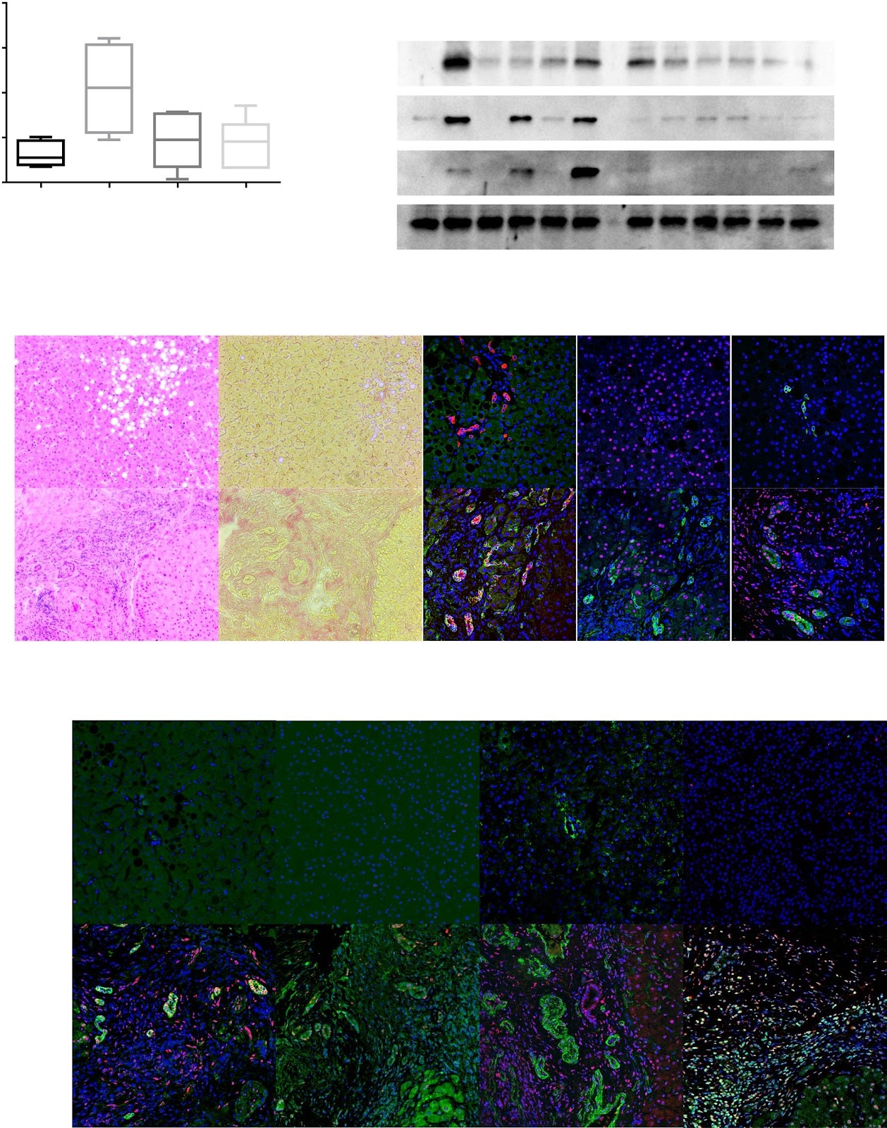
Background Hepatic progenitor cells serve not only as the origin of combined hepatocellular cholangiocarcinoma (cHCC-CCA) but are also responsible for malignancy recurrence after surgical resection. Nucleophosmin 1 (NPM1) has been implicated in cancer metastasis and poor prognosis. This study aimed to determine the expression of NPM1 by hepatic progenitor cells in cHCC-CCA and the effects of targeting NPM1 on hepatic progenitor cells and BEL-7402 cells with characteristics of both progenitor cells and cHCC-CCA. Methods First, NPM1 was detected by RT‒PCR, western blotting, and double-immunofluorescence staining in cHCC-CCA tissues. NPM1 expression was subsequently analysed in rat hepatic progenitor cells cultured in vitro and in interleukin 6 (IL6)-treated cells. The effects and mechanism of NPM1 on hepatic progenitor cells were determined by knocking down NPM1 and performing RNA sequencing analysis. Finally, NSC348884, a small-molecule inhibitor that disrupts NPM1 dimer formation, was used to confirm the function of NPM1 in BEL-7402 cells. Results Both human hepatic progenitor cells in cHCC-CCA tissues and rat in vitro cultured hepatic progenitor cells highly expressed NPM1. IL6, a cytokine involved in the malignant transformation of hepatic progenitor cells, dose-dependently increased NPM1 and PCNA expression. Knocking down NPM1 reduced IL6R transcription (P < 0.0001) and inhibited the proliferation (P = 0.0065) of hepatic progenitor cells by suppressing the mTOR signalling pathway and activating the apoptosis pathway. Furthermore, knocking down NPM1 in hepatic progenitor cells resulted in more apoptotic cells (7.33 ± 0.09% vs. 3.76 ± 0.13%, P < 0.0001) but fewer apoptotic cells in the presence of NSC348884 (47.57 ± 0.49% vs. 63.40 ± 0.05%, P = 0.0008) than in the control cells, suggesting that low-NPM1-expressing cells are more resistant to NSC348884. In addition, NSC348884 induced the apoptosis of BEL-7402 cells with an IC50 of 2.77 μmol/L via the downregulation of the IL-6R and mTOR signalling pathways and inhibited the growth of BEL-7402 cells in a subcutaneous xenograft tumour model (P = 0.0457). Conclusions Targeting NPM1 inhibits proliferation and induces apoptosis in hepatic progenitor cells and BEL-7402 cells, thus serving as a potential therapy for cHCC-CCA.

Read Full Abstract10.1186/s13287-024-03898-8
TGF-β signaling regulates differentiation of MSCs in bone metabolism: disputes among viewpointsGraphical AbstractVerified
Stem Cell Research & Therapy

TGF-β signaling regulates differentiation of MSCs in bone metabolism: disputes among viewpoints

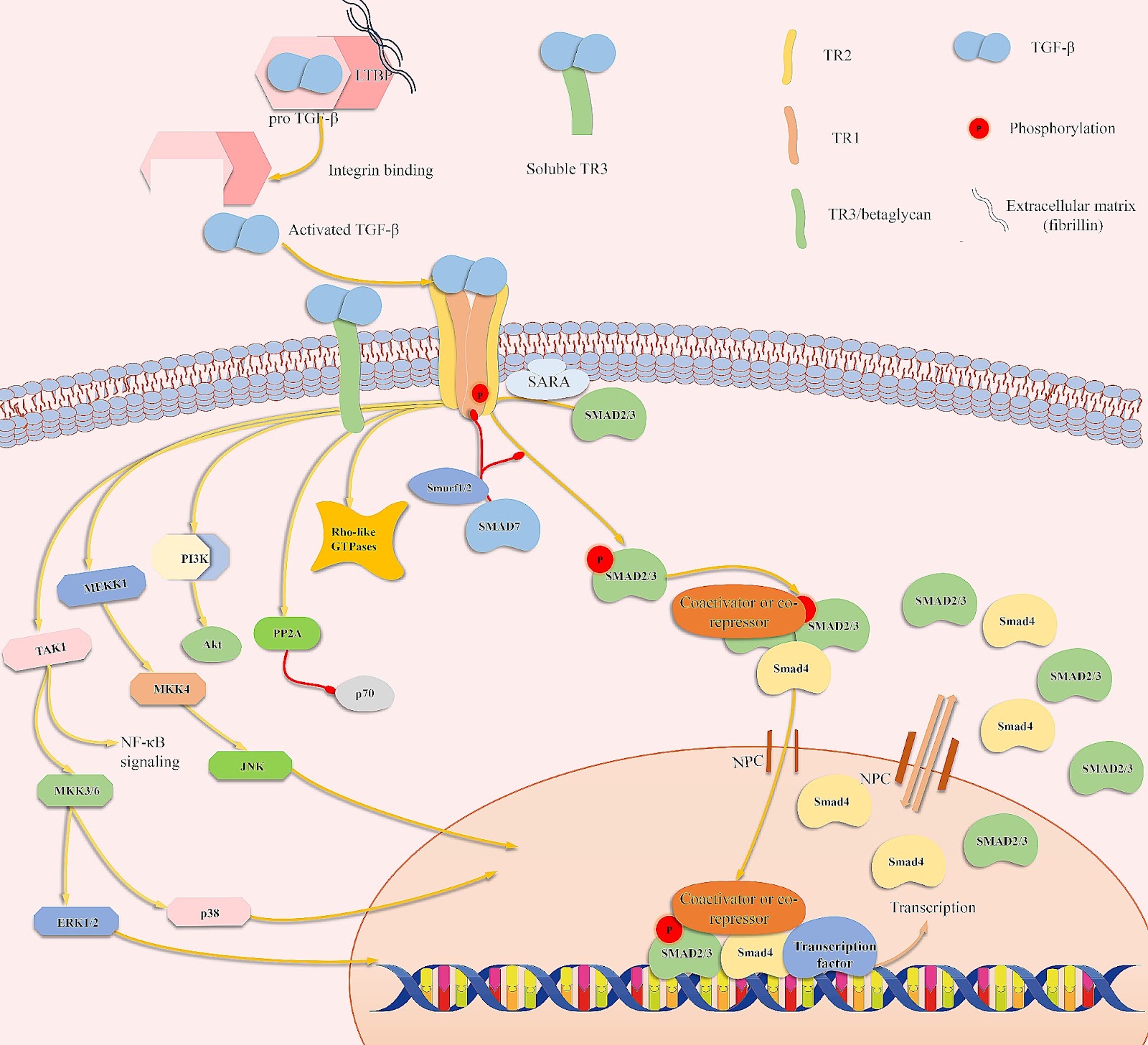
Mesenchymal stem cells (MSCs) are multipotent cells that can differentiate into cells of different lineages to form mesenchymal tissues, which are promising in regard to treatment for bone diseases. Their osteogenic differentiation is under the tight regulation of intrinsic and extrinsic factors. Transforming growth factor β (TGF-β) is an essential growth factor in bone metabolism, which regulates the differentiation of MSCs. However, published studies differ in their views on whether TGF-β signaling regulates the osteogenic differentiation of MSCs positively or negatively. The controversial results have not been summarized systematically and the related explanations are required. Therefore, we reviewed the basics of TGF-β signaling and summarized how each of three isoforms regulates osteogenic differentiation. Three isoforms of TGF-β (TGF-β1/β2/β3) play distinct roles in regulating osteogenic differentiation of MSCs. Additionally, other possible sources of conflicts are summarized here. Further understanding of TGF-β signaling regulation in MSCs may lead to new applications to promote bone regeneration and improve therapies for bone diseases.

Read Full Abstract10.1186/s13287-024-03761-w
Manufacturing, quality control, and GLP-grade preclinical study of nebulized allogenic adipose mesenchymal stromal cells-derived extracellular vesiclesGraphical AbstractVerified
Stem Cell Research & Therapy

Manufacturing, quality control, and GLP-grade preclinical study of nebulized allogenic adipose mesenchymal stromal cells-derived extracellular vesicles

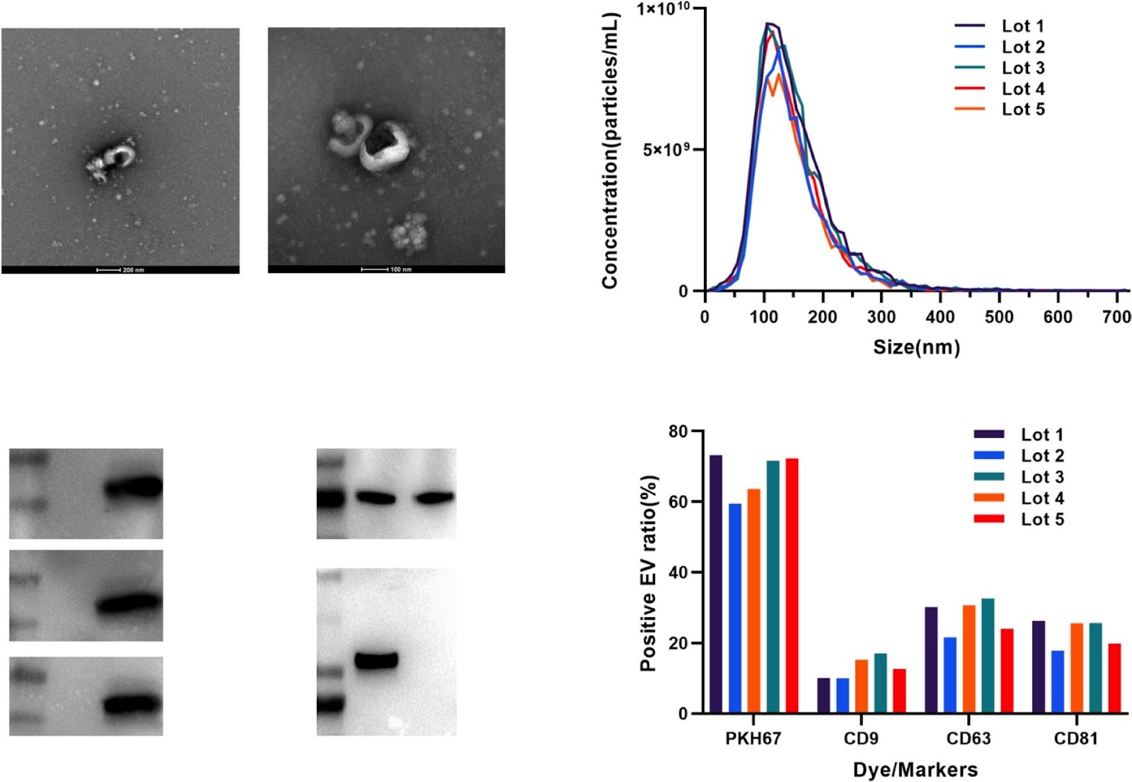
Background Human adipose stromal cells-derived extracellular vesicles (haMSC-EVs) have been shown to alleviate inflammation in acute lung injury (ALI) animal models. However, there are few systemic studies on clinical-grade haMSC-EVs. Our study aimed to investigate the manufacturing, quality control (QC) and preclinical safety of clinical-grade haMSC-EVs. Methods haMSC-EVs were isolated from the conditioned medium of human adipose MSCs incubated in 2D containers. Purification was performed by PEG precipitation and differential centrifugation. Characterizations were conducted by nanoparticle tracking analysis, transmission electron microscopy (TEM), Western blotting, nanoflow cytometry analysis, and the TNF-α inhibition ratio of macrophage [after stimulated by lipopolysaccharide (LPS)]. RNA-seq and proteomic analysis with liquid chromatography tandem mass spectrometry (LC–MS/MS) were used to inspect the lot-to-lot consistency of the EV products. Repeated toxicity was evaluated in rats after administration using trace liquid endotracheal nebulizers for 28 days, and respiratory toxicity was evaluated 24 h after the first administration. In vivo therapeutic effects were assessed in an LPS-induced ALI/ acute respiratory distress syndrome (ARDS) rat model. Results The quality criteria have been standardized. In a stability study, haMSC-EVs were found to remain stable after 6 months of storage at − 80°C, 3 months at − 20 °C, and 6 h at room temperature. The microRNA profile and proteome of haMSC-EVs demonstrated suitable lot-to-lot consistency, further suggesting the stability of the production processes. Intratracheally administered 1.5 × 10^8 particles/rat/day for four weeks elicited no significant toxicity in rats. In LPS-induced ALI/ARDS model rats, intratracheally administered haMSC-EVs alleviated lung injury, possibly by reducing the serum level of inflammatory factors. Conclusion haMSC-EVs, as an off-shelf drug, have suitable stability and lot-to-lot consistency. Intratracheally administered haMSC-EVs demonstrated excellent safety at the tested dosages in systematic preclinical toxicity studies. Intratracheally administered haMSC-EVs improved the lung function and exerted anti-inflammatory effects on LPS-induced ALI/ARDS model rats.

Read Full Abstract10.1186/s13287-024-03708-1
A GMP-compliant manufacturing method for Wharton’s jelly-derived mesenchymal stromal cellsGraphical AbstractVerified
Stem Cell Research & Therapy

A GMP-compliant manufacturing method for Wharton’s jelly-derived mesenchymal stromal cells

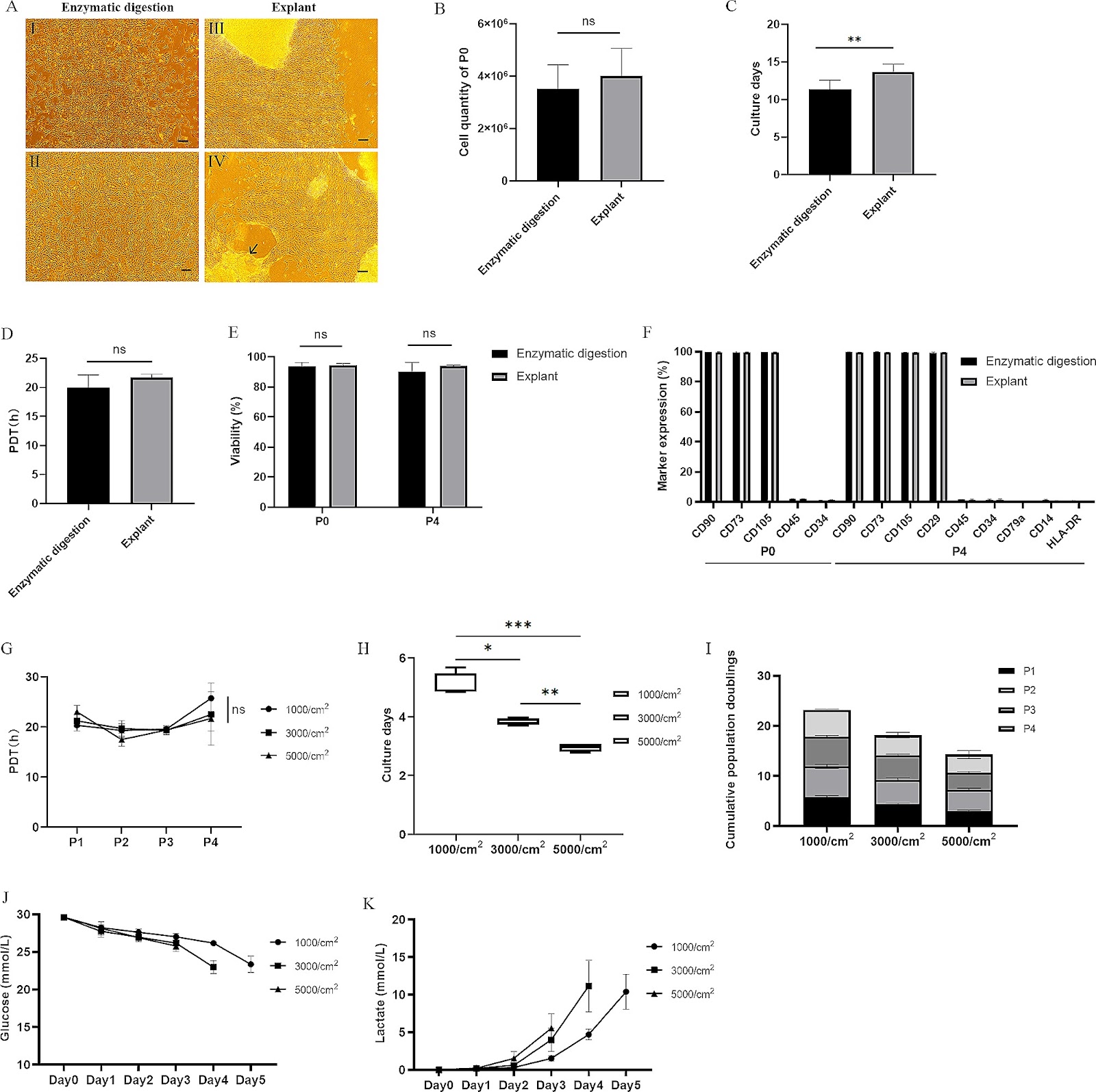
Background Wharton’s jelly-derived mesenchymal stem cells (WJ-MSCs) hold great therapeutic potential in regenerative medicine. Therefore, it is crucial to establish a Good Manufacturing Practice (GMP)-compliant methodology for the isolation and culture of WJ-MSCs. Through comprehensive research, encompassing laboratory-scale experiments to pilot-scale studies, we aimed to develop standardized protocols ensuring the high yield and quality of WJ-MSCs manufacturing. Methods Firstly, optimization of parameters for the enzymatic digestion method used to isolate WJ-MSCs was conducted. These parameters included enzyme concentrations, digestion times, seeding densities, and culture media. Additionally, a comparative analysis between the explant method and the enzymatic digestion method was performed. Subsequently, the consecutive passaging of WJ-MSCs, specifically up to passage 9, was evaluated using the optimized method. Finally, manufacturing processes were developed and scaled up, starting from laboratory-scale flask-based production and progressing to pilot-scale cell factory-based production. Furthermore, a stability study was carried out to assess the storage and use of drug products (DPs). Results The optimal parameters for the enzymatic digestion method were a concentration of 0.4 PZ U/mL Collagenase NB6 and a digestion time of 3 h, resulting in a higher yield of P0 WJ-MSCs. In addition, a positive correlation between the weight of umbilical cord tissue and the quantities of P0 WJ-MSCs has been observed. Evaluation of different concentrations of human platelet lysate revealed that 2% and 5% concentrations resulted in similar levels of cell expansion. Comparative analysis revealed that the enzymatic digestion method exhibited faster outgrowth of WJ-MSCs compared to the explant method during the initial passage. Passages 2 to 5 exhibited higher viability and proliferation ability throughout consecutive passaging. Moreover, scalable manufacturing processes from the laboratory scale to the pilot scale were successfully developed, ensuring the production of high-quality WJ-MSCs. Multiple freeze-thaw cycles of the DPs led to reduced cell viability and viable cell concentration. Subsequent thawing and dilution of the DPs resulted in a significant decrease in both metrics, especially when stored at 20–27 °C.

Read Full Abstract10.1186/s13287-024-03725-0
Exosomes as promising bioactive materials in the treatment of spinal cord injuryGraphical AbstractVerified
Stem Cell Research & Therapy

Exosomes as promising bioactive materials in the treatment of spinal cord injury

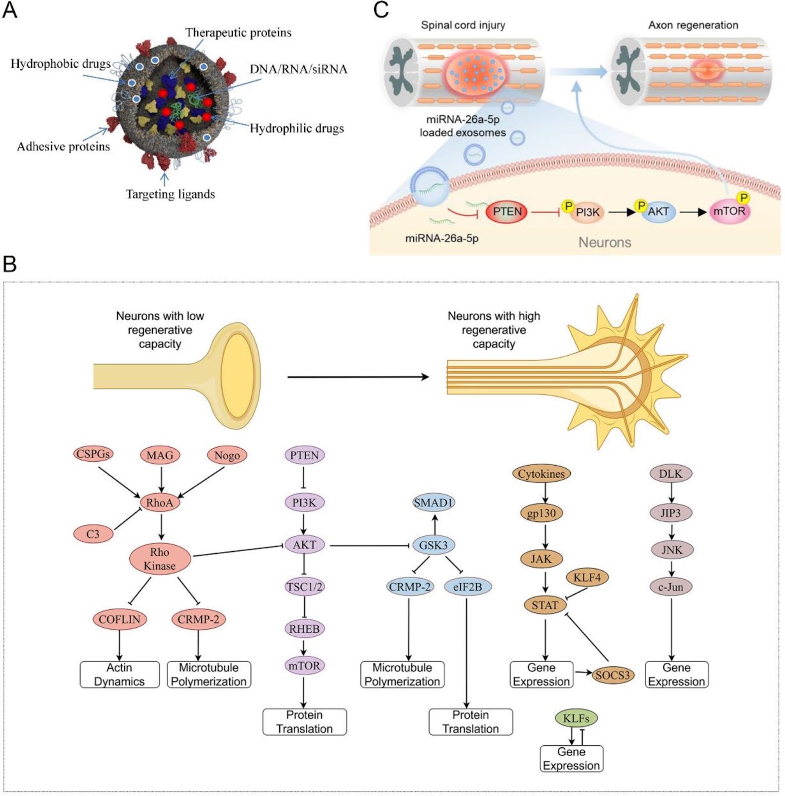
Patients with spinal cord injury (SCI) have permanent devastating motor and sensory disabilities. Secondary SCI is known for its complex progression and presents with sophisticated aberrant inflammation, vascular changes, and secondary cellular dysfunction, which aggravate the primary damage. Since their initial discovery, the potent neuroprotective effects and powerful delivery abilities of exosomes (Exos) have been reported in different research fields, including SCI. In this study, we summarize therapeutic advances related to the application of Exos in preclinical animal studies. Subsequently, we discuss the mechanisms of action of Exos derived from diverse cell types, including neurogenesis, angiogenesis, blood–spinal cord barrier preservation, anti-apoptosis, and anti-inflammatory potential. We also evaluate the relationship between the Exo delivery cargo and signaling pathways. Finally, we discuss the challenges and advantages of using Exos to offer innovative insights regarding the development of efficient clinical strategies for SCI.

Read Full Abstract10.1186/s13287-024-03952-5
Optimized administration of human embryonic stem cell-derived immunity-and-matrix regulatory cells for mouse lung injury and fibrosisGraphical AbstractVerified
Stem Cell Research & Therapy

Optimized administration of human embryonic stem cell-derived immunity-and-matrix regulatory cells for mouse lung injury and fibrosis

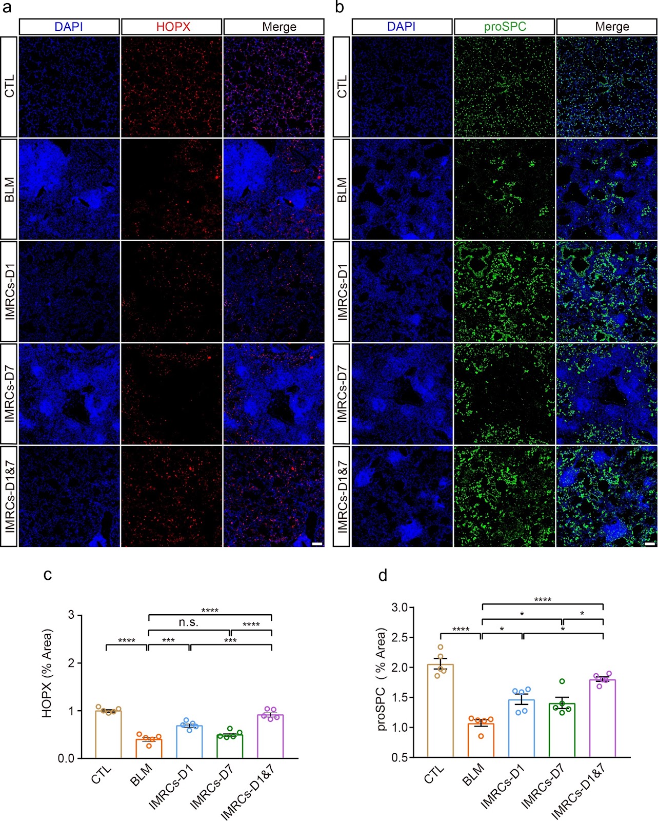
Background Lung injury and pulmonary fibrosis (PF), frequently arising as sequelae of severe and acute lung disease, currently face a dearth of effective therapeutic potions. Mesenchymal stem cells (MSCs) with immunomodulatory and tissue repair functions have immense potential to treat lung injury and PF. However, the optimal route of administration, timing, and frequency of dosing remain elusive. Human embryonic stem cell-derived immunity-and-matrix-regulatory cells (IMRCs) have shown therapeutic potential for lung injury and PF. Methods To ascertain the optimal therapeutic regimen for IMRCs in PF, we conducted an experimental study. Utilizing a mouse model of PF induced by bleomycin (BLM), IMRCs were administered via either a single or double intravenous (IV) or intratracheal (IT) injection on the first and seventh days post-BLM induction. Results Our findings revealed that IV infusion of IMRCs surpassed IT infusion in enhancing survival rates, facilitating body weight recovery, and optimizing Ashcroft and Szapiel scores among the model mice. Notably, IV administration exhibited a more profound ability to mitigate lung inflammation and fibrosis. Moreover, earlier and more frequent administrations of IMRCs were found to be advantageous in enhancing their therapeutic effects. Specifically, early administration with two IV infusions significantly improved body weight, lung organ coefficient, pulmonary ventilation and diffusion functions, and PF. This was accompanied by an increase in alveolar type I and II epithelial cells and a suppression of macrophage infiltration via CD24. Conclusion Collectively, these results suggested that IMRCs infusion ameliorated lung injury by promoting lung regeneration and inhibiting macrophage infiltration in a route, time, and frequency-dependent manner.

Read Full Abstract10.1186/s13287-024-03945-4
Autophagy modulation effect on homotypic transfer of intracellular components via tunneling nanotubes in mesenchymal stem cellsGraphical AbstractVerified
Stem Cell Research & Therapy

Autophagy modulation effect on homotypic transfer of intracellular components via tunneling nanotubes in mesenchymal stem cells

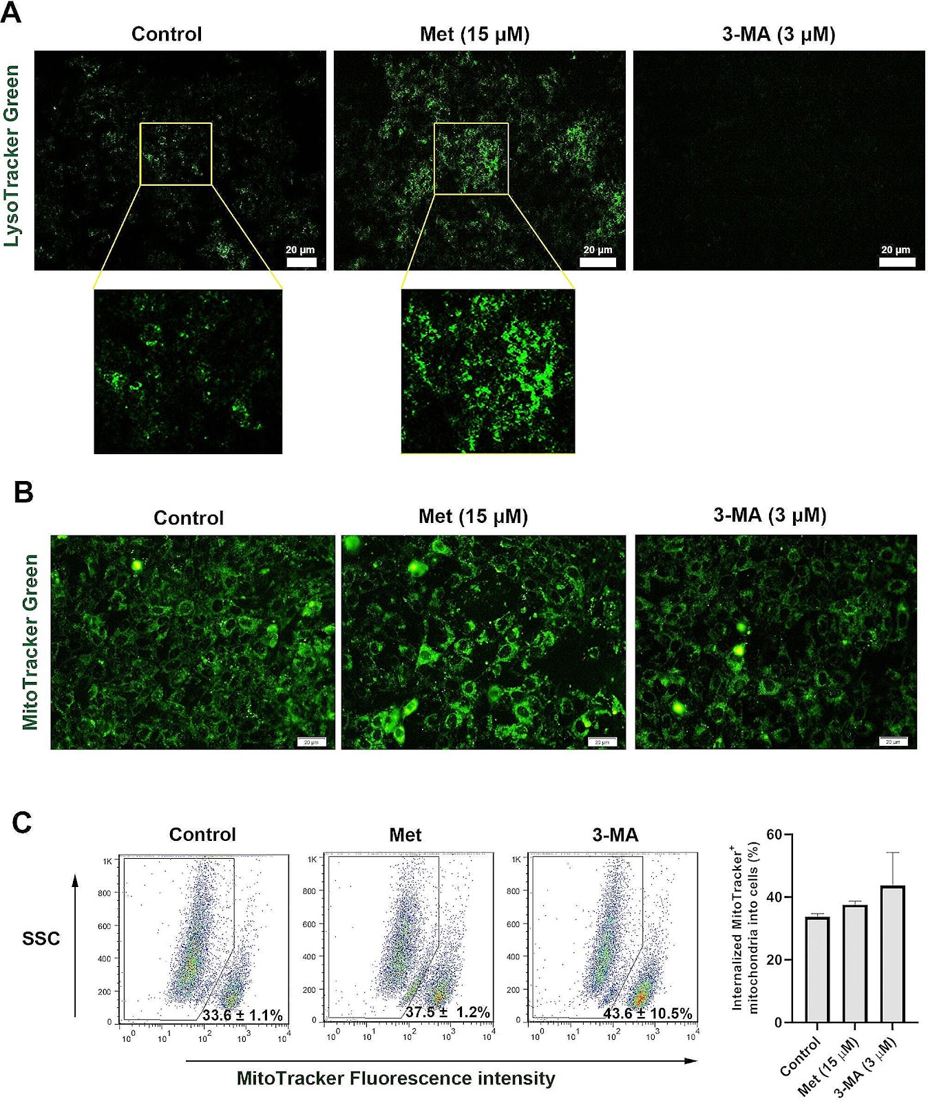
Background Recent studies have proved the role of autophagy in mesenchymal stem cell (MSCs) function and regenerative properties. How and by which mechanism autophagy modulation can affect the juxtacrine interaction of MSCs should be addressed. Here, the role of autophagy was investigated in the formation of tunneling nanotubes (TNTs) and homotypic mitochondrial donation. Methods MSCs were incubated with 15 µM Metformin (Met) and/or 3 µM 3-methyladenine (3-MA) for 48 h. The formation of TNTs was assessed using bright-field and SEM images. The mitochondria density and ΔΨ values were monitored using flow cytometry analysis. Using RT-PCR and protein array, the close interaction and shared mediators between autophagy, apoptosis, and Wnt signaling pathways were also monitored. The total fatty acid profile was assessed using gas chromatography. Result Data indicated the increase of TNT length and number, along with other cell projections after the induction of autophagy while these features were blunted in 3-MA-treated MSCs (p < 0.05). Western blotting revealed the significant reduction of Rab8 and p-FAK in 3-MA-treated MSCs (p < 0.05), indicating the inhibition of TNT assembly and vesicle transport. Likewise, the stimulation of autophagy increased autophagic flux and mitochondrial membrane integrity compared to 3-MA-treated MSCs. Despite these findings, protein levels of mitochondrial membrane Miro1 and 2 were unchanged after autophagy inhibition/stimulation (p > 0.05). We found that the inhibition/stimulation of autophagy can affect the protein, and transcription levels of several mediators related to Wnt and apoptosis signaling

Read Full Abstract10.1186/s13287-024-03813-1
Transplantation of human umbilical cord-derived mesenchymal stem cells improves age-related ovarian functional decline via regulating the local renin–angiotensin system on inflammation and oxidative stressGraphical AbstractVerified
Stem Cell Research & Therapy

Transplantation of human umbilical cord-derived mesenchymal stem cells improves age-related ovarian functional decline via regulating the local renin–angiotensin system on inflammation and oxidative stress

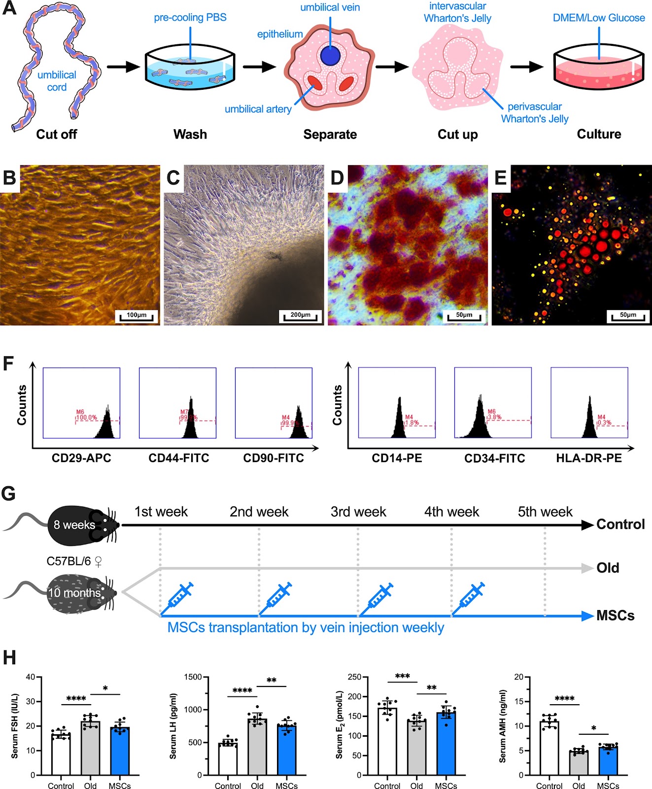
Background Age-related reproductive aging is a natural and irreversible physiological process, and delaying childbearing is increasingly common all over the world. Transplantation of mesenchymal stem cells (MSCs) is considered a new and effective therapy to restore ovarian function, but the relevant mechanisms remain unclear. Recently, it has been found that there is a local Renin–angiotensin system (RAS) in human ovary and it plays a key role. Methods After collecting follicular fluid from women who received oocyte retrieval for pure male factor infertility, the level of RAS components in it were detected, and the correlation analysis by linear regression. Then, the in vivo experiments on female C57BL/6 mice were designed to measure ovarian function, and the transcription and translation levels of RAS pathway were detected by molecular biology methods. Moreover, the role of RAS in regulating inflammation and oxidative stress in the co-culture system were explored in in vitro experiments on KGN cells. Results First, a total of 139 samples of analyzable follicular fluid were obtained. The local RAS of ovary, which is independent of systemic RAS (P > 0.05), is affected by age (Pearson r < 0, P < 0.05) and related to ovarian function, inflammation, oxidative stress indexes and assisted reproduction laboratory outcomes (P < 0.05). Next, the ovary/body weight of aging mice decreased significantly and serum sex hormones levels changed significantly (P < 0.01). The number of functional follicles decreased, while the atresia follicles increased (P < 0.05). After MSCs transplantation, all the above measures have been partially recovered (P < 0.05). Although several RAS components in aging ovary changed, MSCs only improved the expression level of AT1R (P < 0.05). Furthermore, the secretion ability and mitochondrial membrane potential of aging KGN cells decreased, while the intracellular ROS level and the aging cells ratio increased (P < 0.01). All the above measures have been partially recovered when co-cultured with MSCs (P < 0.05). After Ang(1–7) were added into the co-culture system, the above have been more significantly restored compared with Ang II (P < 0.05). Nevertheless, there was no statistical difference in estradiol level no matter which one was added (P > 0.05). Conclusions Together, our findings indicate that a novel possible mechanism to explain how stem cells restore age-related ovarian functional decline.

Read Full Abstract10.1186/s13287-024-03997-6
Osteogenic human MSC-derived extracellular vesicles regulate MSC activity and osteogenic differentiation and promote bone regeneration in a rat calvarial defect modelGraphical AbstractVerified
Stem Cell Research & Therapy

Osteogenic human MSC-derived extracellular vesicles regulate MSC activity and osteogenic differentiation and promote bone regeneration in a rat calvarial defect model

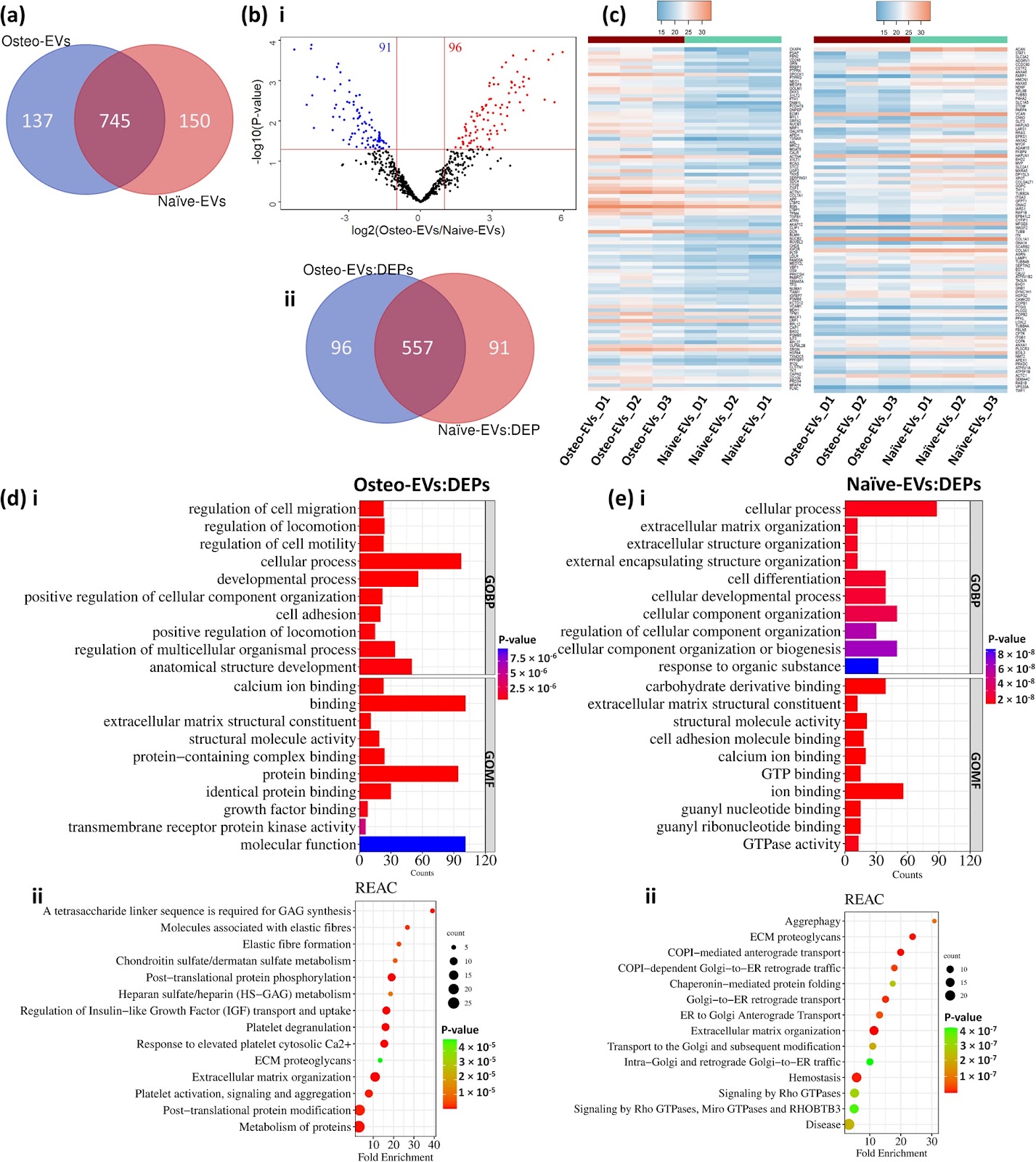
Background There is growing evidence that extracellular vesicles (EVs) play a crucial role in the paracrine mechanisms of transplanted human mesenchymal stem cells (hMSCs). Little is known, however, about the influence of microenvironmental stimuli on the osteogenic effects of EVs. This study aimed to investigate the properties and functions of EVs derived from undifferentiated hMSC (Naïve-EVs) and hMSC during the early stage of osteogenesis (Osteo-EVs). A further aim was to assess the osteoinductive potential of Osteo-EVs for bone regeneration in rat calvarial defects. Methods EVs from both groups were isolated using size-exclusion chromatography and characterized by size distribution, morphology, flow cytometry analysis and proteome profiling. The effects of EVs (10 µg/ml) on the proliferation, migration, and osteogenic differentiation of cultured hMSC were evaluated. Osteo-EVs (50 µg) or serum-free medium (SFM, control) were combined with collagen membrane scaffold (MEM) to repair critical-sized calvarial bone defects in male Lewis rats and the efficacy was assessed using µCT, histology and histomorphometry. Results Although Osteo- and Naïve-EVs have similar characteristics, proteomic analysis revealed an enrichment of bone-related proteins in Osteo-EVs. Both groups enhance cultured hMSC proliferation and migration, but Osteo-EVs demonstrate greater efficacy in promoting in vitro osteogenic differentiation, as evidenced by increased expression of osteogenesis-related genes, and higher calcium deposition. In rat calvarial defects, MEM with Osteo-EVs led to greater and more consistent bone regeneration than MEM loaded with SFM. Conclusions This study discloses differences in the protein profile and functional effects of EVs obtained from naïve hMSC and hMSC during the early stage of osteogenesis, using different methods. The significant protein profile and cellular function of EVs derived from hMSC during the early stage of osteogenesis were further verified

Read Full Abstract10.1186/s13287-024-03639-x
FDFT1 maintains glioblastoma stem cells through activation of the Akt pathwayGraphical AbstractVerified
Stem Cell Research & Therapy

FDFT1 maintains glioblastoma stem cells through activation of the Akt pathway

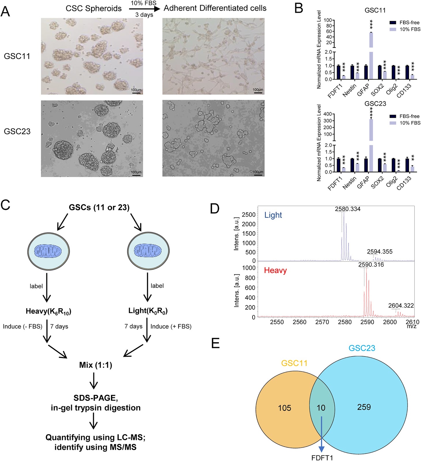
Background Cancer stem cells (CSCs) have unique metabolic characteristics and are hypothesized to contribute significantly to the recurrence and drug resistance of glioblastoma multiforme (GBM). However, the reliance on mitochondrial metabolism and the underlying mechanism of glioblastoma stem cells (GSCs) remains to be elucidated. Methods To quantify differential mitochondrial protein expression between GSCs and differentiated cells, a mass spectrum screen was applied by the Stable Isotope Labeling with Amino Acids in Cell Culture (SILAC) technique. Functional experiments including CCK8, neurosphere formation, flow cytometry, transwell, and wound healing assays were conducted to evaluate GBM cell malignant phenotype. The potential molecular mechanism of FDFT1 was screened by RNA-seq analyses. The candidate target genes were validated through RT-qPCR and western blot analyses. Results As a top candidate, FDFT1 protein expression in GSCs was elevated relative to their differentiated counterparts. Functionally, the knockdown of FDFT1 suppressed the GBM cell proliferation and migration, while simultaneously enhancing sensitivity to temozolomide. Treatment with both the FDFT1 inhibitor (YM-53601) and simvastatin (an HMG-CoA reductase inhibitor) induced apoptosis in GSCs. Mechanistically, FDFT1 was transcriptionally regulated by SREBP2 but not SREBP1. Furthermore, FDFT1 activates the AKT pathway to regulate tumor metabolism and maintain the stemness of tumor cells. Conclusions GSCs exhibit a dependency on FDFT1-mediated mevalonate metabolism. Inhibition of FDFT1 could represent a potent strategy to eliminate GSCs.

Read Full Abstract10.1186/s13287-024-04102-7
Inhibition and reversal of a TGF‑β1 induced myofibroblast phenotype by adipose tissue‑derived paracrine factorsGraphical AbstractVerified
Stem Cell Research & Therapy

Inhibition and reversal of a TGF‑β1 induced myofibroblast phenotype by adipose tissue‑derived paracrine factors

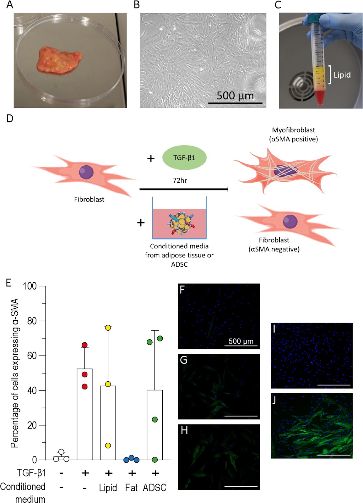
Background Hypertrophic scarring results from myofibroblast differentiation and persistence during wound healing. Currently no effective treatment for hypertrophic scarring exists however, autologous fat grafting has been shown to improve scar elasticity, appearance, and function. The aim of this study was to understand how paracrine factors from adipose tissues and adipose-derived stromal cells (ADSC) affect fibroblast to myofibroblast differentiation. Methods The transforming growth factor-β1 (TGF-β1) induced model of myofibroblast differentiation was used to test the effect of conditioned media from adipose tissue, ADSC or lipid on the proportion of fibroblasts and myofibroblasts. Results Adipose tissue conditioned media inhibited the differentiation of fibroblasts to myofibroblasts but this inhibition was not observed following treatment with ADSC or lipid conditioned media. Hepatocyte growth factor (HGF) was readily detected in the conditioned medium from adipose tissue but not ADSC. Cells treated with HGF, or fortinib to block HGF, demonstrated that HGF was not responsible for the inhibition of myofibroblast differentiation. Conditioned media from adipose tissue was shown to reduce the proportion of myofibroblasts when added to fibroblasts previously treated with TGF-β1, however, conditioned media treatment was unable to significantly reduce the proportion of myofibroblasts in cell populations isolated from scar tissue. Conclusions Cultured ADSC or adipocytes have been the focus of most studies, however, this work highlights the importance of considering whole adipose tissue to further our understanding of fat grafting. This study supports the use of autologous fat grafts for scar treatment and highlights the need for further investigation to determine the mechanism.

Read Full Abstract10.1186/s13287-024-03776-3
Current status of stem cell therapy for type 1 diabetes: a critique and a prospective considerationGraphical AbstractVerified
Stem Cell Research & Therapy

Current status of stem cell therapy for type 1 diabetes: a critique and a prospective consideration

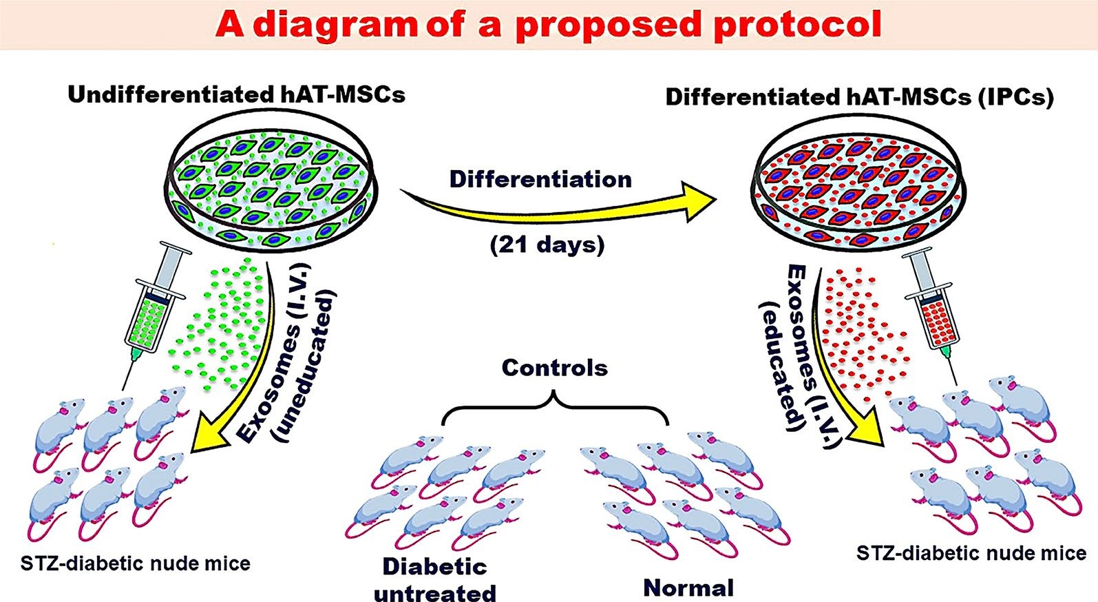
Over the past decade, there had been progress in the development of cell therapy for insulin-dependent diabetes. Nevertheless, important hurdles that need to be overcome still remain. Protocols for the differentiation of pluripotent stem cells into pancreatic progenitors or fully differentiated β-cells have been developed. The resulting insulin-producing cells can control chemically induced diabetes in rodents and were the subject of several clinical trials. However, these cells are immunogenic and possibly teratogenic for their transplantation, and an immunoisolation device and/or immunosuppression is needed. A growing number of studies have utilized genetic manipulations to produce immune evasive cells. Evidence must be provided that in addition to the expected benefit, gene manipulations should not lead to any unforeseen complications. Mesenchymal stem/stromal cells (MSCs) can provide a viable alternative. MSCs are widely available from many tissues. They can form insulin-producing cells by directed differentiation. Experimentally, evidence has shown that the transplantation of allogenic insulin-producing cells derived from MSCs is associated with a muted allogeneic response that does not interfere with their functionality. This can be explained by the immunomodulatory functions of the MSC subpopulation that did not differentiate into insulin-producing cells. Recently, exosomes derived from naive MSCs have been used in the experimental domain to treat diabetes in rodents with varying degrees of success. Several mechanisms for their beneficial functions were proposed including a reduction in insulin resistance, the promotion of autophagy, and an increase in the T regulatory population. However, euglycemia was not achieved in any of these experiments. We suggest that exosomes derived from β-cells or insulin-producing cells (educated) can provide a better therapeutic effect than those derived from undifferentiated cells.

Read Full Abstract10.1186/s13287-024-03636-0
Characteristics of HPC(A) product obtained from a donor with SARS-CoV-2 infection and outcome of autologous transplantGraphical AbstractVerified
Stem Cell Research & Therapy

Characteristics of HPC(A) product obtained from a donor with SARS-CoV-2 infection and outcome of autologous transplant

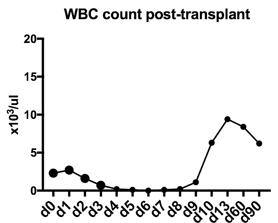
Collection of hematopoietic progenitor cell products [HPC(A)] is deferred if the donor is symptomatic and tests positive for Covid-19. However, donor questionnaires are subjective and may miss minimally symptomatic donors. Alternatively, myalgia associated with Covid-19 infection can be falsely dismissed as an adverse effect of granulocyte stimulating factor (Filgrastim) administered prior to product collection. The likelihood of donors with an underlying acute but minimally symptomatic infection undergoing successful product collection is significant. In these circumstances, it is less known whether Covid-19 infection results in product viremia or alters the clinical outcome of transplant. We aimed to evaluate the above question by studying a donor whose product was collected during acute Covid-19 infection. Aliquots of the product tested negative for SARS-CoV-2 RNA by reverse-transcriptase polymerase chain reaction assay (RT-PCR). Importantly, the donor received an autologous stem cell transplant using the product collected at the time of infection, and their case will be described in this report. We describe one of the very few reports of successful transplant of HPC(A) product collected during acute Covid-19 infection.

Read Full Abstract10.1186/s13287-024-03872-4
The role of biophysical cues and their modulated exosomes in dental diseases: from mechanism to therapyGraphical AbstractVerified
Stem Cell Research & Therapy

The role of biophysical cues and their modulated exosomes in dental diseases: from mechanism to therapy

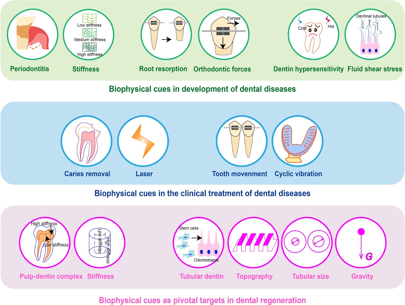
Dental diseases such as caries and periodontitis have been common public health problems. Dental disease treatment can be achieved through stem cell-based dental regeneration. Biophysical cues determine the fate of stem cells and govern the success of dental regeneration. Some studies have manifested exosomes derived from stem cells could not only inherit biophysical signals in microenvironment but also evade some issues in the treatment with stem cells. Nowadays, biophysical cue-regulated exosomes become another promising therapy in dental regenerative medicine. However, methods to improve the efficacy of exosome therapy and the underlying mechanisms are still unresolved. In this review, the association between biophysical cues and dental diseases was summarized. We retrospected the role of exosomes regulated by biophysical cues in curing dental diseases and promoting dental regeneration. Our research also delved into the mechanisms by which biophysical cues control the biogenesis, release, and uptake of exosomes, as well as potential methods to enhance the effectiveness of exosomes. The aim of this review was to underscore the important place biophysical cue-regulated exosomes occupy in the realm of dentistry, and to explore novel targets for dental diseases.

Read Full Abstract10.1186/s13287-024-03990-z
Astilbin improves the therapeutic effects of mesenchymal stem cells in AKI-CKD mice by regulating macrophage polarization through PTGS2-mediated pathwayGraphical AbstractVerified
Stem Cell Research & Therapy

Astilbin improves the therapeutic effects of mesenchymal stem cells in AKI-CKD mice by regulating macrophage polarization through PTGS2-mediated pathway

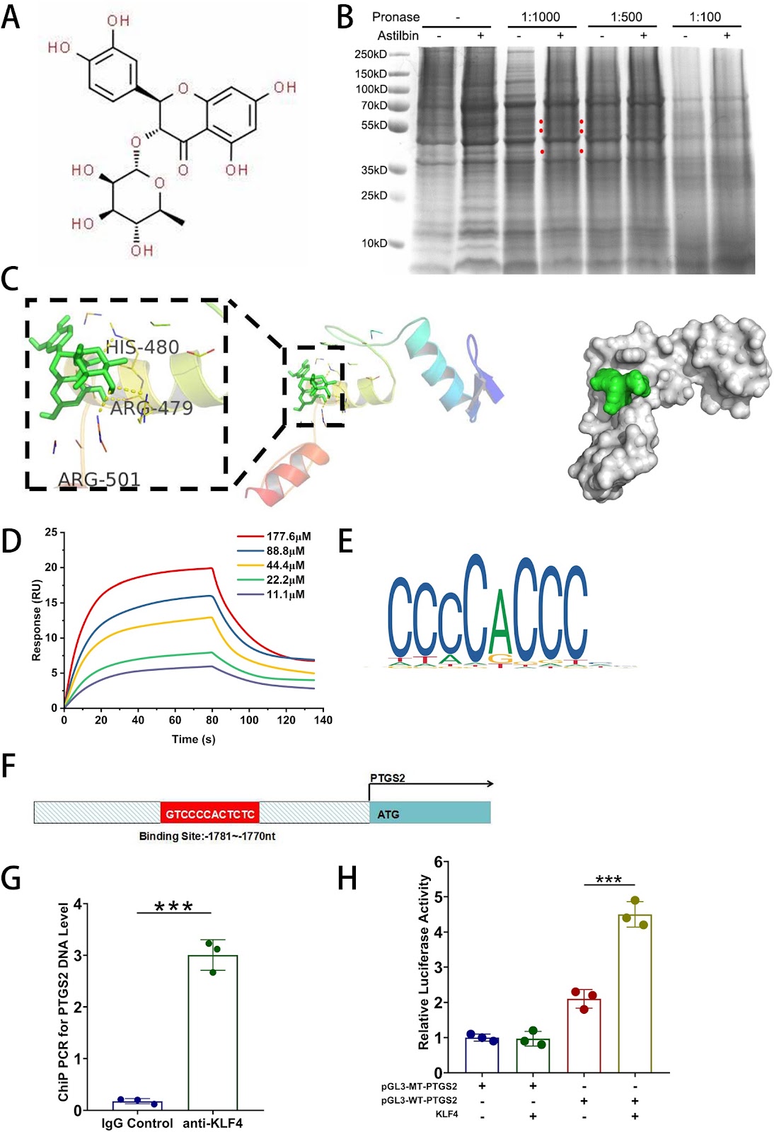
Background Although mesenchymal stem cells (MSCs) have been proven to be appropriate candidates for the treatment of AKI-CKD, their efficacy is limited and variable. Astilbin (AST) had a protective effect on MSCs from oxidative stress via ROS-scavenging, however, whether it can improve MSCs’ renoprotection and the underlying mechanism need to be elucidated. Methods AST-pretreated MSCs were administered intravenously into the ischemia–reperfusion injury mice models and the renal function, pathological changes and inflammation. Were evaluated. In addition, DARTS, molecular docking, surface plasma resonance(SPR), dual-luciferase reporter gene assay and the ChIP-PCR were utilized to explore the potential signaling pathways through which AST exert renal protective effects on MSCs. Results AST-pretreated MSCs markedly improved kidney function, reduced kidney pathological injury and inflammation in AKI and AKI-CKD mice. RNA-seq results showed that PTGS2 related pathway was significantly up-regulated in MSCs after AST pretreatment. DARTS assay, molecular docking and SPR assay revealed that AST could bind with the transcriptional factor of Kruppel-Like Factor 4(KLF4) protein. The promoter of PTGS2 had the binding and transcriptional activation by KLF4. Furthermore, AST pretreatment promoted the secretion of PGE2 in MSCs. And then the westren blot results showed that the protein levels of CD163 and CD206 were upregulated after coculture in AST-pretreated MSCs, indicating that the polarization of RAW264.7 cells towards M2-like macrophages was induced. Knockdown of PTGS2 reversed the ability of AST-pretreated MSCs in converting macrophages to M2 phenotype and reducing their therapeutic effects on AKI-CKD mice. Conclusion AST pretreatment enhances the efficacy of MSCs on AKI and AKI-CKD mice by inducing of M2-like phenotype polarization in macrophages through the PTGS2-mediated pathway. This approach not only provides a novel strategy to strengthen the capability of MSCs but also helps elucidate the beneficial effects of the Chinese herbal medicine AST.

Read Full Abstract10.1186/s13287-024-04025-3
Identification of potential biomarkers for aging diagnosis of mesenchymal stem cells derived from the aged donorsGraphical AbstractVerified
Stem Cell Research & Therapy

Identification of potential biomarkers for aging diagnosis of mesenchymal stem cells derived from the aged donors

Background The clinical application of human bone-marrow derived mesenchymal stem cells (MSCs) for the treatment of refractory diseases has achieved remarkable results. However, there is a need for a systematic evaluation of the quality and safety of MSCs sourced from donors. In this study, we sought to assess one potential factor that might impact quality, namely the age of the donor. Methods We downloaded two data sets from each of two Gene Expression Omnibus (GEO), GSE39035 and GSE97311 databases, namely samples form young (<65 years of age) and old (>65) donor groups. Through, bioinformatics analysis and experimental validation to these retrieved data, we found that MSCs derived from aged donors can lead to differential expression of gene profiles compared with those from young donors, and potentially affect the function of MSCs, and may even induce malignant tumors. Results We identified a total of 337 differentially expressed genes (DEGs), including two upregulated and eight downregulated genes from the databases of both GSE39035 and GSE97311. We further identified 13 hub genes. Six of them, TBX15, IGF1, GATA2, PITX2, SNAI1 and VCAN, were highly expressed in many human malignancies in Human Protein Atlas database. In the MSCs in vitro senescent cell model, qPCR analysis validated that all six hub genes were highly expressed in senescent MSCs. Our findings confirm that aged donors of MSCs have a significant effect on gene expression profiles. The MSCs from old donors have the potential to cause a variety of malignancies. These TBX15, IGF1, GATA2, PITX2, SNAI1, VCAN genes could be used as potential biomarkers to diagnosis aging state of donor MSCs, and evaluate whether MSCs derived from an aged donor could be used for therapy in the clinic. Our findings provide a diagnostic basis for the clinical use of MSCs to treat a variety of diseases. Conclusions Therefore, our findings not only provide guidance for the safe and standardized use of MSCs in the clinic for the treatment of various diseases, but also provide insights into the use of cell regeneration approaches to reverse aging and support rejuvenation.

Read Full Abstract10.1186/s13287-024-03689-1
Rankl genetic deficiency and functional blockade undermine skeletal stem and progenitor cell differentiationGraphical AbstractVerified
Stem Cell Research & Therapy

Rankl genetic deficiency and functional blockade undermine skeletal stem and progenitor cell differentiation

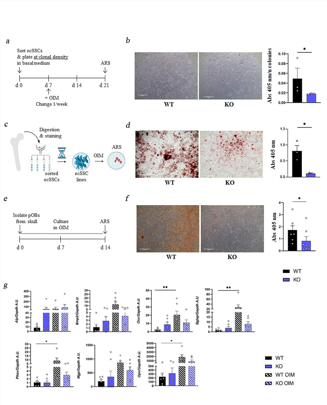
Background Skeletal Stem Cells (SSCs) are required for skeletal development, homeostasis, and repair. The perspective of their wide application in regenerative medicine approaches has supported research in this field, even though so far results in the clinic have not reached expectations, possibly due also to partial knowledge of intrinsic, potentially actionable SSC regulatory factors. Among them, the pleiotropic cytokine RANKL, with essential roles also in bone biology, is a candidate deserving deep investigation. Methods To dissect the role of the RANKL cytokine in SSC biology, we performed ex vivo characterization of SSCs and downstream progenitors (SSPCs) in mice lacking Rankl (Rankl−/−) by means of cytofluorimetric sorting and analysis of SSC populations from different skeletal compartments, gene expression analysis, and in vitro osteogenic differentiation. In addition, we assessed the effect of the pharmacological treatment with the anti-RANKL blocking antibody Denosumab (approved for therapy in patients with pathological bone loss) on the osteogenic potential of bone marrow-derived stromal cells from human healthy subjects (hBMSCs). Results We found that, regardless of the ossification type of bone, osteochondral SSCs had a higher frequency and impaired differentiation along the osteochondrogenic lineage in Rankl−/− mice as compared to wild-type. Rankl−/− mice also had increased frequency of committed osteochondrogenic and adipogenic progenitor cells deriving from perivascular SSCs. These changes were not due to the peculiar bone phenotype of increased density caused by lack of osteoclast resorption (defined osteopetrosis); indeed, they were not found in another osteopetrotic mouse model, i.e., the oc/oc mouse, and were therefore not due to osteopetrosis per se. In addition, Rankl−/− SSCs and primary osteoblasts showed reduced mineralization capacity. Of note, hBMSCs treated in vitro with Denosumab had reduced osteogenic capacity compared to control cultures. Conclusions We provide for the first time the characterization of SSPCs from mouse models of severe recessive osteopetrosis. We demonstrate that Rankl genetic deficiency in murine SSCs and functional blockade in hBMSCs reduce their osteogenic potential. Therefore, we propose that RANKL is an important regulatory factor of SSC features with translational relevance.

Read Full Abstract10.1186/s13287-024-03803-3
Advancements in cell-based therapies for thermal burn wounds: a comprehensive systematic review of clinical trials outcomesGraphical AbstractVerified
Stem Cell Research & Therapy

Advancements in cell-based therapies for thermal burn wounds: a comprehensive systematic review of clinical trials outcomes

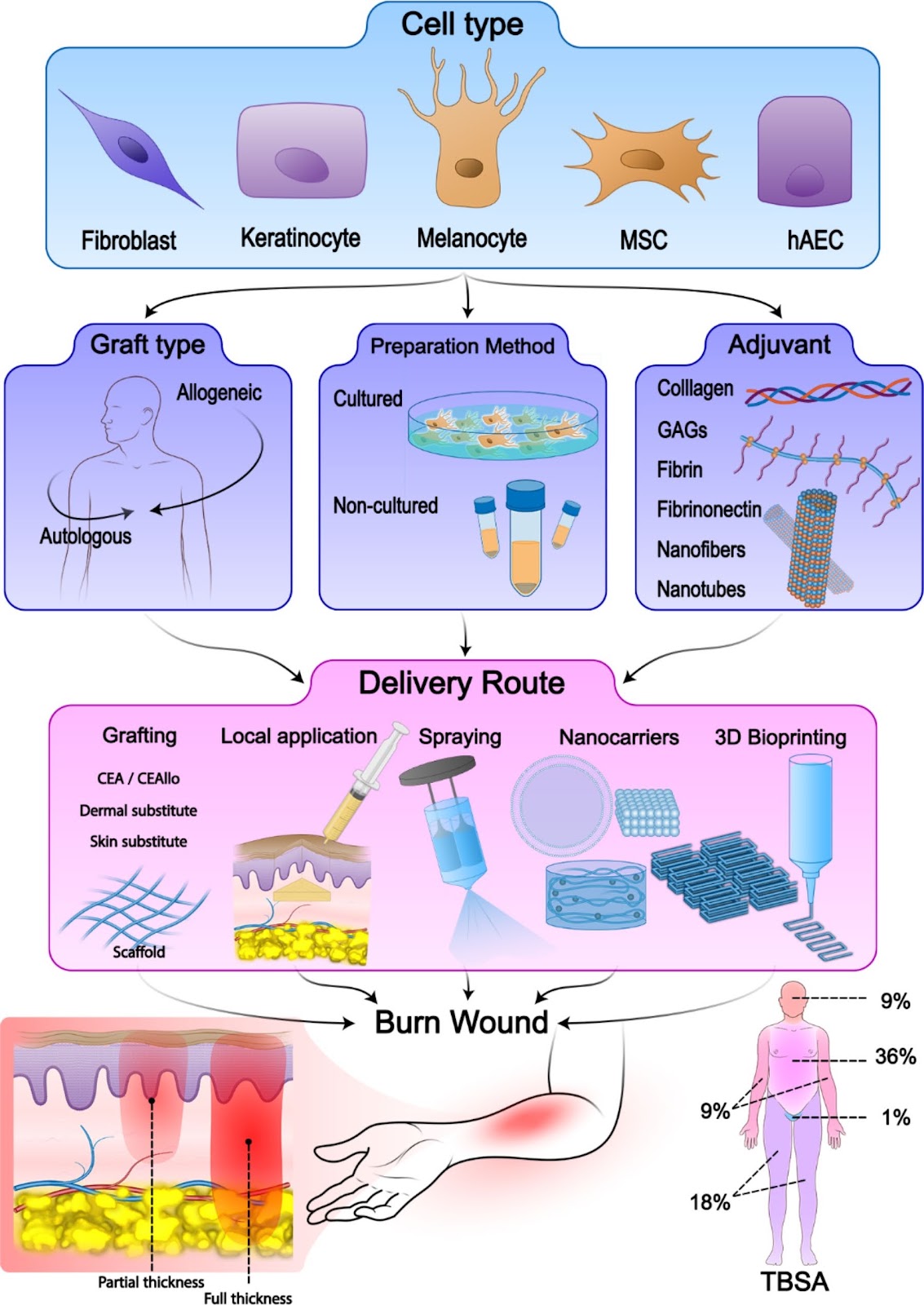
Background Burn trauma is one of the major causes of morbidity and mortality worldwide. The standard management of burn wounds consists of early debridement, dressing changes, surgical management, and split-thickness skin autografts (STSGs). However, there are limitations for the standard management that inclines us to find alternative treatment approaches, such as innovative cell-based therapies. We aimed to systematically review the different aspects of cell-based treatment approaches for burn wounds in clinical trials. Methods A systematic search through PubMed, Medline, Embase, and Cochrane Library databases was carried out using a combination of keywords, including “Cell transplantation”, “Fibroblast”, “Keratinocyte”, “Melanocyte”, or “Stem Cell” with “Burn”, “Burn wound”, or “Burn injury”. Firstly, titles and abstracts of the studies existing in these databases until “February 2024” were screened. Then, the selected studies were read thoroughly, and considering the inclusion and exclusion criteria, final articles were included in this systematic review. Moreover, a manual search was performed through the reference lists of the included studies to minimize the risk of missing reports. Results Overall, 30 clinical trials with 970 patients were included in our study. Considering the type of cells, six studies used keratinocytes, nine used fibroblasts, eight used combined keratinocytes and fibroblasts, one study used combined keratinocytes and melanocytes, five used combined keratinocytes and fibroblasts and melanocytes, and one study used mesenchymal stem cells (MSCs). Evaluation of the preparation type in these studies showed that cultured method was used in 25 trials, and non-cultured method in 5 trials. Also, the graft type of 17 trials was allogeneic, and of 13 other trials was autologous. Conclusions Our study showed that employing cell-based therapies for the treatment of burn wounds have significant results in clinical studies and are promising approaches that can be considered as alternative treatments in many cases. However, choosing appropriate cell-based treatment for each burn wound is essential and depends on the situation of each patient.

Read Full Abstract10.1186/s13287-024-03901-2
Mesenchymal stem cells promote ovarian reconstruction in miceGraphical AbstractVerified
Stem Cell Research & Therapy

Mesenchymal stem cells promote ovarian reconstruction in mice

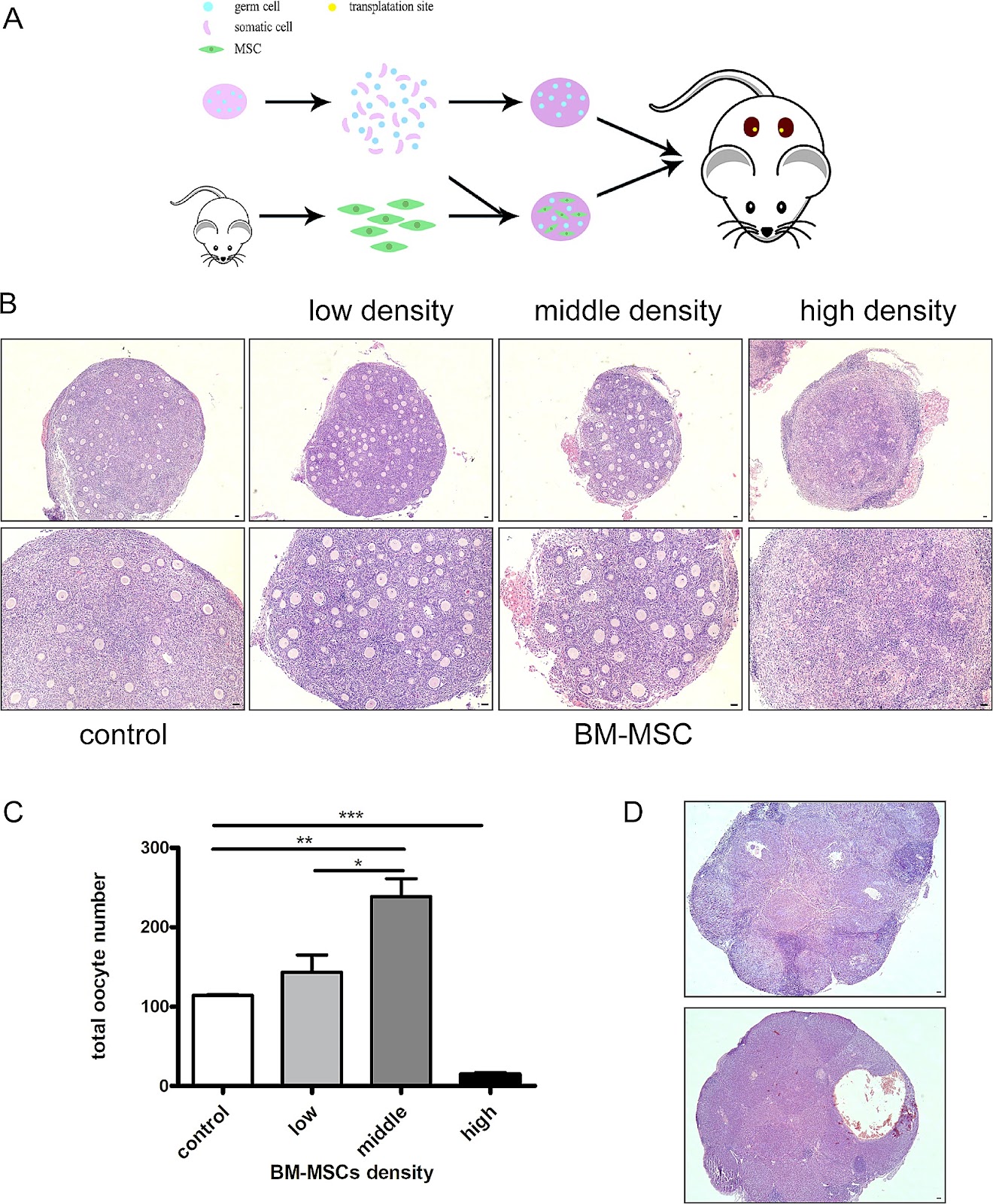
Background Studies have shown that chemotherapy and radiotherapy can cause premature ovarian failure and loss of fertility in female cancer patients. Ovarian cortex cryopreservation is a good choice to preserve female fertility before cancer treatment. Following the remission of the disease, the thawed ovarian tissue can be transplanted back and restore fertility of the patient. However, there is a risk to reintroduce cancer cells in the body and leads to the recurrence of cancer. Given the low success rate of current in vitro culture techniques for obtaining mature oocytes from primordial follicles, an artificial ovary with primordial follicles may be a good way to solve this problem. Methods In the study, we established an artificial ovary model based on the participation of mesenchymal stem cells (MSCs) to evaluate the effect of MSCs on follicular development and oocyte maturation. P2.5 mouse ovaries were digested into single cell suspensions and mixed with bone marrow derived mesenchymal stem cells (BM-MSCs) at a 1:1 ratio. The reconstituted ovarian model was then generated by using phytohemagglutinin. The phenotype and mechanism studies were explored by follicle counting, immunohistochemistry, immunofluorescence, in vitro maturation (IVM), in vitro fertilization (IVF), real-time quantitative polymerase chain reaction (RT-PCR), and Terminal-deoxynucleotidyl transferase mediated nick end labeling(TUNEL) assay. Results Our study found that the addition of BM-MSCs to the reconstituted ovary can enhance the survival of oocytes and promote the growth and development of follicles. After transplanting the reconstituted ovaries under kidney capsules of the recipient mice, we observed normal folliculogenesis and oocyte maturation. Interestingly, we found that BM-MSCs did not contribute to the formation of follicles in ovarian aggregation, nor did they undergo proliferation during follicle growth. Instead, the cells were found to be located around growing follicles in the reconstituted ovary. When theca cells were labeled with CYP17a1, we found some overlapped staining with green fluorescent protein(GFP)-labeled BM-MSCs. The results suggest that BM-MSCs may participate in directing the differentiation of theca layer in the reconstituted ovary. Conclusions The presence of BM-MSCs in the artificial ovary was found to promote the survival of ovarian cells, as well as facilitate follicle formation and development. Since the cells didn’t proliferate in the reconstituted ovary, this

Read Full Abstract10.1186/s13287-024-03718-z
LncRNA SNHG1 enhances cartilage regeneration by modulating chondrogenic differentiation and angiogenesis potentials of JBMMSCs via mitochondrial function regulationGraphical AbstractVerified
Stem Cell Research & Therapy

LncRNA SNHG1 enhances cartilage regeneration by modulating chondrogenic differentiation and angiogenesis potentials of JBMMSCs via mitochondrial function regulation

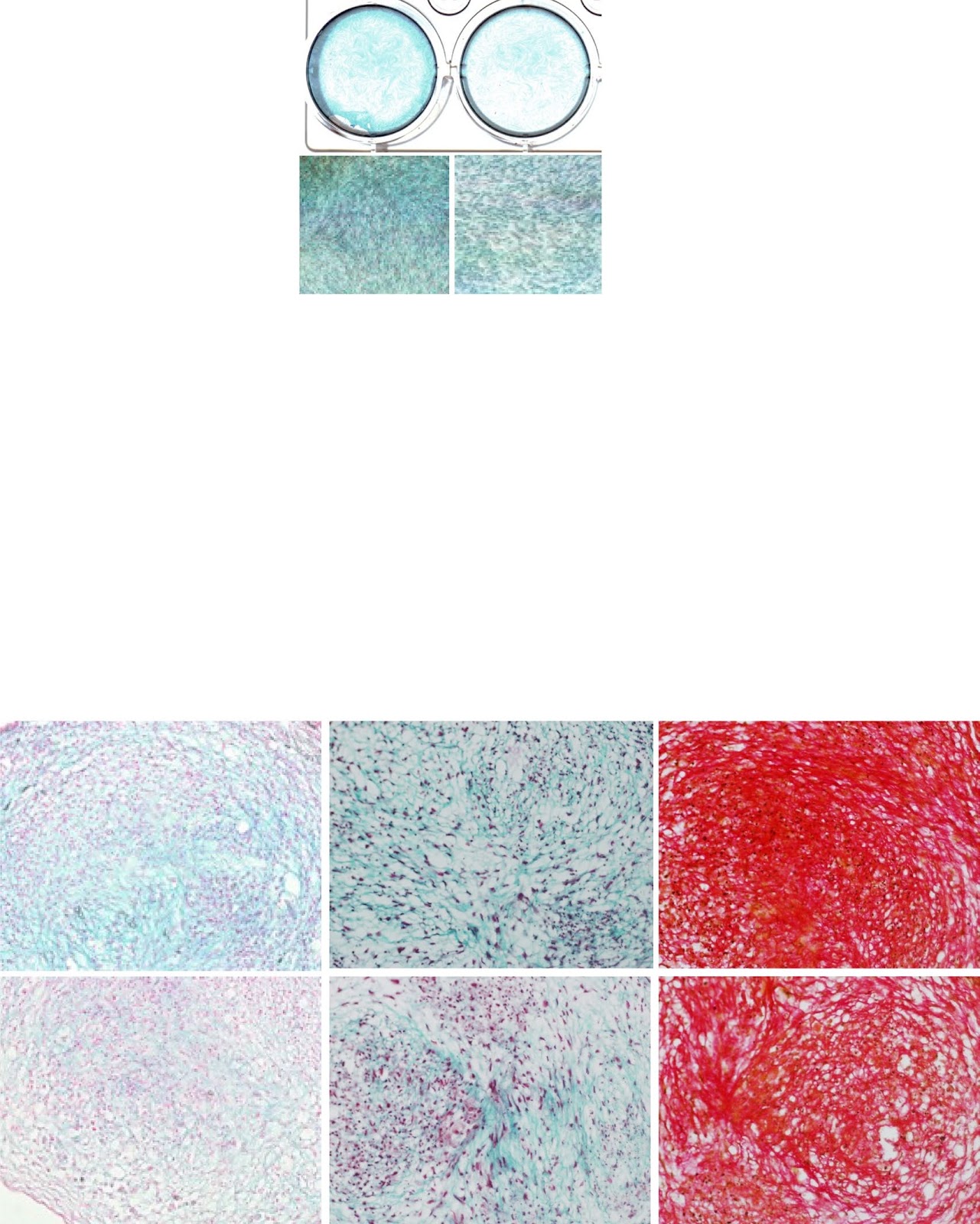
Background Cartilage is a kind of avascular tissue, and it is difficult to repair itself when it is damaged. In this study, we investigated the regulation of chondrogenic differentiation and vascular formation in human jaw bone marrow mesenchymal stem cells (h-JBMMSCs) by the long-chain noncoding RNA small nucleolar RNA host gene 1 (SNHG1) during cartilage tissue regeneration. Methods JBMMSCs were isolated from the jaws via the adherent method. The effects of lncRNA SNHG1 on the chondrogenic differentiation of JBMMSCs in vitro were detected by real-time fluorescence quantitative polymerase chain reaction (RT-qPCR), Pellet experiment, Alcian blue staining, Masson’s trichrome staining, and modified Sirius red staining. RT-qPCR, matrix gel tube formation, and coculture experiments were used to determine the effect of lncRNA SNHG1 on the angiogenesis in JBMMSCs in vitro. A model of knee cartilage defects in New Zealand rabbits and a model of subcutaneous matrix rubber suppositories in nude mice were constructed for in vivo experiments. Changes in mitochondrial function were detected via RT-qPCR, dihydroethidium (DHE) staining, MitoSOX staining, tetramethyl rhodamine methyl ester (TMRM) staining, and adenosine triphosphate (ATP) detection. Western blotting was used to detect the phosphorylation level of signal transducer and activator of transcription 3 (STAT3). Results Alcian blue staining, Masson’s trichrome staining, and modified Sirius Red staining showed that lncRNA SNHG1 promoted chondrogenic differentiation. The lncRNA SNHG1 promoted angiogenesis in vitro and the formation of microvessels in vivo. The lncRNA SNHG1 promoted the repair and regeneration of rabbit knee cartilage tissue. Western blot and alcian blue staining showed that the JAK inhibitor reduced the increase of STAT3 phosphorylation level and staining deepening caused by SNHG1. Mitochondrial correlation analysis revealed that the lncRNA SNHG1 led to a decrease in reactive oxygen species (ROS) levels, an increase in mitochondrial membrane potential

Read Full Abstract10.1186/s13287-024-03793-2