🧬 SinoBioData Academic Portal
📚 Peer-Reviewed Translated Literature

All Biomedical & Clinical Articles (Page 41)

Browse complete peer-reviewed translations from top Chinese biomedical, oncology, and genomics journals. Read verified previews and download full authentic clinical reports.

Published Research Papers

Showing 24 of 1542 peer-reviewed translated articles (Page 41 of 65)

Embryonic stem cells overexpressing high molecular weight FGF2 isoform enhance recovery of pre-ganglionic spinal root lesion in combination with fibrin biopolymer mediated root repairGraphical AbstractVerified
Stem Cell Research & Therapy

Embryonic stem cells overexpressing high molecular weight FGF2 isoform enhance recovery of pre-ganglionic spinal root lesion in combination with fibrin biopolymer mediated root repair

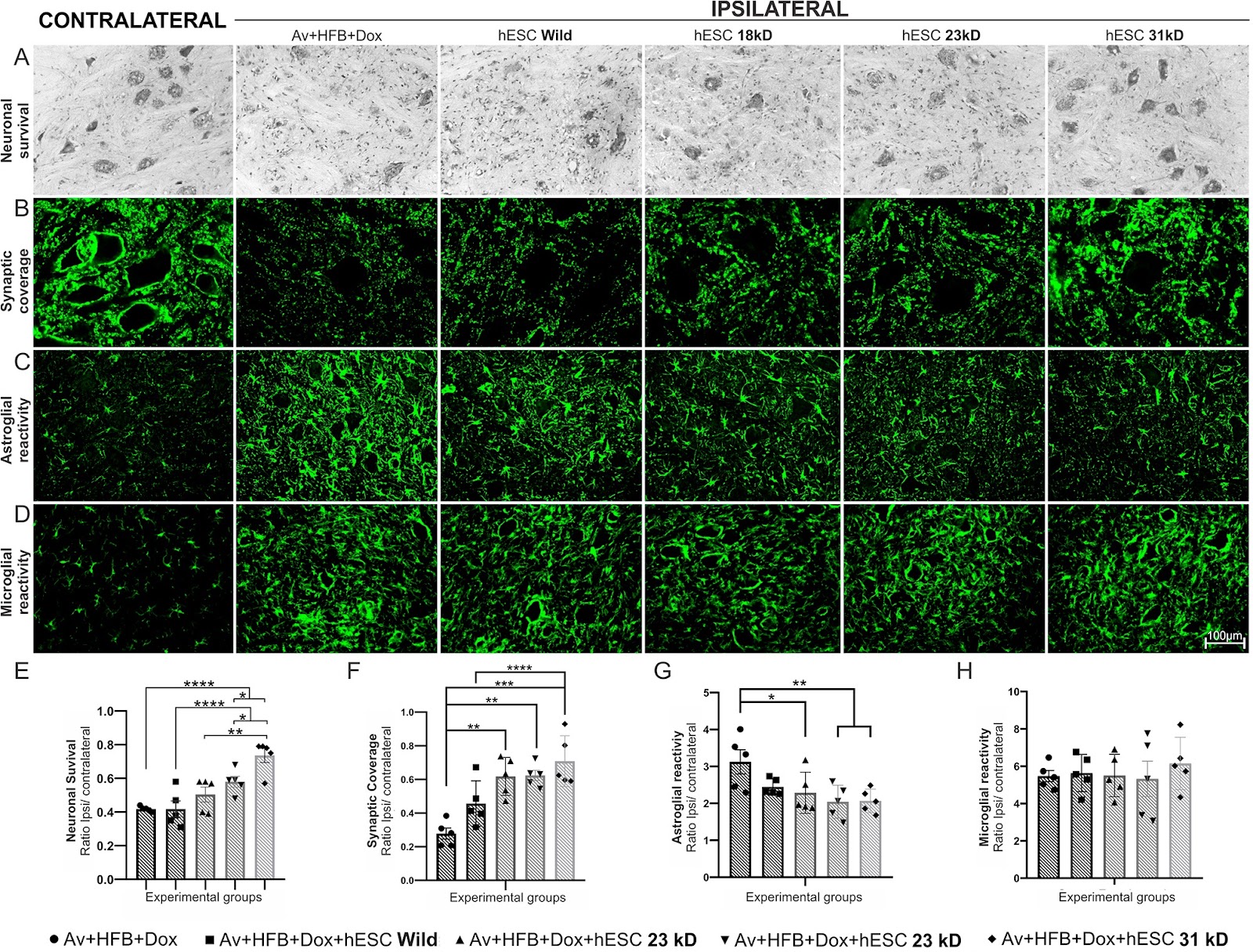
Background Spinal ventral root avulsion results in massive motoneuron degeneration with poor prognosis and high costs. In this study, we compared different isoforms of basic fibroblast growth factor 2 (FGF2), overexpressed in stably transfected Human embryonic stem cells (hESCs), following motor root avulsion and repair with a heterologous fibrin biopolymer (HFB). Methods In the present work, hESCs bioengineered to overexpress 18, 23, and 31 kD isoforms of FGF2, were used in combination with reimplantation of the avulsed roots using HFB. Statistical analysis was conducted using GraphPad Prism software with one-way or two-way ANOVA, followed by Tukey’s or Dunnett’s multiple comparison tests. Significance was set at *p < 0.05, **p < 0.01, ***p < 0.001, and ****p < 0.0001. Results For the first set of experiments, rats underwent avulsion of the ventral roots with local administration of HFB and engraftment of hESCs expressing the above-mentioned FGF2 isoforms. Analysis of motoneuron survival, glial reaction, and synaptic coverage, two weeks after the lesion, indicated that therapy with hESCs overexpressing 31 kD FGF2 was the most effective. Consequently, the second set of experiments was performed with that isoform, so that ventral root avulsion was followed by direct spinal cord reimplantation. Motoneuron survival, glial reaction, synaptic coverage, and gene expression were analyzed 2 weeks post-lesion; while the functional recovery was evaluated by the walking track test and von Frey test for 12 weeks. We showed that engraftment of hESCs led to significant neuroprotection, coupled with immunomodulation, attenuation of astrogliosis, and preservation of inputs to the rescued motoneurons. Behaviorally, the 31 kD FGF2 - hESC therapy enhanced both motor and sensory recovery. Conclusion Transgenic hESCs were an effective delivery platform for neurotrophic factors, rescuing axotomized motoneurons and modulating glial response after proximal spinal cord root injury, while the 31 kD isoform of FGF2 showed superior regenerative properties over other isoforms in addition to the significant functional recovery.
Read Full Abstract10.1186/s13287-024-03676-6
miR-340-3p-modified bone marrow mesenchymal stem cell-derived exosomes inhibit ferroptosis through METTL3-mediated m6A modification of HMOX1 to promote recovery of injured rat uterusGraphical AbstractVerified
Stem Cell Research & Therapy

miR-340-3p-modified bone marrow mesenchymal stem cell-derived exosomes inhibit ferroptosis through METTL3-mediated m6A modification of HMOX1 to promote recovery of injured rat uterus

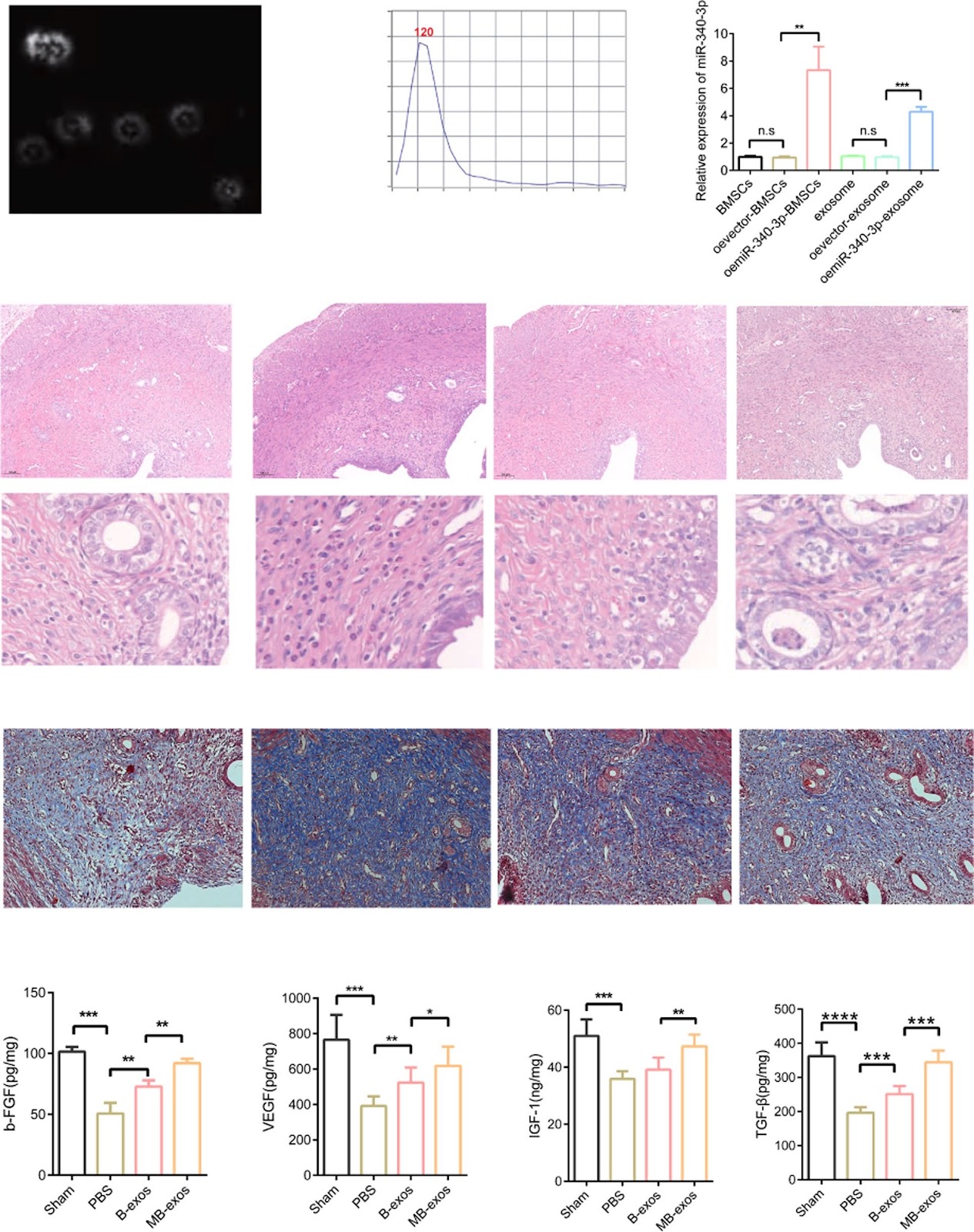
Background Ferroptosis is associated with the pathological progression of hemorrhagic injury and ischemia–reperfusion injury. According to our previous study, exosomes formed through bone marrow mesenchymal stem cells modified with miR-340-3p (MB-exos) can restore damaged endometrium. However, the involvement of ferroptosis in endometrial injury and the effect of MB-exos on ferroptosis remain elusive. Methods The endometrial injury rat model was developed. Exosomes were obtained from the supernatants of bone marrow mesenchymal stromal cells (BMSCs) and miR-340/BMSCs through differential centrifugation. We conducted RNA-seq analysis on endometrial tissues obtained from the PBS and MB-exos groups. Ferroptosis was induced in endometrial stromal cells (ESCs) by treating them with erastin or RSL3, followed by treatment with B-exos or MB-exos. We assessed the endometrial total m6A modification level after injury and subsequent treatment with B-exos or MB-exos by methylation quantification assay. We performed meRIP-qPCR to analyze m6A modification-regulated endogenous mRNAs. Results We reveal that MB-exos facilitate the injured endometrium to recover by suppressing ferroptosis in endometrial stromal cells. The injured endometrium showed significantly upregulated N6-methyladenosine (m6A) modification levels; these levels were attenuated by MB-exos through downregulation of the methylase METTL3. Intriguingly, METTL3 downregulation appears to repress ferroptosis by stabilizing HMOX1 mRNA, thereby potentially elucidating the mechanism through which MB-exos inhibit ferroptosis in ESCs. We identified YTHDF2 as a critical m6A reader protein that contributes to HMOX1 mRNA degradation. YTHDF2 facilitates HMOX1 mRNA degradation by identifying the m6A binding site in the 3′-untranslated regions of HMOX1. In a rat model, treatment with MB-exos ameliorated endometrial injury-induced fibrosis by inhibiting ferroptosis in ESCs. Moreover, METTL3 short hairpin RNA-mediated inhibition of m6A modification enhanced the inhibitory effect of MB-exos on ferroptosis in endometrial injury. Conclusions Thus, these observations provide new insights regarding the molecular mechanisms responsible for endometrial recovery promotion by MB-exos and highlight m6A modification-dependent ferroptosis inhibition as a prospective therapeutic target to attenuate endometrial injury.

Read Full Abstract10.1186/s13287-024-03846-6
Glutamine-αKG axis affects dentin regeneration and regulates osteo/odontogenic differentiation of mesenchymal adult stem cells via IGF2 m6A modificationGraphical AbstractVerified
Stem Cell Research & Therapy

Glutamine-αKG axis affects dentin regeneration and regulates osteo/odontogenic differentiation of mesenchymal adult stem cells via IGF2 m6A modification

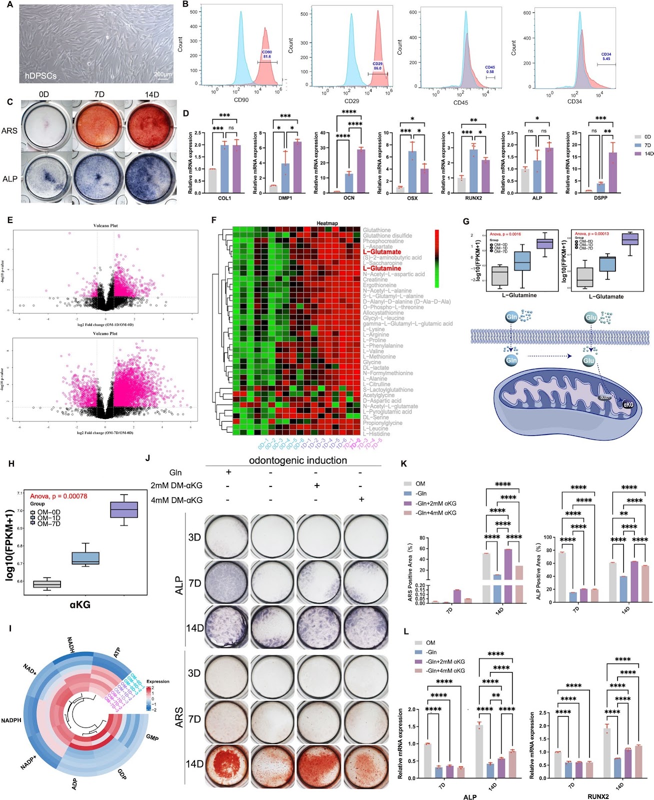
Background Multi-lineage differentiation of mesenchymal adult stem cells (m-ASCs) is crucial for tissue regeneration and accompanied with metabolism reprogramming, among which dental-pulp-derived m-ASCs has obvious advantage of easy accessibility. Stem cell fate determination and differentiation are closely related to metabolism status in cell microenvironment, which could actively interact with epigenetic modification. In recent years, glutamine-α-ketoglutarate (αKG) axis was proved to be related to aging, tumorigenesis, osteogenesis etc., while its role in m-ASCs still lack adequate research evidence. Methods We employed metabolomic analysis to explore the change pattern of metabolites during dental-pulp-derived m-ASCs differentiation. A murine incisor clipping model was established to investigate the influence of αKG on dental tissue repairment. shRNA technique was used to knockdown the expression of related key enzyme-dehydrogenase 1(GLUD1). RNA-seq, m6A evaluation and MeRIP-qPCR were used to dig into the underlying epigenetic mechanism. Results Here we found that the glutamine-αKG axis displayed an increased tendency along with the osteo/odontogenic differentiation of dental-pulp-derived m-ASCs, same as expression pattern of GLUD1. Further, the key metabolite αKG was found able to accelerate the repairment of clipped mice incisor and promote dentin formation. Exogenous DM-αKG was proved able to promote osteo/odontogenic differentiation of dental-pulp-derived m-ASCs, while the inhibition of glutamine-derived αKG level via GLUD1 knockdown had the opposite effect. Under the circumstance of GLUD1 knockdown, extracellular matrix (ECM) function and PI3k-Akt signaling pathway was screened out to be widely involved in the process with insulin-like growth factor 2 (IGF2) participation via RNA-seq. Inhibition of glutamine-αKG axis may affect IGF2 translation efficiency via m6A methylation and can be significantly rescued by αKG supplementation.

Read Full Abstract10.1186/s13287-024-04092-6
Pan PPAR agonist stimulation of induced MSCs produces extracellular vesicles with enhanced renoprotective effect for acute kidney injuryGraphical AbstractVerified
Stem Cell Research & Therapy

Pan PPAR agonist stimulation of induced MSCs produces extracellular vesicles with enhanced renoprotective effect for acute kidney injury

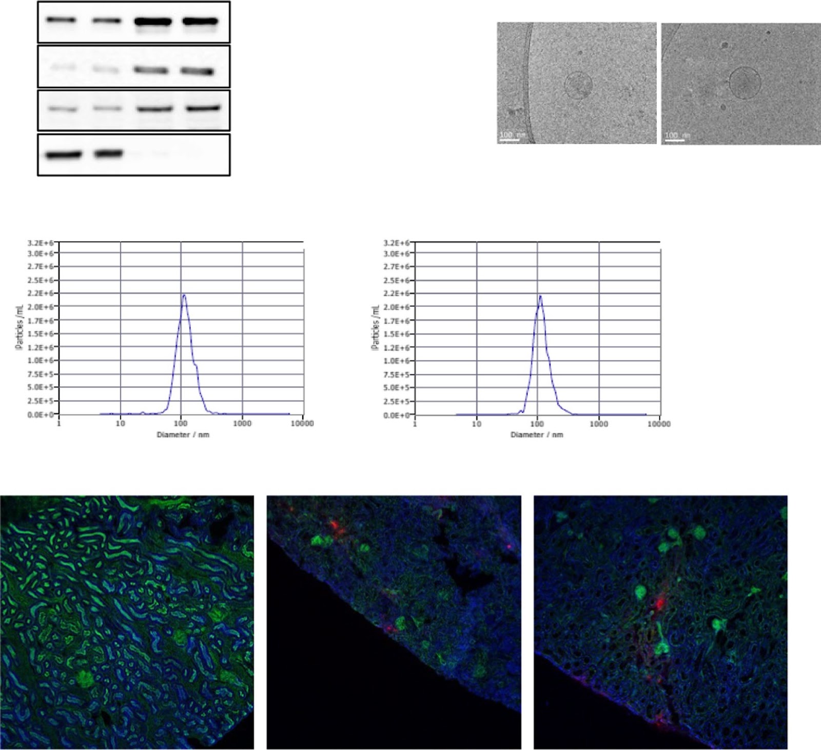
Background Acute kidney injury (AKI) has a complex pathophysiology and imposes serious health concerns worldwide. Extracellular vesicles (EVs) derived from induced mesenchymal stem cells (iMSCs) have been recognized as novel cell-free therapeutics for various inflammatory and degenerative disorders. In this study, we investigated whether iMSCs stimulated with a pan-peroxisome proliferator-activated receptor (PPAR) agonist could enhance the therapeutic efficacy of EVs against AKI. Methods Human iMSCs were primed with or without lanifibranor, a PPAR agonist for 24 h, and EVs were collected after an additional 24 h. The basic characteristics of EVs were evaluated using cryo-transmission electron microscopy imaging, immunoblot detection of EV markers, nanoparticle tracking analysis, and localization in AKI kidneys. In vitro, the potential of the EVs to promote the growth and survival of HK-2 cells undergoing cisplatin-induced apoptosis and anti-inflammatory effects in M1-polarized THP-1 was compared. Subsequently, AKI was induced in BALB/c mice using cisplatin. After 8 and 24 h of cisplatin treatment, iMSC-EVs or pan-PPAR-iMSC-EVs were injected intravascularly. At 96 h after cisplatin administration, the renoprotective effects of iMSC-EVs or pan-PPAR-iMSC-EVs in inhibiting inflammation and apoptosis were compared using serum biochemistry, histology, immunohistochemistry, and gene expression analysis by qPCR. Results Both EV types expressed EV markers and had typical EV morphology, and their localization in the renal tissue was confirmed. The proliferation and survival of HK-2 cells were higher in pan-PPAR-iMSC-EVs than those in iMSC-EVs. In M1-polarized THP-1 cells, the reduction in the mRNA expression of inflammatory cytokines was more significant in pan-PPAR-iMSC-EVs than that in iMSC-EVs. In the mouse model of cisplatin-induced AKI, pan-PPAR-iMSC-EVs markedly enhanced renoprotective effects compared to iMSC-EVs. Specifically, pan-PPAR-iMSC-EVs reduced tissue inflammation, immune cell infiltration, and apoptosis. Pan-PPAR-iMSC-EVs also increased renal capillary density. Conclusion Priming iMSCs with a PPAR agonist significantly improved the therapeutic potential of EVs by reducing inflammation and apoptosis. The reported strategy may contribute to the development of a novel cell-free option for AKI treatment. Trial registration: Not applicable.

Read Full Abstract10.1186/s13287-023-03577-0
Human pancreatic islet-derived stromal cells reveal combined features of mesenchymal stromal cells and pancreatic stellate cellsGraphical AbstractVerified
Stem Cell Research & Therapy

Human pancreatic islet-derived stromal cells reveal combined features of mesenchymal stromal cells and pancreatic stellate cells

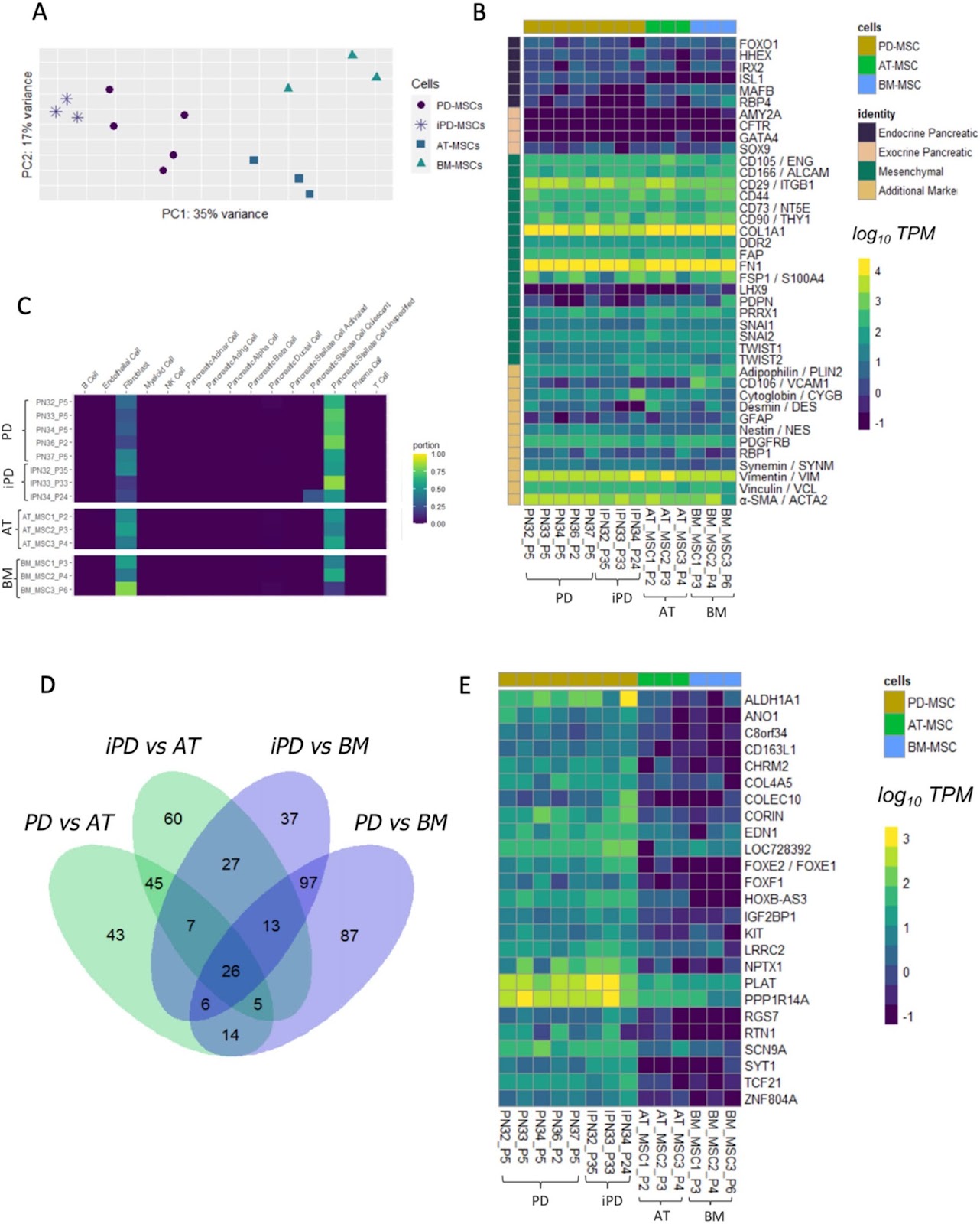
Background Mesenchymal stromal cells (MSCs) are recognized for their potential in regenerative medicine, attributed to their multipotent differentiation capabilities and immunomodulatory properties. Despite this potential, the classification and detailed characterization of MSCs, especially those derived from specific tissues like the pancreas, remains challenging leading to a proliferation of terminology in the literature. This study aims to address these challenges by providing a thorough characterization of human pancreatic islets-derived mesenchymal stromal cells (hPD-MSCs). Methods hPD-MSCs were isolated from donor islets using enzymatic digestion, immortalized through lentiviral transduction of human telomerase reverse transcriptase (hTERT). Cells were characterized by immunostaining, flow cytometry and multilineage differentiation potential into adipogenic and osteogenic lineages. Further a transcriptomic analysis was done to compare the gene expression profiles of hPD-MSCs with other mesenchymal cells. Results We show that hPD-MSCs express the classical MSC features, including morphological characteristics, surface markers expression (CD90, CD73, CD105, CD44, and CD106) and the ability to differentiate into both adipogenic and osteogenic lineages. Furthermore, transcriptomic analysis revealed distinct gene expression profiles, showing notable similarities between hPD-MSCs and pancreatic stellate cells (PSCs). The study also identified specific genes that distinguish hPD-MSCs from MSCs of other origins, including genes associated with pancreatic function (e.g., ISL1) and neural development (e.g., NPTX1, ZNF804A). A novel gene with an unknown function (ENSG00000286190) was also discovered. Conclusions This study enhances the understanding of hPD-MSCs, demonstrating their unique characteristics and potential applications in therapeutic strategies. The identification of specific gene expression profiles differentiates hPD-MSCs from other mesenchymal cells and opens new avenues for research into their role in pancreatic function and neural development.

Read Full Abstract10.1186/s13287-024-03963-2
Pre-clinical evaluation of the efficacy and safety of human induced pluripotent stem cell-derived cardiomyocyte patchGraphical AbstractVerified
Stem Cell Research & Therapy

Pre-clinical evaluation of the efficacy and safety of human induced pluripotent stem cell-derived cardiomyocyte patch

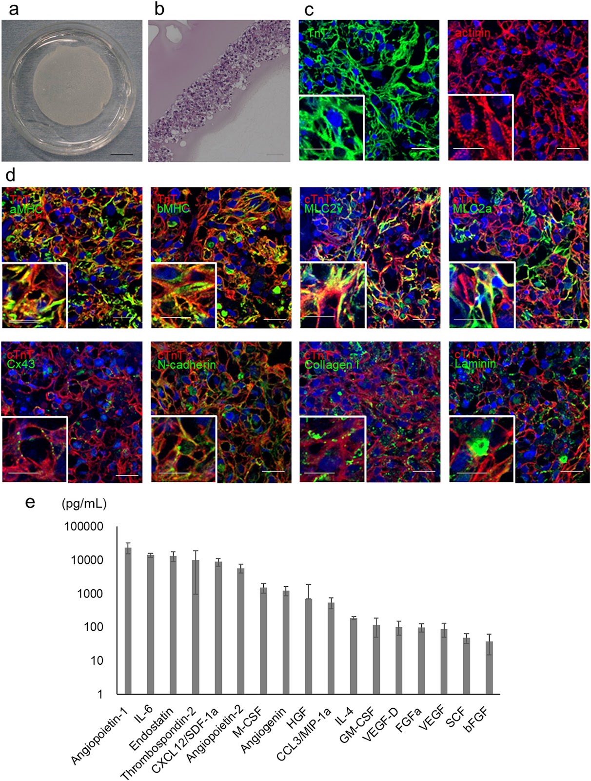
Background Cell- or tissue-based regenerative therapy is an attractive approach to treat heart failure. A tissue patch that can safely and effectively repair damaged heart muscle would greatly improve outcomes for patients with heart failure. In this study, we conducted a preclinical proof-of-concept analysis of the efficacy and safety of clinical-grade human induced pluripotent stem cell-derived cardiomyocyte (hiPSC-CM) patches. Methods A clinical-grade hiPSC line was established using peripheral blood mononuclear cells from a healthy volunteer that was homozygous for human leukocyte antigens. The hiPSCs were differentiated into cardiomyocytes. The obtained hiPSC-CMs were cultured on temperature-responsive culture dishes for patch fabrication. The cellular characteristics, safety, and efficacy of hiPSCs, hiPSC-CMs, and hiPSC-CM patches were analyzed. Results The hiPSC-CMs expressed cardiomyocyte-specific genes and proteins, and electrophysiological analyses revealed that hiPSC-CMs exhibit similar properties to human primary myocardial cells. In vitro and in vivo safety studies indicated that tumorigenic cells were absent. Moreover, whole-genome and exome sequencing revealed no genomic mutations. General toxicity tests also showed no adverse events posttransplantation. A porcine model of myocardial infarction demonstrated significantly improved cardiac function and angiogenesis in response to cytokine secretion from hiPSC-CM patches. No lethal arrhythmias were observed. Conclusions hiPSC-CM patches are promising for future translational research and may have clinical application potential for the treatment of heart failure.

Read Full Abstract10.1186/s13287-024-03690-8
Natural small molecules synergize mesenchymal stem cells for injury repair in vital organs: a comprehensive reviewGraphical AbstractVerified
Stem Cell Research & Therapy

Natural small molecules synergize mesenchymal stem cells for injury repair in vital organs: a comprehensive review

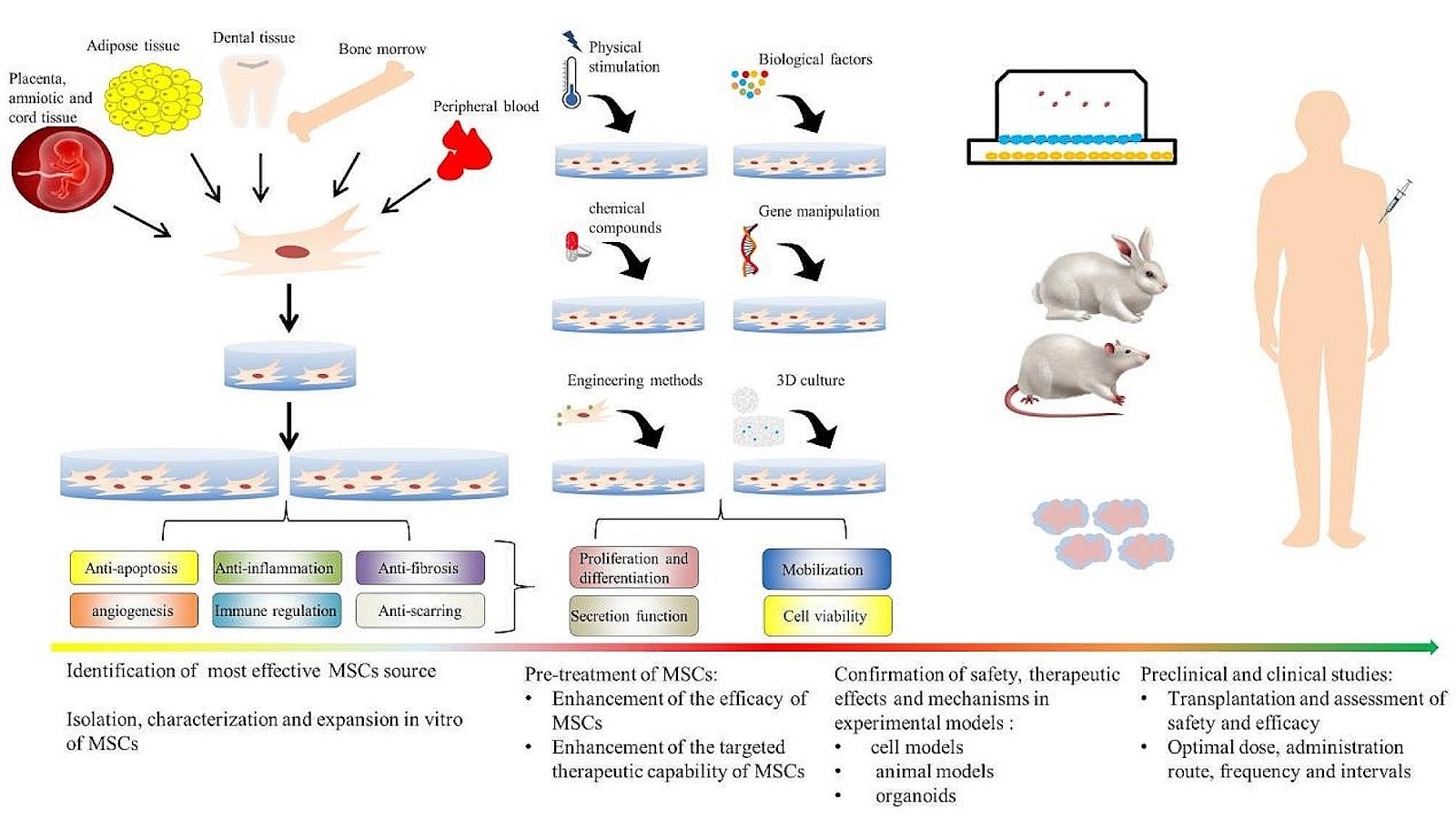
Mesenchymal stem cells (MSCs) therapy is a highly researched treatment that has the potential to promote immunomodulation and anti-inflammatory, anti-apoptotic, and antimicrobial activities. It is thought that it can enhance internal organ function, reverse tissue remodeling, and achieve significant organ repair and regeneration. However, the limited infusion, survival, and engraftment of transplanted MSCs diminish the effectiveness of MSCs-based therapy. Consequently, various preconditioning methods have emerged as strategies for enhancing the therapeutic effects of MSCs and achieving better clinical outcomes. In particular, the use of natural small molecule compounds (NSMs) as a pretreatment strategy is discussed in this narrative review, with a focus on their roles in regulating MSCs for injury repair in vital internal organs. Additionally, the discussion focuses on the future directions and challenges of transforming mesenchymal stem cell research into clinical applications.

Read Full Abstract10.1186/s13287-024-03856-4
Correction: POU3F4 up-regulates Gli1 expression and promotes neuronal differentiation and synaptic development of hippocampal neural stem cellsGraphical AbstractVerified
Stem Cell Research & Therapy

Correction: POU3F4 up-regulates Gli1 expression and promotes neuronal differentiation and synaptic development of hippocampal neural stem cells

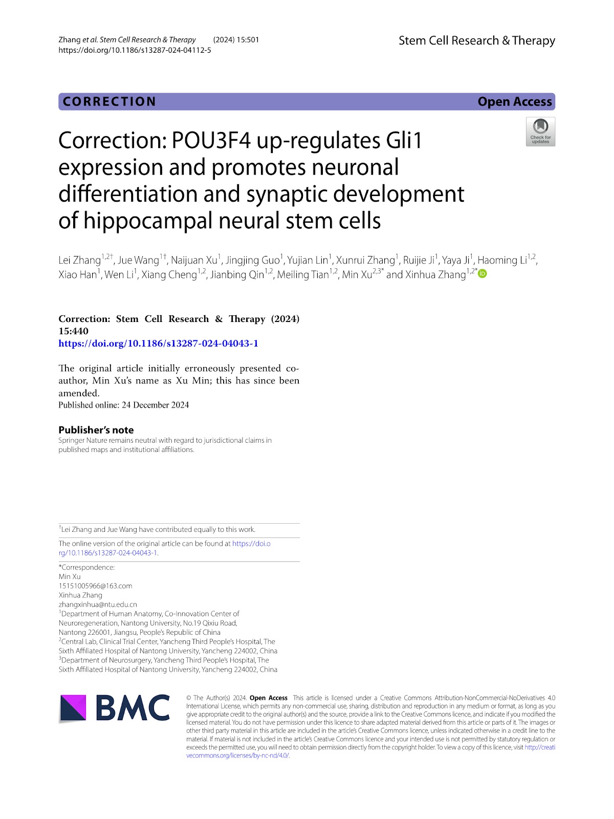
Correction to: Stem Cell Research & Therapy (2024) 15:440. The original article initially erroneously presented co-author, Min Xu's name as Xu Min; this has since been amended.

Read Full Abstract10.1186/s13287-024-04112-5
iPSC-derived lung and lung cancer organoid model to evaluate cisplatin encapsulated autologous iPSC-derived mesenchymal stromal cell-isolated extracellular vesiclesGraphical AbstractVerified
Stem Cell Research & Therapy

iPSC-derived lung and lung cancer organoid model to evaluate cisplatin encapsulated autologous iPSC-derived mesenchymal stromal cell-isolated extracellular vesicles

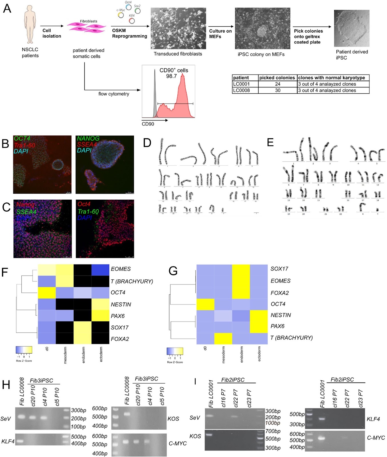
Background Lung cancer remains a leading cause of cancer-related mortality globally. Although recent therapeutic advancements have provided targeted treatment approaches, the development of resistance and systemic toxicity remain primary concerns. Extracellular vesicles (EVs), especially those derived from mesenchymal stromal cells (MSC), have gained attention as promising drug delivery systems, offering biocompatibility and minimal immune responses. Recognizing the limitations of conventional 2D cell culture systems in mimicking the tumor microenvironment, this study aims to describe a proof-of-principle approach for using patient-specific organoid models for both lung cancer and normal lung tissue and the feasibility of employing autologous EVs derived from induced pluripotent stem cell (iPSC)-MSC in personalized medicine approaches. Methods First, we reprogrammed healthy fibroblasts into iPSC. Next, we differentiated patient-derived iPSC into branching lung organoids (BLO) and generated patient-matched lung cancer organoids (LCO) from patient-derived tumor tissue. We show a streamlined process of MSC differentiation from iPSC and EV isolation from iPSC-MSC, encapsulated with 0.07 µg/mL of cytotoxic agent cisplatin and applied to both organoid models. Cytotoxicity of cisplatin and cisplatin-loaded EVs was recorded with LDH and CCK8 tests. Results Fibroblast-derived iPSC showed a normal karyotype, pluripotency staining, and trilineage differentiation. iPSC-derived BLO showed expression of lung markers, like TMPRSS2 and MUC5A while patient-matched LCO showed expression of Napsin and CK5. Next, we compared the effects of iPSC-MSC derived EVs loaded with cisplatin against empty EVs and cisplatin alone in lung cancer organoid and healthy lung organoid models. As expected, we found a cytotoxic effect when LCO were treated with 20 µg/mL cisplatin. Treatment of LCO and BLO with empty EVs resulted in a cytotoxic effect after 24 h. However, EVs loaded with 0.07 µg/mL cisplatin failed to induce any cytotoxic effect in both organoid models. Conclusion We report on a proof-of-principle pipeline towards using autologous or allogeneic iPSC-MSC EVs as drug delivery tests for lung cancer in future. However, due to the time and labor-intensive processes, we conclude that this pipeline might not be feasible for personalized approaches at the moment.

Read Full Abstract10.1186/s13287-024-03862-6
Evaluation of the impact of customized serum-free culture medium on the production of clinical-grade human umbilical cord mesenchymal stem cells: insights for future clinical applicationsGraphical AbstractVerified
Stem Cell Research & Therapy

Evaluation of the impact of customized serum-free culture medium on the production of clinical-grade human umbilical cord mesenchymal stem cells: insights for future clinical applications

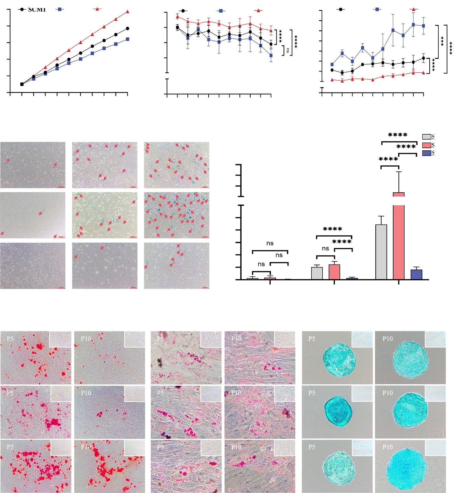
Background The selection of suitable culture medium is critical for achieving good clinical outcomes in cell therapy. To support the commercial application of stem cell therapy, customized culture media not only need to promote stem cell proliferation, but also need to save costs and meet industrial requirements for inter-batch consistency, efficacy, and biosafety. In this study, we developed a series of serum-free media (SFM) and elucidated the effects between different SFM, as well as between SFM and serum-containing meida (SCM), on human umbilical cord mesenchymal stem cells (hUC-MSCs) phenotype and function. We analyze and emphasize from the perspectives of clinical and commercial application why research on customized culture media is critical for the success of enterprises developing novel cellular therapeutics. Methods We cultured hUC-MSCs with identical cell seeding densities in different formulations of SFM and SCM until passage 10 and examined the changes in cell phenotype and function. We analyzed the results with the commercial application requirments of the cellular therapy industry to assess the potential impact of customized culture media on inter-batch consistency, efficacy, stability, biosafety, and cost-effectiveness of industrial-scale cell production. Results hUC-MSCs cultured in SCM and SFM exhibit consistent cell morphology and surface molecule expression, but hUC-MSCs cultured in SFM demonstrate higher activity, superior proliferative capacity, and greater stability. Furthermore, hUC-MSCs cultured in different SFM exhibit differences in cell activity, proliferative capacity, senescent rate, and S/M ratio of cell cycle, while maintaining a normal karyotype after long-term in vitro cultivation. Moreover, [abstract truncated]

Read Full Abstract10.1186/s13287-024-03949-0
Transplantation of human endometrial perivascular stem cells with hydroxy saffron yellow A promotes uterine repair in ratsGraphical AbstractVerified
Stem Cell Research & Therapy

Transplantation of human endometrial perivascular stem cells with hydroxy saffron yellow A promotes uterine repair in rats

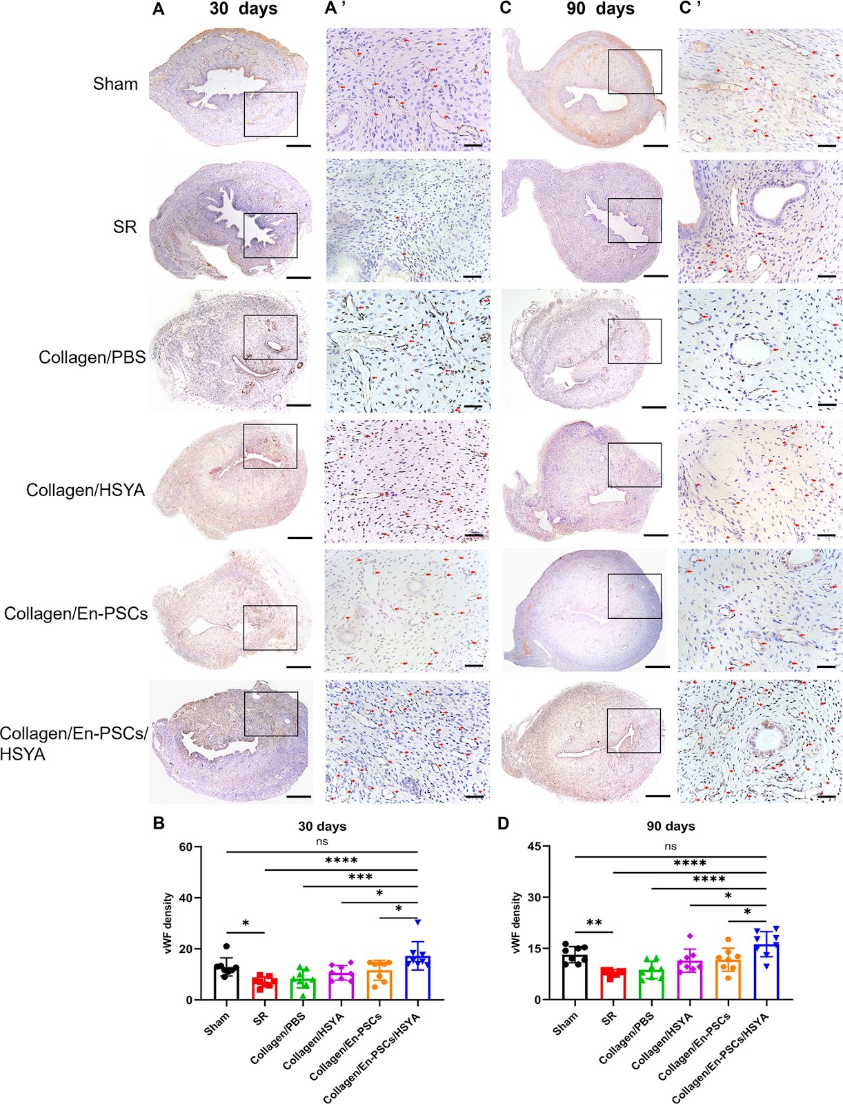
Background Intrauterine adhesions (IUAs) jeopardise uterine function in women, which is a great challenge in the clinic. Previous studies have shown that endometrial perivascular cells (En-PSCs) can improve the healing of scarred uteri and that hydroxysafflor yellow A (HSYA) promotes angiogenesis. The purpose of this study was to observe whether the combination of En-PSCs with HSYA could improve the blood supply and fertility in the rat uterus after full-thickness injury. Methods En-PSCs were sorted by flow cytometry, and the effect of HSYA on the proliferation and angiogenesis of the En-PSCs was detected using CCK-8 and tube formation assays. Based on a previously reported rat IUA model, the rat uteri were sham-operated, spontaneously regenerated, or treated with collagen-loaded PBS, collagen-loaded HSYA, collagen-loaded En-PSCs, or collagen-loaded En-PSCs with HSYA, and then collected at both 30 and 90 days postsurgery. HE staining and Masson staining were used to evaluate uterine structure and collagen fibre deposition, and immunohistochemical staining for α-SMA and vWF was used to evaluate myometrial regeneration and neovascularization in each group. A fertility assay was performed to detect the recovery of pregnancy function in each group. RNA-seq was performed to determine the potential mechanism underlying En-PSCs/HSYA treatment. Immunofluorescence, tube formation assays, and Western blot were used to validate the molecular mechanism involved. Results The transplantation of Collagen/En-PSCs/HSYA markedly promoted uterine repair in rats with full-thickness injury by reducing fibrosis, increasing endometrial thickness, regenerating myometrium, promoting angiogenesis, and facilitated live births. RNA sequencing results suggested that En-PSCs/HSYA activated the NRG1/ErbB4 signaling pathway. In vitro tube formation experiments revealed that the addition of an ErbB inhibitor diminished the tube formation ability of cocultured En-PSCs and HUVECs. Western blot results further showed that elevated levels of NRG1 and ErbB4 proteins were detected in the Collagen/En-PSCs/HSYA group compared to the Collagen/En-PSCs group. These collective results suggested that the beneficial effects of the transplantation of Collagen/En-PSCs/HSYA might be attributed to the modulation of the NRG1/ErbB4 signaling pathway. Conclusions The combination of En-PSCs/HSYA facilitated morphological and functional repair in rats with full-thickness uterine injury and may promote endometrial angiogenesis by regulating the NRG1/ErbB4 signaling pathway.

Read Full Abstract10.1186/s13287-024-03821-1
Correction: Radiochemotherapy-induced DNA repair promotes the biogenesis of gastric cancer stem cellsGraphical AbstractVerified
Stem Cell Research & Therapy

Correction: Radiochemotherapy-induced DNA repair promotes the biogenesis of gastric cancer stem cells

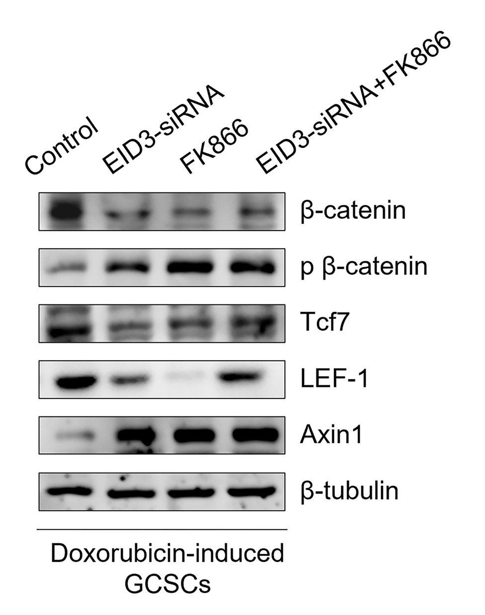
This correction article addresses an inadvertent error in the original publication. In Fig. 5M of the original article, the image of the fourth lane (LEF-1) of the second picture (Doxorubicin-induced GCSCs) was inadvertently replaced with an incorrect version during the upload process. The authors wish to note a correction to the aforementioned picture via the corrected picture ahead in this Correction article. The authors deeply regret that this error occurred and sincerely apologize for any inconvenience.

Read Full Abstract10.1186/s13287-024-03984-x
Dysregulated lncRNAs regulate human umbilical cord mesenchymal stem cell differentiation into insulin-producing cells by forming a regulatory network with mRNAsGraphical AbstractVerified
Stem Cell Research & Therapy

Dysregulated lncRNAs regulate human umbilical cord mesenchymal stem cell differentiation into insulin-producing cells by forming a regulatory network with mRNAs

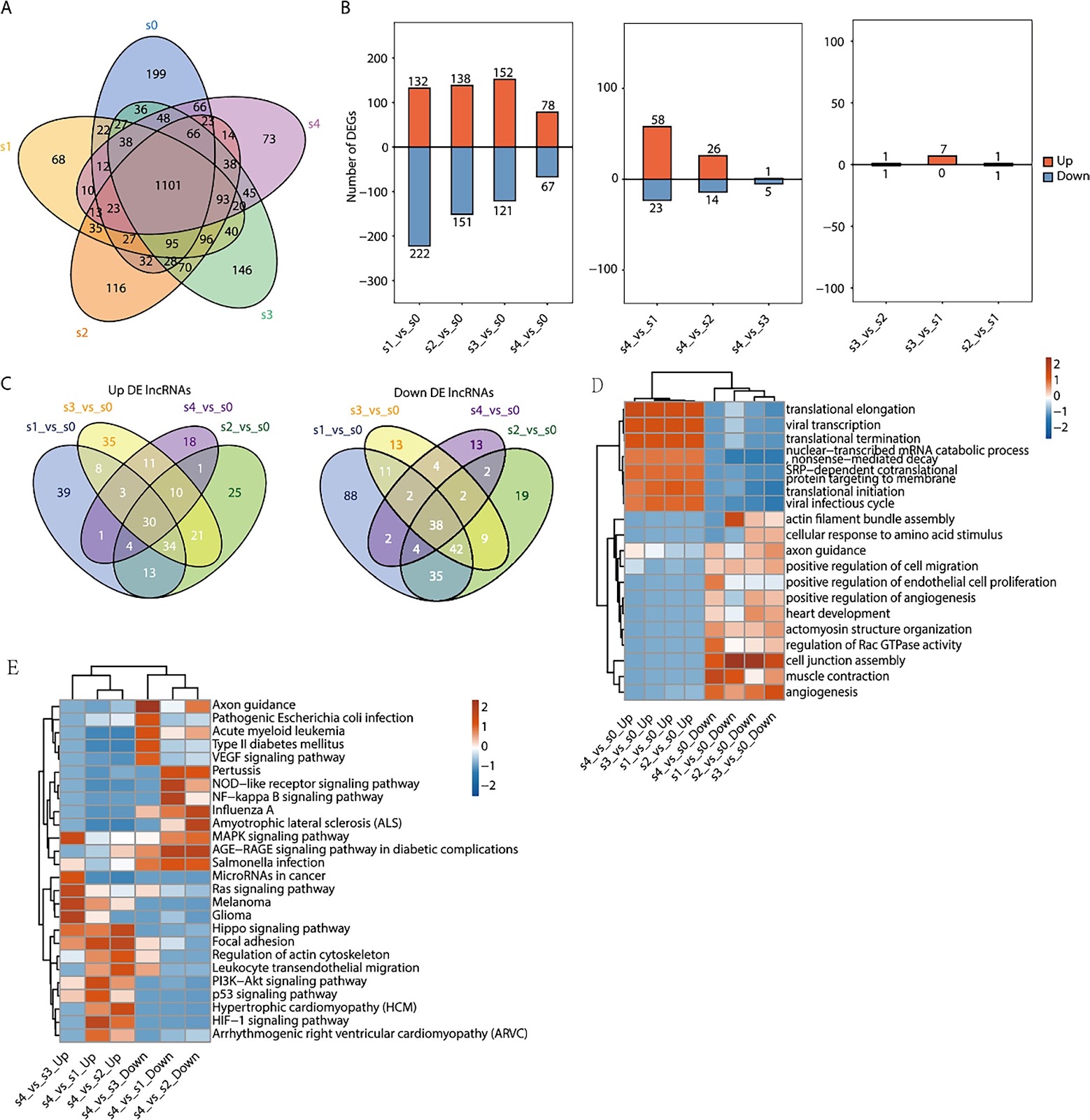
Objective In recent years, cell therapy has emerged as a new research direction in the treatment of diabetes. However, the underlying molecular mechanisms of mesenchymal stem cell (MSC) differentiation necessary to form such treatment have not been clarified. Methods In this study, human umbilical cord mesenchymal stem cells (HUC-MSCs) isolated from newborns were progressively induced into insulin-producing cells (IPCs) using small molecules. HUC-MSC (S0) and four induced stage (S1–S4) samples were prepared. We then performed transcriptome sequencing experiments to obtain the dynamic expression profiles of both mRNAs and long noncoding RNAs (lncRNAs). Results We found that the number of differentially expressed lncRNAs and mRNAs trended downwards during differentiation. Gene Ontology (GO) analysis showed that the target genes of differentially expressed lncRNAs were associated with translation, cell adhesion, and cell connection. Kyoto Encyclopedia of Genes and Genomes (KEGG) analysis revealed that the NF-KB signalling pathway, MAPK signalling pathway, HIPPO signalling pathway, PI3K–Akt signalling pathway, and p53 signalling pathway were enriched in these differentially expressed lncRNA-targeting genes. We also found that the coexpression of the lncRNA CTBP1-AS2 with PROX1 and the lncRNAs AC009014.3 and GS1-72M22.1 with JARID2 mRNA was related to the development of pancreatic beta cells. Moreover, the coexpression of the lncRNAs: XLOC_ 050969, LINC00883, XLOC_050981, XLOC_050925, MAP3K14- AS1, RP11-148K1.12, and CTD2020K17.3 with p53, regulated insulin secretion by pancreatic beta cells. Conclusion In this study, HUC-MSCs combined with small molecule compounds were successfully induced into IPCs. Differentially expressed lncRNAs may regulate the insulin secretion of pancreatic beta cells by regulating multiple signalling pathways. The lncRNAs AC009014.3, Gs1-72m21.1, and CTBP1-AS2 may be involved in the development of pancreatic beta cells, and the lncRNAs: XLOC_050969, LINC00883, XLOC_050981, XLOC_050925, MAP3K14-AS1, RP11-148K1.12, and CTD2020K17.3 may be involved in regulating the insulin secretion of pancreatic beta cells, thus providing a lncRNA catalogue for future research regarding the mechanism of the transdifferentiation of HUC-MSCs into IPCs. It also provides a new theoretical basis for the transplantation of insulin-producing cells into diabetic patients in the future.

Read Full Abstract10.1186/s13287-023-03572-5
Dissecting human adipose tissue heterogeneity using single-cell omics technologiesGraphical AbstractVerified
Stem Cell Research & Therapy

Dissecting human adipose tissue heterogeneity using single-cell omics technologies

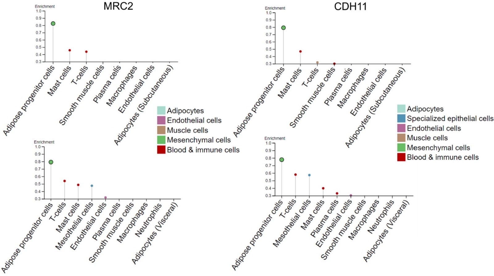
Single-cell omics technologies that profile genes (genomic and epigenomic) and determine the abundance of mRNA (transcriptomic), protein (proteomic and secretomic), lipids (lipidomic), and extracellular matrix (matrisomic) support the dissection of adipose tissue heterogeneity at unprecedented resolution in a temporally and spatially defined manner. In particular, cell omics technologies may provide innovative biomarkers for the identification of rare specific progenitor cell subpopulations, assess transcriptional and proteomic changes affecting cell proliferation and immunomodulatory potential, and accurately define the lineage hierarchy and differentiation status of progenitor cells. Unraveling adipose tissue complexity may also provide for the precise assessment of a dysfunctional state, which has been associated with cancer, as cancer-associated adipocytes play an important role in shaping the tumor microenvironment supporting tumor progression and metastasis, obesity, metabolic syndrome, and type 2 diabetes mellitus. The information collected by single-cell omics has relevant implications for regenerative medicine because adipose tissue is an accessible source of multipotent cells; alternative cell-free approaches, including the use of adipose tissue stromal cell-conditioned medium, extracellular vesicles, or decellularized extracellular matrix, are clinically valid options. Subcutaneous white adipose tissue, which is generally harvested via liposuction, is highly heterogeneous because of intrinsic biological variability and extrinsic inconsistencies in the harvesting and processing procedures. The current limited understanding of adipose tissue heterogeneity impinges on the definition of quality standards appropriate for clinical translation, which requires consistency and uniformity of the administered product. We review the methods used for dissecting adipose tissue heterogeneity and provide an overview of advances in omics technology that may contribute to the exploration of heterogeneity and dynamics of adipose tissue at the single-cell level.

Read Full Abstract10.1186/s13287-024-03931-w
Mesenchymal stem cell therapy for liver transplantation: clinical progress and immunomodulatory propertiesGraphical AbstractVerified
Stem Cell Research & Therapy

Mesenchymal stem cell therapy for liver transplantation: clinical progress and immunomodulatory properties

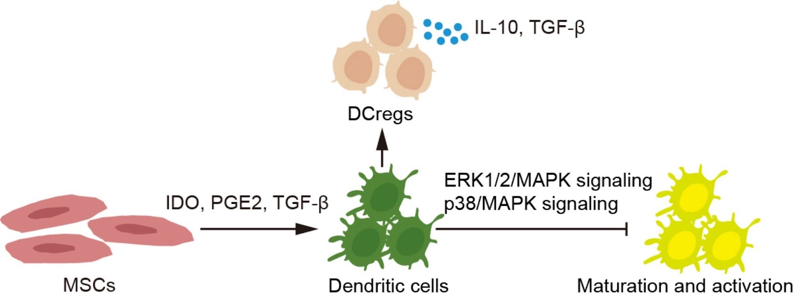
Although liver transplantation (LT) is an effective strategy for end-stage liver diseases, the shortage of donor organs and the immune rejection hinder its widespread implementation in clinical practice. Mesenchymal stem cells (MSCs) transplantation offers a promising approach for patients undergoing liver transplantation due to their immune regulatory capabilities, hepatic protection properties, and multidirectional differentiation potential. In this review, we summarize the potential applications of MSCs transplantation in various LT scenarios. MSCs transplantation has demonstrated effectiveness in alleviating hepatic ischemia-reperfusion injury, enhancing the viability of liver grafts, preventing acute graft-versus-host disease, and promoting liver regeneration in split LT therapy. We also discuss the clinical progress, and explore the immunomodulatory functions of MSCs in response to both adaptive and innate immune responses. Furthermore, we emphasize the interactions between MSCs and different immune cells, including T cells, B cells, plasma cells, natural killer cells, dendritic cells, Kupffer cells, and neutrophils, to provide new insights into the immunomodulatory properties of MSCs in adoptive cell therapy.

Read Full Abstract10.1186/s13287-024-03943-6
Robust bioprocess design and evaluation of commercial media for the serial expansion of human induced pluripotent stem cell aggregate cultures in vertical-wheel bioreactorsGraphical AbstractVerified
Stem Cell Research & Therapy

Robust bioprocess design and evaluation of commercial media for the serial expansion of human induced pluripotent stem cell aggregate cultures in vertical-wheel bioreactors

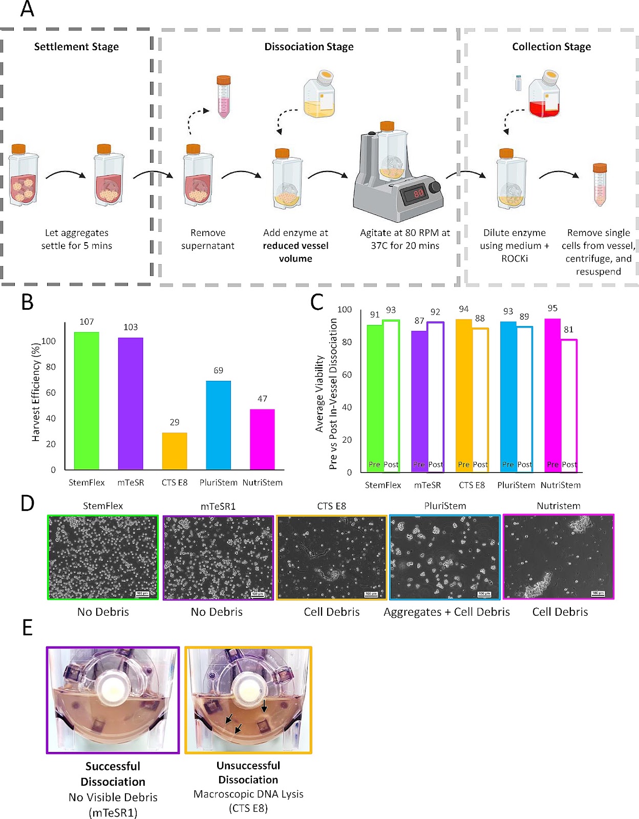
Background: While pluripotent stem cell (PSC) therapies move toward clinical and commercial applications at a rapid rate, manufacturing reproducibility and robustness are notable bottlenecks in regulatory approval. Therapeutic applications of PSCs require large cell quantities to be generated under highly robust, well-defined, and economically viable conditions. Small-scale and short-term process optimization, however, is often performed in a linear fashion that does not account for time needed to verify the bioprocess protocols and analysis methods used. Design of a reproducible and robust bioprocess should be dynamic and include a continuous effort to understand how the process will respond over time and to different stresses before transitioning into large-scale production where stresses will be amplified. Methods: This study utilizes a baseline protocol, developed for the short-term culture of PSC aggregates in Vertical-Wheel® bioreactors, to evaluate key process attributes through long-term (serial passage) suspension culture. This was done to access overall process robustness when performed with various commercially available media and cell lines. Process output variables including growth kinetics, aggregate morphology, harvest efficiency, genomic stability, and functional pluripotency were assessed through short and long-term culture. Results: The robust nature of the expansion protocol was demonstrated over a six-day culture period where spherical aggregate formation and expansion were observed with high-fold expansions for all five commercial media tested. Profound differences in cell growth and quality were revealed only through long-term serial expansion and in-vessel dissociation operations. Some commercial media formulations tested demonstrated maintenance of cell growth

Read Full Abstract10.1186/s13287-024-03819-9
Single-cell sequencing of facial adipose tissue unveils FKBP5 as a therapeutic target for facial infiltrating lipomatosisGraphical AbstractVerified
Stem Cell Research & Therapy

Single-cell sequencing of facial adipose tissue unveils FKBP5 as a therapeutic target for facial infiltrating lipomatosis

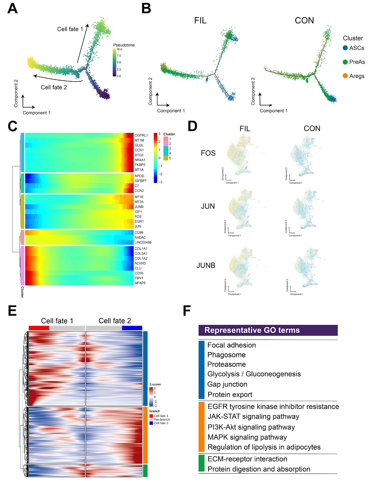
Background Facial infiltrating lipomatosis is characterized by excessive growth of adipose tissue. Its etiology is associated with somatic phosphatidylinositol 3-kinase catalytic subunit alpha (PIK3CA) variants, but the specific mechanisms are not yet fully understood. Methods We collected facial adipose tissue from both FIL patients and non-FIL individuals, isolated the stromal vascular fraction (SVF) and performed single-cell transcriptome sequencing on these samples. Results We mapped out the cellular landscape within the SVF, with a specific focus on a deeper analysis of fibro-adipogenic precursor cells (FAPs). Our analysis revealed that FAPs from FIL patients (FIL-FAPs) significantly overexpressed FK506 binding protein 51 (FKBP5) compared to FAPs from individuals without FIL. Further experiments indicated that FKBP5 is regulated by the PI3K-AKT signaling pathway. The overactivation of this pathway led to an increase in FKBP5 expression. In vitro experiments demonstrated that FKBP5 promoted adipogenic differentiation of FAPs, a process that could be hindered by FKBP5 knockdown or inhibition. Additionally, in vivo assessments confirmed FKBP5’s role in adipogenesis. Conclusions These insights into the pathogenesis of FIL underscore FKBP5 as a promising target for developing non-surgical interventions to manage the excessive adipose tissue growth in FIL.

Read Full Abstract10.1186/s13287-024-03835-9
Mesenchymal stem/stromal cells from human pluripotent stem cell-derived brain organoid enhance the ex vivo expansion and maintenance of hematopoietic stem/progenitor cellsGraphical AbstractVerified
Stem Cell Research & Therapy

Mesenchymal stem/stromal cells from human pluripotent stem cell-derived brain organoid enhance the ex vivo expansion and maintenance of hematopoietic stem/progenitor cells

Background Mesenchymal stem/stromal cells (MSCs) are of great therapeutic value due to their role in maintaining the function of hematopoietic stem/progenitor cells (HSPCs). MSCs derived from human pluripotent stem cells represent an ideal alternative because of their unlimited supply. However, the role of MSCs with neural crest origin derived from HPSCs on the maintenance of HSPCs has not been reported. Methods Flow cytometric analysis, RNA sequencing and differentiation ability were applied to detect the characteristics of stromal cells from 3D human brain organoids. Human umbilical cord blood CD34+ (UCB-CD34+) cells were cultured in different coculture conditions composed of stromal cells and umbilical cord MSCs (UC-MSCs) with or without a cytokine cocktail. The hematopoietic stroma capacity of stromal cells was tested in vitro with the LTC-IC assay and in vivo by cotransplantation of cord blood nucleated cells and stroma cells into immunodeficient mice. RNA and proteomic sequencing were used to detect the role of MSCs on HSPCs. Results The stromal cells, derived from both H1-hESCs and human induced pluripotent stem cells forebrain organoids, were capable of differentiating into the classical mesenchymal-derived cells (osteoblasts, chondrocytes, and adipocytes). These cells expressed MSC markers, thus named pluripotent stem cell-derived MSCs (pMSCs). The pMSCs showed neural crest origin with CD271 expression in the early stage. When human UCB-CD34+ HSPCs were cocultured on UC-MSCs or pMSCs, the latter resulted in robust expansion of UCB-CD34+ HSPCs in long-term culture and efficient maintenance of their transplantability. Comparison by RNA sequencing indicated that coculture of human UCB-CD34+ HSPCs with pMSCs provided an improved microenvironment for HSC maintenance. The pMSCs highly expressed the Wnt signaling inhibitors SFRP1 and SFRP2, indicating that they may help to modulate the cell cycle to promote the maintenance of UCB-CD34+ HSPCs by antagonizing Wnt activation.

Read Full Abstract10.1186/s13287-023-03624-w
Examining the potentials of stem cell therapy in reducing the burden of selected non-communicable diseases in AfricaGraphical AbstractVerified
Stem Cell Research & Therapy

Examining the potentials of stem cell therapy in reducing the burden of selected non-communicable diseases in Africa

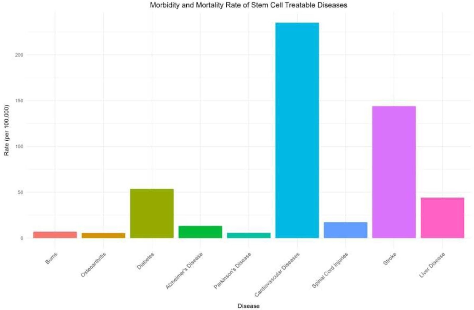
Stem cell therapy (SCT) is a promising solution for addressing health challenges in Africa, particularly non-communicable diseases (NCDs). With their regenerative potential, stem cells have the inherent capacity to differentiate into numerous cell types for tissue repair. Despite infrastructural, ethical, and legal challenges, SCT holds immense promise for managing chronic illnesses and deep-seated tissue injuries. The rising prevalence of NCDs in Africa highlights the need for innovative strategies and treatment options. SCT offers hope in combating conditions like burns, osteoarthritis, diabetes, Alzheimer’s disease, stroke, heart failure and cancer, potentially reducing the burden of NCDs on the continent. Despite SCT’s opportunities in Africa, there are significant obstacles. However, published research on SCT in Africa is scarce, but recent initiatives such as the Basic School on Neural Stem Cells (NSC) express interest in developing NSC research in Africa. SCT research in African regions, notably on neurogenesis, demonstrates a concentration on studying neurological processes in indigenous settings. While progress has been made in South Africa and Nigeria, issues such as brain drain and impediments to innovation remain. Clinical trials have investigated the efficacy of stem cell treatments, emphasising both potential benefits and limitations in implementing these therapies efficiently. Financing research, developing regulatory frameworks, and resolving affordability concerns are critical steps toward realizing the potential of stem cell treatment in Africa.

Read Full Abstract10.1186/s13287-024-03864-4
N-CADHERIN+/CD168− subpopulation determines therapeutic variations of UC-MSCs for cardiac repair after myocardial infarctionGraphical AbstractVerified
Stem Cell Research & Therapy

N-CADHERIN+/CD168− subpopulation determines therapeutic variations of UC-MSCs for cardiac repair after myocardial infarction

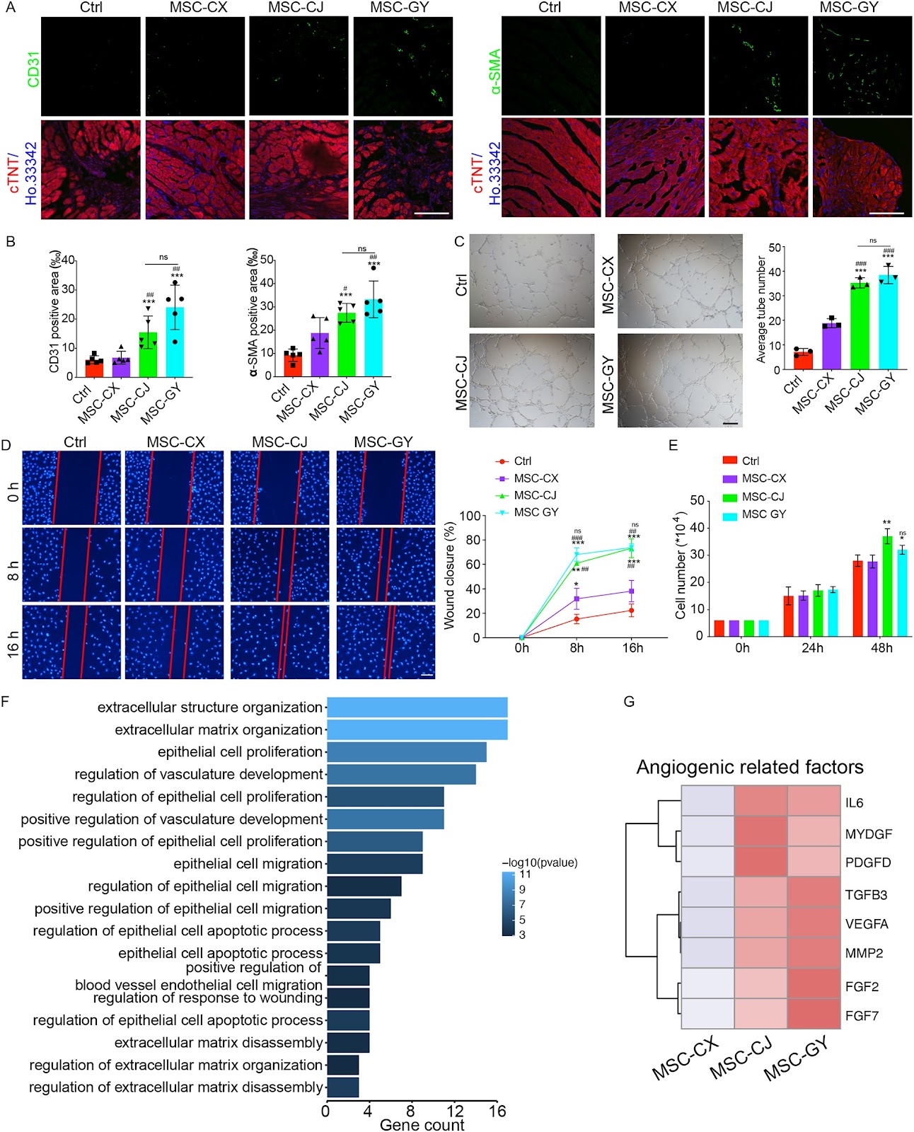
Background The efficiency of mesenchymal stem cells (MSCs) in treating myocardial infarction (MI) remains inconsistent, which limits their therapeutic applications. Therefore, exploring the mechanism for the inconsistent efficacy of MSCs and identification the criteria for screening MSCs are important for improving the efficiency of MSCs. Methods Mouse model after MI was utilized to test the role of MSCs from different donors and the functional subpopulation in improving cardiac function. Heterogeneity of MSCs was identified using single-cell RNA sequencing (scRNA-seq) of MSC-GY. GSEA and Scissor analyses were used to find the functional subpopulations of MSCs that promote angiogenesis. The role of functional subpopulations in promoting angiogenesis was verified by detecting the secretory proteins, the ratio of N-CADHERIN+/CD168− subpopulations in MSCs, and the tube formation, migration, and proliferation of HUVECs after treatment with conditional medium (CM) derived from different MSCs. Results We found that umbilical cord-derived MSCs (UC-MSCs) from different donors have varied therapeutic efficacy in MI mice and UC-MSCs with higher therapeutic effectiveness exhibited the most potent pro-angiogenic effects by secreting elevated levels of angiogenesis-related proteins, such as MYDGF, VEGFA, and FGF2. ScRNA-seq of 10,463 UC-MSCs revealed that the N-CADHERIN+/CD168− subpopulation was closely associated with pro-angiogenic effects, and the ratio of this cell subpopulation was positively correlated with the angiogenic potential of MSCs. We also found that the N-CADHERIN+/CD168− subpopulation was the functional subpopulation of MSCs in improving cardiac function of MI mice. Conclusions Our study identified that the N-CADHERIN+/CD168− subpopulation was the functional subpopulation of MSCs in treating MI, which was essential for the development and utilization of MSCs in MI treatment.

Read Full Abstract10.1186/s13287-024-04032-4
Human intermediate prostate cancer stem cells contribute to the initiation and development of prostate adenocarcinomaGraphical AbstractVerified
Stem Cell Research & Therapy

Human intermediate prostate cancer stem cells contribute to the initiation and development of prostate adenocarcinoma

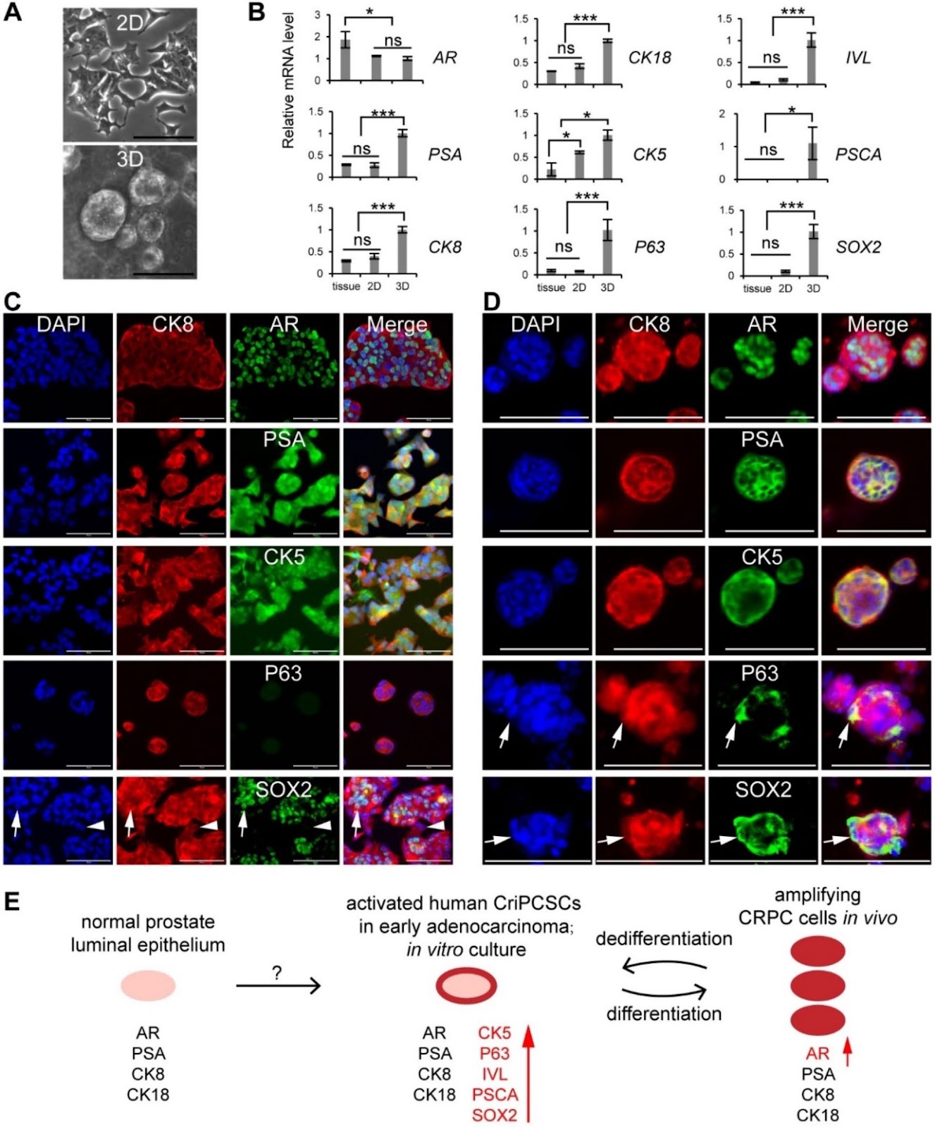
Background Intermediate cells are present in the early stages of human prostate development and adenocarcinoma. While primary cells isolated from benign human prostate tissues or tumors exhibit an intermediate phenotype in vitro, they cannot form tumors in vivo unless genetically modified. It is unclear about the stem cell properties and tumorigenicity of intermediate cells. Methods We developed a customized medium to culture primary human intermediate prostate cells, which were transplanted into male immunodeficient NCG mice to examine tumorigenicity in vivo. We treated the cells with different concentrations of dihydrotestosterone (DHT) and enzalutamide in vitro and surgically castrated the mice after cell transplantation in vivo. Immunostaining, qRT-PCR, RNA sequencing, and western blotting were performed to characterize the cells in tissues and 2D and 3D cultures. Results We found intermediate cells expressing AR+PSA+CK8+CK5+ in the luminal compartment of human prostate adenocarcinoma by immunostaining. We cultured the primary intermediate cells in vitro, which expressed luminal (AR+PSA+CK8+CK18+), basal (CK5+P63+), intermediate (IVL+), and stem cell (CK4+CK13+PSCA+SOX2+) markers. These cells resisted castration in vitro by upregulating the expression of AR, PSA, and proliferation markers KI67 and PCNA. The intermediate cells had high tumorigenicity in vivo, forming tumors in immunodeficient NCG mice in a month without any genetic modification or co-transplantation with embryonic urogenital sinus mesenchyme (UGSM) cells. We named these cells human castration-resistant intermediate prostate cancer stem cells or CriPCSCs and defined the xenograft model as patient primary cell-derived xenograft (PrDX). Human CriPCSCs resisted castration in vitro and

Read Full Abstract10.1186/s13287-024-03917-8
Urine-derived stem cells serve as a robust platform for generating native or engineered extracellular vesiclesGraphical AbstractVerified
Stem Cell Research & Therapy

Urine-derived stem cells serve as a robust platform for generating native or engineered extracellular vesicles

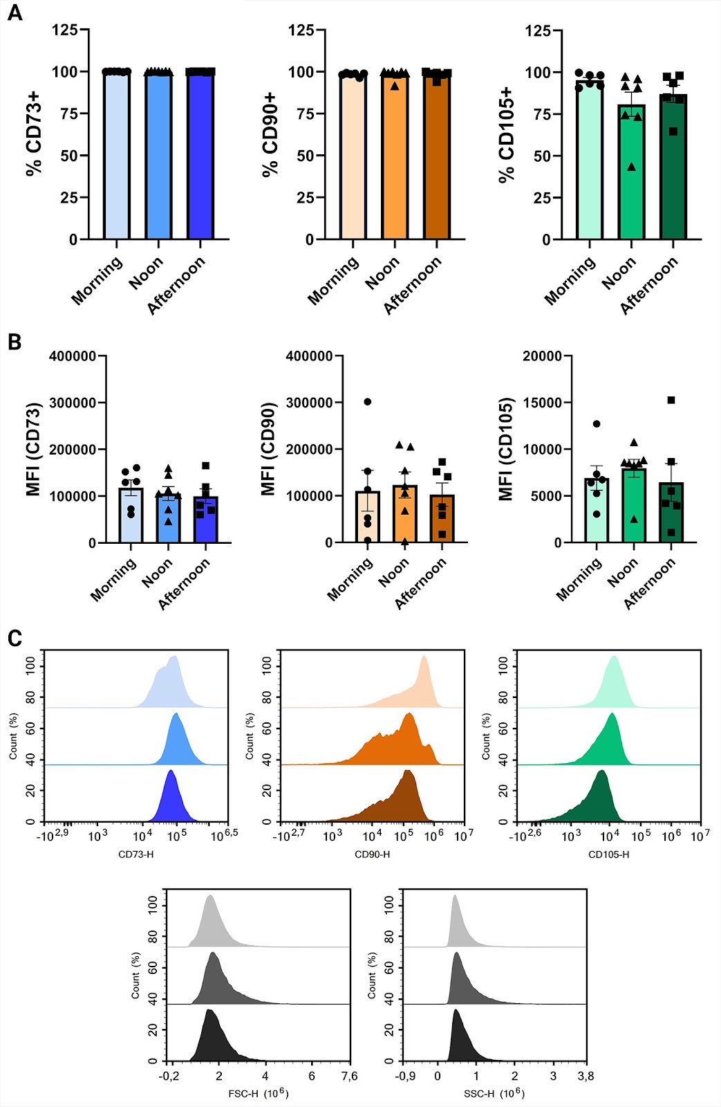
Background Mesenchymal stromal cell (MSC) therapy holds great potential yet efficacy and safety concerns with cell therapy persist. The beneficial effects of MSCs are often attributed to their secretome that includes extracellular vesicles (EVs). EVs carry biologically active molecules, protected by a lipid bilayer. However, several barriers hinder large-scale MSC EV production. A serum-free culturing approach is preferred for producing clinical-grade MSC-derived EVs but this can affect both yield and purity. Consequently, new strategies have been explored, including genetically engineering MSCs to alter EV compositions to enhance potency, increase circulation time or mediate targeting. However, efficient transfection of MSCs is challenging. Typical sources of MSC include adipose tissue and bone marrow, which both require invasive extraction procedures. Here, we investigate the use of urine-derived stem cells (USCs) as a non-invasive and inexhaustible source of MSCs for EV production. Methods We isolated, expanded, and characterized urine-derived stem cells (USCs) harvested from eight healthy donors at three different time points during the day. We evaluated the number of clones per urination, proliferation capacity and conducted flow cytometry to establish expression of surface markers. EVs were produced in chemically defined media and characterized. PEI/DNA transfection was used to genetically engineer USCs using transposon technology. Results There were no differences between time points for clone number, doubling time or viability. USCs showed immunophenotypic characteristics of MSCs, such as expression of CD73, CD90 and CD105, with no difference at the assessed time points, however, male donors had reduced CD73+ cells. Expanded USCs were incubated without growth factors or serum for 72 h without a loss in viability and EVs were isolated. USCs were transfected with high efficiency and after 10 days of selection, pure engineered cell cultures were established. Conclusions Isolation and expansion of MSCs from urine is non-invasive, robust, and without apparent sex-related differences. The sampling time point did not affect any measured markers or USC isolation potential. USCs offer an attractive production platform for EVs, both native and engineered.

Read Full Abstract10.1186/s13287-024-03903-0
Donor and recipient hematopoietic stem and progenitor cells mobilization in liver transplantation patientsGraphical AbstractVerified
Stem Cell Research & Therapy

Donor and recipient hematopoietic stem and progenitor cells mobilization in liver transplantation patients

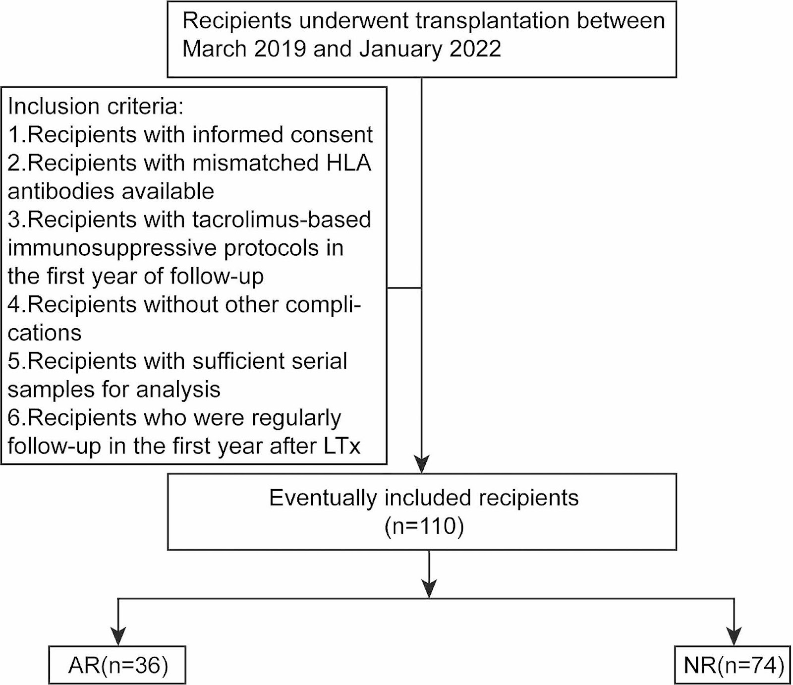
Background Hematopoietic stem and progenitor cells (HSPCs) mobilize from bone marrow to peripheral blood in response to stress. The impact of alloresponse-induced stress on HSPCs mobilization in human liver transplantation (LTx) recipients remains under-investigated. Methods Peripheral blood mononuclear cell (PBMC) samples were longitudinally collected from pre- to post-LTx for one year from 36 recipients with acute rejection (AR), 74 recipients without rejection (NR), and 5 recipients with graft-versus-host disease (GVHD). 28 PBMC samples from age-matched healthy donors were collected as healthy control (HC). Multi-color flow cytometry (MCFC) was used to immunophenotype HSPCs and their subpopulations. Donor recipient-distinguishable major histocompatibility complex (MHC) antibodies determined cell origin. Results Before LTx, patients who developed AR after transplant contained more HSPCs in PBMC samples than HC, while the NR group patients contained fewer HSPCs than HC. After LTx, the HSPC ratio in the AR group sharply decreased and became less than HC within six months, and dropped to a comparable NR level afterward. During the one-year follow-up period, myeloid progenitors (MPs) biased differentiation was observed in all LTx recipients who were under tacrolimus-based immunosuppressive treatment. During both AR and GVHD episodes, the recipient-derived and donor-derived HSPCs mobilized into the recipient’s blood-circulation and migrated to the target tissue, respectively. The HSPCs percentage in blood reduced after the disease was cured. Conclusions A preoperative high HSPC ratio in blood characterizes recipients who developed AR after LTx. Recipients exhibited a decline in blood-circulating HSPCs after transplant, the cells mobilized into the blood and migrated to target tissue during alloresponse.

Read Full Abstract10.1186/s13287-024-03855-5
Correction: Primary explants of the postnatal thymus allow the expansion of clonogenic thymic epithelial cells that constitute thymospheresGraphical AbstractVerified
Stem Cell Research & Therapy

Correction: Primary explants of the postnatal thymus allow the expansion of clonogenic thymic epithelial cells that constitute thymospheres

The original article contains an erroneous attribution of co-author, Gloria Leon-Avila to affiliation #3. Dr Leon-Avila should instead be affiliated to affiliation #2 as shown in this correction article.

Read Full Abstract10.1186/s13287-024-03665-9