🧬 SinoBioData Academic Portal
šŸ“š Peer-Reviewed Translated Literature

All Biomedical & Clinical Articles (Page 38)

Browse complete peer-reviewed translations from top Chinese biomedical, oncology, and genomics journals. Read verified previews and download full authentic clinical reports.

Published Research Papers

Showing 24 of 1542 peer-reviewed translated articles (Page 38 of 65)

Subchondral injection of human umbilical cord mesenchymal stem cells ameliorates knee osteoarthritis by inhibiting osteoblast apoptosis and TGF-beta activityGraphical AbstractVerified
Stem Cell Research & Therapy

Subchondral injection of human umbilical cord mesenchymal stem cells ameliorates knee osteoarthritis by inhibiting osteoblast apoptosis and TGF-beta activity

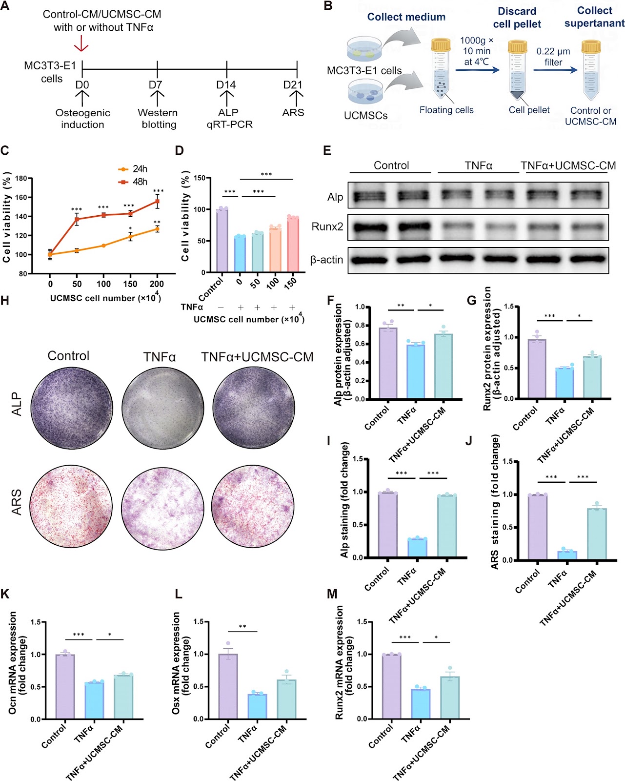
Background Osteoarthritis (OA) is a common degenerative disease caused by multiple pathological mechanisms wherein subchondral bone malfunction plays a substantial role. Recently, subchondral (SC) injection of orthobiologics has been attracting growing interest albeit the mainstream delivery method of mesenchymal stem cells (MSCs) is through intra-articular (IA). This study investigates the effect of SC injection of human umbilical cord mesenchymal stem cells (UCMSCs) on OA and its possible therapeutic mechanism compared to IA injection. Methods Male Sprague-Dawley rats with anterior cruciate ligament transection (ACLT) received saline or UCMSC injections via SC or IA. Consecutive injections once a week for three weeks and withdrawal for another four weeks, followed by Radiographical scanning, histopathological, immunohistochemical, and terminal deoxynucleotidyl transferase (TdT)-mediated dUTP nick-end labelling (TUNEL) staining. Cell counting Kit-8 (CCK-8) assay, alkaline phosphatase (ALP), alizarin red staining (ARS), TUNEL, flow cytometry, quantitative real-time polymerase chain reaction (qRT-PCR) and Western blotting were employed in TNFα-induced MC3T3-E1 cells to illustrate the exact pathogenesis mechanism. Results IA and SC UCMSC injections preserved cartilage, synovium, and subchondral bone parameters like trabecular bone volume fraction (BV/TV). SC injection uniquely improved Trabecular separation (Tb.Sp) and Trabecular number (Tb.N). SC and IA injections of UCMSCs demonstrated equivalent efficacy in promoting osteoblastic bone formation and attenuating aberrant angiogenesis of subchondral bone. In addition, we demonstrated that osteoblast apoptosis and Smad2-dependent TGF-beta (TGF-β) are crucial and interactive subchondral bone pathological features in OA. In vivo and vitro studies further revealed that UCMSCs inhibited excessive TGF-β/pSmad2 signaling to regulate osteoblast apoptosis and bone remodeling, thereby ameliorating OA progression. These findings suggest that SC injection of UCMSCs is a promising therapeutic strategy for OA, potentially offering advantages over IA injection in improving subchondral bone microarchitecture.
Read Full Abstract10.1186/s13287-025-04366-7
Mechanisms and clinical progress of adipose-derived stem cells and their derivatives in the treatment of hair lossGraphical AbstractVerified
Stem Cell Research & Therapy

Mechanisms and clinical progress of adipose-derived stem cells and their derivatives in the treatment of hair loss

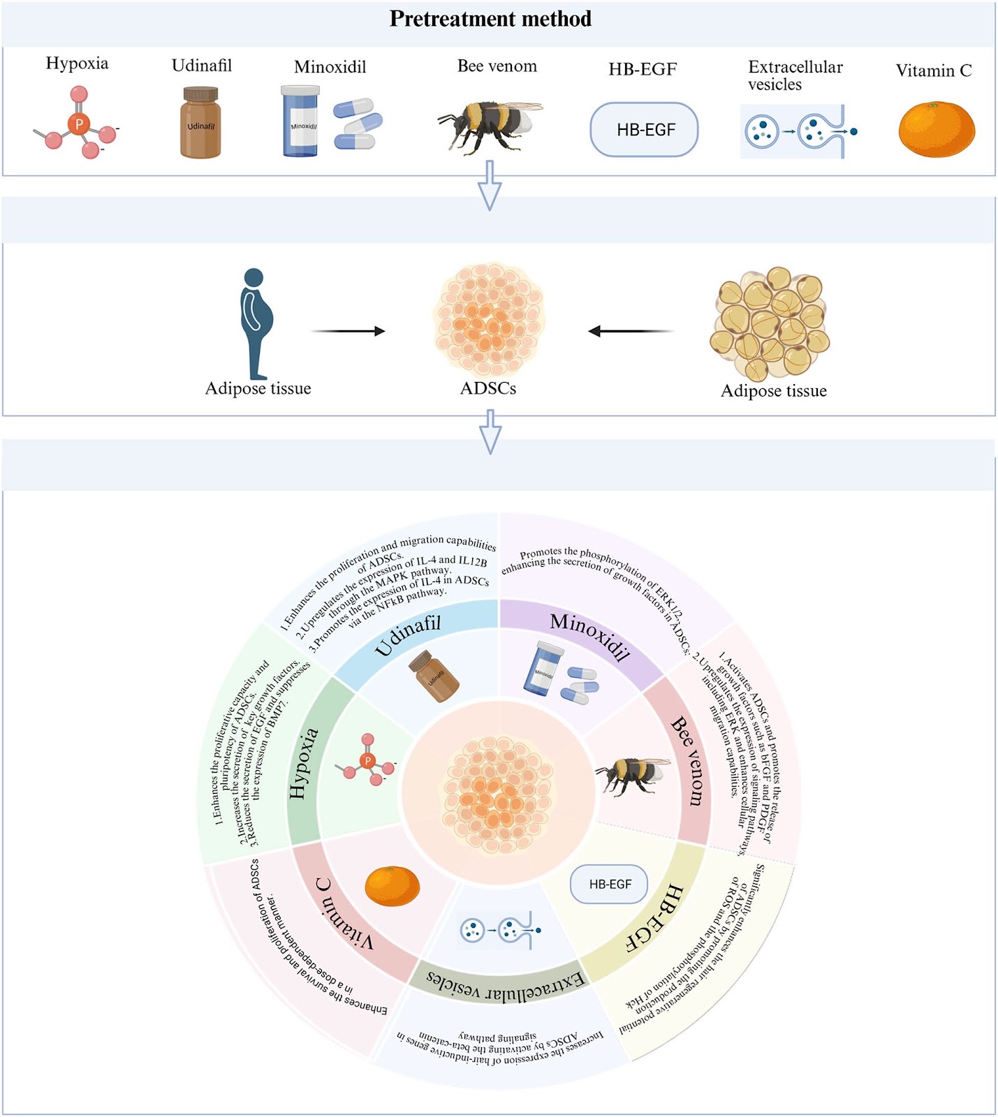
The rising prevalence of alopecia poses a significant challenge for both clinicians and researchers. As the global incidence of hair loss continues to increase, research into hair biology and regenerative mechanisms has gained considerable attention. However, current treatment options for alopecia are often constrained by limited efficacy and notable adverse effects. This underscores an urgent need for innovative therapeutic strategies to address these gaps. Adipose-derived stem cells (ADSCs), a subset of mesenchymal stem cells, represent a promising new approach in the treatment of alopecia. This review provides a detailed examination of the fundamental properties of ADSCs and their derivatives, exploring their mechanisms of action in alopecia therapy. Analysis of the efficacy of ADSCs and their derivatives in both preclinical and clinical settings highlight their potential to stimulate hair regeneration. Additionally, the review discusses various pre-treatment methods designed to enhance the regenerative capacity of ADSCs in hair growth, elucidating the mechanisms involved. The review also addresses the challenges and future directions for the use of ADSCs in alopecia treatment, aiming to offer valuable insights for both theoretical research and clinical practice. Ultimately, this work seeks to contribute to the development of more effective treatment regimens for alopecia.

Read Full Abstract10.1186/s13287-025-04560-7
CXCR5-engineered mesenchymal stromal cells home to spleen and mitigate post-sepsis syndrome by preventing secondary infectionGraphical AbstractVerified
Stem Cell Research & Therapy

CXCR5-engineered mesenchymal stromal cells home to spleen and mitigate post-sepsis syndrome by preventing secondary infection

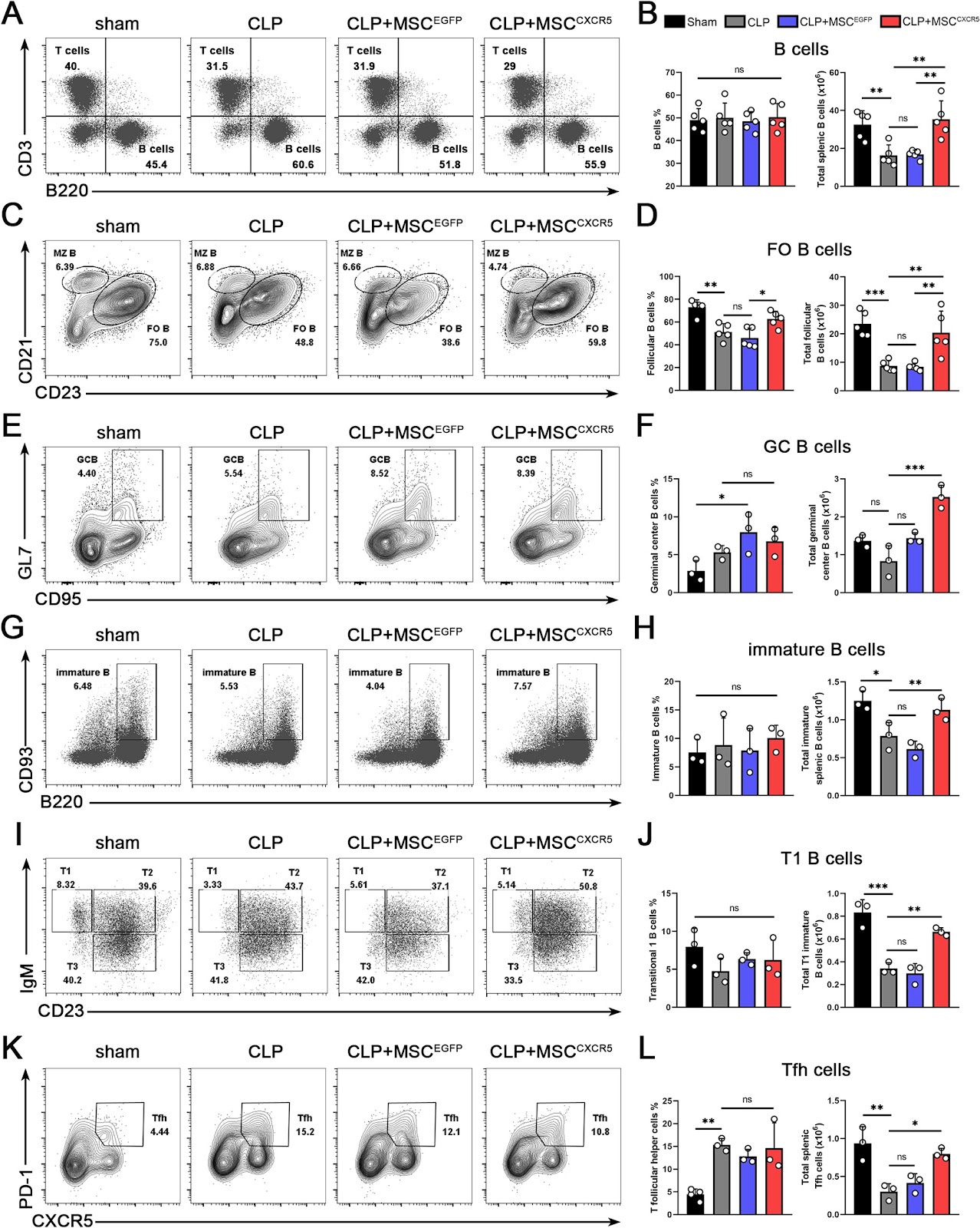
Background Sepsis survivors frequently develop compromised humoral immunity, manifesting as prolonged immunosuppression that increases susceptibility to secondary infections with high associated mortality. While mesenchymal stromal cells (MSCs) have demonstrated efficacy in mitigating initial inflammatory responses and improving early survival rates in murine cecal ligation and puncture (CLP) models, they show limited effectiveness against subsequent secondary infections. Given that lymphopenia represents a primary driver of this infection vulnerability, we sought to investigate whether spleen-targeted MSC therapy could prevent lymphocyte depletion and enhance protection against secondary infections. Methods In this study, we developed CXCR5-overexpressing MSCs (MSCCXCR5) to enhance their targeted migration toward splenic CXCL13. Using a CLP mouse model followed by secondary Staphylococcus aureus (S. aureus) infection, we systematically evaluated the therapeutic potential of MSCCXCR5 in combating post-sepsis immunosuppression. Results MSCCXCR5 demonstrated enhanced splenic migration, specifically homing to CXCL13-rich B-cell zones, and conferred significant protection against both S. aureus secondary infection and sepsis-induced lymphopenia. Mechanistic investigations revealed that MSCCXCR5 treatment preserved follicular and germinal center B-cell populations, maintaining splenic architecture and potentiating antigen-specific B-cell responses to CLP challenge. These effects collectively contributed to markedly improved survival outcomes in CLP mice following secondary infection. Conclusion Our findings demonstrate that MSCCXCR5 confers dual-phase protection in sepsis, improving survival during both the initial hyperinflammatory phase and subsequent immunosuppressive period. This comprehensive

Read Full Abstract10.1186/s13287-025-04751-2
A perfect islet: reviewing recent protocol developments and proposing strategies for stem cell derived functional pancreatic isletsGraphical AbstractVerified
Stem Cell Research & Therapy

A perfect islet: reviewing recent protocol developments and proposing strategies for stem cell derived functional pancreatic islets

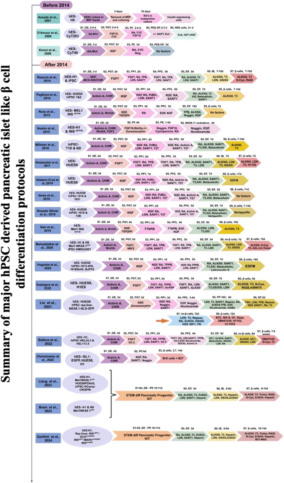
The search for an effective cell replacement therapy for diabetes has driven the development of ā€œperfectā€ pancreatic islets from human pluripotent stem cells (hPSCs). These hPSC-derived pancreatic islet-like β cells can overcome the limitations for disease modelling, drug development and transplantation therapies in diabetes. Nevertheless, challenges remain in generating fully functional and mature β cells from hPSCs. This review underscores the significant efforts made by researchers to optimize various differentiation protocols aimed at enhancing the efficiency and quality of hPSC-derived pancreatic islets and proposes methods for their improvement. By emulating the natural developmental processes of pancreatic embryogenesis, specific growth factors, signaling molecules and culture conditions are employed to guide hPSCs towards the formation of mature β cells capable of secreting insulin in response to glucose. However, the efficiency of these protocols varies greatly among different human embryonic stem cell (hESC) and induced pluripotent stem cell (hiPSC) lines. This variability poses a particular challenge for generating patient-specific β cells. Despite recent advancements, the ultimate goal remains to develop a highly efficient directed differentiation protocol that is applicable across all genetic backgrounds of hPSCs. Although progress has been made, further research is required to optimize the protocols and characterization methods that could ensure the safety and efficacy of hPSC-derived pancreatic islets before they can be utilized in clinical settings.

Read Full Abstract10.1186/s13287-025-04293-7
Correction: Adenovirus-mediated transfer of hepatocyte growth factor gene to human dental pulp stem cells under good manufacturing practice improves their potential for periodontal regeneration in swineGraphical AbstractVerified
Stem Cell Research & Therapy

Correction: Adenovirus-mediated transfer of hepatocyte growth factor gene to human dental pulp stem cells under good manufacturing practice improves their potential for periodontal regeneration in swine

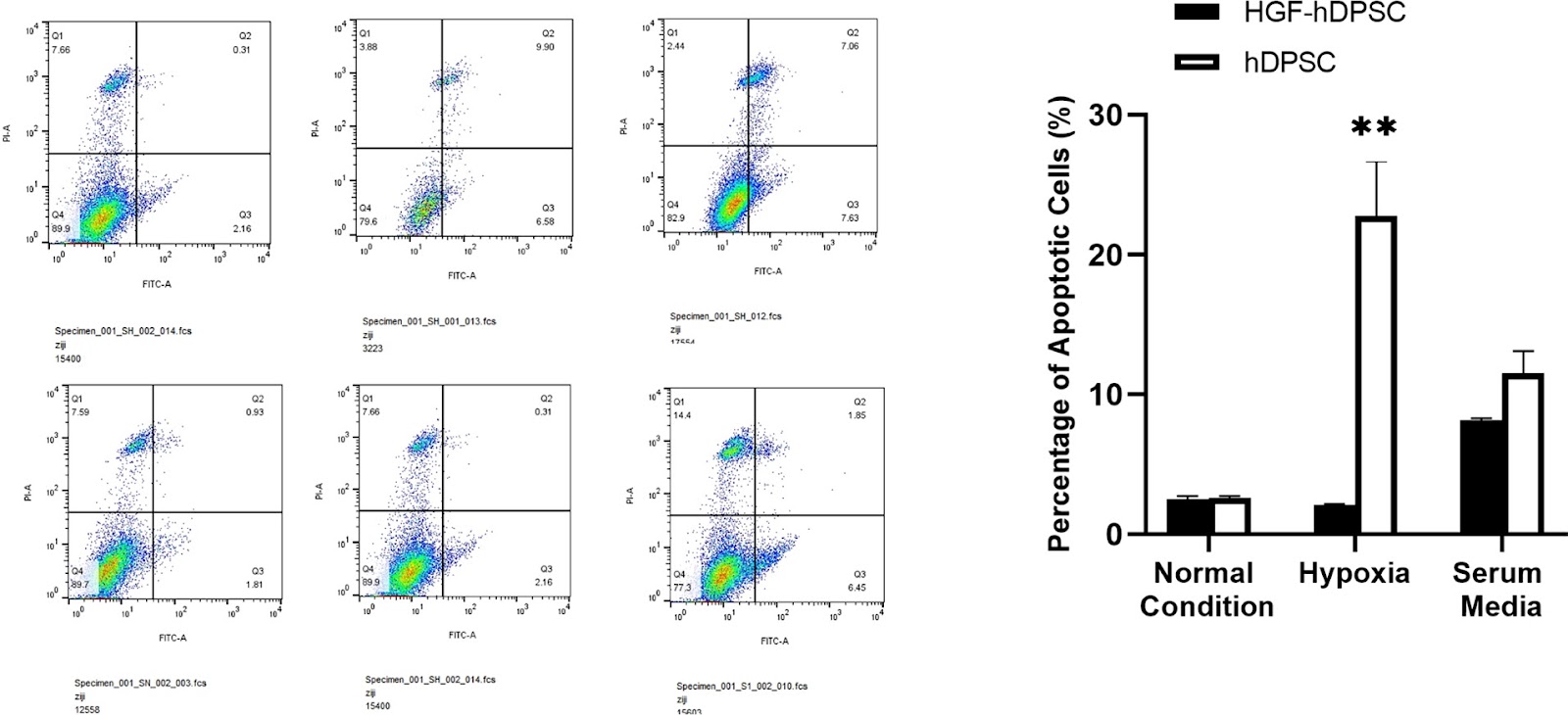
The authors wish to note the following correction: The images in Fig. 2D of our paper, which were intended to show Annexin V staining for apoptosis of human dental pulp stem cells (hDPSCs) and HGF-transfected hDPSCs (HGF-hDPSCs) under hypoxic conditions or serum-free media, were incorrect. The original results demonstrated that more apoptotic cells were observed in the hDPSCs group compared to the HGF-hDPSCs group. However, we inadvertently used images of Annexin V staining for apoptosis in human bone marrow mesenchymal stem cells (hBMSCs) and HGF-transfected hBMSCs (HGF-hBMSCs). Upon reviewing the original experimental records, we discovered that the incorrect images were included during the manuscript preparation process due to insufficient verification. We have now provided the correct images for hDPSCs and HGF-hDPSCs in Fig. 2D (see attachment). We sincerely apologize for this oversight. This error occurred because our research group has been extensively engaged in studying the biological characteristics of HGF gene-transfected mesenchymal stem cells. Unfortunately, due to carelessness, we mistakenly selected the wrong images. Nevertheless, our research consistently demonstrates that the anti-apoptotic ability of mesenchymal stem cells (including rBMSCs, hBMSCs, and hDPSCs) is enhanced under hypoxic conditions or serum-free media following HGF gene transfection. The methodology and results remain consistent with our previous studies. After thoroughly reviewing all data and experimental records, we confirm that this correction does not affect the validity of the original study’s results or conclusions.

Read Full Abstract10.1186/s13287-025-04214-8
Device encapsulated MSCs for adaptive secretome therapy to effectively target ischaemic heart injuryGraphical AbstractVerified
Stem Cell Research & Therapy

Device encapsulated MSCs for adaptive secretome therapy to effectively target ischaemic heart injury

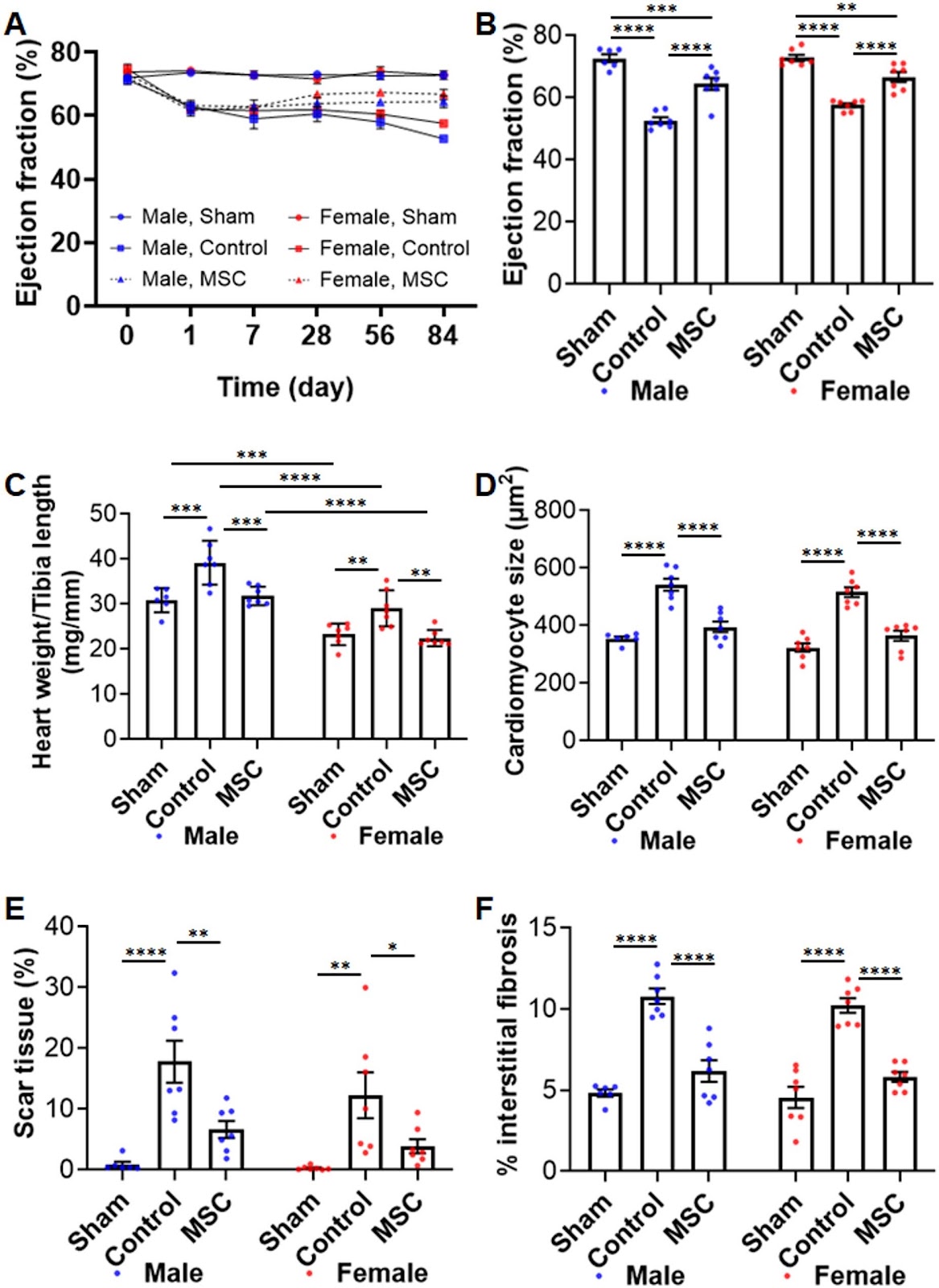
Background: Effective long-term strategies to protect the ischaemic heart remain a significant challenge. Mesenchymal stromal cells (MSCs) offer therapeutic potential primarily through their secretome, a bioactive factor-rich milieu with broad beneficial effects. However, existing delivery methods have not demonstrated sustained cardioprotection. The objective of this study was to evaluate a clinically translatable approach for sustained MSC-secretome delivery to achieve long-term cardioprotection. Methods: Cymerus MSCs, derived from human induced pluripotent stem cells (iPSCs), were encapsulated in a Procyon immunoisolation device and implanted subcutaneously in adult Sprague Dawley rats with chronic myocardial ischaemia-reperfusion injury. A human iPSC-derived engineered cardiac microtissue model was used to simulate ischaemia-reperfusion injury and assess cardioprotective effects in a human context. Proteomic analysis was performed to characterize adaptive changes in MSCs and their secretome post-implantation. Results: The MSC-loaded Procyon device significantly improved cardiac function and reduced adverse left ventricular remodelling over 12 weeks in both young and middle-aged, male and female rats. The encapsulated MSCs remained viable and retained the ability to release therapeutic secretome at 12 weeks post-implantation. In vitro, the MSC secretome protected human engineered cardiac microtissues from simulated ischaemia-reperfusion injury by restoring contractile function, improving cell viability, and reducing oxidative stress. Proteomic profiling of encapsulated MSC identified 179 unique cellular proteins post-implantation, associated with adaptive immune and inflammatory responses as well as wound healing. MSC secretome profiling revealed increased protein diversity associated with tissue repair and immune regulation, suggesting MSCs undergo an adaptive response to ischaemic conditions.

Read Full Abstract10.1186/s13287-025-04847-9
Allogeneic bone marrow-derived mesenchymal stem cells in the aging kidney: secondary results of a Parkinson's disease clinical trialGraphical AbstractVerified
Stem Cell Research & Therapy

Allogeneic bone marrow-derived mesenchymal stem cells in the aging kidney: secondary results of a Parkinson's disease clinical trial

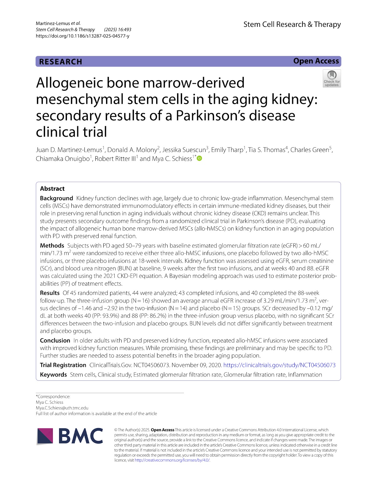
Background Kidney function declines with age, largely due to chronic low-grade inflammation. Mesenchymal stem cells (MSCs) have demonstrated immunomodulatory effects in certain immune-mediated kidney diseases, but their role in preserving renal function in aging individuals without chronic kidney disease (CKD) remains unclear. This study presents secondary outcome findings from a randomized clinical trial in Parkinson's disease (PD), evaluating the impact of allogeneic human bone marrow-derived MSCs (allo-hMSCs) on kidney function in an aging population with PD with preserved renal function. Methods Subjects with PD aged 50–79 years with baseline estimated glomerular filtration rate (eGFR) > 60 mL/min/1.73 m2 were randomized to receive either three allo-hMSC infusions, one placebo followed by two allo-hMSC infusions, or three placebo infusions at 18-week intervals. Kidney function was assessed using eGFR, serum creatinine (SCr), and blood urea nitrogen (BUN) at baseline, 9 weeks after the first two infusions, and at weeks 40 and 88. eGFR was calculated using the 2021 CKD-EPI equation. A Bayesian modeling approach was used to estimate posterior probabilities (PP) of treatment effects. Results Of 45 randomized patients, 44 were analyzed; 43 completed infusions, and 40 completed the 88-week follow-up. The three-infusion group (N=16) showed an average annual eGFR increase of 3.29 mL/min/1.73 m2, versus declines of –1.46 and –2.92 in the two-infusion (N=14) and placebo (N=15) groups. SCr decreased by –0.12 mg/dL at both weeks 40 (PP: 93.9%) and 88 (PP: 86.2%) in the three-infusion group versus placebo, with no significant SCr differences between the two-infusion and placebo groups. BUN levels did not differ significantly between treatment and placebo groups. Conclusion In older adults with PD and preserved kidney function, repeated allo-hMSC infusions were associated with improved kidney function measures. While promising, these findings are preliminary and may be specific to PD. Further studies are needed to assess potential benefits in the broader aging population. Trial Registration ClinicalTrials.Gov. NCT04506073. November 09, 2020. https://clinicaltrials.gov/study/NCT04506073

Read Full Abstract10.1186/s13287-025-04577-y
SPARC-modified mesenchymal stem cells promote recovery of β-cells and insulin secretion by calcium ion homeostasisGraphical AbstractVerified
Stem Cell Research & Therapy

SPARC-modified mesenchymal stem cells promote recovery of β-cells and insulin secretion by calcium ion homeostasis

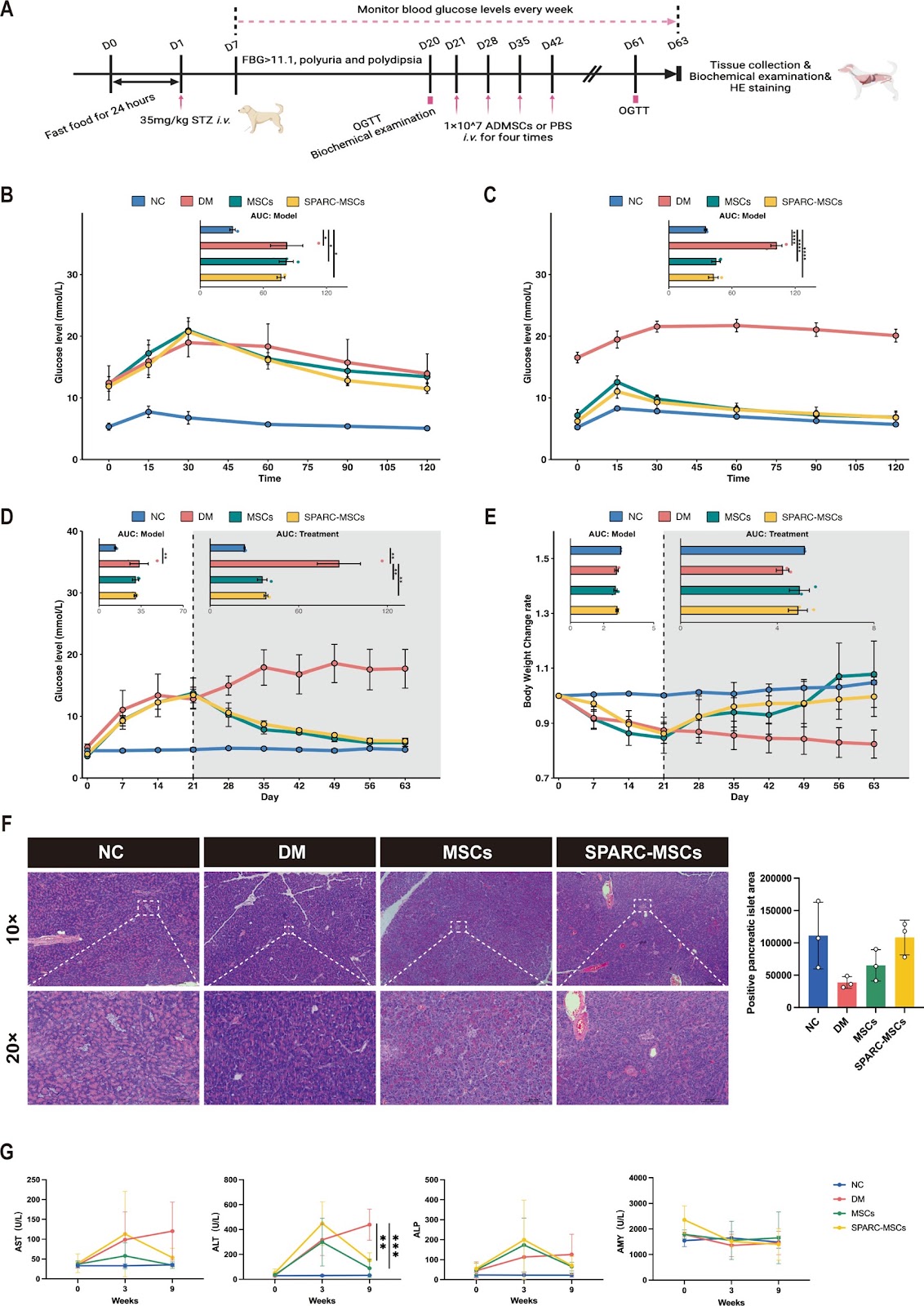
Introduction Type 1 diabetes (T1D) results from the destruction of pancreatic β-cells, leading to insulin deficiency. As insulin therapy does not affect disease progression, advancements in immune regulation therapies have emerged, including the reconstitution of the insulin secretory system. Cysteine-rich acidic secretory protein (SPARC) is an extracellular matrix glycoprotein that regulates cell adhesion, facilitating cell migration, and mediating interactions between cells and their extracellular matrix. SPARC is overexpressed during tissue repair and is involved in β-cells survival. However, the potential of SPARC-modified mesenchymal stem cells (MSCs) to improve insulin secretion has not been thoroughly investigated. This study investigated the therapeutic effects of SPARC-MSCs in vivo and in vitro and assessed whether SPARC enhances survival and insulin secretion after β-cells injury. Methods In vivo, we established T1D models in mice and canine using SPARC-MSCs for cell transplantation. In vitro, MIN6 cells were damaged with STZ, and SPARC-MSC supernatant was co-cultured with MIN6 for various assays. Results Our study demonstrated that SPARC enhanced the regenerative capacity and migratory efficiency of MSCs after H2O2 injury and improved their morphology. In STZ-induced canine and mice diabetes models, SPARC-MSCs therapy significantly reduced hyperglycemia, improved oral glucose tolerance test (OGTT), and reversed weight loss in canine. Biochemical analyses showed improved liver function, and histological examination revealed restored islet area was significantly restored. Transcriptome and proteome sequencing indicated significant enrichment in calcium binding and cell migration pathways. Co-culturing SPARC-MSC supernatant with MIN6 cells after STZ injury restored their regenerative ability, enhancing insulin secretion and ATP content under high glucose stimulation. SPARC treatment also significantly increased intracellular Ca2+ levels in MIN6 cells. Conclusion SPARC significantly promotes cell regeneration and stimulates insulin secretion by increasing intracellular ATP and Ca2+ influx. In diabetic canine and mice models, it alleviated hyperglycemia, improved glucose tolerance, and enhanced pancreatic islet area and insulin secretion.

Read Full Abstract10.1186/s13287-025-04727-2
Human adipose-derived stem cell exosomes reduce mitochondrial DNA common deletion through PINK1/Parkin-mediated mitophagy to improve skin photoagingGraphical AbstractVerified
Stem Cell Research & Therapy

Human adipose-derived stem cell exosomes reduce mitochondrial DNA common deletion through PINK1/Parkin-mediated mitophagy to improve skin photoaging

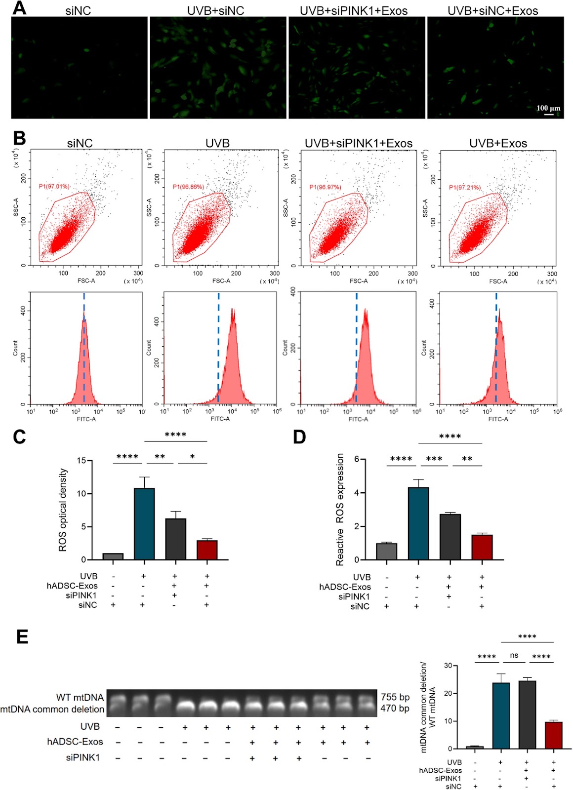
Background: Mitochondrial DNA (mtDNA) deletion and oxidative stress are key contributors to skin photoaging. Mitophagy helps mitigate oxidative stress. Human adipose-derived stem cell exosomes (hADSC-Exos) have been shown to counteract skin photoaging. This study aimed to explore the role and mechanism of hADSC-Exos in addressing skin photoaging. Methods: hADSC-Exos were isolated, and their surface markers were identified. Human dermal fibroblasts (HDFs) and nude mice were exposed to ultraviolet-B (UVB) irradiation, and treated with hADSC-Exos. Oxidative stress and photoaging were assessed through SA-β-gal staining, p21 expression, mtDNA deletion, reactive oxygen species (ROS) levels, and histological analysis. The PINK1, Parkin, LC3b, and p62 protein levels were measured to evaluate mitophagy. The PINK1 small-interfering RNA (siPINK1) was then used in HDFs to investigate the role of hADSC-Exos in mitophagy. Results: In UVB-exposed HDFs and nude mice, the number of SA-β-gal-positive cells, along with levels of p21, ROS, and mtDNA deletion, were significantly increased, but these effects were reduced by hADSC-Exos. Moreover, hADSC-Exos treatment significantly elevated PINK1 and Parkin levels, as well as the LC3bII/I ratio, while reducing p62 expression. In photoaged HDFs treated with hADSC-Exos, PINK1 knockout using siRNA decreased the LC3bII/I ratio and levels of PINK1 and Parkin, while increasing p62, ROS, and mtDNA deletion compared to the negative control (NC) group. Conclusion: hADSC-Exos can mitigate skin photoaging by promoting PINK1/Parkin-mediated mitophagy, thereby reducing mtDNA deletion and oxidative stress.

Read Full Abstract10.1186/s13287-025-04475-3
Hyaluronic acid–ornithine crosslinked hydrogel as a superior 3D culture platform for high-quality exosome production in advanced wound healingGraphical AbstractVerified
Stem Cell Research & Therapy

Hyaluronic acid–ornithine crosslinked hydrogel as a superior 3D culture platform for high-quality exosome production in advanced wound healing

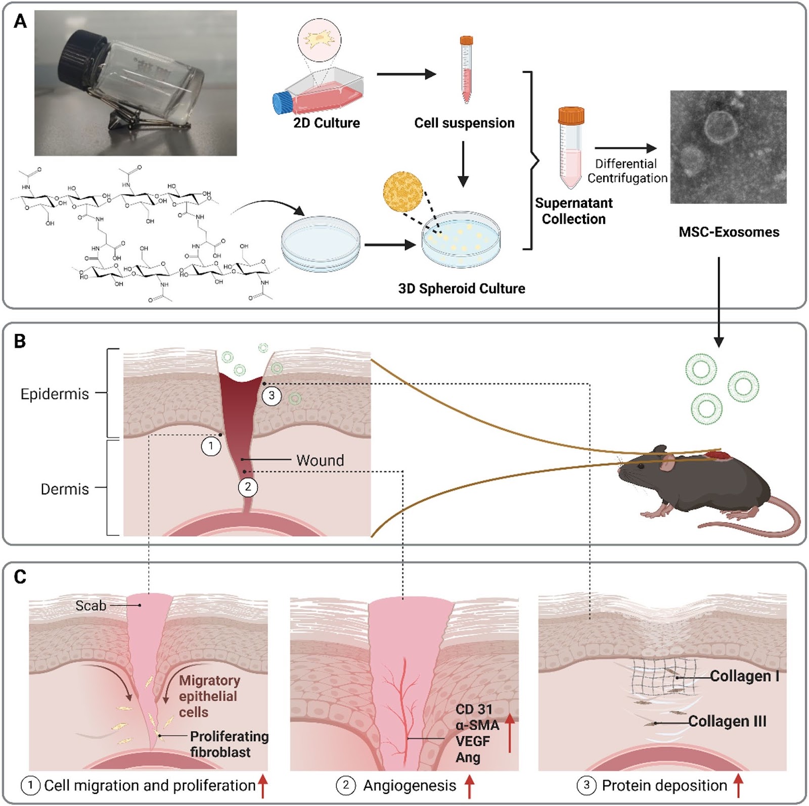
Background: Extracellular vesicle (EV)-based cell-free therapies have emerged as a powerful alternative to stem cell transplantation in regenerative medicine, owing to their ability to promote tissue repair while avoiding safety concerns associated with live-cell therapies. However, traditional two-dimensional (2D) cell cultures used for EV production are constrained by low exosome (Exo) yields and limited biological activity. Objective: This study introduces a novel and scalable three-dimensional (3D) culture platform based on a hyaluronic acid (HA) and L-ornithine methyl ester (Orn) hydrogel to enhance the production and therapeutic efficacy of stem cell-derived exosomes. Methods: The HA-Orn hydrogel was fabricated via a simple and mild crosslinking strategy, forming a biomimetic matrix that promotes spontaneous spheroid formation. Exosomes derived from 3D cultures (3D-Exo) were compared with those from 2D cultures (2D-Exo) in terms of yield, molecular composition, and biological functions. Results: 3D-Exo exhibited significantly increased yield and superior functional properties, including enhanced stimulation of cell proliferation, migration, angiogenesis, and extracellular matrix remodeling. In vivo, 3D-Exo treatment accelerated wound closure and reduced inflammation in a mouse skin injury model, demonstrating robust therapeutic efficacy and safety. Mechanistic studies revealed distinct miRNA expression profiles and activation of regenerative signaling pathways in 3D-Exo. Conclusion: This work presents a cost-effective, scalable, and bioinspired 3D culture system for high-yield and functionally enhanced Exo production. The HA-Orn hydrogel platform offers significant translational potential for advancing cell-free regenerative therapies, particularly in the context of wound healing.

Read Full Abstract10.1186/s13287-025-04635-5
From gut to liver: organoids as platforms for next-generation toxicology assessment vehicles for xenobioticsGraphical AbstractVerified
Stem Cell Research & Therapy

From gut to liver: organoids as platforms for next-generation toxicology assessment vehicles for xenobiotics

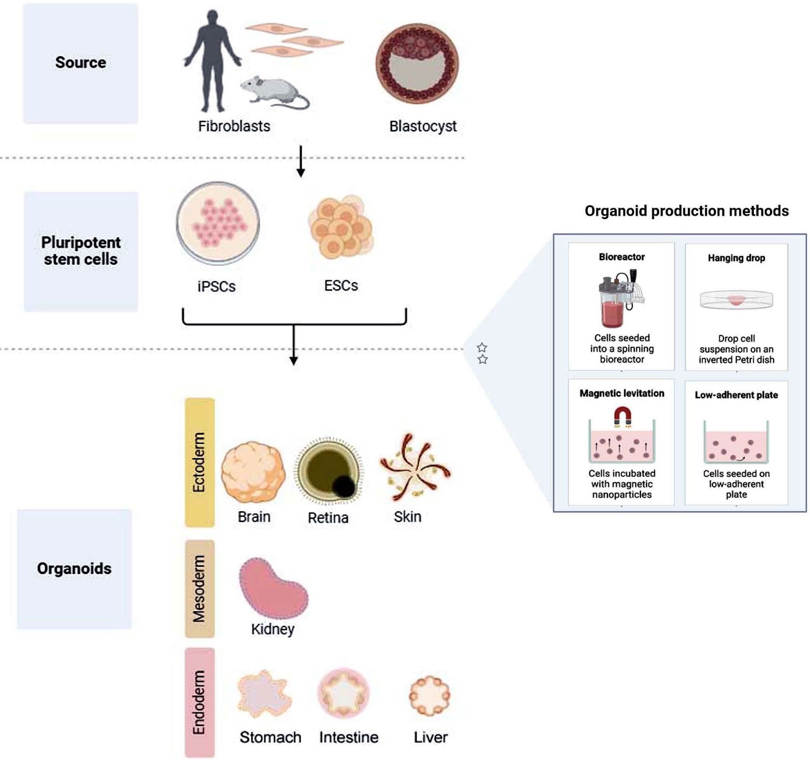
Traditional toxicological assessment relied heavily on 2D cell cultures and animal models of study, which were inadequate for the precise prediction of human response to chemicals. Researchers have now shifted focus on organoids for toxicological assessment. Organoids are 3D structures produced from stem cells that mimic the shape and functionality of human organs and have a number of advantages compared to traditional models of study. They have the capacity to replicate the intricate cellular microenvironment and in vivo interactions. They offer a physiologically pertinent platform that is useful for the researchers to monitor cellular responses in a more realistic manner and evaluate drug toxicity. Additionally, organoids can be created from cells unique to a patient, allowing for individualized toxicological research and providing understanding of the inter-individual heterogeneity in drug responses. Recent developments in the use of gut and liver organoids for assessment of the xenobiotics (environmental toxins and drugs) is reviewed in this article. Gut organoids can reveal potential damage to the digestive system and how xenobiotics affect nutrient absorption and barrier function. Liver is the primary site of detoxification and metabolism of xenobiotics, usually routed from the gut. Hence, these are linked and crucial for evaluating chemical or pollutant induced organ toxicity, forecasting their metabolism and pharmacokinetics. When incorporated into the drug development process, organoid models have the potential to improve the accuracy and efficiency of drug safety assessments, leading to safer and more effective treatments. We also discuss the limitations of using organoid-based toxicological assays, and future prospects, including the need for standardized protocols for overcoming reproducibility issues.

Read Full Abstract10.1186/s13287-025-04264-y
Mesenchymal stem cell therapy for end-stage liver disease: adversity and opportunityGraphical AbstractVerified
Stem Cell Research & Therapy

Mesenchymal stem cell therapy for end-stage liver disease: adversity and opportunity

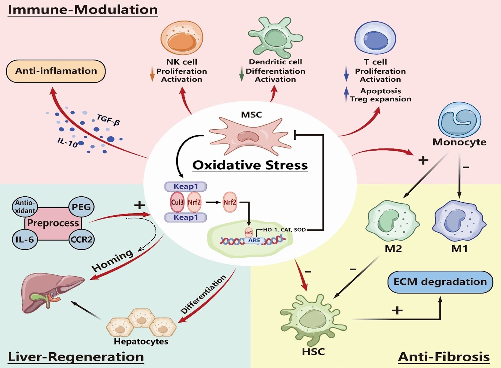
End-stage liver disease (ESLD) is one of the predominant diseases contributing to high morbidity and mortality worldwide, with etiologies including alcoholic liver disease, viral hepatitis, non-alcoholic fatty liver disease, and metabolic-associated liver disease. Currently, liver transplantation remains the only effective treatment, however, its clinical application is significantly limited by donor shortages, immune rejection, and high medical costs. Among the five types of stem cells that have been experimentally applied to liver diseases, mesenchymal stem cells (MSCs) have emerged as the most extensively studied, with the largest number of experimental and clinical research platforms worldwide. This review compiles findings from 25 preclinical and clinical studies on MSCs in the treatment of ESLD, aiming to elucidate the core mechanisms of action and then outline both the challenges in MSC clinical translation and the novel opportunities arising from cutting-edge research.

Read Full Abstract10.1186/s13287-025-04788-3
Human platelet lysate produced from leukoreduction filter contents enables sufficient MSC growthGraphical AbstractVerified
Stem Cell Research & Therapy

Human platelet lysate produced from leukoreduction filter contents enables sufficient MSC growth

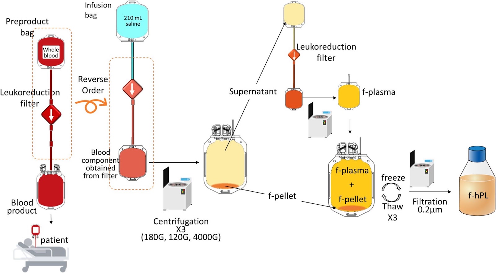
Background Stem cell therapy holds significant potential for promoting recovery, with numerous products currently under development. Blood-derived supplements are often essential for successful stem cell expansion, with fetal bovine serum (FBS) being the most commonly used supplement. However, FBS has drawbacks, including the risk of immune responses, ethical concerns about animal welfare, and potential zoonotic infections. Human platelet lysate (hPL), derived from lysed platelets, contains various growth factors and has been proposed as an alternative to FBS. However, obtaining sufficient human platelets for clinical use remains challenging. Leukoreduction filters, used during blood transfusion manufacturing to remove leukocytes, also retain significant amounts of platelets and plasma. This study investigates the feasibility and efficacy of filter-derived hPL (f-hPL) for mesenchymal stem cell (MSC) expansion. Methods Leukoreduction filters were collected after their use in the manufacturing of whole blood transfusion products. Each filter was reverse-perfused with saline to extract residual blood contents. Platelets (f-platelet) and supernatant were separated by multiple centrifugation steps. f-Platelet were lysed with varying concentrations of fresh frozen plasma (FFP) to determine the optimal protein concentration for the lysate solution. Then, plasma left in the leukoreduction filters were used to generate lysate solution (f-plasma) at optimal protein concentration. f-Platelet (1.1 Ɨ 10^9/mL) and f-plasma (27 mg/mL protein) were combined in a freezing bag and subjected to three freeze-thaw cycles to produce f-hPL. Both small- and large-scale f-hPL were manufactured, and MSCs expansion and quality assessments were perfomed to evaluate the efficacy of f-hPL. Results A total of 3.5 ± 0.6 Ɨ 10^10 f-platelets were obtained from a single leukoreduction filter, yielding a collection efficiency of 37.1 ± 5.3%. The optimal protein concentration of lysate solution for cell expansion was > 27 mg/mL. Subsequently, six leukoreduction filters used to produce enough f-platelet and p-plasma for 100 mL of f-hPL. MSCs cultured in medium supplemented with 10% f-hPL demonstrated superior expansion, with cell proliferation rates 20% higher than those observed with commercial hPL and 300% higher than those cultured with FBS. The expanded MSCs met the International Society for Cell & Gene Therapy criteria for cell surface markers and differentiation potential.

Read Full Abstract10.1186/s13287-025-04329-y
Fucoxanthin protects placenta-derived human mesenchymal stem cells against oxidative stress-induced apoptosis by modulating genes involved in DNA damage repair, ER stress response, p53-induced apoptosisGraphical AbstractVerified
Stem Cell Research & Therapy

Fucoxanthin protects placenta-derived human mesenchymal stem cells against oxidative stress-induced apoptosis by modulating genes involved in DNA damage repair, ER stress response, p53-induced apoptosis

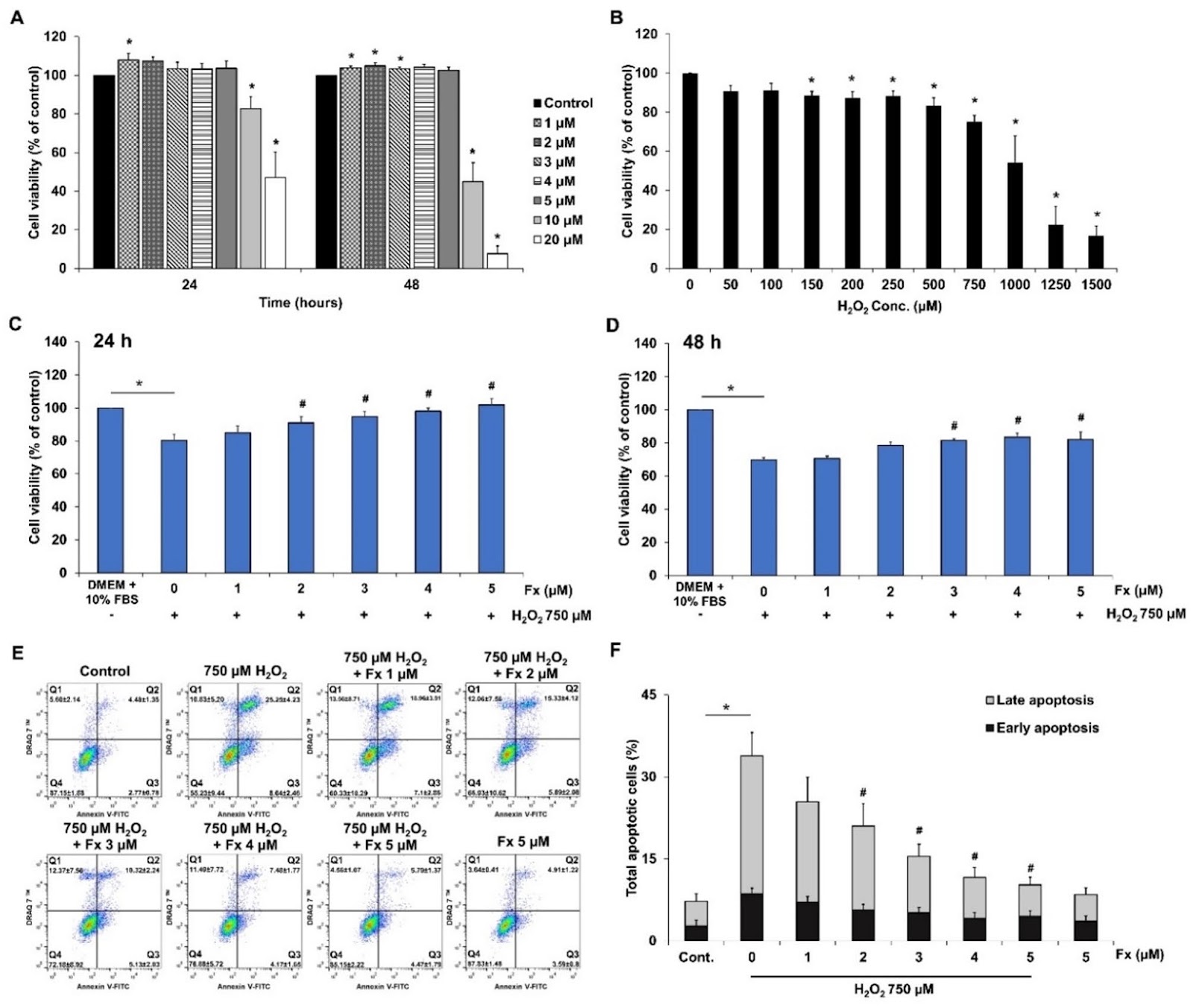
Human mesenchymal stem cells (hMSCs) hold significant promise in regenerative medicine due to their ability to reduce inflammation and promote tissue repair. However, their therapeutic potential is often compromised by their high susceptibility to apoptosis under oxidative stress, prevalent in the microenvironment of the target tissues. Our previous study showed that fucoxanthin, a carotenoid derived from brown algae, can improve the viability of placenta-derived mesenchymal stem cells (PL-MSCs) by reducing intracellular ROS levels through the activation of the PI3K/Akt/Nrf-2 signaling pathway. In this study, we further investigate the mechanisms underlying the protective effect of fucoxanthin against oxidative stress-induced apoptosis in PL-MSCs, using an in vitro model. PL-MSCs were cultured with 750 µM H2O2 to induce oxidative stress and treated with various concentrations of fucoxanthin for 48 h. The effect of fucoxanthin on PL-MSC apoptosis under oxidative stress conditions was determined using CCK-8, Annexin V/DRAQ7ā„¢ apoptosis assays, as well as the expression of apoptosis-related genes and proteins. The effect of fucoxanthin on the transcriptome of PL-MSCs under oxidative stress conditions was also assessed by high-throughput Nanostring analysis. The results showed that fucoxanthin significantly decreased the apoptosis of PL-MSCs under oxidative stress in a dose-dependent manner by reducing the expression of pro-apoptotic proteins and inhibiting their activation, while increasing the expression of anti-apoptotic proteins in these cells. Furthermore, fucoxanthin also downregulates the expression of genes associated with the endoplasmic reticulum stress, p53-induced apoptosis, while increasing the expression of genes involved in the regulation of the cell cycle, DNA damage repair, cytokine signaling, nucleotide synthesis, PI3K/mTOR pathway and AMPK pathway in PL-MSCs under oxidative stress conditions. Taken together, the findings provide compelling evidence that fucoxanthin protects PL-MSCs against oxidative stress-induced apoptosis by modulating the expression of various genes involved in DNA damage repair, ER stress response, p53-induced apoptosis in these cells. This suggests that fucoxanthin could be used as a potential supplement to enhance the therapeutic efficacy of MSC-based therapies.

Read Full Abstract10.1186/s13287-025-04629-3
Morphometric prediction of mesenchymal stromal cell-like immunosuppressive capacity of human hair follicle dermal sheath cup cells: an implication for regenerative medicine in hair loss diseasesGraphical AbstractVerified
Stem Cell Research & Therapy

Morphometric prediction of mesenchymal stromal cell-like immunosuppressive capacity of human hair follicle dermal sheath cup cells: an implication for regenerative medicine in hair loss diseases

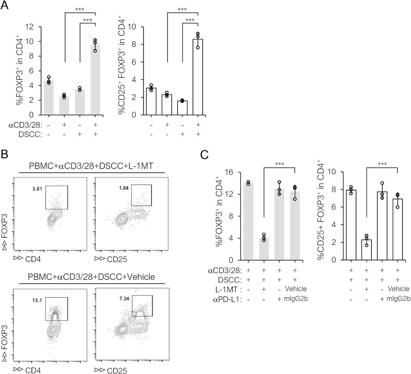
Background Human hair follicle dermal sheath cup cells (DSCCs) hold promise as a cell source of regenerative medicine treatment for hair loss owing to their ability to secrete growth factors and/or signal pathway activators. The therapeutic effect of autologous DSCCs transplantation for male/female pattern hair loss (PHL) was demonstrated in a phase III equivalent clinical study. Intralesional inflammation has been implicated in the pathophysiology of various hair loss diseases, including PHL. As DSCCs possess mesenchymal stem/stromal cell (MSC)-like properties and MSCs are immunosuppressive, we investigated whether they exhibit immunoregulatory capabilities comparable to MSCs and developed an in vitro morphometric assay to predict this capability. Methods DSCCs were isolated via microdissection and propagated in vitro. Their conformity to MSC criteria was assessed based on cell surface antigen expression and differentiation potential. Furthermore, immunoregulatory capabilities were assessed by co-culturing DSCCs with anti-CD3/28 antibody-stimulated peripheral blood mononuclear cells (PBMCs) and examining the suppression mechanisms through pharmacological intervention. Multiple lots of DSCCs derived from various donors and manufacturing conditions were cultured and analyzed by phase-contrast microscopy to obtain their morphometric profiles. Parameters correlating with the expression levels of immunomodulatory factors were used to create a predictive model. Additional DSCC lots were manufactured to validate the predictive model. Results Similar to MSCs, cell differentiation assays revealed that DSCCs exhibited multipotency, and they did not express co-stimulatory molecules in response to immunogenic stimuli, suggesting low immunogenicity. Moreover, co-culture experiments with allogeneic PBMCs revealed that DSCCs reduced T cell proliferation (from 78 to 5%) and

Read Full Abstract10.1186/s13287-025-04764-x
CD317 stabilizes TNFR1 and confers the anti-inflammatory functions of MSCs via NF-ĪŗB/TSG6 pathwayGraphical AbstractVerified
Stem Cell Research & Therapy

CD317 stabilizes TNFR1 and confers the anti-inflammatory functions of MSCs via NF-ĪŗB/TSG6 pathway

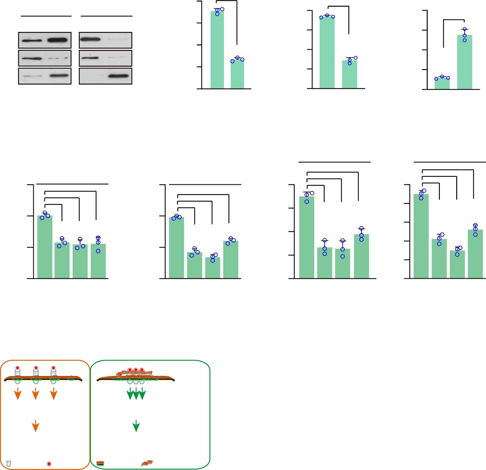
Background Although both pre-clinical and clinical studies show promising outcomes, resulting in rapid growth of clinical trials of MSC-based therapies in recent years, the heterogeneity and therapeutic inconsistency of MSCs have severely hampered their clinical applications. Purifying homogenous MSC populations with enhanced specific functions represents one promising approach. We have demonstrated recently that the CD317+ MSCs have enhanced anti-inflammatory functions and improved therapeutic efficacy and consistency. Methods In the current study, we performed both in vitro and in vivo investigations to delineate whether and how CD317 regulates the immune modulation function of MSCs. Results Our data here indicate that the CD317 directly contributes to the immune suppression function of MSCs stimulated by TNF-α through up-regulating TSG6 via CD317/lipid-raft/TNFR1 complex. The CD317 stabilizes the TNFR1 complex, resulting in hyper-activation of the NF-κB pathway and up-regulation of TSG6, which confers the therapeutic effects of MSCs on the mouse model of ALI (acute lung injury) and IBD (inflammatory bowel disease). Conclusions Thus, the CD317 stabilizes TNFR1 and confers the anti-inflammatory functions of MSCs via NF-κB/TSG6 Pathway.

Read Full Abstract10.1186/s13287-025-04527-8
Clinical application of mesenchymal stem cells in immunosenescence: a qualitative review of their potential and challengesGraphical AbstractVerified
Stem Cell Research & Therapy

Clinical application of mesenchymal stem cells in immunosenescence: a qualitative review of their potential and challenges

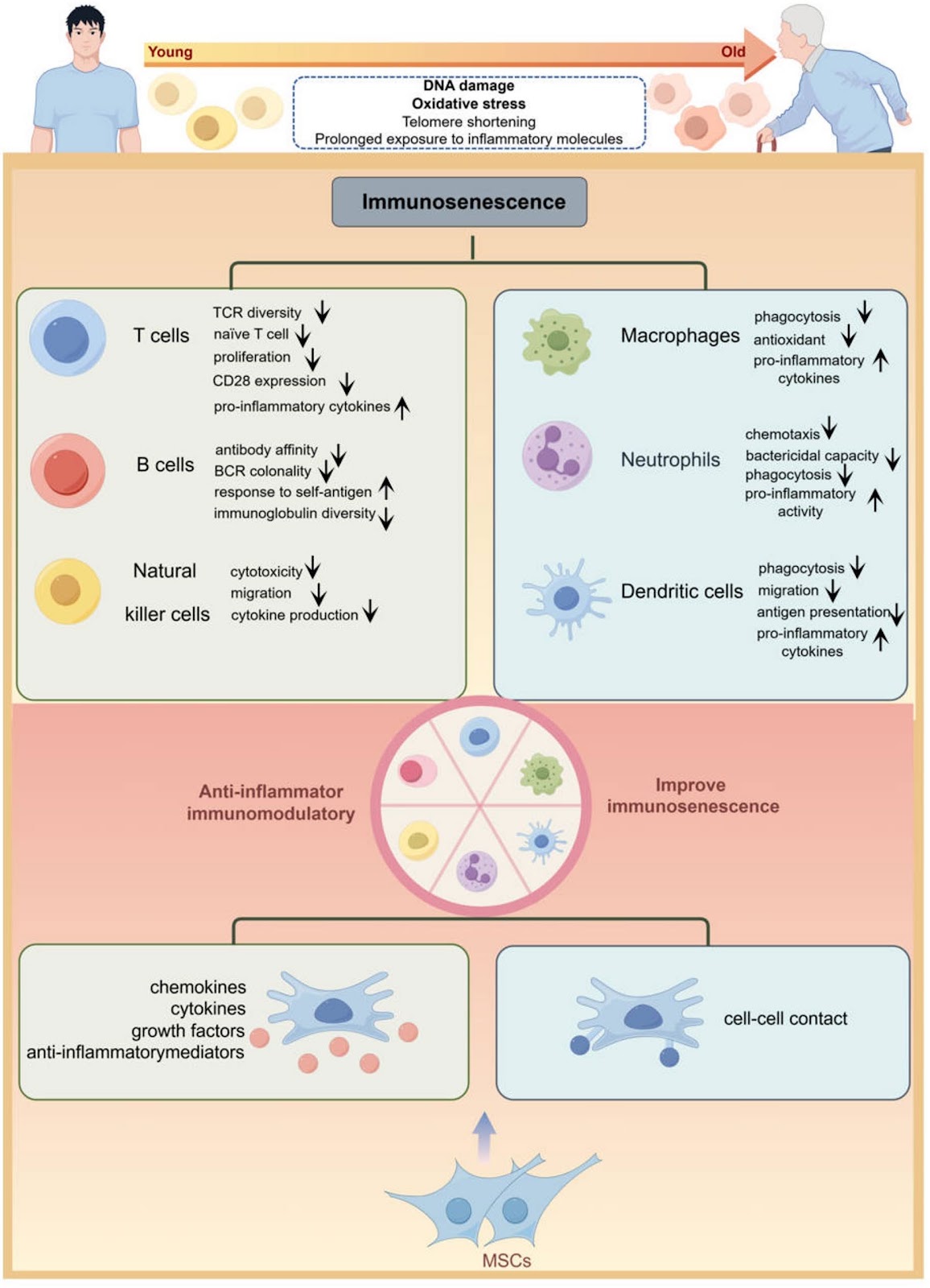
Aging leads to a gradual decline in immune function, termed immunosenescence, which significantly elevates the susceptibility to infections, cancers, and other aging-related diseases. Recent advancements have shed light on the molecular underpinnings of immune aging and pioneered novel therapeutic interventions to counteract its effects. Mesenchymal stem cells (MSCs)-a type of multipotent stromal cells with regenerative potential, low immunogenicity, and strong immunomodulatory properties-are increasingly recognized as a promising therapeutic option to reverse or alleviate immunosenescence-related dysfunction. This review systematically summarizes recent discoveries on how MSCs counteract immune aging, particularly their ability to rejuvenate aged immune cells and restore immune homeostasis. It also addresses key challenges, such as variations in MSC sources, donor variability, and the lack of standardized protocols, while proposing future directions to enhance therapeutic precision. Although preclinical and clinical studies highlight the potential of MSC-based strategies for delaying immunosenescence, critical issues remain unresolved, including long-term safety and efficacy, optimizing cell delivery systems, and elucidating context-specific mechanisms. Addressing these challenges will accelerate the development of MSC-based therapies to combat aging-associated immune decline.

Read Full Abstract10.1186/s13287-025-04360-z
Three-dimensional midbrain organoids: a next-generation tool for Parkinson's disease modelling and drug discoveryGraphical AbstractVerified
Stem Cell Research & Therapy

Three-dimensional midbrain organoids: a next-generation tool for Parkinson's disease modelling and drug discovery

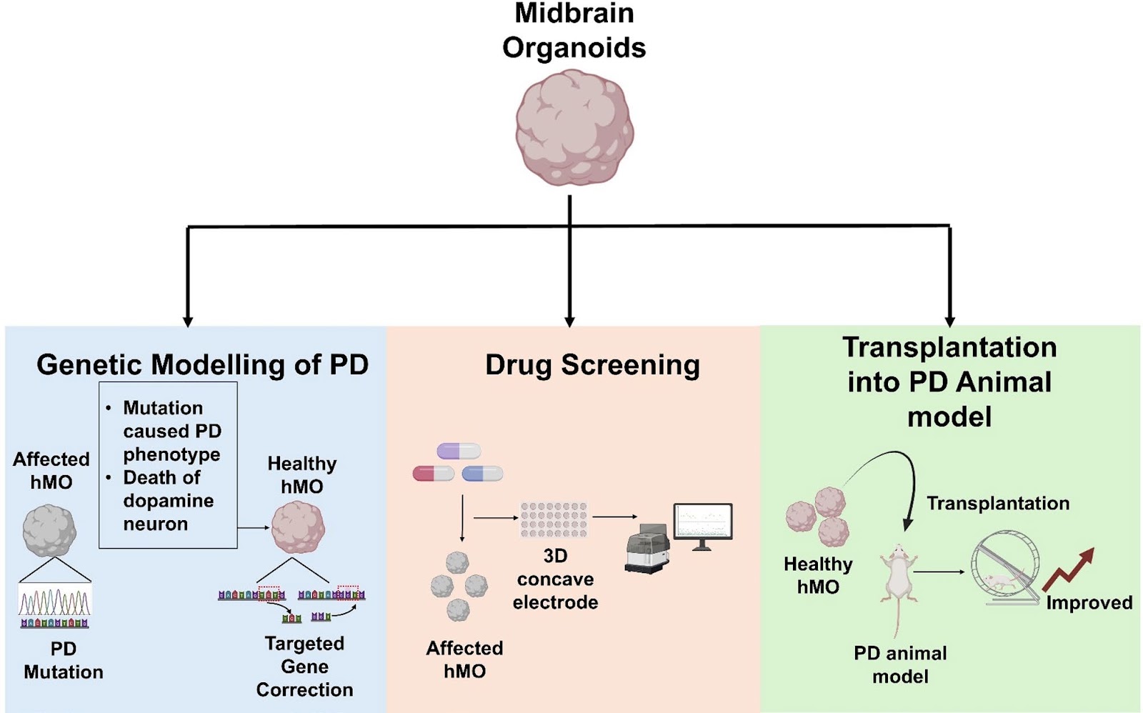
Parkinson's disease (PD), a progressive neurodegenerative disorder marked by dopaminergic (DA) neuron loss and Lewy body formation, lacks therapies to halt neurodegeneration. Current models, including 2D cultures and animal studies, fail to fully recapitulate human midbrain complexity, underscoring the need for advanced human-relevant disease modelling systems. Midbrain organoids (MOs), three-dimensional (3D) stem cell-derived neuronal structures mimicking midbrain architecture, have emerged as transformative tools for modelling PD. These organoids replicate key pathological hallmarks and enable disease mechanistic studies and drug screening for PD. Recent advances of research in MOs include genetic modelling of PD-linked mutations (e.g., LRRK2, GBA1, DNAJC6), optogenetics-assisted α-synuclein (α-syn) protein aggregation systems, and high-throughput drug testing platforms. MOs also show promise for cell replacement therapy, with successful integration and functional recovery in animal PD models. However, challenges such as batch variability, limited vascularization, incomplete neuronal maturation, and high costs hinder reproducibility and scalability. Future directions focus on integrating vascular networks, microglia co-cultures, automated workflows, and assembloid technologies to enhance pathophysiological relevance and translational potential in PD. By addressing these limitations, research in MOs could revolutionize PD research, offering critical insights into disease mechanisms and accelerating therapeutic discovery for PD patients.

Read Full Abstract10.1186/s13287-025-04660-4
L-Glutamate enables the EGFR-MEK-ERK-mTFB2 axis to enhance mitochondrial biogenesis in intestinal stem cellsGraphical AbstractVerified
Stem Cell Research & Therapy

L-Glutamate enables the EGFR-MEK-ERK-mTFB2 axis to enhance mitochondrial biogenesis in intestinal stem cells

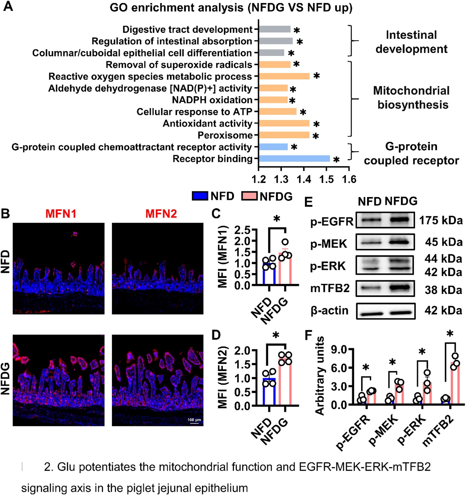
Background Intestinal stem cells (ISCs) sustain epithelial homeostasis through rapid mitochondrial metabolism, however, how they sense nutrient signals to regulate mitochondrial function remains unclear. Methods We examined the role of L-glutamate (Glu) in regulating cell mitochondrial biosynthesis using in vivo piglets, ex vivo porcine intestinal organoids (IOs), and in vitro IPEC-J2 cells. Results Glu enhanced jejunal development in weaned piglets. Isobaric tags for relative and absolute quantitation (iTRAQ) analysis revealed the significant enrichment of mitochondrial functions and activation of EGFR-MEK-ERK-mTFB2 signaling pathway in the jejunum. In vitro, 5 mM Glu promotes mitochondrial biosynthesis and potentiates the EGFR-MEK-ERK-mTFB2 axis. Whereas inhibition of EGFR with Osimertinib and silencing EGFR abolished these effects in IOs and IPEC-J2 cells. Colocalization and biochemical studies demonstrated interaction between Glu and EGFR in IOs. Conclusions Glu promotes mitochondrial biogenesis and ISC expansion by activating the EGFR–MEK–ERK–mTFB2 axis, highlighting a nutrient-sensing mechanism that couples energy availability to ISC function.

Read Full Abstract10.1186/s13287-025-04718-3
Overexpression of SOX4 in MSCs inhibits cellular senescence and enhances therapeutic efficacy in systemic lupus erythematosusGraphical AbstractVerified
Stem Cell Research & Therapy

Overexpression of SOX4 in MSCs inhibits cellular senescence and enhances therapeutic efficacy in systemic lupus erythematosus

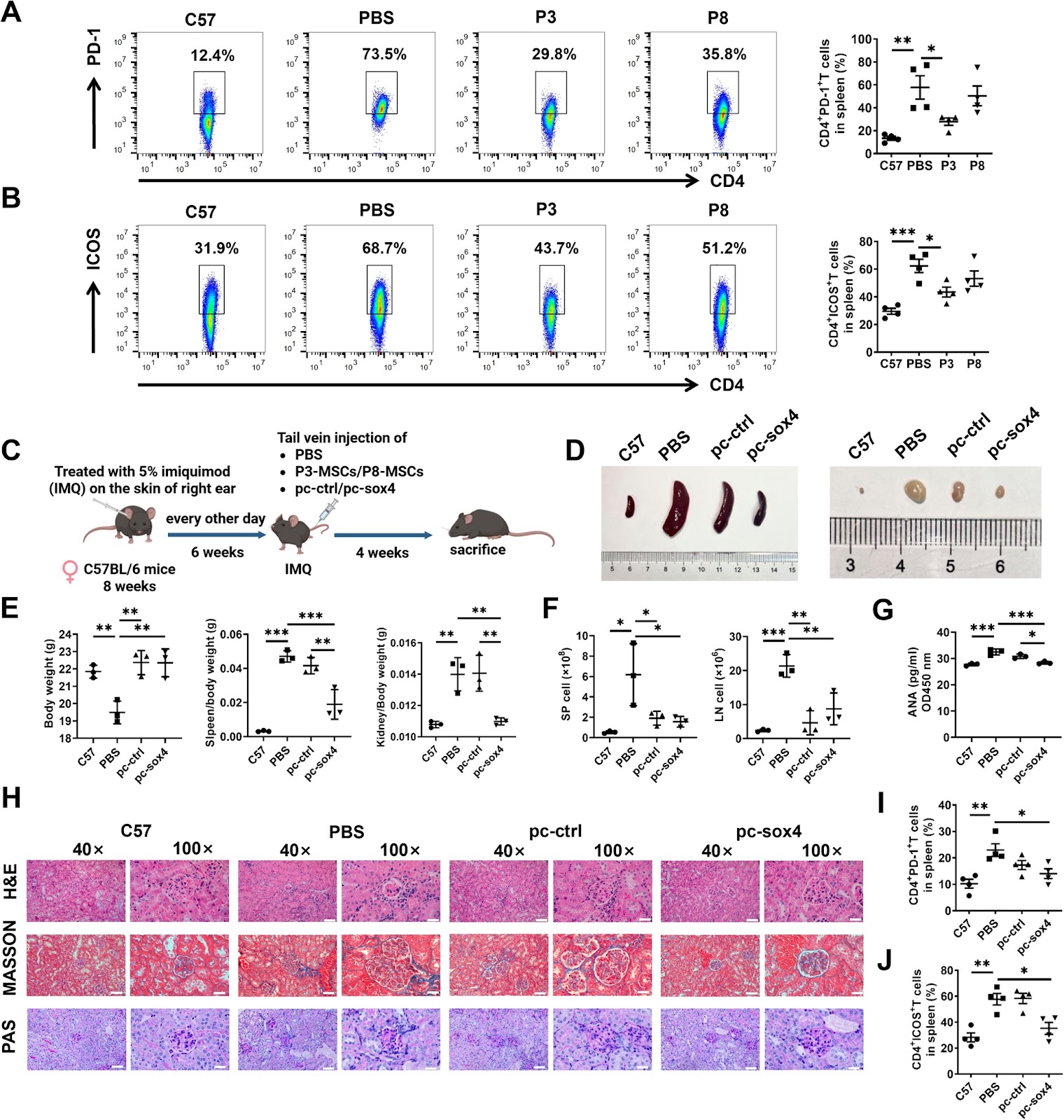
Background Mesenchymal stem cells (MSCs) are widely used in treating autoimmune diseases. However, replicative senescence limits the quantity and quality of MSCs during population doublings in vitro. Transcription factor SOX4 is a crucial regulator of cell fate and stemness. This study aims to explore the role of SOX4 in senescence of MSCs and enhance their therapeutic efficacy in systemic lupus erythematosus (SLE). Methods In early-passage MSCs (P3), late-passage MSCs (P8), SOX4 downregulated P3-MSCs or SOX4 overexpressed P8-MSCs, cell morphology, mitochondrial reactive oxygen species (mtROS), senescence-associated β-galactosidase (SA-β-Gal) activity, cell proliferation rate, senescence-associated secretory phenotype (SASP) factors, cell cycle suppressors, the immunosuppressive effects on T cell activation and proliferation and the expression levels of SOX4 were determined. Imiquimod induced SLE mice were transplanted with P3-MSCs and P8-MSCs or control and SOX4 overexpressed P8-MSCs, and clinical symptoms were assessed. Results Compared to P3-MSCs, P8-MSCs display a senescent phenotype, increased mtROS, SA-β-Gal activity, SASP factors, and cell cycle suppressors p53, p21, and p16. Additionally, P8-MSCs have a reduced immunosuppressive function on T cell activation and proliferation, and express lower levels of SOX4. Downregulation of SOX4 in P3-MSCs promotes cellular senescence and impairs their immunosuppressive function. Conversely, overexpression of SOX4 in P8-MSCs ameliorates cellular senescence and enhances their immunosuppressive function. Furthermore, transplantation of P3-MSCs or SOX4-overexpressing P8-MSCs demonstrates greater therapeutic significantly efficacy in SLE mice compared to P8-MSCs.

Read Full Abstract10.1186/s13287-025-04525-w
aFGF gene-modified adipose-derived mesenchymal stem cells promote healing of full-thickness skin defects in diabetic ratsGraphical AbstractVerified
Stem Cell Research & Therapy

aFGF gene-modified adipose-derived mesenchymal stem cells promote healing of full-thickness skin defects in diabetic rats

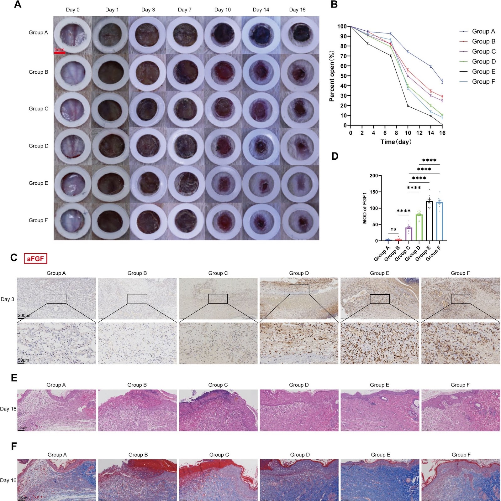
Background Chronic diabetic wounds pose a significant clinical challenge due to the limited efficacy of current treatments. This study aimed to investigate the role and potential mechanisms of adipose-derived mesenchymal stem cells (ADSCs) overexpressing acidic fibroblast growth factor (aFGF) in diabetic wound healing in a rat model. Methods ADSCs were genetically modified to achieve stable overexpression of aFGF. Varying doses of aFGF-ADSCs (1Ɨ10⁶, 2Ɨ10⁶, 3Ɨ10⁶, 4Ɨ10⁶) were injected into the muscular tissue surrounding diabetic rat wounds. We assessed aFGF expression and its impact on various stages of wound healing, including angiogenesis, inflammatory response, epithelialization, and collagen deposition. Transcriptomic sequencing was performed to explore the underlying mechanisms driving enhanced wound healing. Results Lentiviral transduction successfully induced stable aFGF overexpression in ADSCs. In vivo experiments revealed that varying doses of aFGF-ADSCs markedly enhanced wound healing in diabetic rats in a dose-dependent manner. The dose of 3Ɨ10⁶ aFGF-ADSCs demonstrated the most significant effect. In the 3Ɨ10⁶ aFGF-ADSCs group, expression levels of aFGF, CD31, and CD163 were significantly higher than in other groups (p < 0.05), while CD86 expression was significantly lower (p < 0.05). Conclusion Single doses of aFGF-ADSCs comprehensively improved various aspects of wound repair in diabetic rats, offering a potential new approach for treating chronic diabetic wounds. The mechanism of action involves promoting angiogenesis, modulating inflammatory responses, accelerating epithelialization, and optimizing collagen deposition.

Read Full Abstract10.1186/s13287-025-04241-5
Bone marrow mesenchymal stem cells derived cytokines associated with AKT/IAPs signaling ameliorate Alzheimer’s disease developmentGraphical AbstractVerified
Stem Cell Research & Therapy

Bone marrow mesenchymal stem cells derived cytokines associated with AKT/IAPs signaling ameliorate Alzheimer’s disease development

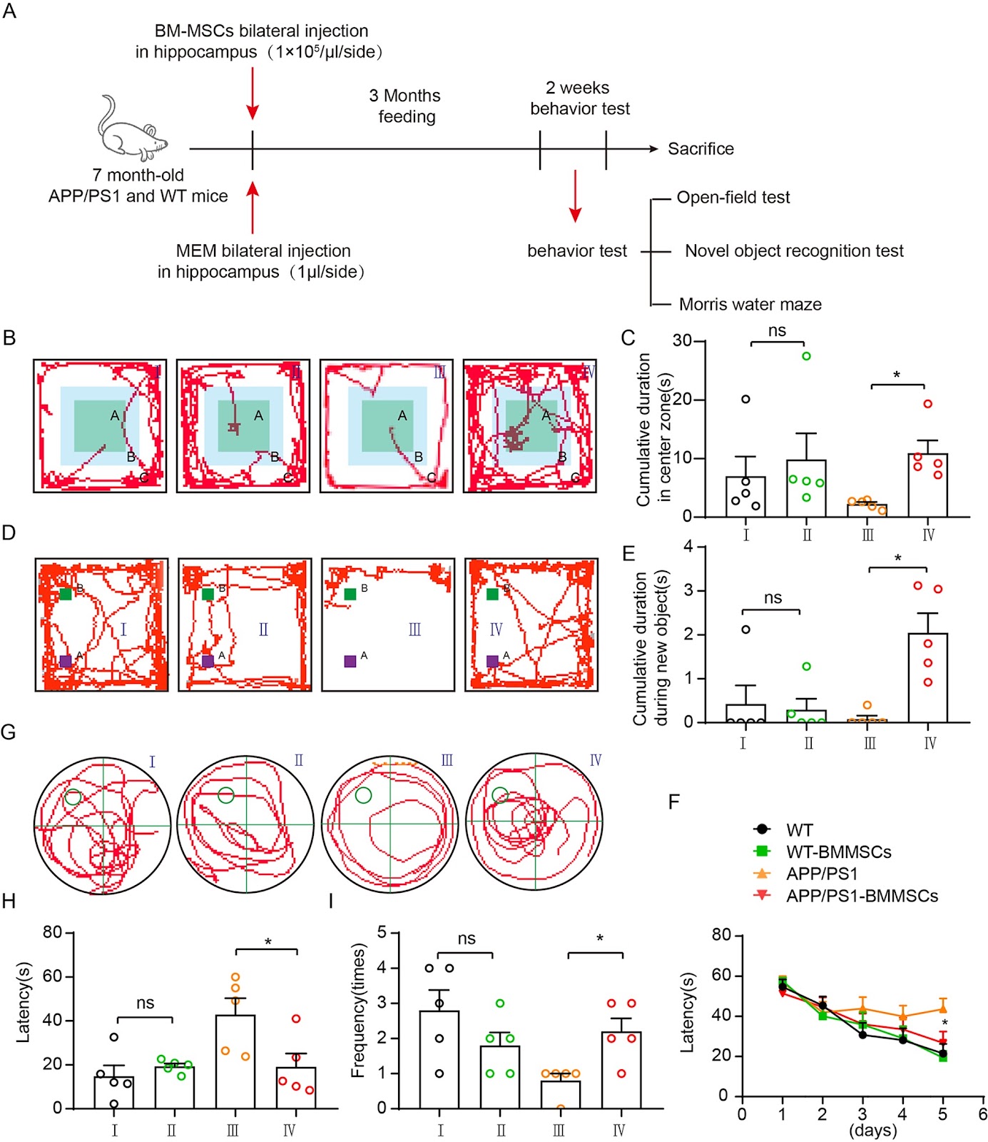
Background Alzheimer’s disease (AD) is a progressive neurodegenerative condition affecting around 50 million people worldwide. Bone marrow-derived mesenchymal stem cells (BMMSCs) have emerged as a promising source for cellular therapy due to their ability to differentiate into multiple cell types and their paracrine effects. However, the direct injection of BMMSCs can lead to potential unpredictable impairments, prompting a renewed interest in their paracrine effects for AD treatment. The specific mechanism and central role of cytokines in this process have not been fully elucidated. Methods Mouse BMMSCs were isolated, validated, and then transplanted intracerebrally into APP/PS1 female mice. The behavioral tests, including open-field test, novel object recognition test, and Morris water maze were performed, followed by β-amyloidosis plaque and neuron apoptosis analyses. Then the tissue RNA sequencing and mBMMSC cytokine analysis were performed. A cytokine antibody array for BMMSCs and the brain slice models were performed with AD model tissues were used to elucidate the molecular mechanisms. Finally, APP/PS1 mice were administrated with cytokine mixture for cognitive recovery. Results Our results demonstrated that BMMSCs significantly improved cognitive function, reduced beta-amyloid plaque deposition, and decreased apoptotic neurons through the activation of the AKT signaling pathway. Using a cytokine antibody array, we identified three highly expressed AKT pathway regulated neuroprotective factors in BMMSCs: IGF1, VEGF, and Periostin2. These cytokines were found to upregulate inhibitors of apoptosis family proteins (IAPs) and suppress Caspase-3 activity in brain slices induced with beta amyloidosis (Aβ), okadaic acid (OA), and lipopolysaccharide (LPS). When injection of this cytokine mixture to APP/PS1 mice also resulted in a mitigation of cognitive impairment. Conclusions These findings suggest that the secretory factors IGF1, VEGF, and Periostin2 derived from BMMSCs play a crucial role in neuroprotection by modulating the AKT/IAPs pathway to restore neuronal function. These cytokine sets could be a potential therapeutic strategy for AD and lay the groundwork for promising clinical applications.

Read Full Abstract10.1186/s13287-025-04131-w
MSCs engineered with secreted Klotho alleviate blood–brain barrier disruption and reduce neuroinflammation more effectively than MSCs in experimental autoimmune encephalomyelitisGraphical AbstractVerified
Stem Cell Research & Therapy

MSCs engineered with secreted Klotho alleviate blood–brain barrier disruption and reduce neuroinflammation more effectively than MSCs in experimental autoimmune encephalomyelitis

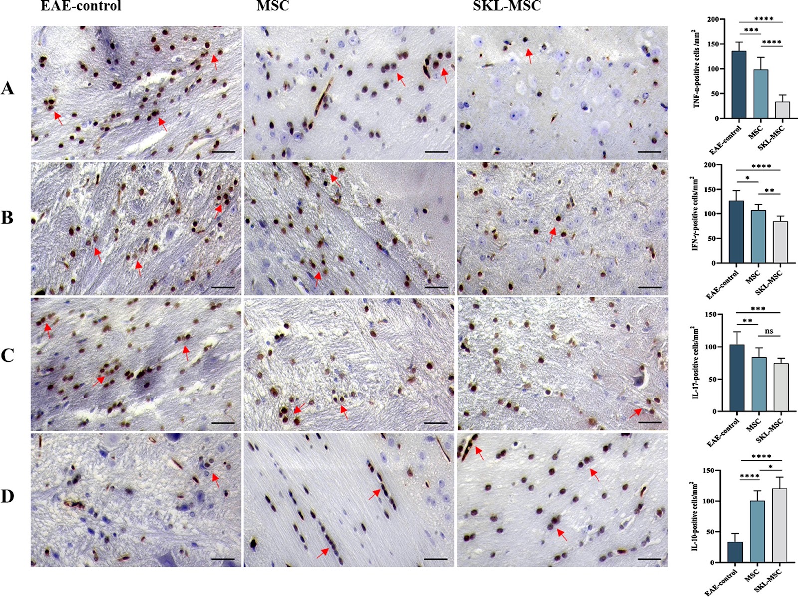
Background The anti-aging protein, Klotho, has been shown to exert neuroprotective effects in neurodegenerative disorders. This study was designed to evaluate the effects of MSCs engineered with secreted Klotho (SKL-MSCs) on neuroinflammation in experimental autoimmune encephalomyelitis (EAE) mouse model and to investigate underlying molecular mechanisms. Methods EAE was induced in female C57BL/6 mice, and animals were then randomized to receive PBS, MSCs, or SKL-MSCs at the onset of disease. BBB permeability assay was performed. The mRNA and protein expression of inflammatory factors was detected in the brain of animals by real-time PCR and immunohistochemistry, respectively. The mRNA and protein expression of BBB-associated factors was detected in the brain of animals by real-time PCR and Western blotting, respectively. Results The results showed that SKL-MSCs slowed EAE progression and attenuated the disease severity more effectively than MSCs. SKL-MSCs also decreased the expression of TNF-α, IFN-γ, and IL-17 but increased the expression of IL-10 more potently than MSCs in the brain of EAE animals. Furthermore, SKL-MSCs reduced BBB permeability more significantly than MSCs, which was accompanied by decreased levels of BBB-associated factors, ICAM-1, VCAM-1, MMP-9, and CCL2, in the brain of EAE animals. However, in mice treated with MSCs, the reduction in the expression of BBB-associated factors was limited to ICAM-1 and MMP-9. Conclusions Our study highlighted the significantly greater therapeutic power of SKL-MSCs compared with MSCs in attenuating EAE disease severity and reducing neuroinflammation, which might be mediated through a more marked reduction in the BBB permeability and BBB-associated factors expression levels in the brain of animals.

Read Full Abstract10.1186/s13287-025-04428-w
FTO-mediated m6A modification regulates the osteogenic differentiation of ADSCs by targeting FOXO1Graphical AbstractVerified
Stem Cell Research & Therapy

FTO-mediated m6A modification regulates the osteogenic differentiation of ADSCs by targeting FOXO1

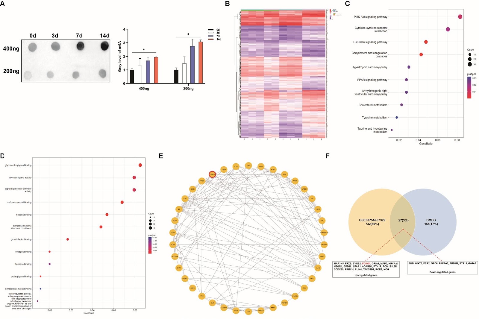
Using adipose-derived stem cells (ADSCs) has recently become a crucial approach for treating bone defects owing to their ease of accessibility and substantial differentiation potential. N6-methyladenosine (m6A) modification greatly influences biological processes and determines the differentiation fate of stem cells. However, the specific mechanisms by which m6A modification influences the osteogenic differentiation of ADSCs remain unclear. We identified FOXO1 as the key m6A-modified gene during the osteogenesis of ADSCs. Furthermore, demethylase FTO enhanced RUNX2 expression while inhibiting PPARG expression by modifying FOXO1, thereby facilitating ADSC osteogenesis. FTO knockdown inhibited ADSC migration and proliferation and impaired osteogenesis by suppressing FOXO1. At the mechanistic level, we first revealed that FTO was exported to the cytoplasm and then directly bound with FOXO1 mRNA at its 1760th bp site. Consistent use of non-steroidal anti-inflammatory drugs (NSAIDs) containing FTO inhibitors impeded ADSC-mediated bone formation both in vivo and in vitro. In summary, our study reveals the role of m6A modification based on the FTO–FOXO1–RUNX2/PPARG axis in regulating the osteogenic differentiation of ADSCs, thereby improving the clinical use of ADSCs and providing strategies for related drug applications in bone regeneration.

Read Full Abstract10.1186/s13287-025-04862-w