🧬 SinoBioData Academic Portal
📚 Peer-Reviewed Translated Literature

All Biomedical & Clinical Articles (Page 39)

Browse complete peer-reviewed translations from top Chinese biomedical, oncology, and genomics journals. Read verified previews and download full authentic clinical reports.

Published Research Papers

Showing 24 of 1542 peer-reviewed translated articles (Page 39 of 65)

Combining sodium-glucose co-transporter-2 inhibitor with mesenchymal stem cells and brown adipose tissue (BAT) and white adipose tissue (WAT) transplantation to mitigate the progression of diabetic kidney disease: a pre-clinical approachGraphical AbstractVerified
Stem Cell Research & Therapy

Combining sodium-glucose co-transporter-2 inhibitor with mesenchymal stem cells and brown adipose tissue (BAT) and white adipose tissue (WAT) transplantation to mitigate the progression of diabetic kidney disease: a pre-clinical approach

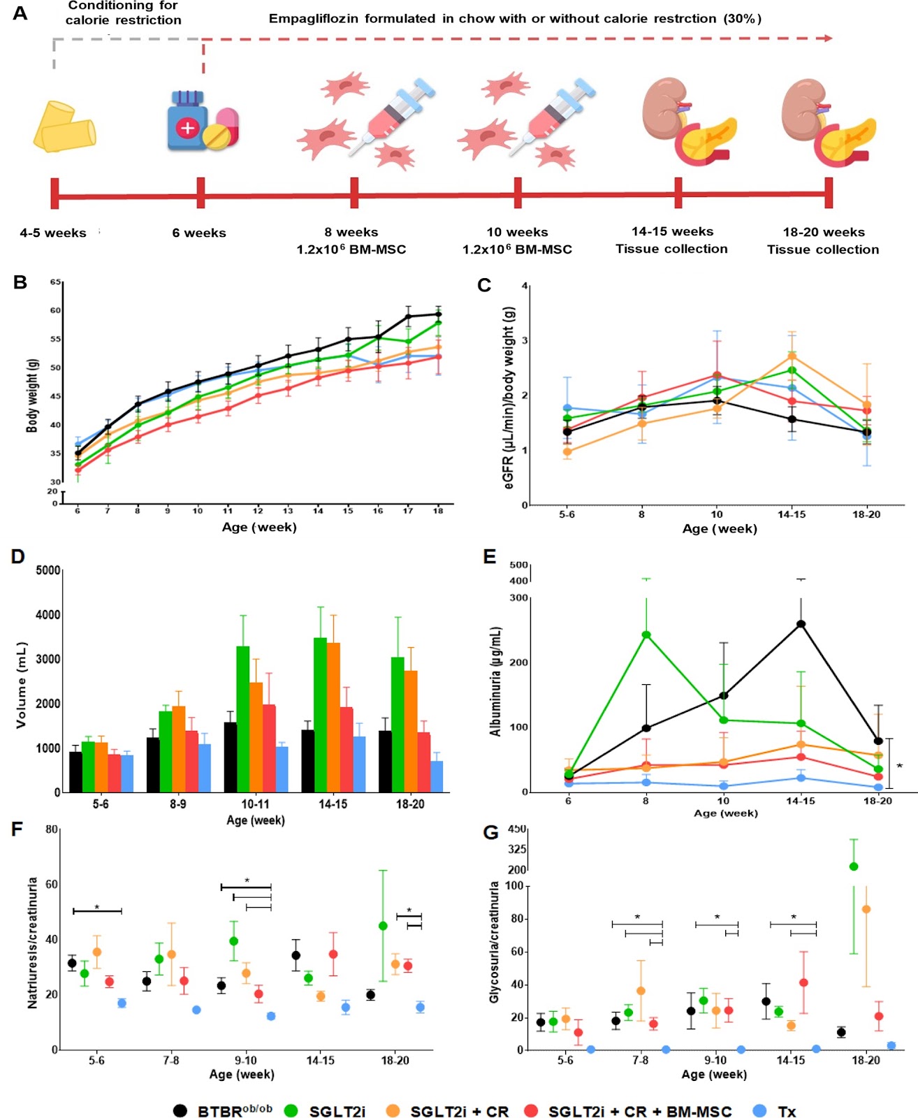
Introduction The increasing prevalence of Diabetes Mellitus (DM) correlates with a rising incidence of Diabetic Kidney Disease (DKD). DKD, a multifactorial condition, is characterized by activation of the renin–angiotensin–aldosterone system (RAAS), with angiotensin II playing a significant role in podocyte injury. While conventional treatments show potential in mitigating DKD progression, a combination of strategies is required to both impede its development and repair damaged structures. Methods In this study, we explored the brown adipose tissue (BAT) and white adipose tissue (WAT) transplantation, and the use of bone marrow mesenchymal stem cell therapy (BM-MSC) combined with sodium-glucose co-transporter-2 (SGLT2) inhibitor treatment and calorie restriction in the BTBRob/ob model, recognized as a robust representation of DKD featuring hyperglycemia, obesity, time-dependent albuminuria, and histological changes. Results Our primary findings revealed enhanced blood glucose control through combined cell therapy, diminished mesangial matrix expansion, alleviated tissue oxidative stress, preserved podocyte numbers, and an upregulation of podocyte structural markers and components of the RAAS renoprotective axis. Conclusion BM-MSC therapy demonstrates considerable promise as a combined treatment for mitigating DKD progression, with similar findings observed for BAT and WAT transplantation.
Read Full Abstract10.1186/s13287-025-04358-7
Dental pulp stem cell-derived intracellular vesicles prevent orthodontic relapse by inhibiting PI3K/Akt/NF-ÎșB-mediated osteoclast activityGraphical AbstractVerified
Stem Cell Research & Therapy

Dental pulp stem cell-derived intracellular vesicles prevent orthodontic relapse by inhibiting PI3K/Akt/NF-ÎșB-mediated osteoclast activity

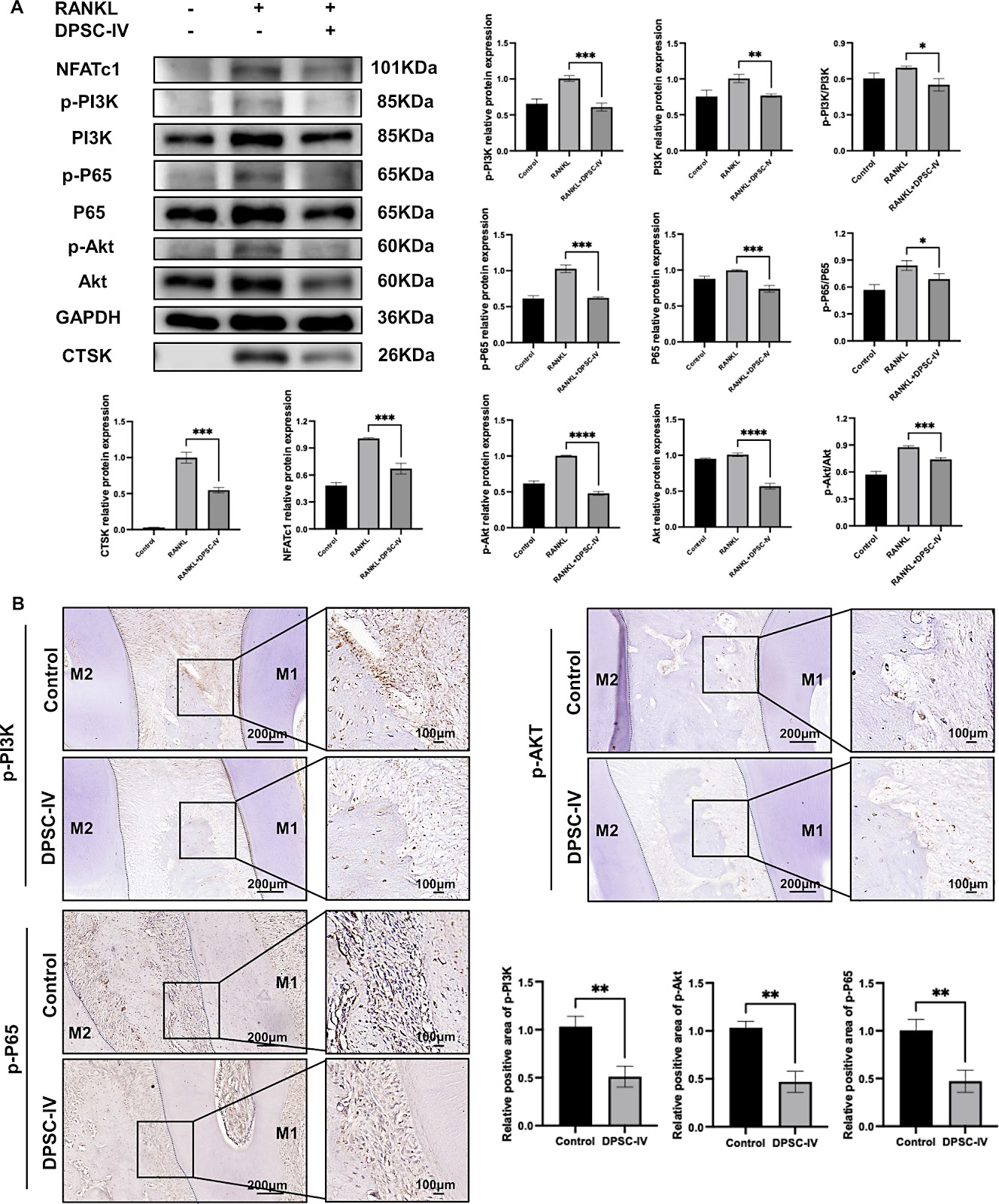
Background Orthodontic relapse, the undesired deviation of teeth from their corrected positions, remains a significant challenge in clinical orthodontics. Incomplete periodontal bone remodeling has been identified as a key factor in this process. Despite decades of research, currently there are no effective strategies to prevent relapse. Methods We isolated and identified dental pulp stem cell-derived intracellular vesicles (DPSC-IV) from human dental pulp tissue. To investigate its effect, DPSC-IV was added to osteoblast or osteoclast differentiation medium. During the orthodontic retention period, DPSC-IV was administrated to rats by subgingival injection. Relapse distance and relapse rate were calculated to evaluate DPSC-IV's ability to prevent relapse. Additionally, Western blot analysis were used to examine DPSC-IV's inhibitory effect on osteoclast differentiation. Results DPSC-IV significantly promoted osteoblast differentiation and inhibited osteoclast differentiation. Application of DPSC-IV during retention resulted in a significant reduction in both relapse distance and relapse rate, with improved periodontal structure and decreased osteoclast activity. This effect was mediated by the PI3K/Akt/NF-ÎșB signaling pathway and could be reversed by the PI3K activator insulin-like growth factor-1 (IGF-1). Conclusion This study highlights the potential of DPSC-IV as a novel preventive approach against orthodontic relapse, offering a novel strategy for maintaining long-term orthodontic stability.

Read Full Abstract10.1186/s13287-025-04146-3
Geometrical constraints dictate assembly and phenotype of human iPSC-derived motoneuronal spheroidsGraphical AbstractVerified
Stem Cell Research & Therapy

Geometrical constraints dictate assembly and phenotype of human iPSC-derived motoneuronal spheroids

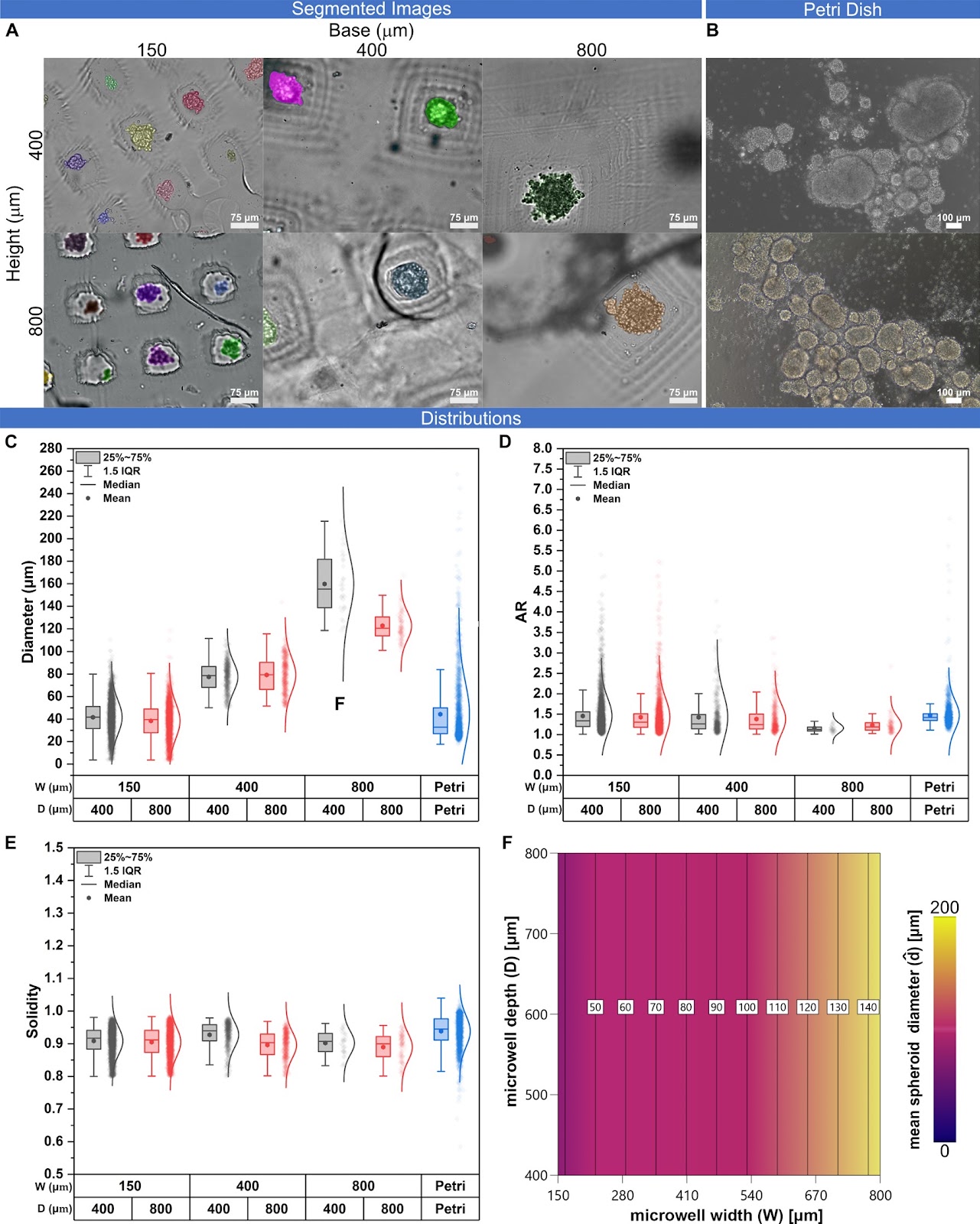
Background Neuronal spheroids represent an easy and versatile solution to model neuronal tissue in vitro. Conventional approaches to generate spheroids lack accurate size control, scalability, and customizability. This is even more exacerbated in case of pluripotent stem cell (PSC) derived spheroids, which remain challenging to standardize. Microwell devices address these limitations, providing an optimal balance between accessibility and scalability. With the aim of optimizing culture conditions, we parametrically investigated the role of microwell geometry on the formation and maturation of iPSC-derived motor neuron precursor (MNP) spheroids. Methods We developed a customizable mold device using Digital Light Processing (DLP) 3D printing to fabricate agarose microwell arrays with distinct aspect ratios for culturing hiPSC-derived MNP spheroids with high reproducibility. We generated nine different pyramidal microwell array geometries for culturing size-controlled spheroids in the 40–140 ÎŒm diameter range. We then evaluated the differential expression of genes related to cell proliferation and motor-neuron differentiation as function of microwell geometry and spheroid size. Results Our results indicate that spheroid size is significantly influenced by the microwell geometry, reliably due to cell partitioning at the seeding stage. Expression of proliferation and differentiation markers, such as motor neuron and pancreas homeobox 1 (MNX1) and Islet-1 (ISL1) transcription factors, is also dependent on microwell geometry and spheroid morphological descriptors. Conclusion Our approach enables the scalable production of size-controlled MNP spheroids and underscores the effect of geometrical confinement on regulating motor neuron differentiation.

Read Full Abstract10.1186/s13287-025-04547-4
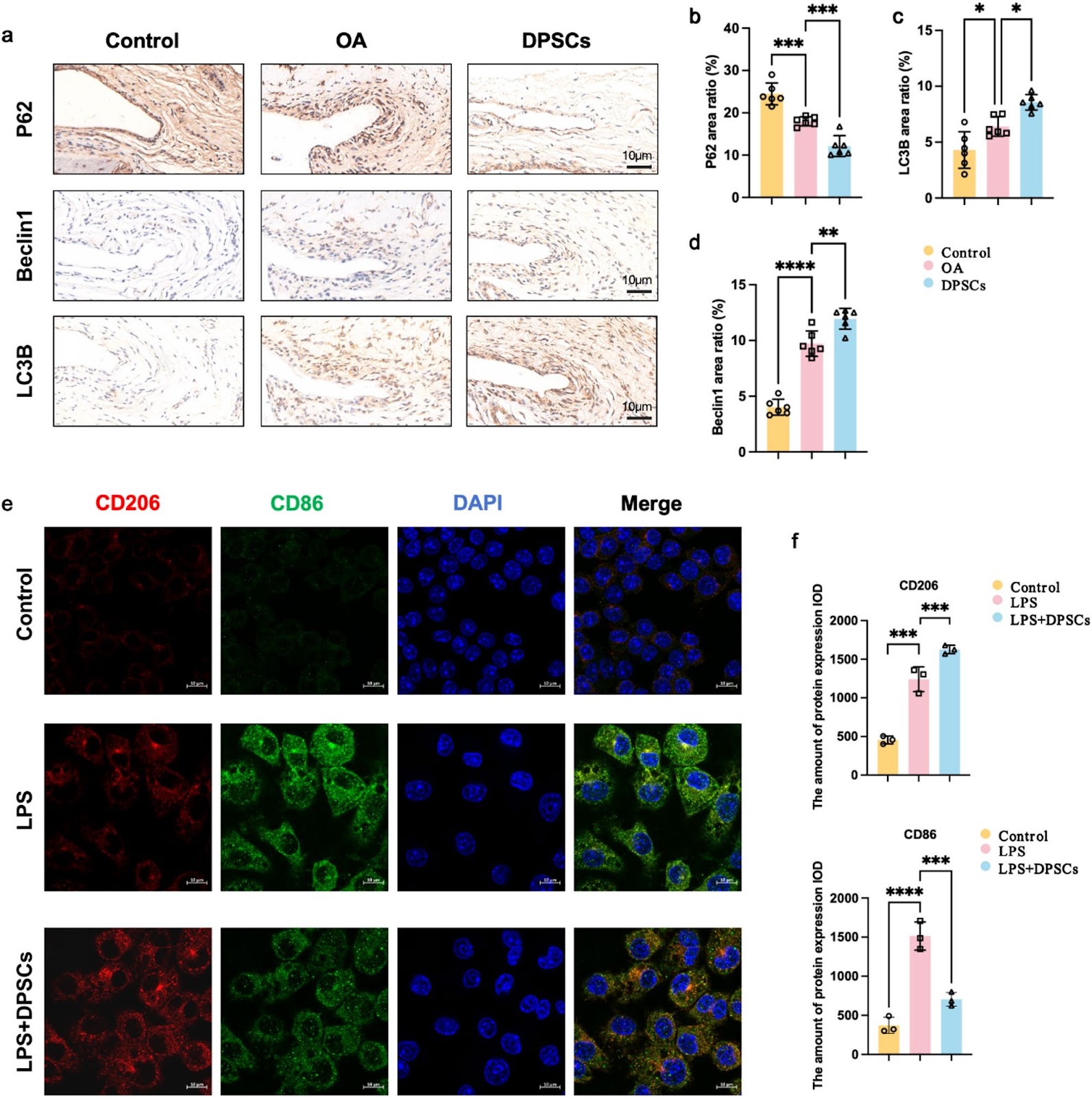
Correction: DPSCs modulate synovial macrophage polarization and efferocytosis via PINK1/Parkin-dependent mitophagyGraphical AbstractVerified
Stem Cell Research & Therapy

Correction: DPSCs modulate synovial macrophage polarization and efferocytosis via PINK1/Parkin-dependent mitophagy

This correction article addresses an error in the scale of the control group image in Fig. 4E of the original article. The corrected image is provided. The original article can be found online at https://doi.org/10.1186/s13287-025-04468-2.

Read Full Abstract10.1186/s13287-025-04565-2
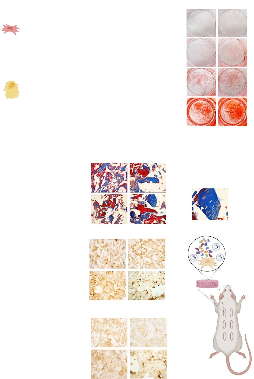
Paracrine activity of Smurf1-silenced mesenchymal stem cells enhances bone regeneration and reduces bone loss in postmenopausal osteoporosisGraphical AbstractVerified
Stem Cell Research & Therapy

Paracrine activity of Smurf1-silenced mesenchymal stem cells enhances bone regeneration and reduces bone loss in postmenopausal osteoporosis

Background: Osteoporosis (OP), characterized by reduced bone mass and mineral density, is a global metabolic disorder that severely impacts the quality of life in affected individuals. Although current pharmacological treatments are effective, their long-term use is often associated with adverse effects, highlighting the need for safer, more sustainable therapeutic strategies. This study investigates the pro-osteogenic and anti-resorptive potential of the secretome from Smurf1-silenced mesenchymal stem cells (MSCs) as a promising cell-free therapy for bone regeneration. Methods: Conditioned media (CM) from Smurf1-silenced rat (rCM-Smur1) and human MSCs (hCM-Smurf1) was collected and analyzed. Pro-osteogenic potential was assessed by measuring in vitro mineralization in human and rat MSCs cultures. In vivo, studies were conducted using a rat ectopic bone formation model and a post-menopausal osteoporotic mouse model. Additionally, primary human osteoporotic MSCs were preconditioned with hCM-Smurf1, and their osteogenic capacity was compared to that induced by BMP2 treatment. Ex vivo, human bone explants were treated with hCM-Smurf1 to assess anti-resorptive effects. Proteomic analysis of the soluble and vesicular CM fractions identified key proteins involved in bone regeneration. Results: CM from Smurf1-silenced MSCs significantly enhanced mineralization in vitro and bone formation in vivo. Preconditioning human osteoporotic MSCs with hCM-Smurf1 significantly increases in vitro mineralization, with levels comparable to those achieved with BMP2 treatment. Additionally, in ex vivo human bone cultures, treatment with hCM-Smurf1 significantly reduced RANKL expression without affecting OPG levels, indicating an anti-resorptive effect. In vivo, CM from Smurf1-silenced MSCs significantly increased bone formation in a rat ectopic model, and its local administration reduced trabecular bone loss by 50% in a post-menopausal osteoporotic mouse model after a single administration within just four weeks. Proteomic analysis revealed both soluble and vesicular fractions of hCM-Smurf1 were enriched with proteins essential for ossification and extracellular matrix organization, enhancing osteogenic differentiation. Conclusions: The Smurf1-silenced MSCs’ secretome shows potent osteogenic and anti-resorptive effects, significantly enhancing bone formation and reducing bone loss. This study provides compelling evidence for the therapeutic potential of Smurf1-silenced MSC-derived secretome as a non-toxic and targeted treatment for osteoporosis. These findings warrant further in vivo studies and clinical trials to validate its therapeutic efficacy and safety.

Read Full Abstract10.1186/s13287-025-04165-0
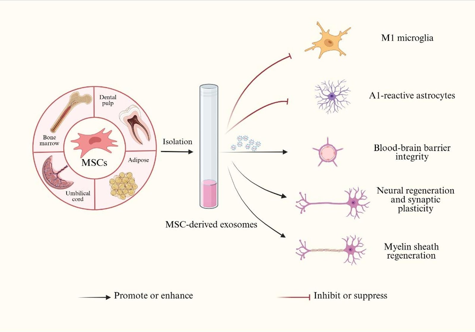
Mesenchymal stem cell-derived exosomes–a promising therapeutic approach to improve neurocognitive disorders in chronic obstructive pulmonary diseaseGraphical AbstractVerified
Stem Cell Research & Therapy

Mesenchymal stem cell-derived exosomes–a promising therapeutic approach to improve neurocognitive disorders in chronic obstructive pulmonary disease

Chronic obstructive pulmonary disease (COPD) is a leading cause of morbidity and mortality worldwide and is often accompanied by neurocognitive disorders. It seriously affects the quality of life and treatment outcome of patients. COPD-induced neurocognitive disorders (COPD-NCDs) are driven by systemic inflammation, blood-brain barrier (BBB) disruption, and chronic hypoxia, but there is currently no effective treatment to prevent or reverse cognitive decline. Mesenchymal stem cell-derived exosomes (MSC-Exos) are nanoscale extracellular vesicles with unique bioactivity, which have shown great potential in the fields of nervous system diseases, respiratory diseases, bone and joint diseases. Different from traditional cell therapies, MSC-Exos have the advantages of non-immunogenicity, non-tumorigenicity, high stability and biocompatibility. In addition, MSC-Exos can also cross the BBB, regulate neuroinflammation, promote neurogenesis, enhance myelination and improve synaptic plasticity, thereby addressing the multifaceted pathogenesis of central nervous system (CNS) diseases. In this review, we first summarize the pathogenic mechanism of COPD-NCDs, and then summarize the multiple mechanisms of MSC-Exos improving NCDs based on the efficacy of MSC-Exos on other CNS diseases, emphasizing the theoretical basis and unique potential of MSC-Exos as a treatment for COPD-NCDs. Finally, we prospected the future research directions and potential problems of applying MSC-Exos to treat COPD-NCDs, future research should focus on optimizing the large-scale preparation of MSC-Exos, exploring their long-term safety, and advancing clinical translation to address the unmet needs of COPD-NCDs patients.

Read Full Abstract10.1186/s13287-025-04457-5
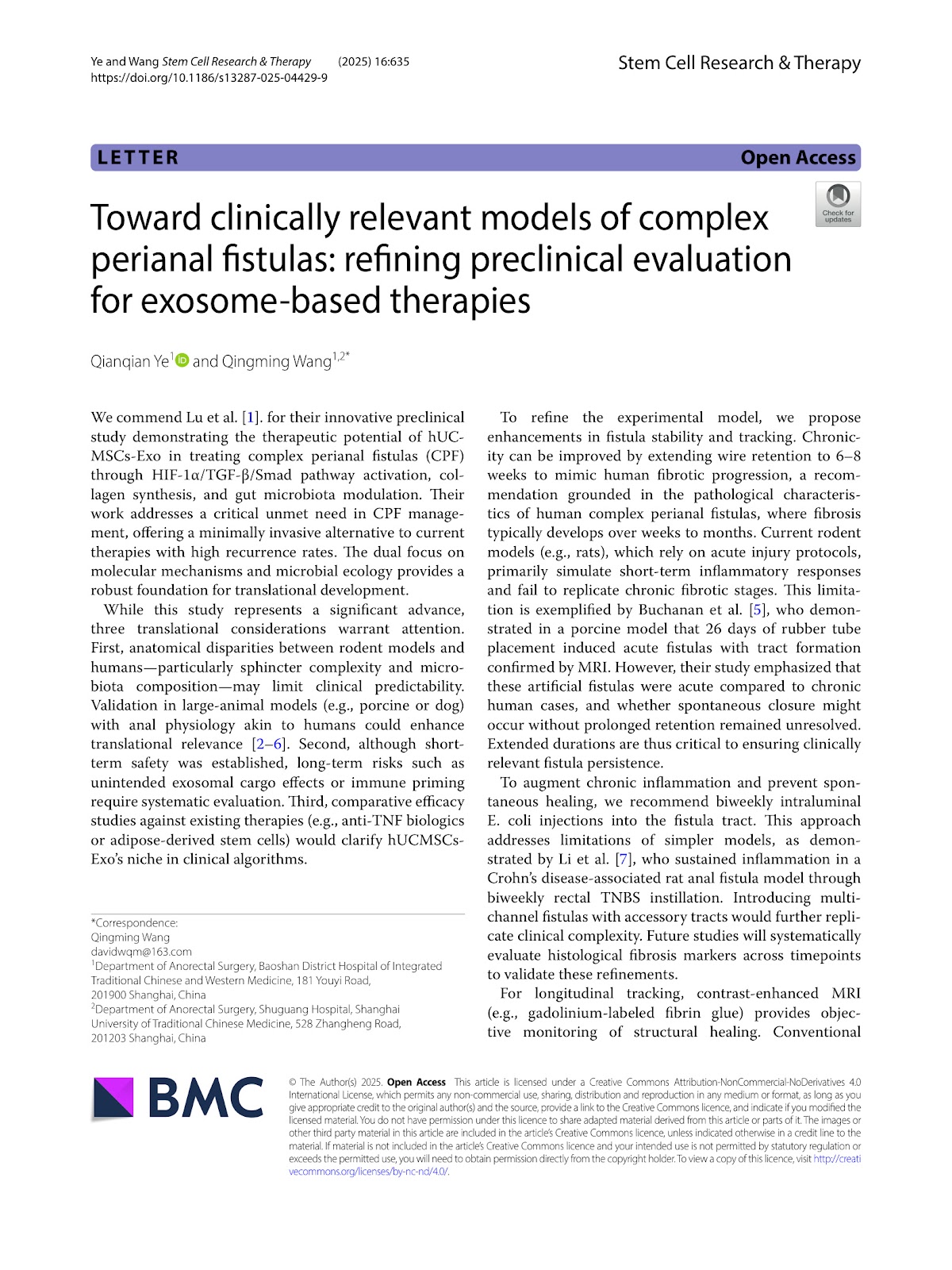
Toward clinically relevant models of complex perianal fistulas: refining preclinical evaluation for exosome-based therapiesGraphical AbstractVerified
Stem Cell Research & Therapy

Toward clinically relevant models of complex perianal fistulas: refining preclinical evaluation for exosome-based therapies

This letter commends Lu et al. for their innovative preclinical study demonstrating the therapeutic potential of hUCMSCs-Exo in treating complex perianal fistulas (CPF) through HIF-1α/TGF-ÎČ/Smad pathway activation, collagen synthesis, and gut microbiota modulation. While acknowledging the study's significant advances, the authors highlight three translational considerations: anatomical disparities between rodent models and humans, long-term safety concerns, and the need for comparative efficacy studies. To refine the experimental model, they propose enhancements in fistula stability and tracking, including extending wire retention to 6–8 weeks, biweekly intraluminal E. coli injections, and using contrast-enhanced MRI for longitudinal monitoring. These refinements aim to better mimic human fibrotic progression and improve clinical predictability.

Read Full Abstract10.1186/s13287-025-04429-9
Exosomes from adipose-derived stem cells accelerate wound healing by increasing the release of IL-33 from macrophagesGraphical AbstractVerified
Stem Cell Research & Therapy

Exosomes from adipose-derived stem cells accelerate wound healing by increasing the release of IL-33 from macrophages

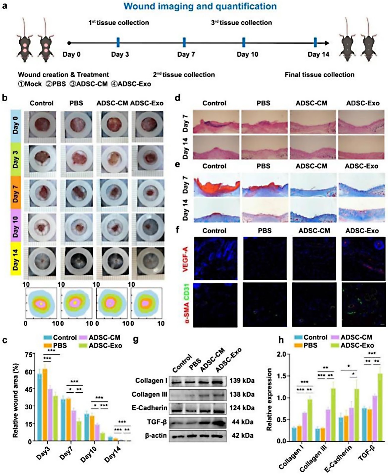
Background Mesenchymal stem cell (MSC) -derived exosomes, especially adipose-derived mesenchymal stem cell exosomes (ADSC-Exos), have emerged as a promising alternative for skin damage repair with anti-inflammatory, angiogenic and cell proliferation effects while overcoming some of the limitations of MSC. However, the mechanism by which ADSC-Exos regulates inflammatory cells during wound healing remains unclear. This study investigated how ADSC-Exos regulate macrophages to promote wound healing. Methods ADSC-Exos were isolated using ultracentrifugation, with subsequent quantification of exosomes particle number. To investigate their role in wound healing, the effects of ADSC-Exos on inflammation, angiogenesis, collagen deposition and macrophage polarization were evaluated through immunohistochemical staining, immunofluorescence and western blotting. Changes in gene expression associated with ADSC-Exos-induced macrophage polarization were analyzed using qPCR. RNA sequencing was performed to identify differentially expressed genes affected by ADSC-Exos. The critical role of IL-33 in the wound healing process was further confirmed using Il33−/− mice. Additionally, co-culture experiments were conducted to explore the effects of IL-33 on keratinocyte proliferation, collagen deposition and epithelialization. Results ADSC-Exos inhibited the expression of TNF-α and IL-6, induced M2 macrophage polarization, promoted collagen deposition and angiogenesis, and accelerated wound healing. RNA sequencing identified IL-33 as a key mediator in this process. In Il33−/− mice, impaired wound healing and decreased M2 macrophage polarization were observed. The co-culture experiments showed that IL-33 enhanced keratinocyte function through activation of the Wnt/ÎČ-catenin signaling pathway. These findings highlight the therapeutic potential of ADSC-Exos in wound healing by modulating IL-33. Conclusions ADSC-Exos promote wound healing by regulating macrophage polarization and enhancing IL-33 release which drives keratinocyte proliferation, collagen deposition and epithelialization via the Wnt/ÎČ-catenin

Read Full Abstract10.1186/s13287-025-04203-x
Migrasomes derived from human umbilical cord mesenchymal stem cells: a new therapeutic agent for ovalbumin-induced asthma in miceGraphical AbstractVerified
Stem Cell Research & Therapy

Migrasomes derived from human umbilical cord mesenchymal stem cells: a new therapeutic agent for ovalbumin-induced asthma in mice

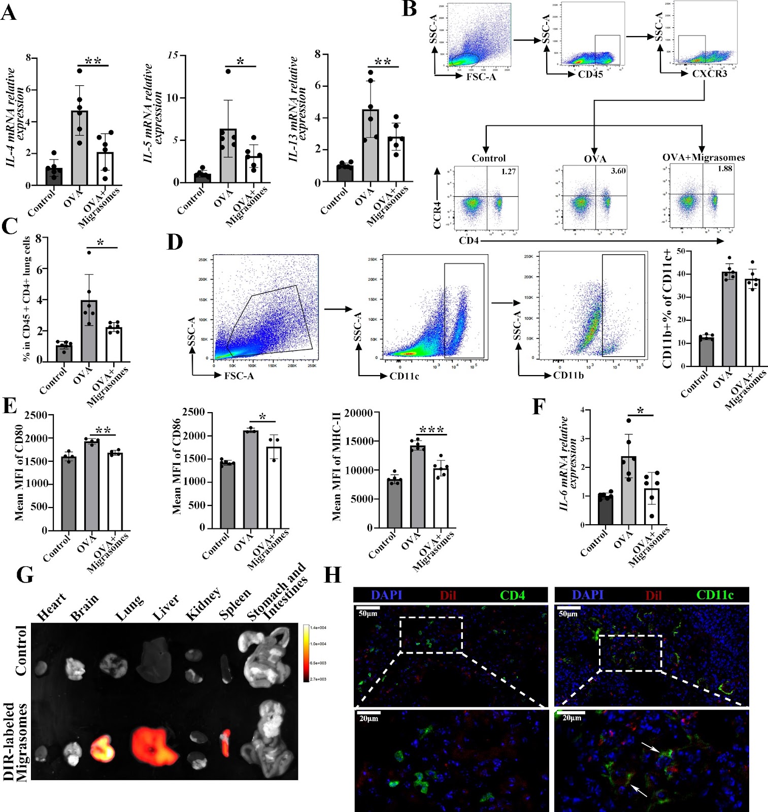
Background  Asthma is a prevalent respiratory disease, and its management remains largely unsatisfactory. Mesenchymal stem cells (MSCs) have been demonstrated to be efficacious in reducing airway inflammation in experimental allergic diseases, representing a potential alternative treatment for asthma. Migrasomes are recently identified extracellular vesicles (EVs) generated in migrating cells and facilitate intercellular communication. The objective of this study was to investigate the therapeutic effects of migrasomes obtained from MSC in a model of asthma. Methods  Migrasomes produced by human umbilical cord MSCs (hUCMSCs) were isolated by sequential centrifugation. Characterization of hUCMSC-derived migrasomes were carried out by transmission electron microscopy and western blot analysis. The therapeutic effects of migrasomes on airway inflammation in ovalbumin (OVA)-induced asthmatic mice were evaluated by hematoxylin-eosin (HE) and periodic-acid schiff (PAS) staining, and their mechanism were further testified by immunofluorescent staining, real-time PCR and flow cytometry. Results  Here, we showed that inhibition of migrasomes’ production dramatically impaired the anti-inflammatory effects of hUCMSCs in OVA animals, as evidenced by a notable increase in both the infiltration of inflammatory cells and the number of epithelial goblet cells. We successfully isolated hUCMSC-migrasomes, which were morphologically intact and positive for the specific migrasomes markers. The administration of hUCMSC-migrasomes was observed to significantly ameliorate the symptoms of airway inflammation and mucus production in asthmatic mice. Additionally, the expression of Th2 cytokines (IL-4, IL-5 and IL-13) were found to be reduced, while the activation of dendritic cells (DCs) was inhibited. HUCMSC-migrasomes could possibly be delivered to lung region after injection, and were able to be taken in by DCs both in vivo and in vitro. Notably, in vitro, migraosmes decreased the capacity of

Read Full Abstract10.1186/s13287-025-04145-4
Research on the mechanism of human umbilical cord mesenchymal stem cells and their extracellular vesicles in the treatment of common reproductive diseasesGraphical AbstractVerified
Stem Cell Research & Therapy

Research on the mechanism of human umbilical cord mesenchymal stem cells and their extracellular vesicles in the treatment of common reproductive diseases

Reproductive system disorders significantly contribute to infertility, and traditional or conventional treatments often have limited efficacy in addressing this issue. In recent years, stem cell therapy has emerged as an alternative therapeutic strategy owing to its various advantages. Human umbilical cord mesenchymal stem cells (hUC-MSCs) are pivotal in tissue repair owing to their robust proliferative capacity, potent immunomodulatory effects, low immunogenicity, and paracrine actions. Extracellular vesicles (EVs), the primary mediators of paracrine functions, exhibit therapeutic effects similar to those of hUC-MSCs. Consequently, numerous researchers have investigated the application of hUC-MSCs and their EVs in treating reproductive disorders. These cells have the potential to restore fertility by mitigating oxidative stress, excessive autophagy, and ferroptosis in tissues, while promoting the expression of anti-inflammatory factors and vascular remodeling. However, hUC-MSCs present significant limitations compared to EVs, including higher tumorigenicity and low infusion efficiency. Consequently, EVs may emerge as the primary alternative therapy, while hUC-MSCs hold promise as a therapeutic option with potential applications in regenerative medicine.

Read Full Abstract10.1186/s13287-025-04773-w
Combination of rapamycin and adipose-derived mesenchymal stromal cells enhances therapeutic potential for osteoarthritisGraphical AbstractVerified
Stem Cell Research & Therapy

Combination of rapamycin and adipose-derived mesenchymal stromal cells enhances therapeutic potential for osteoarthritis

Background The regenerative potential of mesenchymal stromal/stem cells (MSCs) has been extensively studied in clinical trials in the past decade. However, despite the promising regenerative properties documented in preclinical studies, for instance in osteoarthritis (OA), the therapeutic translation of these results in patients has not been fully conclusive. One factor contributing to this therapeutic barrier could be the presence of senescent cells in OA joints. Methods This study evaluated a novel approach to OA treatment by combining adipose tissue-derived MSCs (AD-MSCs) with rapamycin, a clinically approved immunosuppressive drug with anti-senescence properties. First, rapamycin effects on senescence and fibrosis markers were investigated in freshly isolated OA chondrocytes by immunostaining. Next, the in vitro differentiation capacities of AD-MSCs, their regulatory immune functions on activated immune cells and their regenerative effects on OA chondrocyte signature were assessed in the presence of rapamycin. Results In OA chondrocytes, rapamycin reduced the senescence marker p15INK4B and the fibrosis marker COL1A1 without affecting the expression of the master chondrogenic markers SOX9 and COL2. Rapamycin also enhanced AD-MSC differentiation into chondrocytes and reduced their differentiation into adipocytes. In addition, rapamycin improved AD-MSC immunoregulatory functions by promoting the expression of immunosuppressive factors, such as IDO1, PTGS2 and also CD274 (encoding PD-L1). Finally, RNA sequencing analysis showed that in the presence of rapamycin, AD-MSCs displayed improved chondroprotective regenerative effects on co-cultured OA chondrocytes. Conclusions Our findings suggest that the rapamycin and AD-MSC combination enhances the therapeutic efficacy of these cells in senescence-driven degenerative diseases such as OA, notably by improving their anti-fibrotic and anti-inflammatory properties.

Read Full Abstract10.1186/s13287-024-04090-8
Mesenchymal stem cell-derived extracellular vesicles for disease therapy by regulating ferroptosis: focus on diabetes mellitus and diabetic complicationsGraphical AbstractVerified
Stem Cell Research & Therapy

Mesenchymal stem cell-derived extracellular vesicles for disease therapy by regulating ferroptosis: focus on diabetes mellitus and diabetic complications

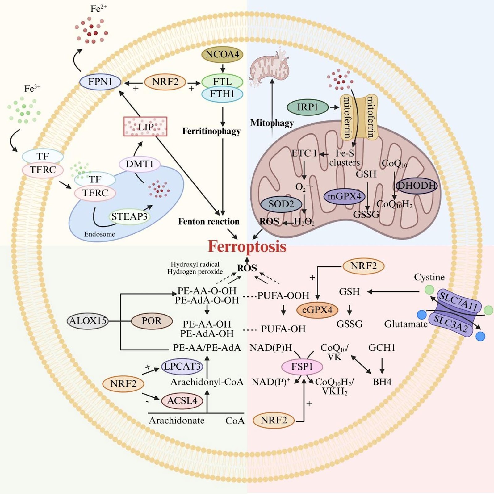
Ferroptosis is a novel form of programmed cell death, which has been demonstrated to play a pivotal role in various pathological processes due to its association with iron overload, lipid peroxidation, and dysregulation of the antioxidant system. In recent years, mesenchymal stem cell-derived extracellular vesicles (MSC-EVs) have garnered significant attention as a promising cell-free therapeutic strategy for modulating ferroptosis. This article elucidates the biological characteristics of MSC-EVs and the molecular mechanisms underlying ferroptosis, with a focus on how MSC-EVs regulate ferroptosis through three key pathways: iron metabolism, lipid metabolism, and the antioxidant defense system. Additionally, the therapeutic potential of both natural and engineered MSC-EVs in treating ferroptosis-related diseases is discussed, particularly highlighting their efficacy in diabetes mellitus and diabetic complications. Finally, this article evaluates the challenges and opportunities in translating MSC-EVs-based ferroptosis modulation therapies into clinical applications, providing valuable insights for future research and therapeutic development.

Read Full Abstract10.1186/s13287-025-04852-y
The therapeutic potential of mesenchymal stem cells in intestinal diseases: from mechanisms to clinical translationGraphical AbstractVerified
Stem Cell Research & Therapy

The therapeutic potential of mesenchymal stem cells in intestinal diseases: from mechanisms to clinical translation

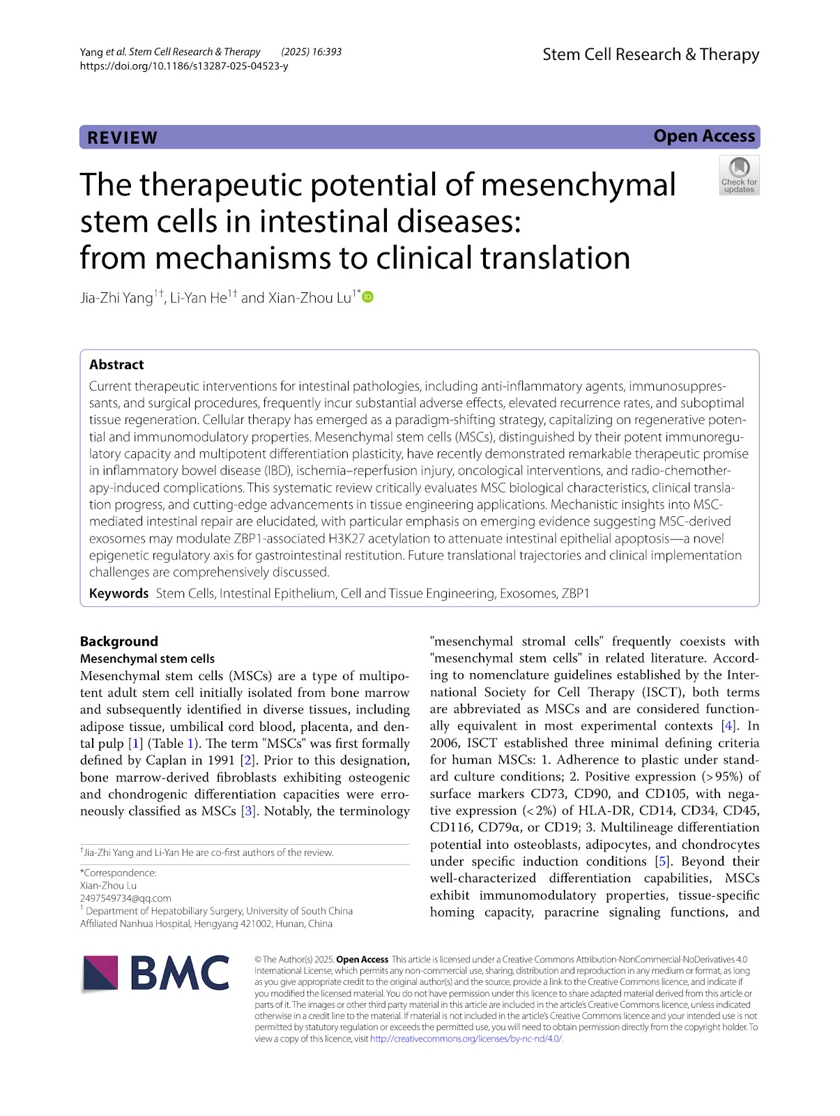
Current therapeutic interventions for intestinal pathologies, including anti-inflammatory agents, immunosuppressants, and surgical procedures, frequently incur substantial adverse effects, elevated recurrence rates, and suboptimal tissue regeneration. Cellular therapy has emerged as a paradigm-shifting strategy, capitalizing on regenerative potential and immunomodulatory properties. Mesenchymal stem cells (MSCs), distinguished by their potent immunoregulatory capacity and multipotent differentiation plasticity, have recently demonstrated remarkable therapeutic promise in inflammatory bowel disease (IBD), ischemia–reperfusion injury, oncological interventions, and radio-chemotherapy-induced complications. This systematic review critically evaluates MSC biological characteristics, clinical translation progress, and cutting-edge advancements in tissue engineering applications. Mechanistic insights into MSC-mediated intestinal repair are elucidated, with particular emphasis on emerging evidence suggesting MSC-derived exosomes may modulate ZBP1-associated H3K27 acetylation to attenuate intestinal epithelial apoptosis—a novel epigenetic regulatory axis for gastrointestinal restitution. Future translational trajectories and clinical implementation challenges are comprehensively discussed.

Read Full Abstract10.1186/s13287-025-04523-y
Targeting prominin-2/BACH1/GLS pathway to inhibit oxidative stress-induced ferroptosis of bone mesenchymal stem cellsGraphical AbstractVerified
Stem Cell Research & Therapy

Targeting prominin-2/BACH1/GLS pathway to inhibit oxidative stress-induced ferroptosis of bone mesenchymal stem cells

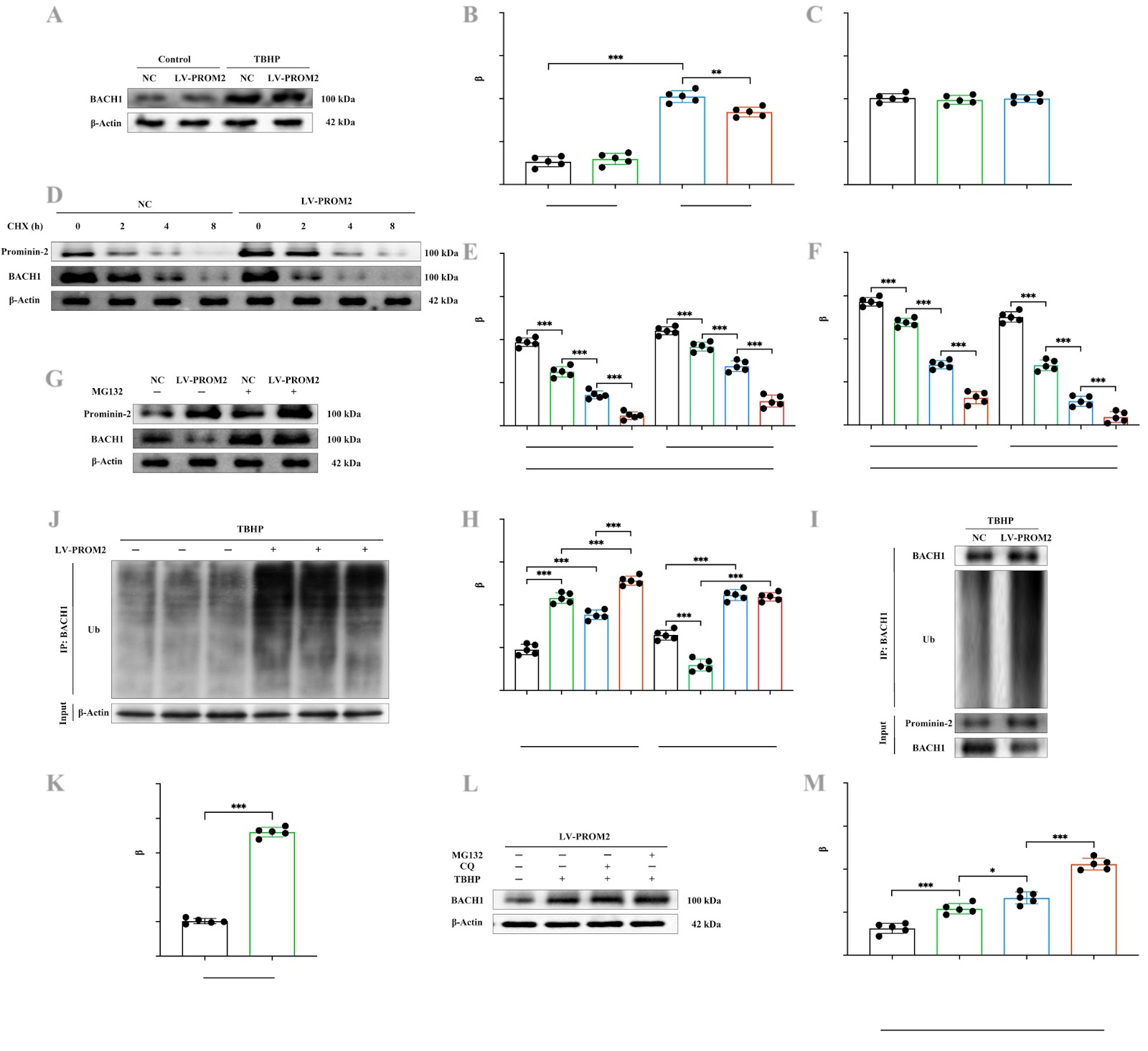
Suppressing bone mesenchymal stem cell (BMSC) ferroptosis is expected to optimize BMSCs-based therapy for intervertebral disc degeneration (IVDD). Our previous study revealed that Prominin-2 could protect against ferroptosis by decreasing cellular Fe2+ content and inhibiting transcription regulator protein BACH1 (BACH1) expression. In this study we probed the molecular mechanisms underlying the Prominin-2/BACH1 pathway in BMSC ferroptosis. Using an array of in vitro and in vivo experiments we found that heat shock factor protein 1 (HSF1) activates PROM2 (encoding protein Prominin-2) transcription and elevated Prominin-2 expression. Furthermore, we showed that Prominin-2 attenuates ferroptosis induced by tert-butyl hydroperoxide (TBHP) through promoting BACH1 ubiquitination and degradation. Inhibition of BACH1 expression reversed TBHP-stimulated down expression of glutaminase kidney isoform, mitochondrial (GLS), which plays a crucial role in protecting BMSCs against ferroptosis. Targeting the Prominin-2/BACH1 axis has also been shown to improve BMSC survival post-transplantation and mitigate IVDD progression by inhibiting ferroptosis. Our results support a new mechanistic insight into the regulation of the Prominin-2/BACH1/GLS pathway in BMSC ferroptosis. These finding could lead to potential therapeutic targets to improve the survival of engrafted BMSCs under oxidative stress circumstances.

Read Full Abstract10.1186/s13287-025-04326-1
Donor-dependent regulation of type II and X collagen deposition by early modulation of miR-335-5p and miR-1246 during chondrogenic commitmentGraphical AbstractVerified
Stem Cell Research & Therapy

Donor-dependent regulation of type II and X collagen deposition by early modulation of miR-335-5p and miR-1246 during chondrogenic commitment

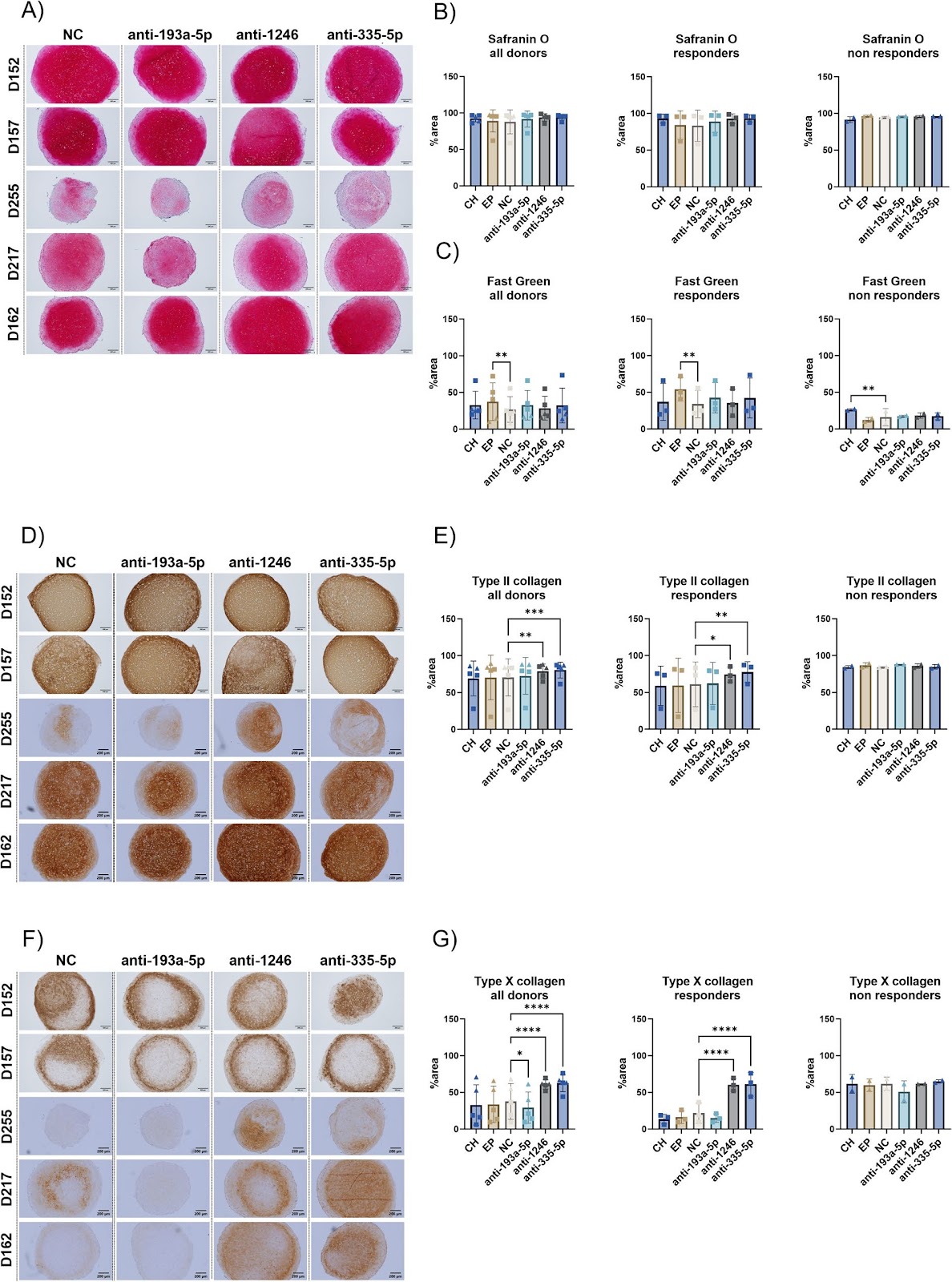
Background Identification of biomarkers to predict the risk of healing delays are of huge clinical interest since 10% of fracture patients progress to delayed or non-union. During endochondral ossification, which takes place in mechanically unstable regions, the bone regenerates through a cartilage intermediate. We previously identified miR-1246, miR-335-5p and miR-193a-5p as fracture-related biomarkers in patient serum, but they appear not to have a functional role in an in vitro model of direct ossification. However, their involvement in other processes related to fracture healing cannot be ruled out and the most common healing process in fracture repair is secondary healing by way of endochondral ossification. Therefore, this study aims to explore the role of miR-1246, miR-335-5p and miR-193a-5p during in vitro endochondral differentiation of human bone marrow-derived mesenchymal stromal cells (BMSCs). Methods The activity of miR-1246, miR-335-5p, and miR-193a-5p was transiently inhibited just before pellet formation and the start of chondrogenic differentiation in human BMSCs (n=5 donors), serving as a model for early endochondral ossification. The effect of miRNA inhibition was assessed by histology (Safranin O/Fast Green), immunohistochemistry (type II and type X collagen), and gene expression analysis by bulk RNA sequencing and RT-qPCR. Results Inhibition of miR-1246 and miR-335-5p enhanced chondrogenic and hypertrophic differentiation in BMSCs from three out of five donors, while miR-193a-5p inhibition had minimal effect. Donors were categorized as “responders” or “non-responders” based on histological and gene expression profiles. RNA sequencing and RT-qPCR identified differentially expressed genes, including a 1.6 and 1.5-fold upregulation of GDF5 and CCN5 respectively (p<0.05) and downregulation of SKIL (fold change: 1.3, p=0.0563) after miR-335-5p inhibition, while the same genes were unaltered by miRNA inhibition in non-responders, suggesting donor-specific responses to miRNA inhibition during early chondrogenesis.

Read Full Abstract10.1186/s13287-025-04589-8
The role of stem cell-derived exosomes in regulating pyroptosis for disease therapyGraphical AbstractVerified
Stem Cell Research & Therapy

The role of stem cell-derived exosomes in regulating pyroptosis for disease therapy

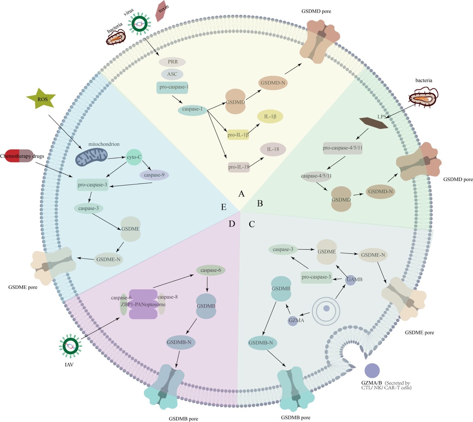
Pyroptosis, a form of programmed cell death, is widely involved in the occurrence and development of various diseases. Its mechanism relies primarily on the activation of pyroptosis proteins, making their expression levels crucial biological markers for assessing the degree of pyroptosis. In the progression of diseases, regulating pyroptosis can alleviate tissue damage and promote repair; in cancer treatment, inducing pyroptosis in cancer cells is also considered a potential therapeutic strategy. In recent years, acellular therapies have garnered significant attention in clinical research, with extracellular vesicles (EVs) (such as exosomes) emerging as novel acellular therapeutic tools. Exosomes exhibit remarkable potential for the treatment of various diseases, particularly in regulating pyroptosis. Owing to their diverse biological functions, exosomes derived from different sources of mesenchymal stem cells (MSCs) play distinct roles in treating different diseases. This review systematically summarizes the role and application prospects of MSC-derived exosomes in regulating pyroptosis for disease treatment. Studies have indicated that MSC-derived exosomes not only precisely regulate the process of pyroptosis but also offer new insights and methods for future disease therapies, and therefore, MSC-derived exosomes possess significant clinical translational value.

Read Full Abstract10.1186/s13287-025-04519-8
Single-cell RNA sequencing identifies PD-L1+ mesenchymal stem cells with enhanced immunomodulatory capacity and alleviated the degree of ectopic new bone formation in ankylosing spondylitisGraphical AbstractVerified
Stem Cell Research & Therapy

Single-cell RNA sequencing identifies PD-L1+ mesenchymal stem cells with enhanced immunomodulatory capacity and alleviated the degree of ectopic new bone formation in ankylosing spondylitis

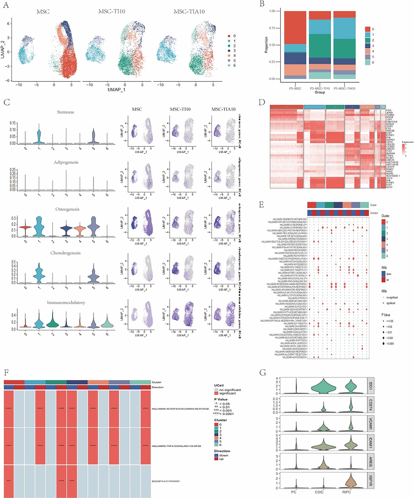
Background This study systematically evaluated the immunomodulatory function of PD-L1-positive mesenchymal stem cells (PD-L1(+) MSCs) using single-cell RNA sequencing (scRNA-seq) and investigated their roles in suppressing inflammation and regulating pathological bone formation in curdlan-induced SKG ankylosing spondylitis (AS) mouse models. Methods scRNA-seq identified MSC subpopulations with high immunomodulatory capacity and key biomarker PD-L1 for subpopulation classification. In vitro co-culture experiments were conducted to evaluate the effects of MSC subpopulations on T-cell proliferation and TNF-α levels. In vivo experiments were performed in forty-eight SKG mouse models to analyze the effects of MSC subpopulations on joint inflammation scores, T-cell subset proportions, inflammatory cytokines, histopathology, and pathological bone formation. Results scRNA-seq revealed significant heterogeneity in MSCs under inflammatory stimulation, with the immunomodulatory subpopulation exhibiting high expression of PD-L1 and IDO. In vitro experiments demonstrated that PD-L1(+) MSCs significantly suppressed T-cell proliferation and reduced TNF-α levels. Joint redness and swelling scores showed that the PD-L1(+) MSC group exhibited the most significant improvement in arthritis, while the IL-17Ai, PD-L1(-) MSC, and MSC groups also effectively reduced inflammation, with significantly lower scores than the model control(MC) group. Histological analysis revealed severe inflammatory cell infiltration in the MC group, while the IL-17Ai, PD-L1(+) MSC, and MSC groups exhibited reduced infiltration. Immunohistochemical analysis further confirmed these findings, with PD-L1(+) MSCs exhibiting a significant reduction in TNF-α and IL-17A-positive cells (P < 0.0001 and P < 0.01, respectively). PD-L1(+) MSCs regulated immune responses by reducing Th17 cell proportions, increasing Th2 and Treg cell proportions, and significantly lowering pro-inflammatory cytokines IFN-Îł, IL-17A, and TNF-α. MicroCT analysis indicated that the PD-L1(+) MSC, MSC, and IL-17Ai group effectively suppressed pathological bone formation.

Read Full Abstract10.1186/s13287-025-04701-y
Prostatic lineage differentiation from human embryonic stem cells through inducible expression of NKX3-1Graphical AbstractVerified
Stem Cell Research & Therapy

Prostatic lineage differentiation from human embryonic stem cells through inducible expression of NKX3-1

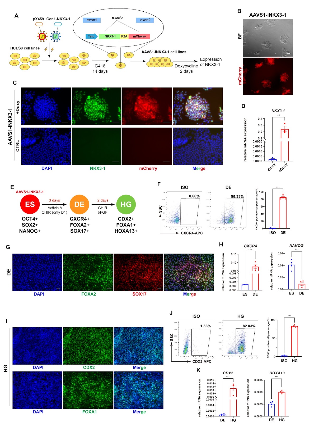
Background Understanding the lineage differentiation of human prostate not only is crucial for basic research on human developmental biology but also significantly contributes to the management of prostate-related disorders. Current knowledge mainly relies on studies on rodent models, lacking human-derived alternatives despite clinical samples may provide a snapshot at certain stage. Human embryonic stem cells can generate all the embryonic lineages including the prostate, and indeed a few studies demonstrate such possibility based on co-culture or co-transplantation with urogenital mesenchyme into mouse renal capsule. Methods To establish a stepwise protocol to obtain prostatic organoids in vitro from human embryonic stem cells, we apply chemicals and growth factors by mimicking the regulation network of transcription factors and signal transduction pathways, and construct cell lines carrying an inducible NKX3-1 expressing cassette, together with three-dimensional culture system. Unpaired t test was applied for statistical analyses. Results We first successfully generate the definitive endoderm, hindgut, and urogenital sinus cells. The embryonic stem cell-derived urogenital sinus cells express prostatic key transcription factors AR and FOXA1, but fail to express NKX3-1. Therefore, we construct NKX3-1-inducible cell line by homologous recombination, which is eventually able to yield AR, FOXA1, and NKX3-1 triple-positive urogenital prostatic lineage cells through stepwise differentiation. Finally, combined with 3D culture we successfully derive prostate-like organoids with certain structures and prostatic cell populations. Conclusions This study reveals the crucial role of NKX3-1 in prostatic differentiation and offers the inducible NKX3-1 cell line, as well as provides a stepwise differentiation protocol to generate human prostate-like organoids, which should facilitate the studies on prostate development and disease pathogenesis.

Read Full Abstract10.1186/s13287-024-03886-y
Mesenchymal stem cell-derived exosomes ameliorate gentamicin-induced vestibular hair cell injury by regulating the SNARE pathway and enhancing autophagyGraphical AbstractVerified
Stem Cell Research & Therapy

Mesenchymal stem cell-derived exosomes ameliorate gentamicin-induced vestibular hair cell injury by regulating the SNARE pathway and enhancing autophagy

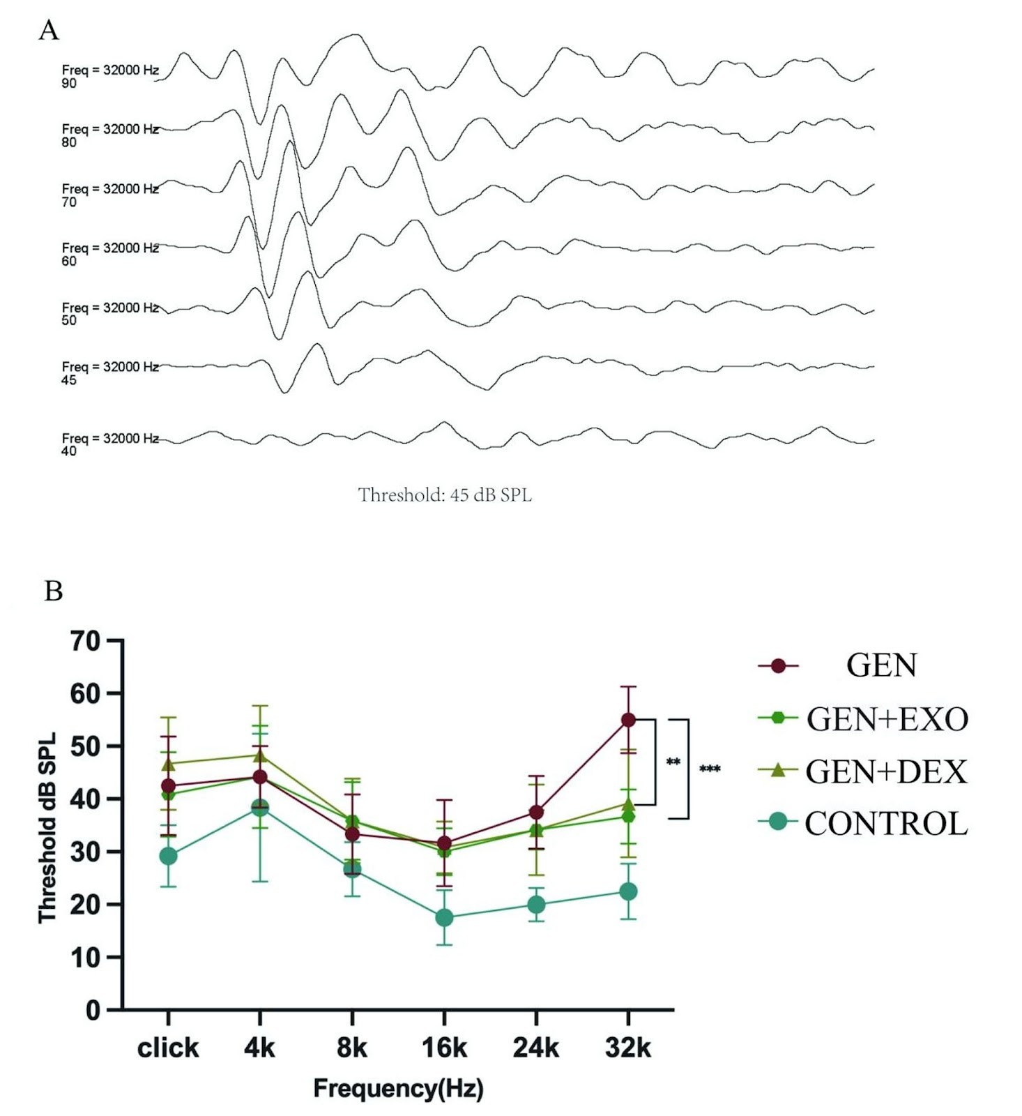
Objective To investigate the delivery efficiency of human umbilical cord mesenchymal stem cell-derived exosomes (hucMSC-EXOs) via intratympanic injection into vestibular end organs, evaluate their protective effects against gentamicin-induced vestibular dysfunction and hearing loss on gentamicin-induced vestibular dysfunction and hearing loss, and explore their regulatory mechanisms on hair cell apoptosis and autophagy. Methods Exosome characteristics were identified by transmission electron microscopy, nanoparticle tracking analysis, and Western blot. PKH26 labeling was used to trace their distribution in the vestibule. SD rats were randomly divided into four groups: control group, gentamicin group (GEN group), gentamicin + exosome group (GEN + EXO group), and gentamicin + dexamethasone group (GEN + DEX group). On day 6 after administration, vestibular function was assessed via open-field test and beam balance test. On day 7, high-frequency hearing (32 kHz) was detected by auditory brainstem response (ABR). The quantity and structural changes of hair cells were analyzed by immunofluorescence staining and scanning electron microscopy. Proteomics was used to analyze differentially expressed proteins in vestibular tissues treated with dexamethasone or hucMSC-EXOs. The regulatory effects on Caspase-3 (apoptosis) and LC3 (autophagy) were validated by immunofluorescence. Results hucMSC-EXOs administered via intratympanic injection were found to target the utricle, saccule, and crista ampullaris. Behavioral studies showed that the GEN + EXO group exhibited significant suppression of gentamicin-induced reduction in total movement distance (p < 0.05) and movement speed (p < 0.05, superior to the GEN + DEX group), with a 60.5% reduction in beam balance test passage time (p < 0.05). ABR results revealed that the auditory threshold at 32 kHz in the GEN + EXO group was 18.3 dB SPL lower than that in the injury group (p < 0.01), with no statistical difference compared to the GEN + DEX group. Hair cell counting showed significant protective effects of exosomes in reducing hair cell loss in the utricular striola (+25%), saccular striola (+44%), and central crista ampullaris

Read Full Abstract10.1186/s13287-025-04819-z
Enhancing myelinogenesis through LIN28A rescues impaired cognition in PWMI miceGraphical AbstractVerified
Stem Cell Research & Therapy

Enhancing myelinogenesis through LIN28A rescues impaired cognition in PWMI mice

Background: In premature newborn infants, preterm white matter injury (PWMI) causes motor and cognitive disabilities. Accumulating evidence suggests that PWMI may result from defected differentiation of oligodendrocyte precursor cells (OPCs) and impaired maturation of oligodendrocytes. However, the underlying mechanisms remain unclear. Methods: Using RNAscope, we analyzed the expression level of RNA-binding protein LIN28A in individual OPCs. Knockout of one or both alleles of Lin28a in OPCs was achieved by administrating tamoxifen to NG2CreER::Ai14::Lin28aflox/+ or NG2CreER::Ai14::Lin28aflox/flox mice. Lentivirus expressing FLEX-Lin28a was used in NG2CreER mice to overexpress LIN28A in OPCs. A series of behavioral tests were performed to assess the cognitive functions of mice. Two-tailed unpaired t-tests was carried out for statistical analysis between groups. Results: We found that the expression of Lin28a was decreased in OPCs in a PWMI mouse model. Knockout of one or both alleles of Lin28a in OPCs postnatally resulted in reduced OPC differentiation, decreased myelinogenesis and impaired cognitive functions. Supplementing LIN28A in OPCs postnatally was able to promote OPC differentiation and enhance myelinogenesis, thus rescuing the cognitive functions in PWMI mice. Conclusion: Our study reveals that LIN28A is critical in regulating postnatal myelinogenesis. Overexpression of LIN28A in OPCs rescues cognitive deficits in PWMI mice by promoting myelinogenesis, thus providing a potential strategy for the treatment of PWMI.

Read Full Abstract10.1186/s13287-025-04267-9
Mesenchymal stromal cell secretome reduces lung injury and thrombo-inflammation induced by SARS-CoV-2 spike proteinGraphical AbstractVerified
Stem Cell Research & Therapy

Mesenchymal stromal cell secretome reduces lung injury and thrombo-inflammation induced by SARS-CoV-2 spike protein

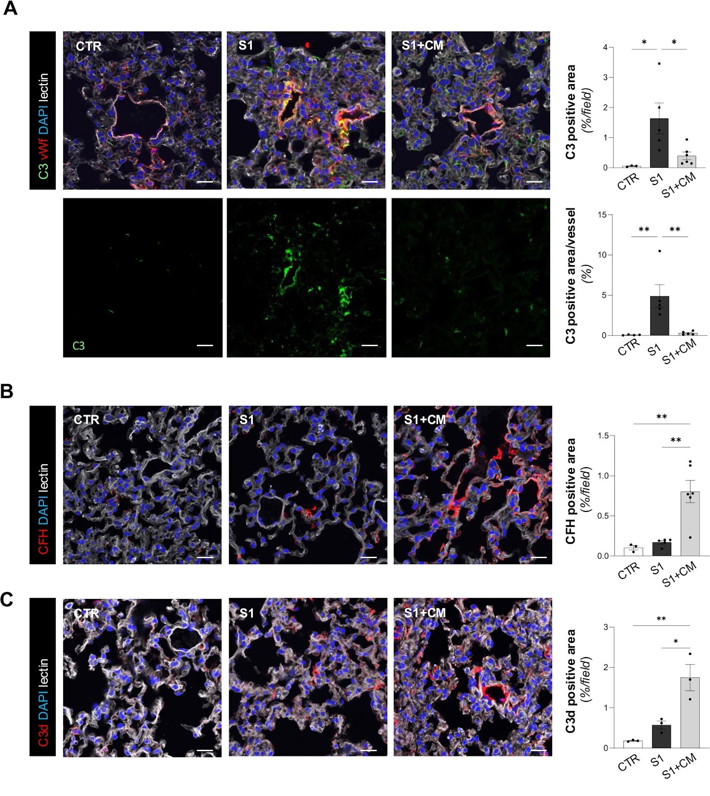
Severe COVID-19 is characterized by thrombo-inflammatory processes within the lung microvasculature. In pursuit of effective treatments, clinical studies explored mesenchymal stromal cells (MSCs) as a promising approach due to their anti-inflammatory, immunomodulatory, and regenerative properties, through their paracrine action. Here, we tested the conditioned medium (CM) derived from human umbilical cord (UC)-MSCs in acute lung injury induced by the spike protein subunit 1 (S1) in ACE2-humanized male mice. Injection of CM significantly limited S1-induced lung injury, edema, and fibrosis. This was associated with reduced vascular dysfunction, in terms of restored thrombomodulin levels and decreased von Willebrand (vWF) expression. By preserving endothelial glycocalyx, CM reduced complement C3 accumulation, favoring factor H binding on the lung microvasculature. Reduced oxidative stress, nuclear NF-ÎșB p65 accumulation, and inflammatory cell infiltration were also observed in response to CM in S1-injected mice. In vitro, CM counteracted thrombo-inflammation by preserving thrombomodulin, as well as limiting vWF expression, due to endothelial glycocalyx recovery. CM reduced nuclear translocation of NF-ÎșB p65 and its downstream targets, ICAM-1 and P-selectin, translating in decreased C3 deposits, platelet aggregation, and leukocyte adhesion on S1-challenged endothelial cells. Collectively, these data indicate that UC-MSC-derived secretome represents a promising therapy in COVID-19 due to its potent anti-thrombotic and anti-inflammatory effects on lung microcirculation.

Read Full Abstract10.1186/s13287-025-04472-6
LincRNA-ASAO promotes dental pulp repair through interacting with PTBP1 to increase ALPL alternative splicingGraphical AbstractVerified
Stem Cell Research & Therapy

LincRNA-ASAO promotes dental pulp repair through interacting with PTBP1 to increase ALPL alternative splicing

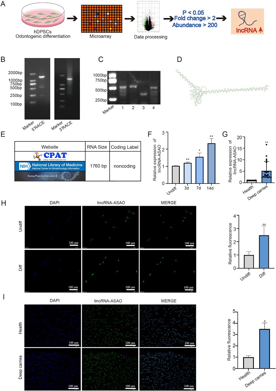
Background Alternative splicing not only expands the genetic encoding of genes but also determines cellular activities. This study aimed to elucidate the regulation mechanism and biological functions of lincRNA-ASAO in the process of odontogenesis-related genes alternative splicing mediated odontogenic differentiation of hDPSCs. Methods RACE, RNA-seq, FISH and bioinformatics techniques were used to identify novel lincRNA-ASAO. ALP staining, alizarin red staining, qRT-PCR and western blot were used to identify the role of lincRNA-ASAO in regulating the odontoblast differentiation of hDPSCs. The binding protein PTBP1 of lincRNA-ASAO was screened by RNA-Pull-down, protein profiling and bioinformatics. The target gene ALPL of lincRNA-ASAO/PTBP1 was identified by RNA-seq, bioinformatics technology and DNA agarose gel electrophoresis. FISH, IF, PAR-CLIP and bioinformatics techniques were used to determine the roles of lincRNA-ASAO, PTBP1 and ALPL pre-mRNA in the odontoblast differentiation of hDPSCs. Results We identified a novel lincRNA-ASAO that could promote the odontogenic differentiation of human Dental Pulp Stem Cells (hDPSCs). And, the interaction between lincRNA-ASAO and alternative splicing factor PTBP1 promoted the odontoblast differentiation of hDPSCs. In addition, lincRNA-ASAO forms duplexes with ALPL pre-mRNA, targeting PTBP1 to exonic splicing silencer (ESS) of ALPL and regulating exon 2 skipping. Notably, lincRNA-ASAO/PTBP1 regulated ALPL production to increase the type 2 splice variant, which promoted the odontoblast differentiation of hDPSCs. Conclusions We have identified the novel lincRNA-ASAO, which can promote the odontoblast differentiation of hDPSCs. The mechanism study found that lincRNA-ASAO/PTBP1 mediated the exon 2 skipping of ALPL pre-mRNA, resulting in the type 2 splice variant of ALPL. Our results enrich the understanding of lncRNAs and alternative splicing in regulating the odontoblast differentiation of hDPSCs, and provide clues to improve the clinical therapeutic potential of hDPSCs for dental pulp restoration.

Read Full Abstract10.1186/s13287-025-04274-w
Trained human bone marrow mesenchymal stem cells restore tissue immuno-microenvironment in fulminant hepatic failure miceGraphical AbstractVerified
Stem Cell Research & Therapy

Trained human bone marrow mesenchymal stem cells restore tissue immuno-microenvironment in fulminant hepatic failure mice

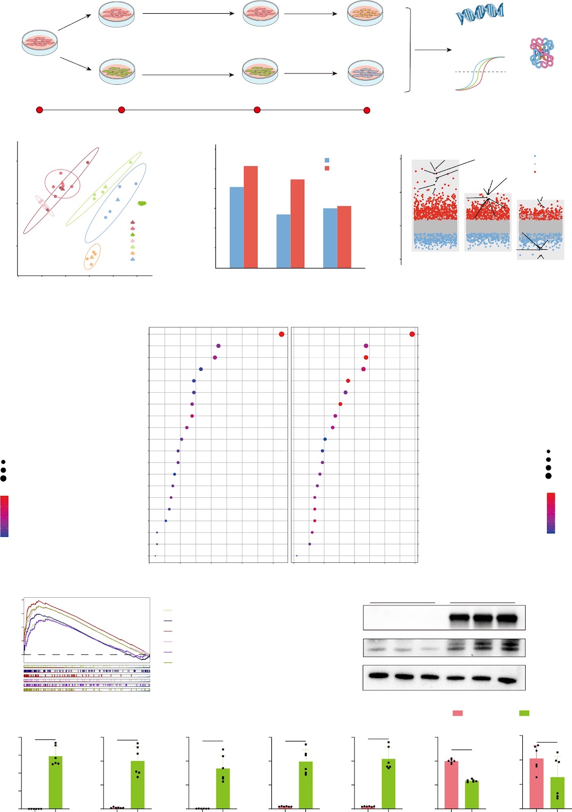
Background Trained immunity with human bone marrow mesenchymal stem cells (hBMSC) is a promising approach to liver regeneration. This study aimed to clarify the trained-hBMSC (T-hBMSC) in restoring tissue immuno-microenvironment in fulminant hepatic failure (FHF) mice. Methods hBMSC trained with tumor necrosis factor-α and interferon-Îł were phenotypically characterized in vitro. FHF mouse models were established in male Balb/c mice via tail vein injection of concanavalin A. The therapeutic potential of T-hBMSC was evaluated through transplantation into FHF mice. Transcriptomic analysis was performed to elucidate the mechanism of liver regeneration post-transplantation of T-hBMSC. Results T-hBMSC with the characteristics of trilineage differentiation potential showed that pro-inflammatory (IL1ÎČ, IL8, both p < 0.0001) and immunoregulatory genes (PDL1, IDO1, both p < 0.0001) were significantly upregulated compared to untrained-hBMSC (UT-hBMSC). Time-trajectory analysis revealed downregulation of pro-inflammatory genes (IL6, IL8, and IL1α) and upregulation of immunomodulatory genes (IDO1) in T-hBMSC upon mimic-stimulation, characterized by distinct transcriptional programs. The liver function (ALT, AST) and inflammatory cytokines (IL6, MCP1, both p < 0.01) levels were significantly improved in the T-hBMSC-treated mice. The survival status of the T-hBMSC group was superior to the UT-hBMSC group, although there was no statistical significance. Histological analysis confirmed reduced necrosis and fewer infiltrating CD45+ immune cells in the T-hBMSC-treated mice. Significant downregulation of immune response (TNF & IL-17 signaling pathways and neutrophil chemotaxis) and upregulation of metabolic pathways were observed in the T-hBMSC group, associated with enhanced liver regeneration. The proportion of anti-inflammatory F4/80+CD163+ macrophages was increased in the liver of T-hBMSC group.

Read Full Abstract10.1186/s13287-025-04540-x
Retraction Note: Human fetal skin-derived stem cell secretome enhances radiation-induced skin injury therapeutic effects by promoting angiogenesisGraphical AbstractVerified
Stem Cell Research & Therapy

Retraction Note: Human fetal skin-derived stem cell secretome enhances radiation-induced skin injury therapeutic effects by promoting angiogenesis

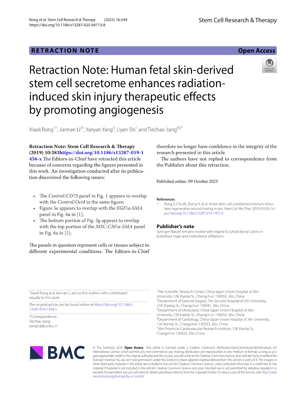
The Editors-in-Chief have retracted this article because of concerns regarding the figures presented in this work. An investigation conducted after its publication discovered the following issues: The Control/CD73 panel in Fig. 1 appears to overlap with the Control/Oct4 in the same figure; Figure 5e appears to overlap with the EGF/α-SMA panel in Fig. 6a in [1]; The bottom portion of Fig. 5g appears to overlap with the top portion of the MSC-CM/α-SMA panel in Fig. 6a in [1]; The panels in question represent cells or tissues subject to different experimental conditions. The Editors-in-Chief therefore no longer have confidence in the integrity of the research presented in this article. The authors have not replied to correspondence from the Publisher about this retraction.

Read Full Abstract10.1186/s13287-025-04713-8