🧬 SinoBioData Academic Portal
šŸ“š Peer-Reviewed Translated Literature

All Biomedical & Clinical Articles (Page 42)

Browse complete peer-reviewed translations from top Chinese biomedical, oncology, and genomics journals. Read verified previews and download full authentic clinical reports.

Published Research Papers

Showing 24 of 1542 peer-reviewed translated articles (Page 42 of 65)

Xenogenous implanted dental follicle stem cells promote periodontal regeneration through inducing the N2 phenotype of neutrophilsGraphical AbstractVerified
Stem Cell Research & Therapy

Xenogenous implanted dental follicle stem cells promote periodontal regeneration through inducing the N2 phenotype of neutrophils

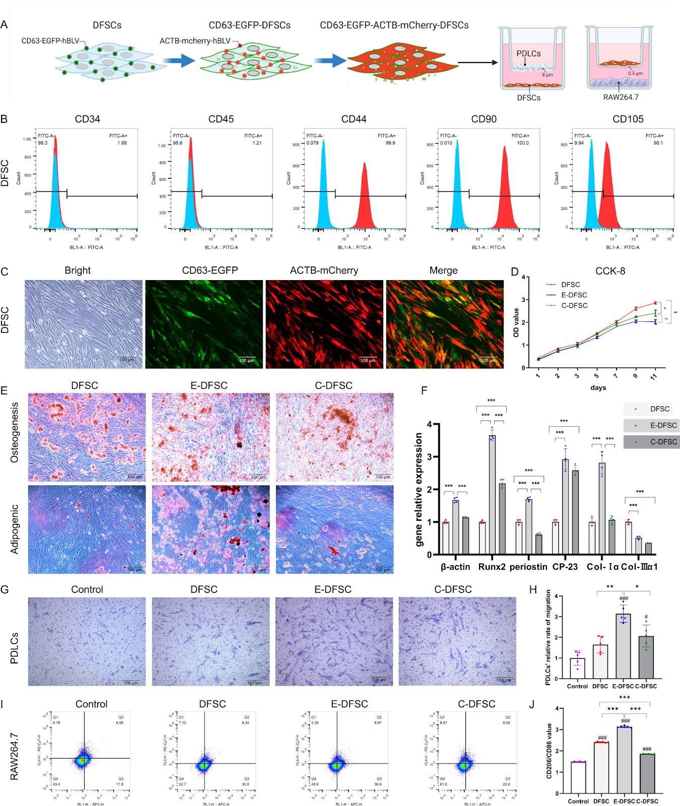
Background Periodontal tissue loss is the main reason for tooth mobility and loss caused by periodontal disease. Dental follicle stem cells (DFSCs) have significant therapeutic potential in periodontal regeneration, which maybe mainly depends on their potent immunomodulatory capacity. Consequently, this study aims to elucidate the impact of implanted xenogenous DFSCs on innate immune responses during early and late stages in the periodontal defect repair period. Methods To trace and investigate the immunomodulation mechanisms of DFSCs in vivo, DFSCs were engineered (E-DFSCs) using lentiviral vectors expressing CD63-enhanced green fluorescent protein (CD63-EGFP) and β-Actin-mCherry protein (ACTB-mCherry) to exhibit green and red fluorescence. The biological characteristics and functions of E-DFSCs were verified by proliferation, differentiation, and co-culture experiments in vitro. In vivo, the periodontal regeneration capacity of E-DFSCs was detected by implantation of murine periodontal defect model, and the response of innate immune cells was detected at the 1st, 3rd, and 5th days (early stage) and 4th week (late stage) after implantation. Results In vitro assessments showed that E-DFSCs retain similar properties to their non-engineered counterparts but exhibit enhanced macrophage immunomodulation capability. In mice models, four-week micro-CT and histological evaluations indicated that E-DFSCs have equivalent efficiency to DFSCs in periodontal defect regeneration. At the early stage of repair in mice periodontal defect, fluorescence tracking showed that implanted E-DFSCs might primarily activate endogenous cells through direct contact and indirect actions, and most of these cells are myeloperoxidase-positive neutrophils. Additionally, compared with the control group, the neutrophilic infiltration and conversion of N2-type were significantly increased in the E-DFSC group. At the late stage of defect regeneration, more M2-type
Read Full Abstract10.1186/s13287-024-03882-2
Kupffer cells abrogate homing and repopulation of allogeneic hepatic progenitors in injured liver siteGraphical AbstractVerified
Stem Cell Research & Therapy

Kupffer cells abrogate homing and repopulation of allogeneic hepatic progenitors in injured liver site

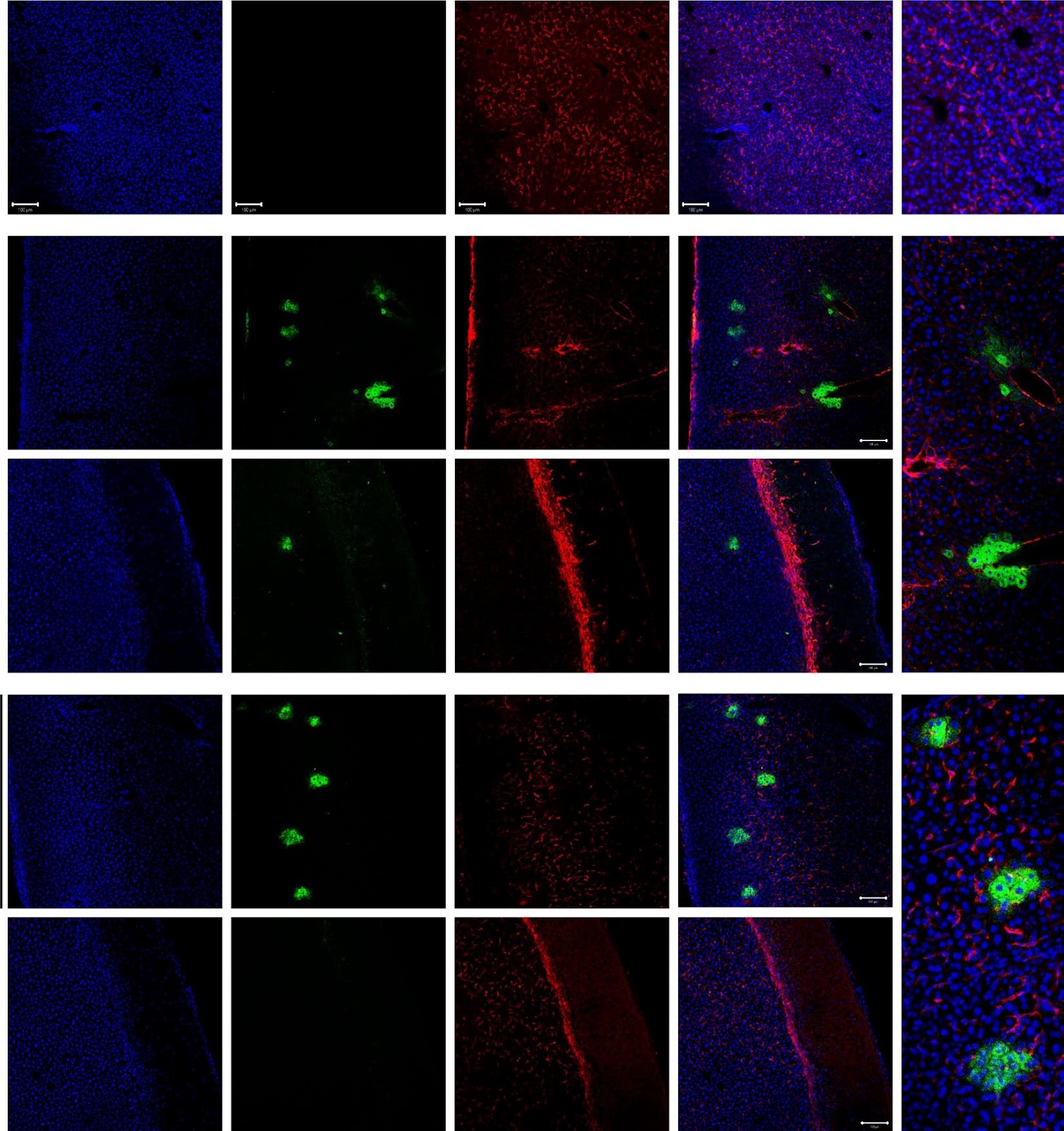
Background Allogeneic hepatocyte transplantation is an emerging approach to treat acute liver defects. However, durable engraftment of the transplanted cells remains a daunting task, as they are actively cleared by the recipient’s immune system. Therefore, a detailed understanding of the innate or adaptive immune cells-derived responses against allogeneic transplanted hepatic cells is the key to rationalize cell-based therapies. Methods Here, we induced an acute inflammatory regenerative niche (3–96 h) on the surface of the liver by the application of cryo-injury (CI) to systematically evaluate the innate immune response against transplanted allogeneic hepatic progenitors in a sustained micro-inflammatory environment. Results The resulting data highlighted that the injured site was significantly repopulated by alternating numbers of innate immune cells, including neutrophils, monocytes and Kupffer cells (KCs), from 3 to 96 h. The transplanted allo-HPs, engrafted 6 h post-injury, were collectively eliminated by the innate immune response within 24 h of transplantation. Selective depletion of the KCs demonstrated a delayed recruitment of monocytes from day 2 to day 6. In addition, the intrasplenic engraftment of the hepatic progenitors 54 h post-transplantation was dismantled by KCs, while a time-dependent better survival and translocation of the transplanted cells into the injured site could be observed in samples devoid of KCs. Conclusion Overall, this study provides evidence that KCs ablation enables a better survival and integration of allo-HPs in a sustained liver inflammatory environment, having implications for rationalizing the cell-based therapeutic interventions against liver defects.

Read Full Abstract10.1186/s13287-024-03656-w
Harnessing three-dimensional porous chitosan microsphere embedded with adipose-derived stem cells to promote nerve regenerationGraphical AbstractVerified
Stem Cell Research & Therapy

Harnessing three-dimensional porous chitosan microsphere embedded with adipose-derived stem cells to promote nerve regeneration

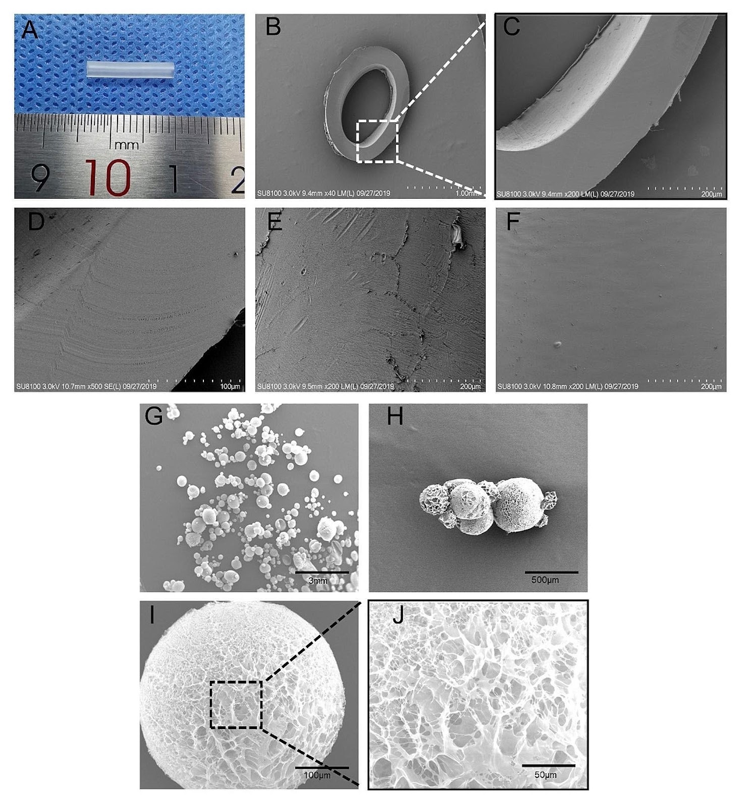
Background Nerve guide conduits are a promising strategy for reconstructing peripheral nerve defects. Improving the survival rate of seed cells in nerve conduits is still a challenge and microcarriers are an excellent three-dimensional (3D) culture scaffold. Here, we investigate the effect of the 3D culture of microcarriers on the biological characteristics of adipose mesenchymal stem cells (ADSCs) and to evaluate the efficacy of chitosan nerve conduits filled with microcarriers loaded with ADSCs in repairing nerve defects. Methods In vitro, we prepared porous chitosan microspheres by a modified emulsion cross-linking method for loading ADSCs and evaluated the growth status and function of ADSCs. In vivo, ADSCs-loaded microcarriers were injected into chitosan nerve conduits to repair a 12 mm sciatic nerve defect in rats. Results Compared to the conventional two-dimensional (2D) culture, the prepared microcarriers were more conducive to the proliferation, migration, and secretion of trophic factors of ADSCs. In addition, gait analysis, neuro-electrophysiology, and histological evaluation of nerves and muscles showed that the ADSC microcarrier-loaded nerve conduits were more effective in improving nerve regeneration. Conclusions The ADSCs-loaded chitosan porous microcarrier prepared in this study has a high cell engraftment rate and good potential for peripheral nerve repair.

Read Full Abstract10.1186/s13287-024-03753-w
Good manufacturing practice production of human corneal limbus-derived stromal stem cells and in vitro quality screening for therapeutic inhibition of corneal scarringGraphical AbstractVerified
Stem Cell Research & Therapy

Good manufacturing practice production of human corneal limbus-derived stromal stem cells and in vitro quality screening for therapeutic inhibition of corneal scarring

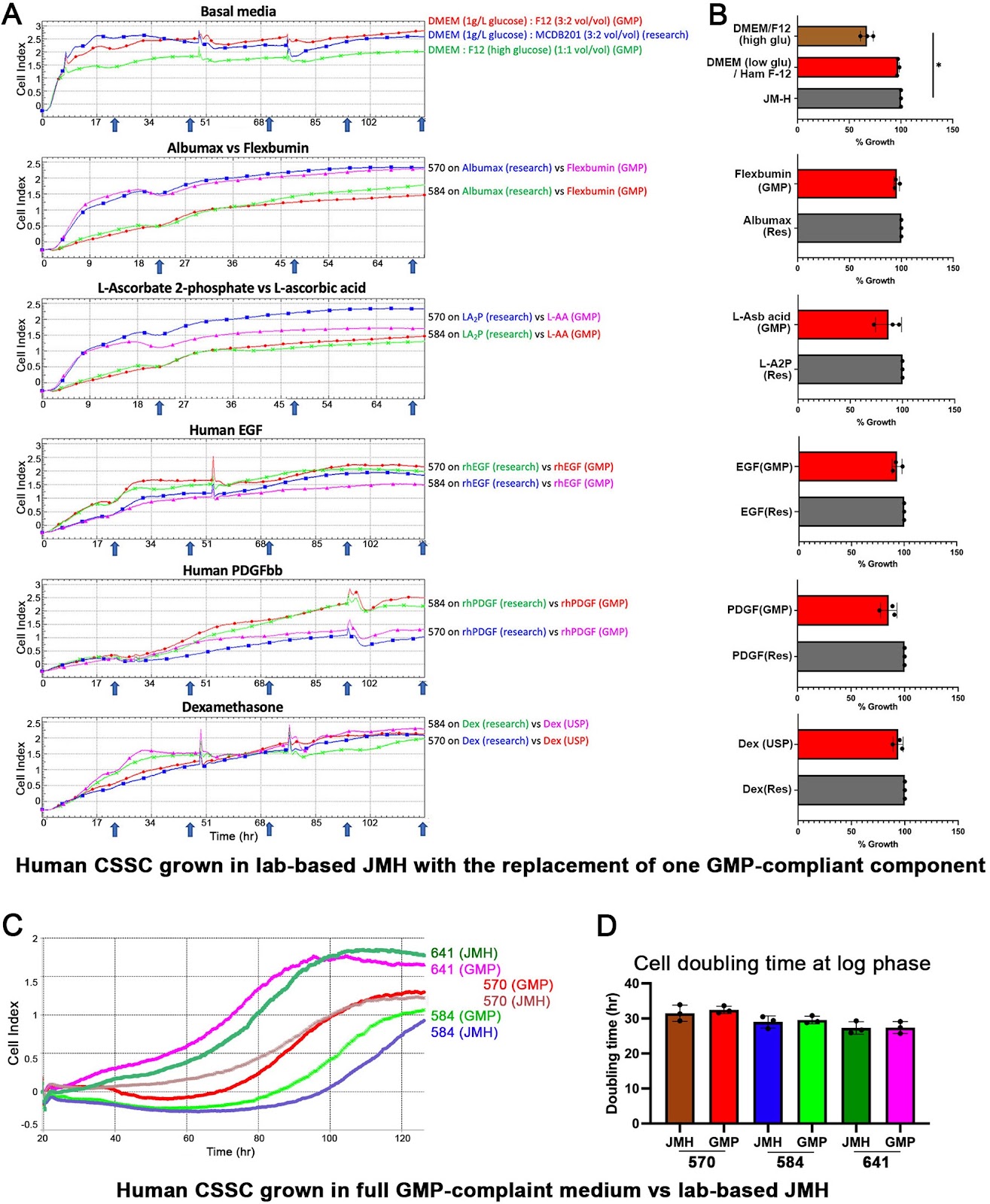
Background Mesenchymal stem cells in the adult corneal stroma (named corneal stromal stem cells, CSSCs) inhibit corneal inflammation and scarring and restore corneal clarity in pre-clinical corneal injury models. This cell therapy could alleviate the heavy reliance on donor materials for corneal transplantation to treat corneal opacities. Herein, we established Good Manufacturing Practice (GMP) protocols for CSSC isolation, propagation, and cryostorage, and developed in vitro quality control (QC) metric for in vivo anti-scarring potency of CSSCs in treating corneal opacities. Methods A total of 24 donor corneal rims with informed consent were used—18 were processed for the GMP optimization of CSSC culture and QC assay development, while CSSCs from the remaining 6 were raised under GMP-optimized conditions and used for QC validation. The cell viability, growth, substrate adhesion, stem cell phenotypes, and differentiation into stromal keratocytes were assayed by monitoring the electric impedance changes using xCELLigence real-time cell analyzer, quantitative PCR, and immunofluorescence. CSSC’s conditioned media were tested for the anti-inflammatory activity using an osteoclastogenesis assay with mouse macrophage RAW264.7 cells. In vivo scar inhibitory outcomes were verified using a mouse model of anterior stromal injury caused by mechanical ablation using an Algerbrush burring. Results By comparatively assessing various GMP-compliant reagents with the corresponding non-GMP research-grade chemicals used in the laboratory-based protocols, we finalized GMP protocols covering donor limbal stromal tissue processing, enzymatic digestion, primary CSSC culture, and cryopreservation. In establishing the in vitro QC metric, two parameters—stemness stability of ABCG2 and nestin and anti-inflammatory ability (rate of inflammation)—were factored into a novel formula to calculate a Scarring Index (SI) for each CSSC batch. Correlating with the in vivo scar inhibitory outcomes, the CSSC batches with SI < 10 had a predicted 50% scar reduction potency, whereas cells with SI > 10 were ineffective to inhibit scarring. Conclusions We established a full GMP-compliant protocol for donor CSSC cultivation, which is essential toward clinical-grade cell manufacturing. A novel in vitro QC–in vivo potency correlation was developed to predict the anti-scarring efficacy of donor CSSCs in treating corneal opacities. This method is applicable to other cell-based therapies and pharmacological treatments.

Read Full Abstract10.1186/s13287-023-03626-8
Regenerative medicine in the treatment of specific dermatologic disorders: a systematic review of randomized controlled clinical trialsGraphical AbstractVerified
Stem Cell Research & Therapy

Regenerative medicine in the treatment of specific dermatologic disorders: a systematic review of randomized controlled clinical trials

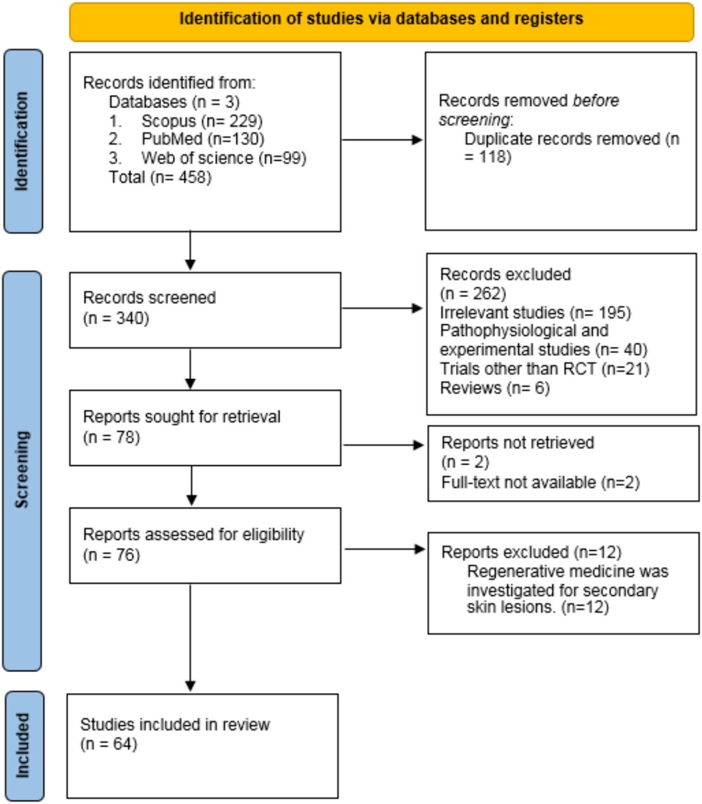
Aims and objectives: The aim of this study is to systematically review randomized controlled clinical trials (RCTs) studying various types of regenerative medicine methods (such as platelet-rich plasma, stromal vascular fraction, cell therapy, conditioned media, etc.) in treating specific dermatologic diseases. Rejuvenation, scarring, wound healing, and other secondary conditions of skin damage were not investigated in this study. Method: Major databases, including PubMed, Scopus, and Web of Science, were meticulously searched for RCTs up to January 2024, focusing on regenerative medicine interventions for specific dermatologic disorders (such as androgenetic alopecia, vitiligo, alopecia areata, etc.). Key data extracted encompassed participant characteristics and sample sizes, types of regenerative therapy, treatment efficacy, and adverse events. Results: In this systematic review, 64 studies involving a total of 2888 patients were examined. Women constituted 44.8% of the study population, while men made up 55.2% of the participants, with an average age of 27.64 years. The most frequently studied skin diseases were androgenetic alopecia (AGA) (45.3%) and vitiligo (31.2%). The most common regenerative methods investigated for these diseases were PRP and the transplantation of autologous epidermal melanocyte/keratinocyte cells, respectively. Studies reported up to 68.4% improvement in AGA and up to 71% improvement in vitiligo. Other diseases included in the review were alopecia areata, melasma, lichen sclerosus et atrophicus (LSA), inflammatory acne vulgaris, chronic telogen effluvium, erosive oral lichen planus, and dystrophic epidermolysis bullosa. Regenerative medicine was found to be an effective treatment option in all of these studies, along with other methods. The regenerative medicine techniques investigated in this study comprised the transplantation of autologous epidermal melanocyte/keratinocyte cells, isolated melanocyte transplantation, cell transplantation from hair follicle origins, melanocyte–keratinocyte suspension in PRP, conditioned media injection, a combination of PRP and basic fibroblast growth factor, intravenous injection of mesenchymal stem cells, concentrated growth factor, stromal vascular fraction (SVF), a combination of PRP and SVF, and preserving hair grafts in PRP. Conclusion: Regenerative medicine holds promise as a treatment for specific dermatologic disorders. To validate our findings, it is recommended to conduct numerous clinical trials focusing on various skin conditions. In our study, we did not explore secondary skin lesions like scars or ulcers. Therefore, assessing the effectiveness of this treatment method for addressing these conditions would necessitate a separate study.

Read Full Abstract10.1186/s13287-024-03800-6
Essential role of p21Waf1/Cip1 in the modulation of post-traumatic hippocampal Neural Stem Cells responseGraphical AbstractVerified
Stem Cell Research & Therapy

Essential role of p21Waf1/Cip1 in the modulation of post-traumatic hippocampal Neural Stem Cells response

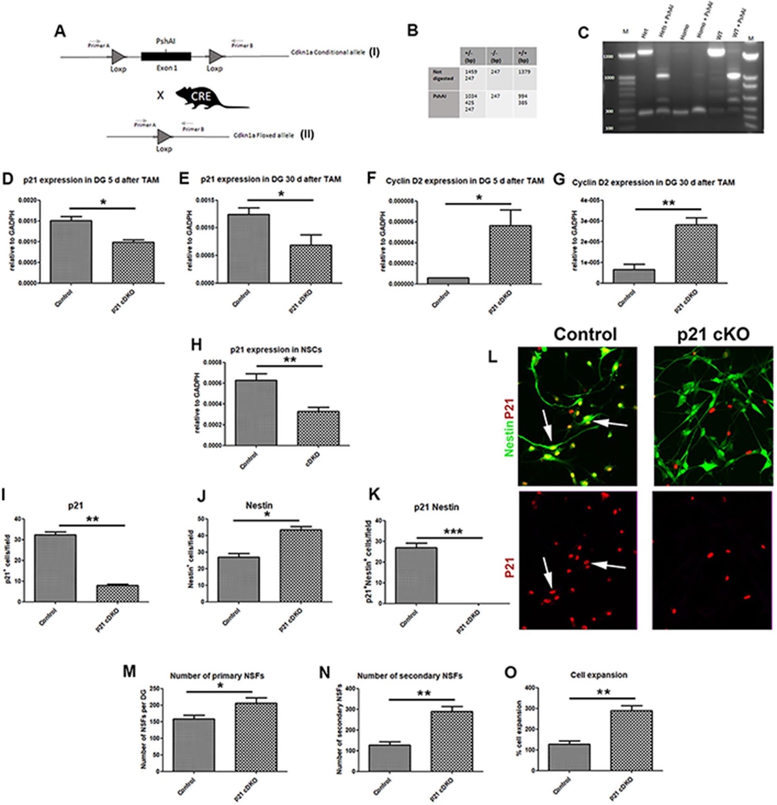
Background Traumatic Brain Injury (TBI) represents one of the main causes of brain damage in young people and the elderly population with a very high rate of psycho-physical disability and death. TBI is characterized by extensive cell death, tissue damage and neuro-inflammation with a symptomatology that varies depending on the severity of the trauma from memory loss to a state of irreversible coma and death. Recently, preclinical studies on mouse models have demonstrated that the post-traumatic adult Neural Stem/Progenitor cells response could represent an excellent model to shed light on the neuro-reparative role of adult neurogenesis following damage. The cyclin-dependent kinase inhibitor p21Waf1/Cip1 plays a pivotal role in modulating the quiescence/activation balance of adult Neural Stem Cells (aNSCs) and in restraining the proliferation progression of progenitor cells. Based on these considerations, the aim of this work is to evaluate how the conditional ablation of p21Waf1/Cip1 in the aNSCS can alter the adult hippocampal neurogenesis in physiological and post-traumatic conditions. Methods We designed a novel conditional p21Waf1/Cip1 knock-out mouse model, in which the deletion of p21Waf1/Cip1 (referred as p21) is temporally controlled and occurs in Nestin-positive aNSCs, following administration of Tamoxifen. This mouse model (referred as p21 cKO mice) was subjected to Controlled Cortical Impact to analyze how the deletion of p21 could influence the post-traumatic neurogenic response within the hippocampal niche. Results The data demonstrates that the conditional deletion of p21 in the aNSCs induces a strong increase in activation of aNSCs as well as proliferation and differentiation of neural progenitors in the adult dentate gyrus of the hippocampus, resulting in an enhancement of neurogenesis and the hippocampal-dependent working memory. However, following traumatic brain injury, the increased neurogenic response of aNSCs in p21 cKO mice leads to a fast depletion of the aNSCs pool, followed by declined neurogenesis and impaired hippocampal functionality. Conclusions These data demonstrate for the first time a fundamental role of p21 in modulating the post-traumatic hippocampal neurogenic response, by the regulation of the proliferative and differentiative steps of aNSCs/progenitor populations after brain damage.

Read Full Abstract10.1186/s13287-024-03787-0
Transcriptional landscape of the interaction of human Mesenchymal Stem Cells with Glioblastoma in bioprinted co-culturesGraphical AbstractVerified
Stem Cell Research & Therapy

Transcriptional landscape of the interaction of human Mesenchymal Stem Cells with Glioblastoma in bioprinted co-cultures

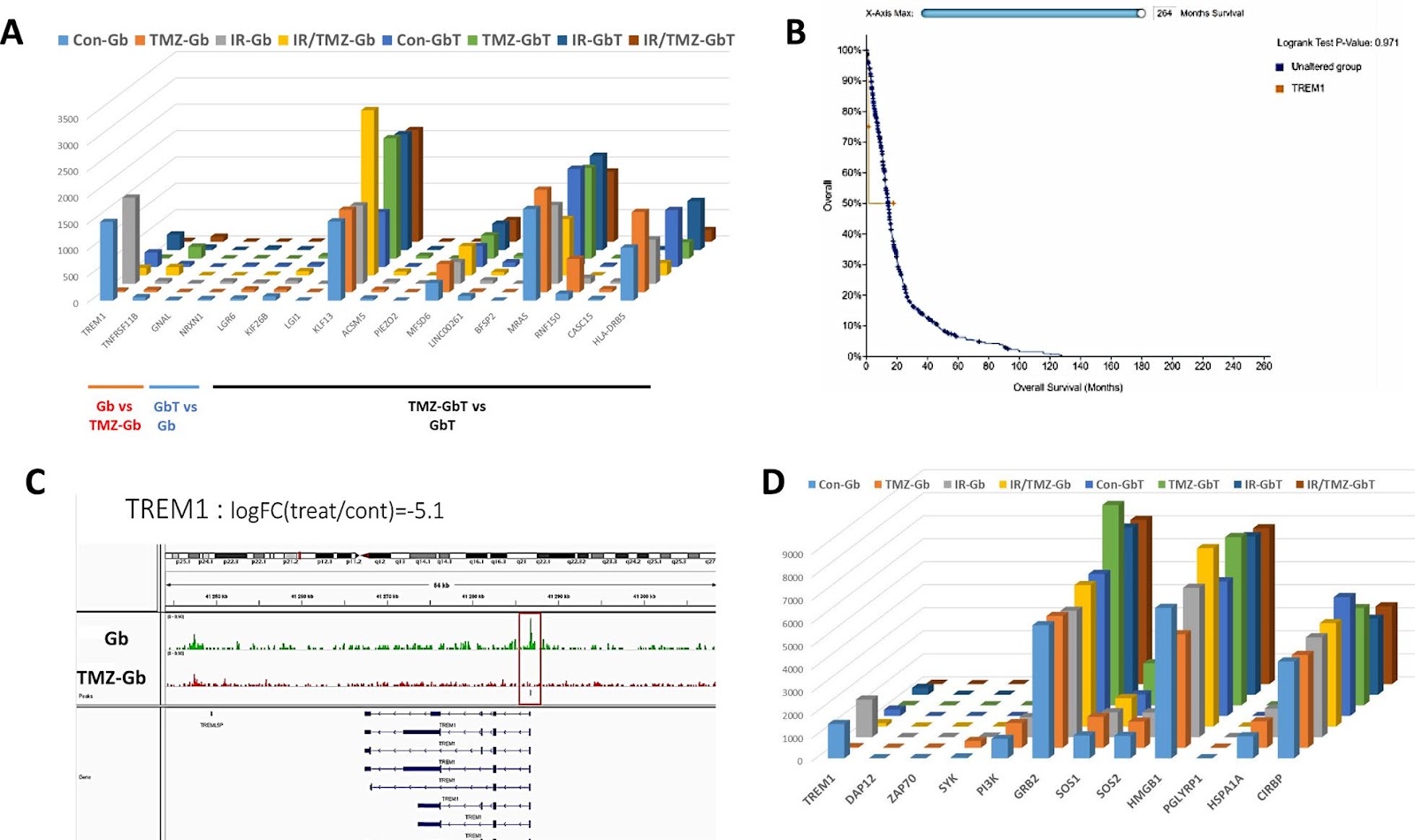
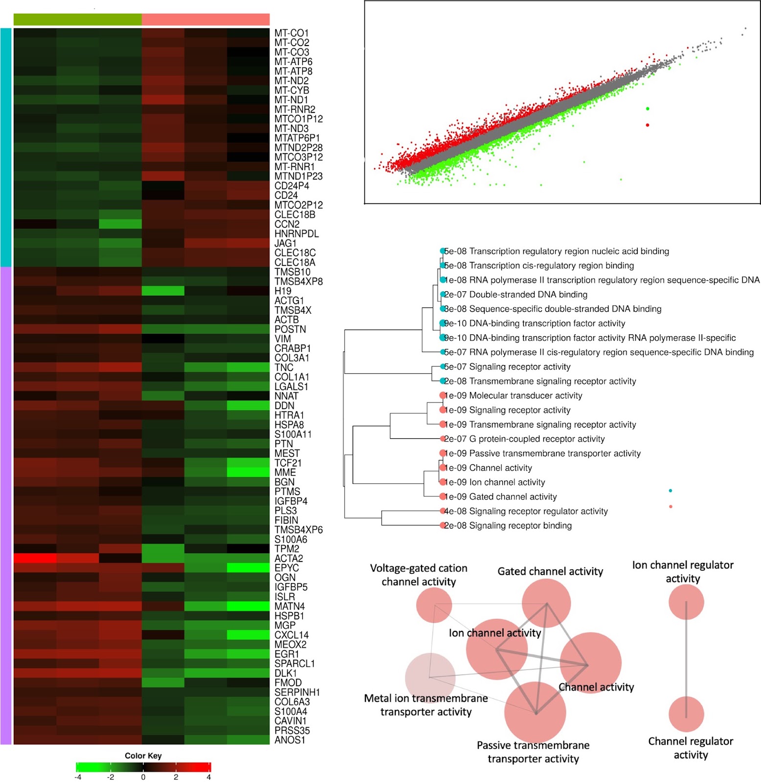
Background The interaction between mesenchymal stem cells (MSC) and Glioblastoma (GBM), although potentially of the highest importance, is ill-understood. This is due, in part, to the lack of relevant experimental models. The similarity between the in vitro situations and the in vivo situation can be improved by 3D co-culture as it reproduces key cell–cell interactions between the tumor microenvironment (TME) and cancer cells. Methods MSC Can acquired characteristics of cancer associated fibroblasts (CAF) by being cultured with conditioned medium from GBM cultures and thus are called MSCCAF. We co Cultured MSCCAF with patient derived GBM in a scaffold 3D bioprinted model. We studied the response to current GBM therapy (e.g. Temozolomide +/Radiation) on the co cultures by bulk transcriptomic (RNA Seq) and epigenetic (ATAC Seq) analyses Results The transcriptomic modifications induced by standard GBM treatment in bioprinted scaffolds of mono- or co-cultures of GBM ± MSC can be analyzed. We found that mitochondrial encoded OXPHOS genes are overexpressed under these conditions and are modified by both co-culture and treatment (chemotherapy ± radiation). We have identified two new markers of MSC/GBM interactions, one epigenetically regulated (i.e. TREM-1) associated with an increased overall survival in GBM patients and another implicated in post-transcriptional regulation (i.e. the long non-coding RNA, miR3681HG), which is associated with a reduced overall survival in GBM patients.

Read Full Abstract10.1186/s13287-024-04022-6
Phenotypic and transcriptomic profiling of induced pluripotent stem cell (iPSC)-derived NK cells and their cytotoxicity against cancersGraphical AbstractVerified
Stem Cell Research & Therapy

Phenotypic and transcriptomic profiling of induced pluripotent stem cell (iPSC)-derived NK cells and their cytotoxicity against cancers

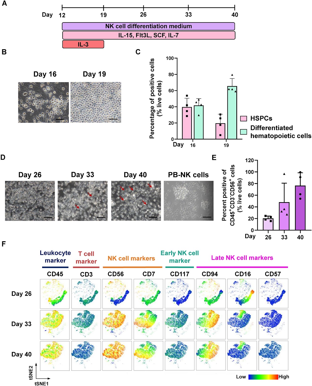
Background: Adoptive immunotherapy using natural killer (NK) cells has attracted considerable interest in numerous clinical trials targeting both hematological and solid tumors. Traditionally, NK cells are primarily derived from either peripheral blood (PB) or umbilical cord blood (UCB). However, these methods can lead to variability and heterogeneity within the NK cell population. In contrast, induced pluripotent stem cell (iPSC)-derived NK (iNK) cells provide a more controlled and uniform cellular population, suitable for large-scale clinical applications. This makes iNK cells a promising option for developing ā€œoff-the-shelfā€ immunotherapeutic products. Nevertheless, current NK cell differentiation protocols, which rely on embryoid body (EB) cultures, are labor-intensive and susceptible to unwanted heterogeneity during differentiation. Here, we developed a more efficient approach for generating iNK cells by employing a monolayer and feeder-free differentiation protocol, alongside optimized culture media. Methods: The iNK cells were generated using a two-step in vitro monolayer feeder-free system following NK cell development. To evaluate their maturity, phenotypic analysis was performed using flow cytometry, comparing with PB-NK cells and the NK-92 cell line. Additionally, single-cell RNA sequencing was performed to examine their transcriptomic profiles. The cytotoxic activity of the iNK cells was evaluated by co-culturing with cholangiocarcinoma (CCA) and breast cancer (BCA) cell lines in both monolayer (2D) and tumor spheroid (3D) co-culture systems. Results: We successfully differentiated iPSCs into mesoderm (ME), hematopoietic stem/progenitor cells (HSPCs), and NK cells. The resulting iNK cells exhibited typical NK cell markers such as CD45, CD56, and CD16, and expressed key functional proteins, including both activating and inhibitory receptors. Single-cell RNA sequencing confirmed that the transcriptomic profile of our iNK cells closely resembles that of PB-NK cells. Importantly, our iNK cells demonstrated strong cytotoxic abilities against various CCA and BCA cell lines, surpassing the NK-92 cell line in both monolayer cultures and tumor spheroid cultures. Conclusion: This study highlights the potential of iPSCs as an effective alternative cell source for generating NK cells. Using a two-step in vitro monolayer feeder-free system, we successfully generated iNK cells that not only expressed key NK cell markers and their receptors but also displayed a transcriptomic profile closely resembling PB-NK cells. Furthermore, iNK cells exhibited cytotoxicity against CCA and BCA cell lines comparable to that of PB-NK cells. This approach could pave the way for off-the-shelf NK cell products, potentially enhancing the effectiveness of adoptive NK cell therapy.

Read Full Abstract10.1186/s13287-024-04029-z
Identification of CD141+ vasculogenic precursor cells from human bone marrow and their endothelial engagement in the arteriogenesis by co-transplantation with mesenchymal stem cellsGraphical AbstractVerified
Stem Cell Research & Therapy

Identification of CD141+ vasculogenic precursor cells from human bone marrow and their endothelial engagement in the arteriogenesis by co-transplantation with mesenchymal stem cells

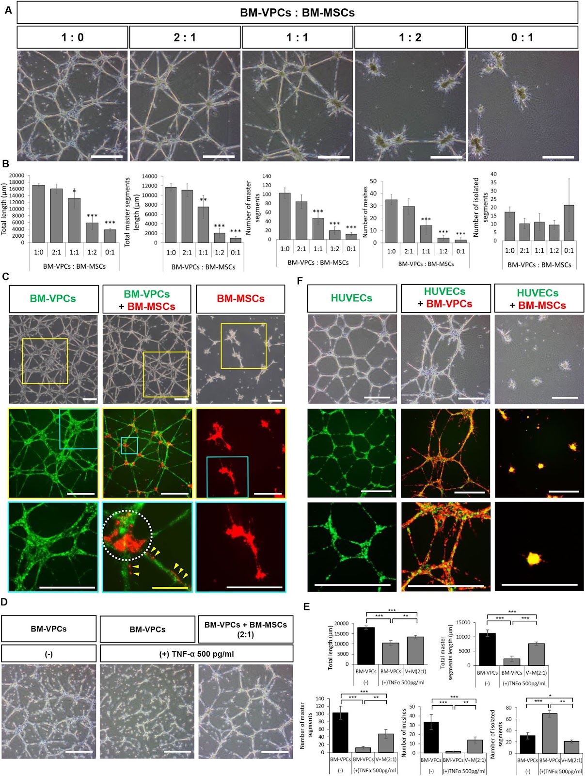
Background Critical limb ischemia (CLI) is a condition characterized by insufficient blood flow to the lower limbs, resulting in severe ischemia and potentially leading to amputation. This study aims to identify novel vasculogenic precursor cells (VPCs) in human bone marrow and evaluate their efficacy in combination with bone marrow-derived mesenchymal stem cells (BM-MSCs) for the treatment of CLI. Methods Ex vivo cultured VPCs and BM-MSCs from bone marrow were characterized and their effects on neovascularization and long-term tissue regeneration were tested in a mouse CLI model. Results VPCs, expressing high levels of hepatocyte growth factor and c-MET, were identified from human bone marrow aspirates. These cells exhibited strong vasculogenic capacity in vitro but possessed a cellular phenotype distinct from those of previously reported endothelial precursor cells in circulation or cord blood. They also expressed most surface markers of BM-MSCs and demonstrated multipotent differentiation ability. Screening of 376 surface markers revealed that VPCs uniquely display CD141 (thrombomodulin). CD141+VPCs are present in BM aspirates as a rare population and can be expanded ex vivo with a population doubling time of approximately 20 h, generating an elaborate vascular network even under angiogenic factor-deficient conditions and recruiting BM-MSCs to the network as pericyte-like cells. Intramuscular transplantation of a combination of human CD141+VPCs and BM-MSCs at a ratio of 2:1 resulted in limb salvage, blood flow recovery, and regeneration of large vessels in the femoral artery-removed CLI model, with an efficacy superior to that of singular transplantation. Importantly, large arteries and arterioles in dual cell transplantation expressed human CD31 in the intima and human α-smooth muscle actin in media layer at 4 weeks post-transplantation, indicating direct engagement of transplanted cells in arteriogenesis. Conclusions CD141+VPCs represent a novel bone marrow-derived vasculogenic precursor cell population with potent therapeutic potential for CLI when combined with BM-MSCs, offering a promising cell-based strategy for vascular regeneration.

Read Full Abstract10.1186/s13287-024-03994-9
The effect of exogenous mitochondria in enhancing the survival and volume retention of transplanted fat tissue in a nude mice modelGraphical AbstractVerified
Stem Cell Research & Therapy

The effect of exogenous mitochondria in enhancing the survival and volume retention of transplanted fat tissue in a nude mice model

Background: Despite the pivotal role of fat grafting in plastic, reconstructive, and aesthetic surgery, inconsistent survival rates of transplanted adipose tissue, primarily due to early ischemic and hypoxic insults, remain a significant challenge. The infusion of healthy mitochondria has emerged as a promising intervention to support tissue recovery from ischemic, hypoxic, and other types of damages across various organ systems. Objectives: This study aims to evaluate the impact of supplementing human adipose tissue grafts with healthy exogenous mitochondria on their volume and mass retention rates when transplanted into the subcutaneous layers of nude mice. This approach seeks to improve and optimize fat grafting techniques. Methods: Human adipose tissues were preconditioned with exogenous mitochondria (10 µg/mL), a combination of exogenous mitochondria and the inhibitor Dyngo-4a, Dyngo-4a alone, or PBS, and then transplanted into the subcutaneous tissue of 24 nude mice. Samples were harvested at 1 and 3 months post-transplantation for analysis of mass and volume retention. The structural morphology and integrity of the adipose tissues were assessed using Hematoxylin and Eosin (H&E) staining. Results: Mitochondrial preconditioning significantly enhanced the retention of mass and volume in fat grafts, demonstrating superior structural morphology and integrity compared to the control group. Conclusions: This study highlights the potential of exogenous mitochondrial augmentation in fat transplantation to significantly improve fat graft survival, thereby optimizing the success of fat grafting procedures.

Read Full Abstract10.1186/s13287-024-03938-3
Development and application of haploid embryonic stem cellsGraphical AbstractVerified
Stem Cell Research & Therapy

Development and application of haploid embryonic stem cells

Haploid cells are a kind of cells with only one set of chromosomes. Compared with traditional diploid cells, haploid cells have unique advantages in gene screening and drug-targeted therapy, due to their phenotype being equal to the genotype. Embryonic stem cells are a kind of cells with strong differentiation potential that can differentiate into various types of cells under specific conditions in vitro. Therefore, haploid embryonic stem cells have the characteristics of both haploid cells and embryonic stem cells, which makes them have significant advantages in many aspects, such as reproductive developmental mechanism research, genetic screening, and drug-targeted therapy. Consequently, establishing haploid embryonic stem cell lines is of great significance. This paper reviews the progress of haploid embryonic stem cell research and briefly discusses the applications of haploid embryonic stem cells.

Read Full Abstract10.1186/s13287-024-03727-y
NQO1 promotes osteogenesis and suppresses angiogenesis in DPSCs via MAPK pathway modulationGraphical AbstractVerified
Stem Cell Research & Therapy

NQO1 promotes osteogenesis and suppresses angiogenesis in DPSCs via MAPK pathway modulation

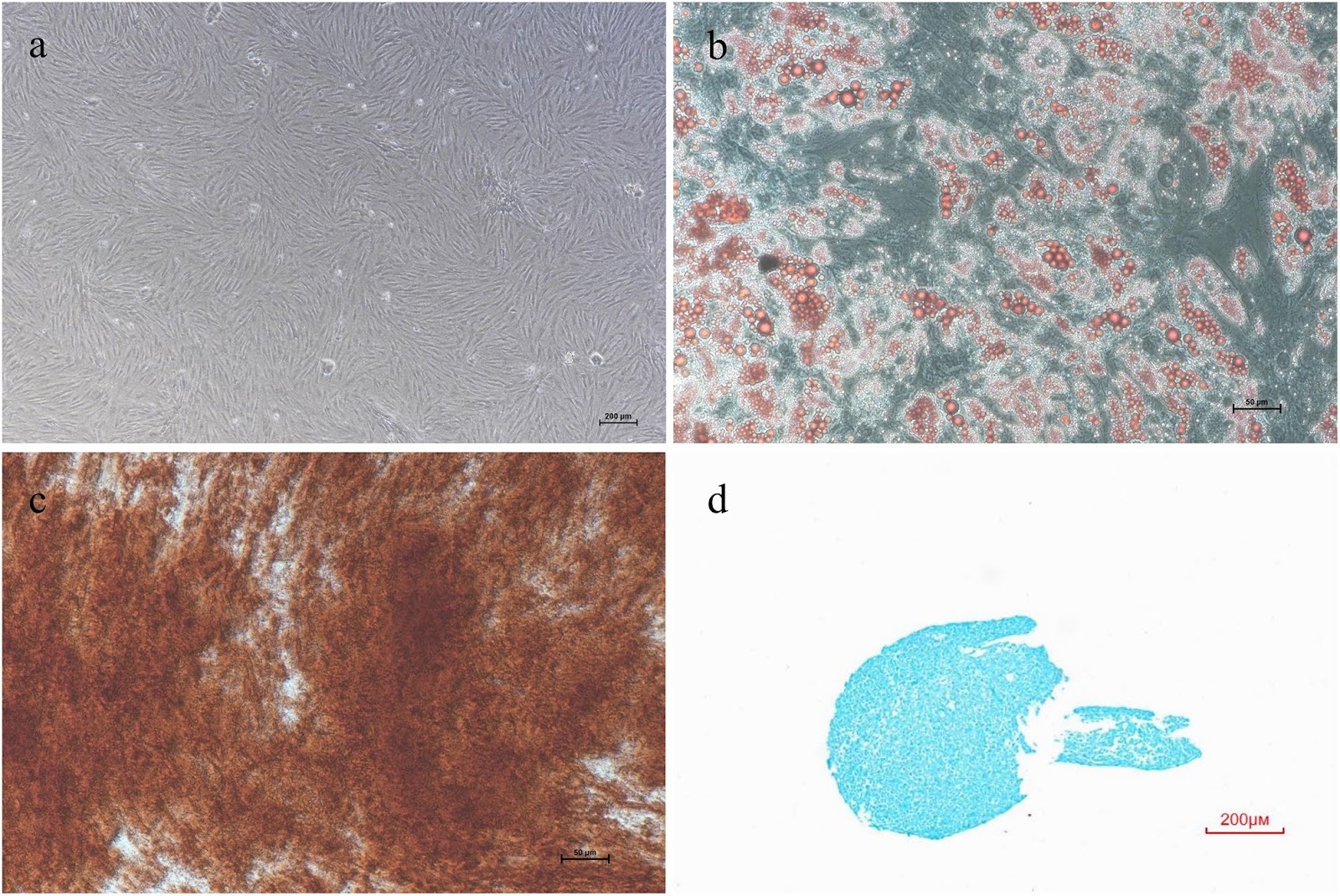
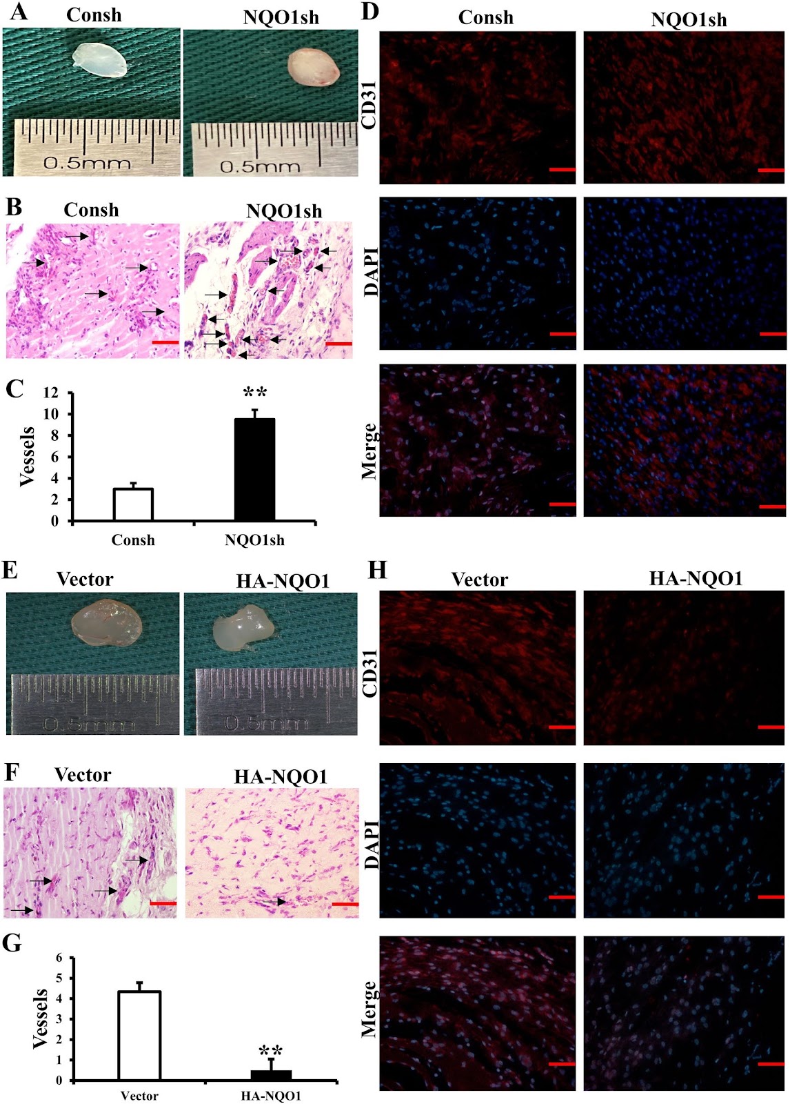
Background Influence on stem cells' angiogenesis and osteogenesis of NAD(P)H Quinone Dehydrogenase 1(NQO1) has been established, but its impact on dental pulp stem cells (DPSCs) is unexplored. An important strategy for the treatment of arteriosclerosis is to inhibit calcium deposition and to promote vascular repair and angiogenesis. This study investigated the function and mechanism of NQO1 on angiogenesis and osteogenesis of DPSCs, so as to provide a new ideal for the treatment of arteriosclerosis. Methods Co-culture of human DPSCs and human umbilical vein endothelial cells (HUVECs) was used to detect the angiogenesis ability. Alkaline phosphatase (ALP) activity, alizarin red staining (ARS), and transplantation of HA/tri-calcium phosphate with DPSCs were used to detect osteogenesis. Results NQO1 suppressed in vitro tubule formation, migration, chemotaxis, and in vivo angiogenesis, as evidenced by reduced CD31 expression. It also enhanced ALP activity, ARS, DSPP expression and osteogenesis and boosted mitochondrial function in DPSCs. CoQ10, an electron transport chain activator, counteracted the effects of NQO1 knockdown on these processes. Additionally, NQO1 downregulated MAPK signaling, which was reversed by CoQ10 supplementation in DPSCs-NQO1sh. Conclusions NQO1 inhibited angiogenesis and promoted the osteogenesis of DPSCs by suppressing MAPK signaling pathways and enhancing mitochondrial respiration.

Read Full Abstract10.1186/s13287-024-03929-4
KCNJ16-depleted kidney organoids recapitulate tubulopathy and lipid recovery upon statins treatmentGraphical AbstractVerified
Stem Cell Research & Therapy

KCNJ16-depleted kidney organoids recapitulate tubulopathy and lipid recovery upon statins treatment

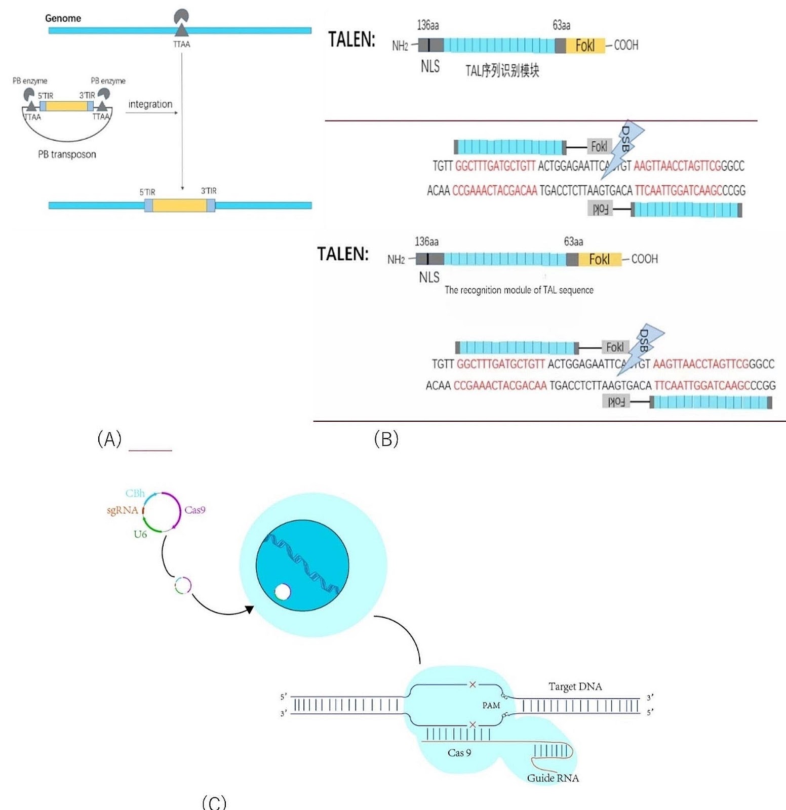
Background The KCNJ16 gene has been associated with a novel kidney tubulopathy phenotype, viz. disturbed acid–base homeostasis, hypokalemia and altered renal salt transport. KCNJ16 encodes for Kir5.1, which together with Kir4.1 constitutes a potassium channel located at kidney tubular cell basolateral membranes. Preclinical studies provided mechanistic links between Kir5.1 and tubulopathy, however, the disease pathology remains poorly understood. Here, we aimed at generating and characterizing a novel advanced in vitro human kidney model that recapitulates the disease phenotype to investigate further the pathophysiological mechanisms underlying the tubulopathy and potential therapeutic interventions. Methods We used CRISPR/Cas9 to generate KCNJ16 mutant (KCNJ16+/āˆ’ and KCNJ16āˆ’/āˆ’) cell lines from healthy human induced pluripotent stem cells (iPSC) KCNJ16 control (KCNJ16WT). The iPSCs were differentiated following an optimized protocol into kidney organoids in an air–liquid interface. Results KCNJ16-depleted kidney organoids showed transcriptomic and potential functional impairment of key voltage-dependent electrolyte and water-balance transporters. We observed cysts formation, lipid droplet accumulation and fibrosis upon Kir5.1 function loss. Furthermore, a large scale, glutamine tracer flux metabolomics analysis demonstrated that KCNJ16āˆ’/āˆ’ organoids display TCA cycle and lipid metabolism impairments. Drug screening revealed that treatment with statins, particularly the combination of simvastatin and C75, prevented lipid droplet accumulation and collagen-I deposition in KCNJ16āˆ’/āˆ’ kidney organoids. Conclusions Mature kidney organoids represent a relevant in vitro model for investigating the function of Kir5.1. We discovered novel molecular targets for this genetic tubulopathy and identified statins as a potential therapeutic strategy for KCNJ16 defects in the kidney.

Read Full Abstract10.1186/s13287-024-03881-3
Correction: Murine skin-derived multipotent papillary dermal fibroblast progenitors show germline potential in vitroGraphical AbstractVerified
Stem Cell Research & Therapy

Correction: Murine skin-derived multipotent papillary dermal fibroblast progenitors show germline potential in vitro

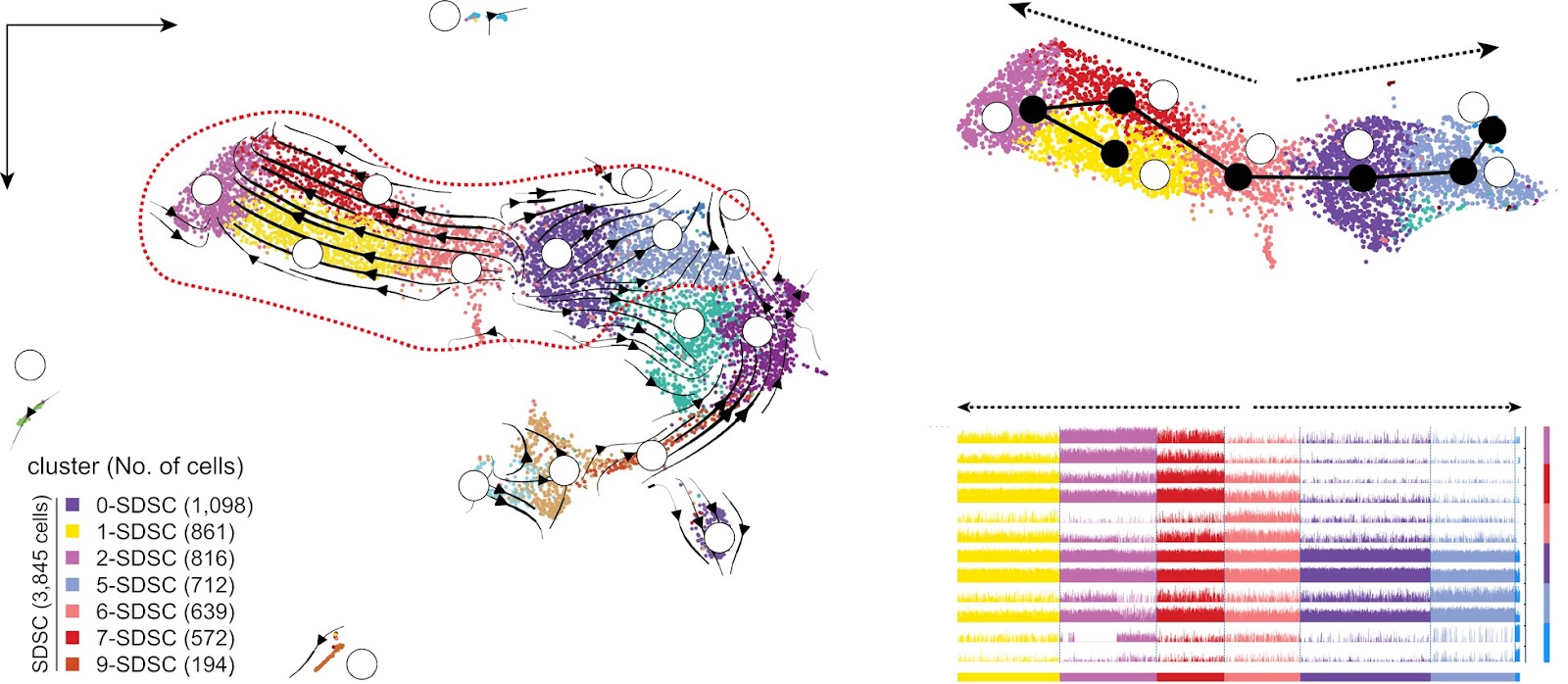
Correction to: Stem Cell Research & Therapy (2023) 14:17. The authors note that during the preparation of the manuscript, the track plot included the expression trends of 14 genes (with Tk1 and Pclaf repeated twice), but only 13 gene symbols were labeled, resulting in a mismatch and repeat of the trackplot with the image on the right. This error occurred during the typesetting process of the original figures. The authors have corrected the annotations in Fig. 2C as shown ahead in this correction article, apologise for the error, and confirm that the overall results and conclusions are not affected by this change.

Read Full Abstract10.1186/s13287-024-03838-6
Critical contribution of mitochondria in the development of cardiomyopathy linked to desmin mutationGraphical AbstractVerified
Stem Cell Research & Therapy

Critical contribution of mitochondria in the development of cardiomyopathy linked to desmin mutation

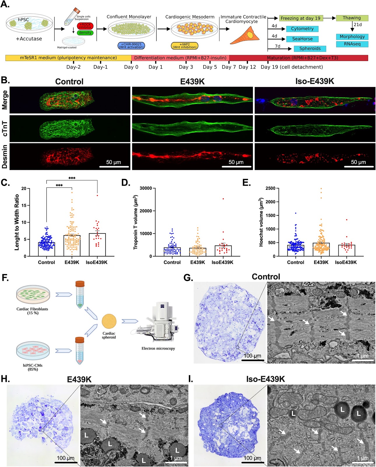
Background Beyond the observed alterations in cellular structure and mitochondria, the mechanisms linking rare genetic mutations to the development of heart failure in patients affected by desmin mutations remain unclear due in part, to the lack of relevant human cardiomyocyte models. Methods To shed light on the role of mitochondria in these mechanisms, we investigated cardiomyocytes derived from human induced pluripotent stem cells carrying the heterozygous DESE439K mutation that were either isolated from a patient or generated by gene editing. To increase physiological relevance, cardiomyocytes were either cultured on an anisotropic micropatterned surface to obtain elongated and aligned cardiomyocytes, or as a cardiac spheroid to create a micro-tissue. Moreover, when applicable, results from cardiomyocytes were confirmed with heart biopsies of suddenly died patient of the same family harboring DESE439K mutation, and post-mortem heart samples from five control healthy donors. Results The heterozygous DESE439K mutation leads to dramatic changes in the overall cytoarchitecture of cardiomyocytes, including cell size and morphology. Most importantly, mutant cardiomyocytes display altered mitochondrial architecture, mitochondrial respiratory capacity and metabolic activity reminiscent of defects observed in patient’s heart tissue. Finally, to challenge the pathological mechanism, we transferred normal mitochondria inside the mutant cardiomyocytes and demonstrated that this treatment was able to restore mitochondrial and contractile functions of cardiomyocytes. Conclusions This work highlights the deleterious effects of DESE439K mutation, demonstrates the crucial role of mitochondrial abnormalities in the pathophysiology of desmin-related cardiomyopathy, and opens up new potential therapeutic perspectives for this disease.

Read Full Abstract10.1186/s13287-023-03619-7
Transplantation of the LRP1high subpopulation of human umbilical cord-derived mesenchymal stem cells improves ovarian function in mice with premature ovarian failure and aged miceGraphical AbstractVerified
Stem Cell Research & Therapy

Transplantation of the LRP1high subpopulation of human umbilical cord-derived mesenchymal stem cells improves ovarian function in mice with premature ovarian failure and aged mice

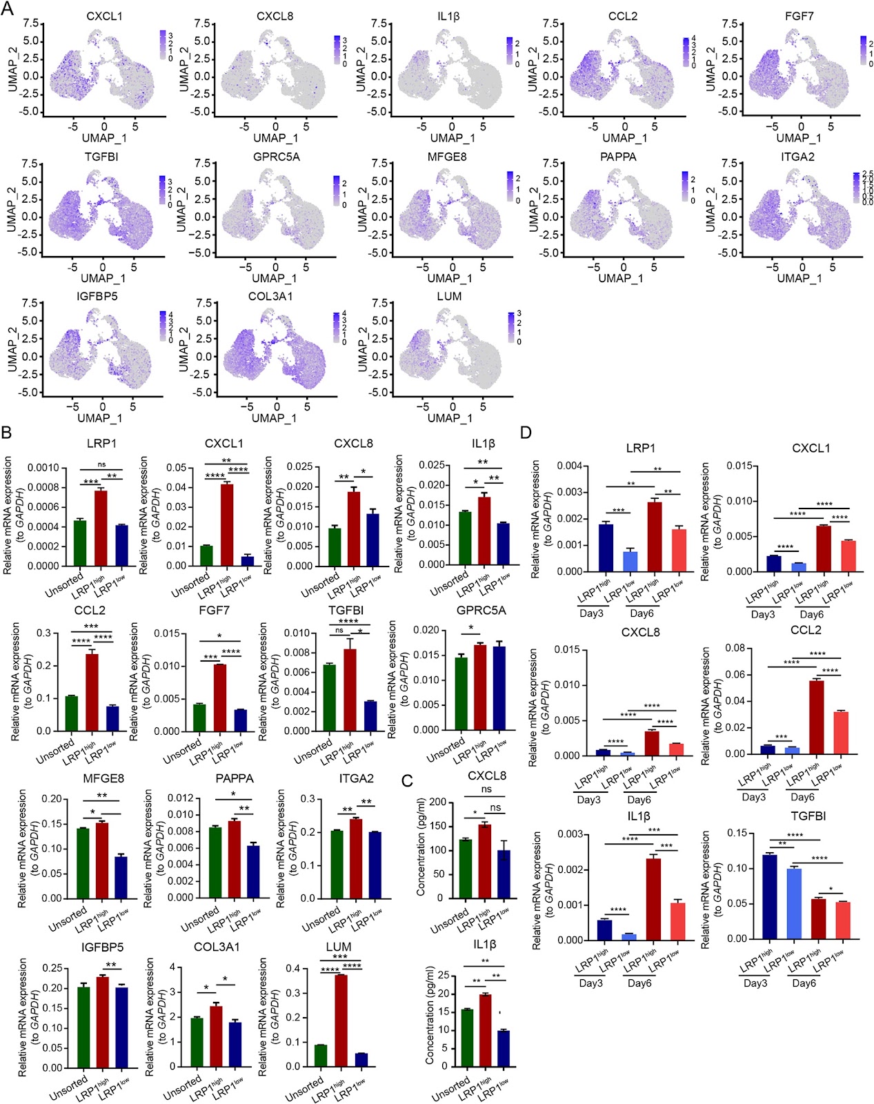
Background Premature ovarian failure (POF) has a profound impact on female reproductive and psychological health. In recent years, the transplantation of umbilical cord-derived mesenchymal stem cells (UC-MSCs) has demonstrated unprecedented potential in the treatment of POF. However, the heterogeneity of human UC-MSCs remains a challenge for their large-scale clinical application. Therefore, it is imperative to identify specific subpopulations within UC-MSCs that possess the capability to improve ovarian function, with the aim of reducing the uncertainty arising from the heterogeneity while achieving more effective treatment of POF. Methods 10Ɨ Genomics was performed to investigate the heterogeneity of human UC-MSCs. We used LRP1 as a marker and distinguished the potential therapeutic subpopulation by flow cytometry, and determined its secretory functions. Unsorted UC-MSCs, LRP1high and LRP1low subpopulation was transplanted under the ovarian capsules of aged mice and CTX-induced POF mice, and therapeutic effects was evaluated by assessing hormone levels, estrous cycles, follicle counts, and embryo numbers. RNA sequencing on mouse oocytes and granulosa cells after transplantation was performed to explore the mechanism of LRP1high subpopulation on mouse oocytes and granulosa cells. Results We identified three distinct functional subtypes, including mesenchymal stem cells, multilymphoid progenitor cells and trophoblasts. Additionally, we identified the LRP1high subpopulation, which improved ovarian function in aged and POF mice. We elucidated the unique secretory functions of the LRP1high subpopulation, capable of secreting various chemokines, cytokines, and growth factors. Furthermore, LRP1 plays a crucial role in regulating the ovarian microenvironment, including tissue repair and extracellular matrix remodeling. Consistent with its functions, the transcriptomes of oocytes and granulosa cells after transplantation revealed that the LRP1high subpopulation improves ovarian function by modulating the extracellular matrix of oocytes, NAD metabolism, and mitochondrial function in granulosa cells. Conclusion Through exploration of the heterogeneity of UC-MSCs, we identified the LRP1high subpopulation capable of improving ovarian function in aged and POF mice by secreting various factors and remodeling the extracellular matrix. This study provides new insights into the targeted exploration of human UC-MSCs in the precise treatment of POF.

Read Full Abstract10.1186/s13287-024-03660-0
Therapeutic efficacy and in vivo distribution of human umbilical cord-derived mesenchymal stem cell spheroids transplanted via B-Ultrasound-guided percutaneous portal vein puncture in rhesus monkey models of liver fibrosisGraphical AbstractVerified
Stem Cell Research & Therapy

Therapeutic efficacy and in vivo distribution of human umbilical cord-derived mesenchymal stem cell spheroids transplanted via B-Ultrasound-guided percutaneous portal vein puncture in rhesus monkey models of liver fibrosis

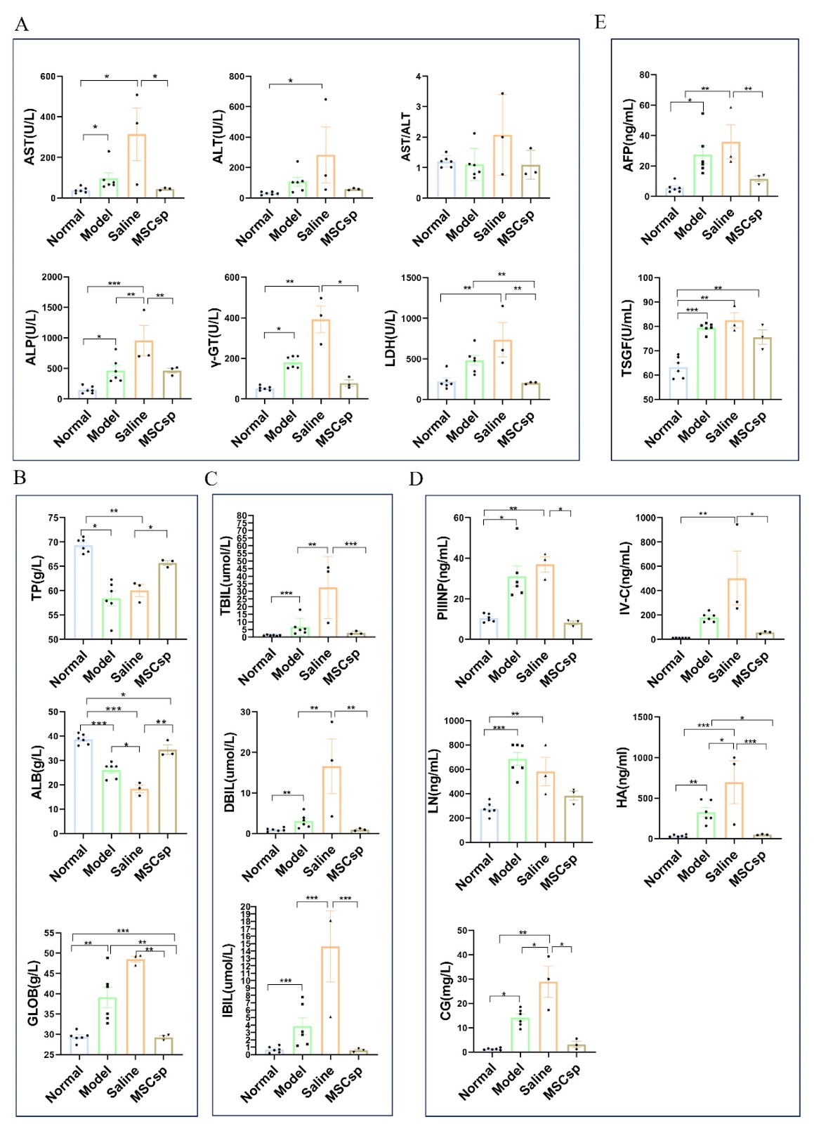
Background Liver fibrosis can progress to end-stage cirrhosis and liver cancer. Mesenchymal stem cells (MSCs) were considered the most promising therapeutic strategy, but most of the MSCs injected intravenously traditionally are trapped in the lungs, rapidly reducing their survival ability. MSC spheroids cultured in 3D have shown higher tolerance to fluid shear stress and better survival than dissociated MSCs. Simulating the route of orthotopic liver transplantation, transplanting MSC spheroids into the liver via hepatic portal vein may impact superior therapeutic effects. Methods In the present study, human umbilical cord-derived MSC spheroids (hUC-MSCsp) were transplanted into rhesus monkey models of liver fibrosis via B-ultrasound-guided percutaneous portal vein puncture with minimized body invasion. The therapeutic effect is evaluated through hematology, ultrasound, and pathology. To study the effect of hUC-MSCsp on gene expression in rhesus monkeys with liver injury, transcriptome sequencing analysis was performed on the livers of rhesus monkeys. The distribution of transplanted hUC-MSCsp was traced with RNA scope technology. Results We found that hUC-MSCsp significantly restored liver function, including ALT, AST, ALB, GLOB and bilirubin. hUC-MSCsp also significantly reduced liver collagen deposition and inflammatory infiltration, and promote dismission

Read Full Abstract10.1186/s13287-024-03934-7
Epicardial EMT and cardiac repair: an updateGraphical AbstractVerified
Stem Cell Research & Therapy

Epicardial EMT and cardiac repair: an update

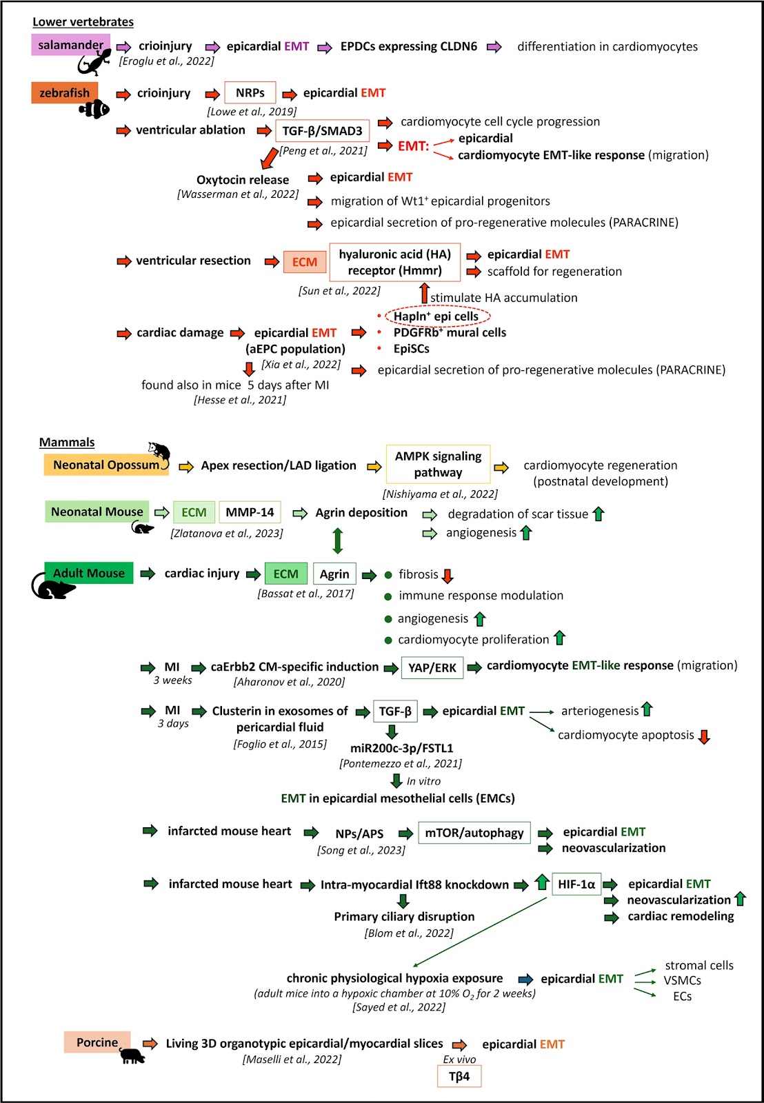
Epicardial epithelial-to-mesenchymal transition (EMT) plays a pivotal role in both heart development and injury response and involves dynamic cellular changes that are essential for cardiogenesis and myocardial repair. Specifically, epicardial EMT is a crucial process in which epicardial cells lose polarity, migrate into the myocardium, and differentiate into various cardiac cell types during development and repair. Importantly, following EMT, the epicardium becomes a source of paracrine factors that support cardiac growth at the last stages of cardiogenesis and contribute to cardiac remodeling after injury. As such, EMT seems to represent a fundamental step in cardiac repair. Nevertheless, endogenous EMT alone is insufficient to stimulate adequate repair. Redirecting and amplifying epicardial EMT pathways offers promising avenues for the development of innovative therapeutic strategies and treatment approaches for heart disease. In this review, we present a synthesis of recent literature highlighting the significance of epicardial EMT reactivation in adult heart disease patients.

Read Full Abstract10.1186/s13287-024-03823-z
Chitosan based extruded nanofibrous bioscaffold for local delivery of mesenchymal stem cells to improve diabetic wound healingGraphical AbstractVerified
Stem Cell Research & Therapy

Chitosan based extruded nanofibrous bioscaffold for local delivery of mesenchymal stem cells to improve diabetic wound healing

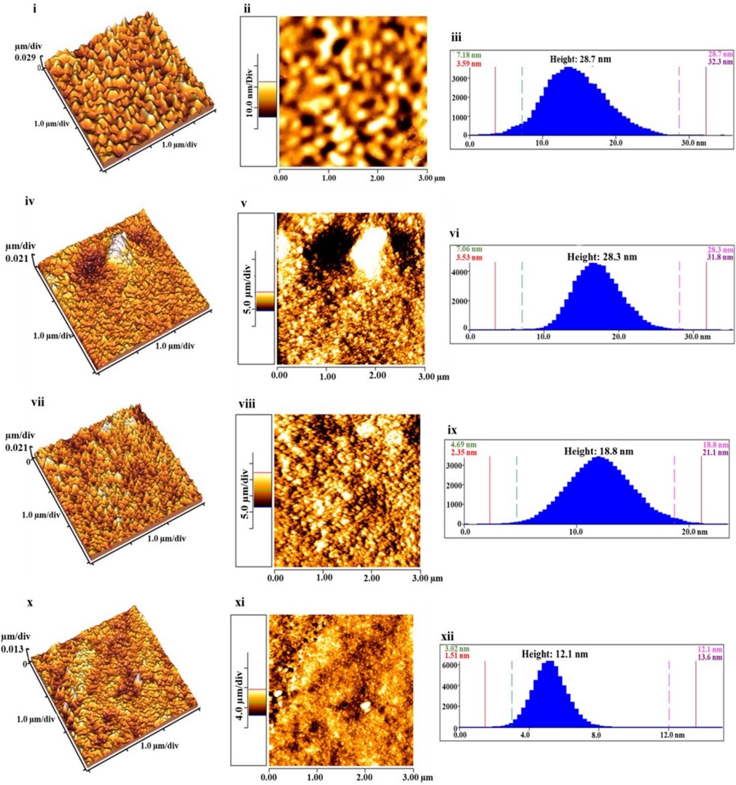
Background Mesenchymal stem cells (MSCs)-based treatment strategy has shown promise in bolstering the healing process of chronic wounds in diabetic patients, who are at risk of amputation and mortality. To overcome the drawbacks of suboptimal cell retention and diminished cell viability at the injury site, a novel nanofibrous biomaterial-based scaffold was developed by using a controlled extrusion of a polymeric solution to deliver the cells (human adipose-derived MSCs (ADMSCs) and placenta-derived MSCs (PLMSCs)) locally to the animal model of diabetic ulcers. Methods The physicochemical and biological properties of the nano-bioscaffold were characterized in terms of microscopic images, FTIR spectroscopy, tensile testing, degradation and swelling tests, contact angle measurements, MTT assay, and cell attachment evaluation. To evaluate the therapeutic efficacy, a study using an excisional wound model was conducted on diabetic rats. Results The SEM and AFM images of scaffolds revealed a network of uniform nanofibers with narrow diameters between 100-130 nm and surface roughness less than 5 nm, respectively. ADMSCs and PLMSCs had a typical spindle-shaped or fibroblast-like morphology when attached to the scaffold. Desired characteristics in terms of swelling, hydrophilicity, biodegradation rate, and biocompatibility were achieved with the CS70 formulation. The wound healing process was accelerated according to wound closure rate assay upon treatment with MSCs loaded scaffold resulting in increased re-epithelialization, neovascularization, and less inflammatory reaction. Our findings unequivocally demonstrated that the cell-loaded nano-bioscaffold exhibited more efficacy compared with its acellular counterpart. In summation, our study underscores the potential of this innovative cellular scaffold as a viable solution for enhancing the healing of diabetic ulcers. Conclusion The utilization of MSCs in a nanofibrous biomaterial framework demonstrates significant promise, providing a novel avenue for advancing wound care and diabetic ulcer management.

Read Full Abstract10.1186/s13287-024-03772-7
Amelioration of diabetic nephropathy in mice by a single intravenous injection of human mesenchymal stromal cells at early and later disease stages is associated with restoration of autophagyGraphical AbstractVerified
Stem Cell Research & Therapy

Amelioration of diabetic nephropathy in mice by a single intravenous injection of human mesenchymal stromal cells at early and later disease stages is associated with restoration of autophagy

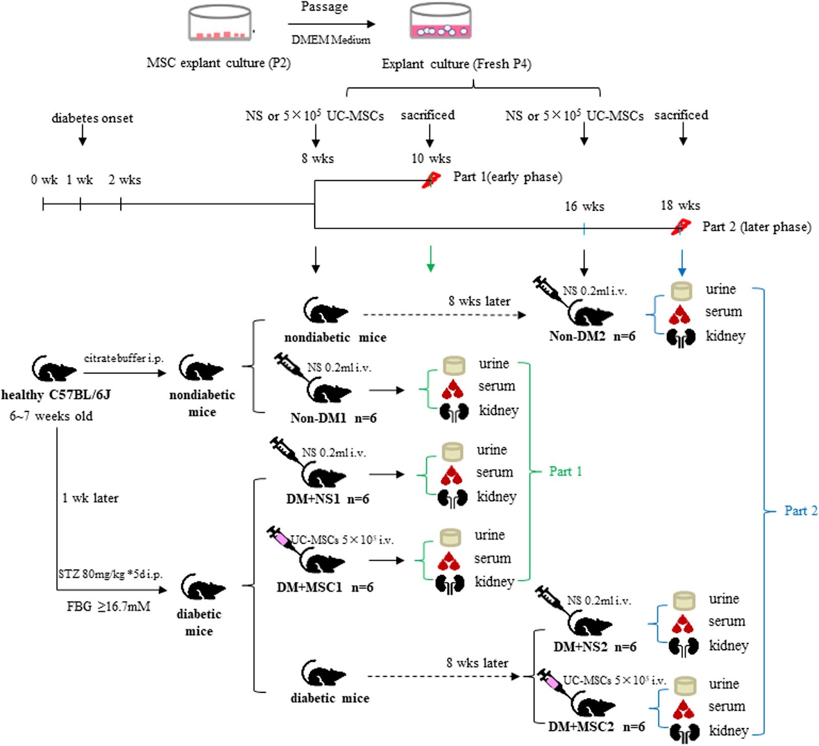
Background and aims Mesenchymal stromal cells (MSCs) a potentially effective disease-modulating therapy for diabetic nephropathy (DN) but their clinical translation has been hampered by incomplete understanding of the optimal timing of administration and in vivo mechanisms of action. This study aimed to elucidate the reno-protective potency and associated mechanisms of single intravenous injections of human umbilical cord-derived MSCs (hUC-MSCs) following shorter and longer durations of diabetes. Methods A streptozotocin (STZ)-induced model of diabetes and DN was established in C57BL/6 mice. In groups of diabetic animals, human (h)UC-MSCs or vehicle were injected intravenously at 8 or 16 weeks after STZ along with vehicle-injected non-diabetic animals. Diabetes-related kidney abnormalities was analyzed 2 weeks later by urine and serum biochemical assays, histology, transmission electron microscopy and immunohistochemistry. Serum concentrations of pro-inflammatory and pro-fibrotic cytokines were quantified by ELISA. The expression of autophagy-related proteins within the renal cortices was investigated by immunoblotting. Bio-distribution of hUC-MSCs in kidney and other organs was evaluated in diabetic mice by injection of fluorescent-labelled cells. Results Compared to non-diabetic controls, diabetic mice had increases in urine albumin creatinine ratio (uACR), mesangial matrix deposition, podocyte foot process effacement, glomerular basement membrane thickening and interstitial fibrosis as well as reduced podocyte numbers at both 10 and 18 weeks after STZ. Early (8 weeks) hUC-MSC injection was associated with reduced uACR and improvements in multiple glomerular and renal interstitial abnormalities as well as reduced serum IL-6, TNF-α, and TGF-β1 compared to vehicle-injected animals. Later (16 weeks) hUC-MSC injection also resulted in reduction of diabetes-associated renal abnormalities and serum TGF-β1 but not of serum IL-6 and TNF-α. At both time-points, the kidneys of vehicle-injected diabetic mice had higher ratio of p-mTOR to mTOR, increased abundance of p62, lower abundance of ULK1 and Atg12, and reduced ratio of LC3B to LC3A compared to non-diabetic animals, consistent with diabetes-associated suppression of autophagy. These changes were largely reversed in the kidneys of hUC-MSC-injected mice. In contrast, neither early nor later hUC-MSC injection had effects on blood glucose and body weight of diabetic animals. Small numbers of CM-Dil-labeled hUC-MSCs remained detectable in kidneys, lungs and liver of diabetic mice at 14 days after intravenous injection. Conclusions Single intravenous injections of hUC-MSCs ameliorated glomerular abnormalities and interstitial fibrosis in a mouse model of STZ-induced diabetes without affecting hyperglycemia, whether administered at relatively short or longer duration of diabetes. At both time-points, the reno-protective effects of hUC-MSCs were associated with reduced circulating TGF-β1 and restoration of intra-renal autophagy.

Read Full Abstract10.1186/s13287-024-03647-x
Effects of xenogeneic transplantation of umbilical cord-derived mesenchymal stem cells combined with irbesartan on renal podocyte damage in diabetic ratsGraphical AbstractVerified
Stem Cell Research & Therapy

Effects of xenogeneic transplantation of umbilical cord-derived mesenchymal stem cells combined with irbesartan on renal podocyte damage in diabetic rats

Background The leading cause of end-stage renal disease (ESRD) is diabetic nephropathy (DN). Podocyte damage is an early event in the development of DN. Currently, there is no effective treatment strategy that can slow the progression of DN or reverse its onset. The role of mesenchymal stem cells (MSCs) transplantation in diabetes and its complications has been extensively studied, and diabetic nephropathy has been a major focus. Irbesartan exerts reno-protective effects independent of lowering blood pressure, can reduce the incidence of proteinuria in rats, and is widely used clinically. However, it remains undetermined whether the combined utilization of the angiotensin II receptor antagonist irbesartan and MSCs could enhance efficacy in addressing DN. Methods A commonly used method for modeling type 2 diabetic nephropathy (T2DN) was established using a high-fat diet and a single low-dose injection of STZ (35 mg/kg). The animals were divided into the following 5 groups: (1) the control group (CON), (2) the diabetic nephropathy group (DN), (3) the mesenchymal stem cells treatment group (MSCs), (4) the irbesartan treatment group (Irb), and (5) the combined administration group (MSC+Irb). MSCs (2Ɨ10^6 cells/rat) were injected every 10 days through the tail vein for a total of three injections; irbesartan (30 mg/kg/d) was administered by gavage. Additionally, the safety and homing of mesenchymal stem cells were verified using positron emission tomography (PET) imaging. Results The combination treatment significantly reduced the UACR, kidney index, IGPTT, HOMA-IR, BUN, serum creatine, and related inflammatory factor levels and significantly improved renal function parameters and the expression of proteins related to glomerular podocyte injury in rats. Moreover, MSCs can homing target to damaged kidneys. Conclusions Compared to the administration of MSCs or irbesartan alone, the combination of MSCs and irbesartan exerted better protective effects on glomerular podocyte injury, providing new ideas for the clinical application of mesenchymal stem cells.

Read Full Abstract10.1186/s13287-024-03844-8
Correction: Neural stem cells genetically-modified to express neprilysin reduce pathology in Alzheimer transgenic modelsGraphical AbstractVerified
Stem Cell Research & Therapy

Correction: Neural stem cells genetically-modified to express neprilysin reduce pathology in Alzheimer transgenic models

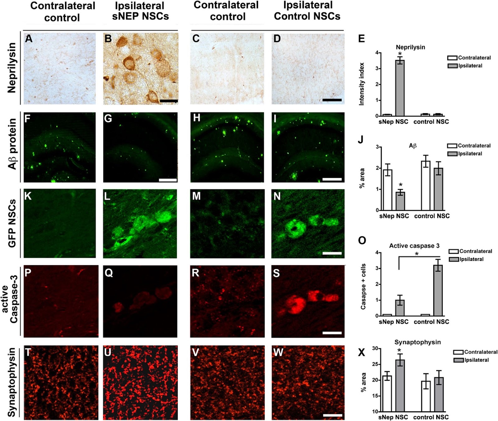
The original article contains an artifact in Fig. 6H that obscures the view of the lower-middle portion of the image. The correct original image for Fig. 6H can be viewed in this Correction article.

Read Full Abstract10.1186/s13287-024-03702-7
Comparative effectiveness of mesenchymal stem cell versus bone-marrow mononuclear cell transplantation in heart failure: a meta-analysis of randomized controlled trialsGraphical AbstractVerified
Stem Cell Research & Therapy

Comparative effectiveness of mesenchymal stem cell versus bone-marrow mononuclear cell transplantation in heart failure: a meta-analysis of randomized controlled trials

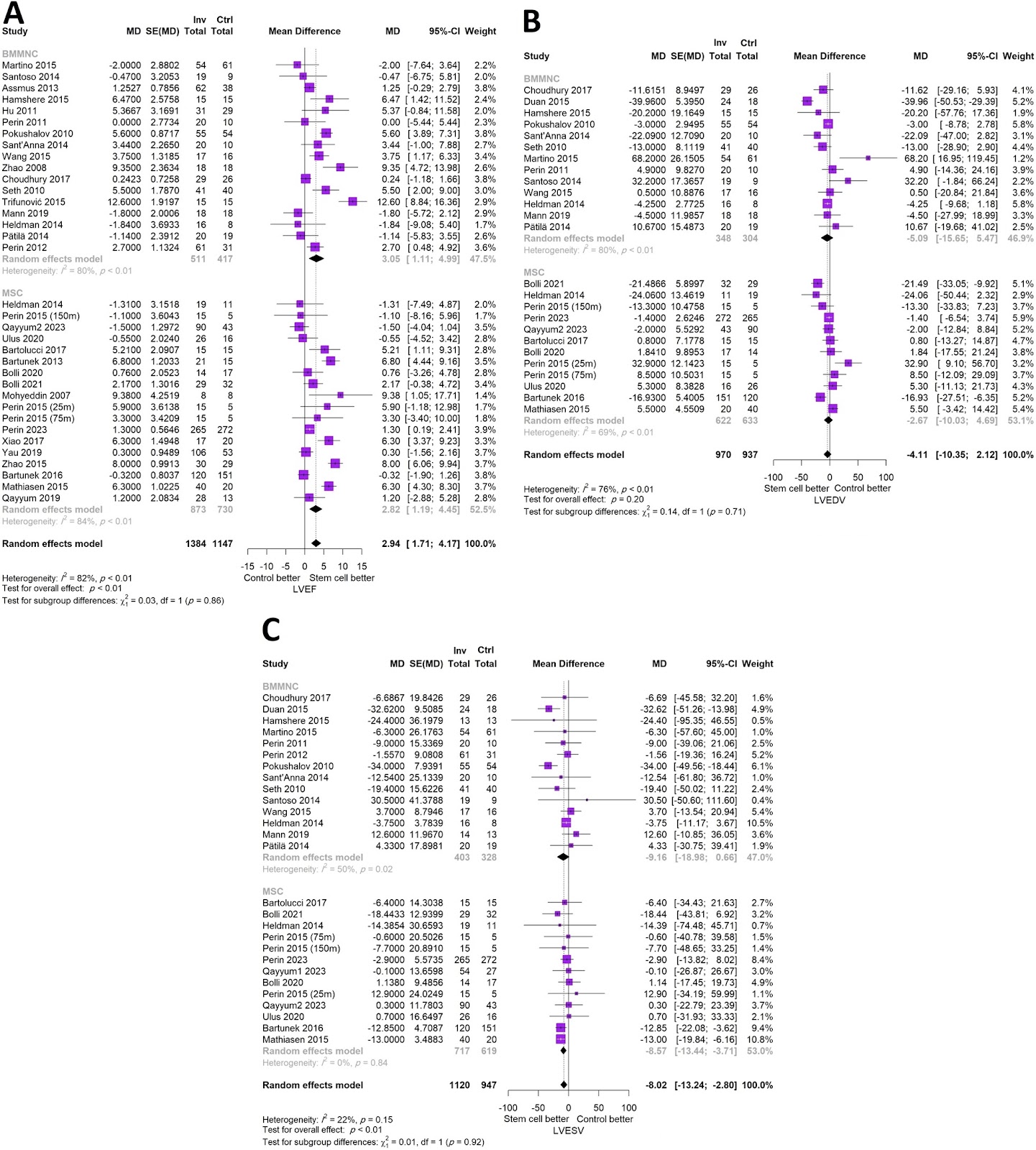
Background There is no clear evidence on the comparative effectiveness of bone-marrow mononuclear cell (BMMNC) vs. mesenchymal stromal cell (MSC) stem cell therapy in patients with chronic heart failure (HF). Methods Using a systematic approach, eligible randomized controlled trials (RCTs) of stem cell therapy (BMMNCs or MSCs) in patients with HF were retrieved to perform a meta-analysis on clinical outcomes (major adverse cardiovascular events (MACE), hospitalization for HF, and mortality) and echocardiographic indices (including left ventricular ejection fraction (LVEF)) were performed using the random-effects model. A risk ratio (RR) or mean difference (MD) with corresponding 95% confidence interval (CI) were pooled based on the type of the outcome and subgroup analysis was performed to evaluate the potential differences between the types of cells. Results The analysis included a total of 36 RCTs (1549 HF patients receiving stem cells and 1252 patients in the control group). Transplantation of both types of cells in patients with HF resulted in a significant improvement in LVEF (BMMNCs: MD (95% CI) = 3.05 (1.11; 4.99) and MSCs: MD (95% CI) = 2.82 (1.19; 4.45), between-subgroup p = 0.86). Stem cell therapy did not lead to a significant change in the risk of MACE (MD (95% CI) = 0.83 (0.67; 1.06), BMMNCs: RR (95% CI) = 0.59 (0.31; 1.13) and MSCs: RR (95% CI) = 0.91 (0.70; 1.19), between-subgroup p = 0.12). There was a marginally decreased risk of all-cause death (MD (95% CI) = 0.82 (0.68; 0.99)) and rehospitalization (MD (95% CI) = 0.77 (0.61; 0.98)) with no difference among the cell types (p > 0.05). Conclusion Both types of stem cells are effective in improving LVEF in patients with heart failure without any noticeable difference between the cells. Transplantation of the stem cells could not decrease the risk of major adverse cardiovascular events compared with controls. Future trials should primarily focus on the impact of stem cell transplantation on clinical outcomes of HF patients to verify or refute the findings of this study.

Read Full Abstract10.1186/s13287-024-03829-7
Comparative analysis of regulations and studies on stem cell therapies: focusing on induced pluripotent stem cell (iPSC)-based treatmentsGraphical AbstractVerified
Stem Cell Research & Therapy

Comparative analysis of regulations and studies on stem cell therapies: focusing on induced pluripotent stem cell (iPSC)-based treatments

Stem cell therapies have emerged as a promising approach in regenerative medicine, demonstrating potential in personalized medicine, disease modeling, and drug discovery. Therapies based on induced pluripotent stem cells (iPSCs) particularly stand out for their ability to differentiate into various cell types while avoiding ethical concerns. However, the development and application of these therapies are influenced by varying regulatory frameworks across countries. This study provides a comparative analysis of regulations and research on stem cell therapies in key regions: The European Union (EU), Switzerland, South Korea, Japan, and the United States. First, the study reviews the regulatory frameworks on stem cell therapies. The EU and Switzerland maintain rigorous guidelines that prioritize safety and ethical considerations, which can hinder innovation. In contrast, the United States adopts a more flexible regulatory stance, facilitating the rapid development of stem cell therapies. South Korea and Japan take a balanced approach by incorporating practices from both regimes. These regulatory differences reflect each country’s unique priorities and impact the pace and scope of stem cell therapy development. Moreover, the study examines global trends in clinical trials on stem cell treatments based on data obtained from two sources: ClinicalTrials.gov and ICTRP. Findings indicate a significant growth in the number of clinical trials since 2008, particularly in that involving iPSCs. Therapeutic studies involving iPSCs predominantly target conditions affecting the cardiovascular and nervous systems which are considered vital. The results put emphasis on the safety of stem cell treatments. Meanwhile, the number of such trials also varies by country. The United States and Japan, where relatively flexible guidelines on stem cell research are adopted, are in a leading position. However, countries in the EU fall behind with rigorous regulations imposed. This reflects the need for more flexible regulatory guidance for active development of stem cell therapies. The findings underscore the importance of legal frameworks in facilitating innovation while ensuring safety. Regulatory agencies in different countries should collaborate to achieve a balanced global standard to ensure the safe and efficient advancement of stem cell therapies. Global regulatory convergence will promote international collaboration in research and the applicability of new treatments.

Read Full Abstract10.1186/s13287-024-04065-9