🧬 SinoBioData Academic Portal
šŸ“š Peer-Reviewed Translated Literature

All Biomedical & Clinical Articles (Page 43)

Browse complete peer-reviewed translations from top Chinese biomedical, oncology, and genomics journals. Read verified previews and download full authentic clinical reports.

Published Research Papers

Showing 24 of 1542 peer-reviewed translated articles (Page 43 of 65)

Mesenchymal stromal/stem cells promote intestinal epithelium regeneration after chemotherapy-induced damageGraphical AbstractVerified
Stem Cell Research & Therapy

Mesenchymal stromal/stem cells promote intestinal epithelium regeneration after chemotherapy-induced damage

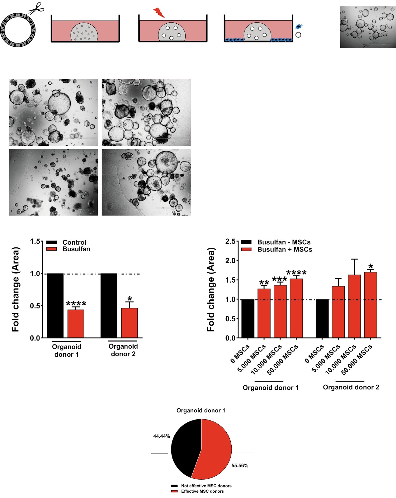
Background Allogeneic hematopoietic stem cell transplantation (HSCT) is a curative treatment for leukemia and a range of non-malignant disorders. The success of the therapy is hampered by occurrence of acute graft-versus-host disease (aGvHD); an inflammatory response damaging recipient organs, with gut, liver, and skin being the most susceptible. Intestinal GvHD injury is often a life-threatening complication in patients unresponsive to steroid treatment. Allogeneic mesenchymal stromal/stem cell (MSC) infusions are a promising potential treatment for steroid-resistant aGvHD. Data from our institution and others demonstrate rescue of approximately 40–50% of aGvHD patients with MSCs in Phase I, II studies and minor side effects. Although promising, better understanding of MSC mode of action and patient response to MSC-based therapy is essential to improve this lifesaving treatment. Methods Single cell human small intestine organoids were embedded in Matrigel, grown for 5 days and treated with busulfan for 48 h. Organoids damaged by treatment with busulfan or control organoids were co-cultured with 5000, 10,000, and 50,000 MSCs for 24 h, 48 h or 7 days and the analyses such as surface area determination, proliferation and apoptosis assessment, RNA sequencing and proteomics were performed. Results Here, we developed a 3D co-culture model of human small intestinal organoids and MSCs, which allows to study the regenerative effects of MSCs on intestinal epithelium in a more physiologically relevant setting than existing in vitro systems. Using this model we mimicked chemotherapy-mediated damage of the intestinal epithelium. The treatment with busulfan, the chemotherapeutic commonly used as conditioning regiment before the HSCT, affected pathways regulating epithelial to mesenchymal transition, proliferation, and apoptosis in small intestinal organoids, as shown by transcriptomic and proteomic analysis. The co-culture of busulfan-treated intestinal organoids with MSCs reversed the effects of busulfan on the transcriptome and proteome of intestinal epithelium, which we also confirmed by functional evaluation of proliferation and apoptosis. Conclusions Collectively, we demonstrate that our in vitro co-culture system is a new valuable tool to facilitate the investigation of the molecular mechanisms behind the therapeutic effects of MSCs on damaged intestinal epithelium. This could benefit further optimization of the use of MSCs in HSCT patients.

Read Full Abstract10.1186/s13287-024-03738-9
Adipose-derived stem cells in immune-related skin disease: a review of current research and underlying mechanismsGraphical AbstractVerified
Stem Cell Research & Therapy

Adipose-derived stem cells in immune-related skin disease: a review of current research and underlying mechanisms

Adipose-derived stem cells (ASCs) are a critical adult stem cell subpopulation and are widely utilized in the fields of regenerative medicine and stem cell research due to their abundance, ease of harvest, and low immunogenicity. ASCs, which are homologous with skin by nature, can treat immune-related skin diseases by promoting skin regeneration and conferring immunosuppressive effects, with the latter being the most important therapeutic mechanism. ASCs regulate the immune response by direct cell–cell communication with immune cells, such as T cells, macrophages, and B cells. In addition to cell–cell interactions, ASCs modulate the immune response indirectly by secreting cytokines, interleukins, growth factors, and extracellular vesicles. The immunomodulatory effects of ASCs have been exploited to treat many immune-related skin diseases with good therapeutic outcomes. This article reviews the mechanisms underlying the immunomodulatory effects of ASCs, as well as progress in research on immune-related skin diseases.

Read Full Abstract10.1186/s13287-023-03561-8
The nuclei of human adult stem cells can move within the cell and generate cellular protrusions to contact other cellsGraphical AbstractVerified
Stem Cell Research & Therapy

The nuclei of human adult stem cells can move within the cell and generate cellular protrusions to contact other cells

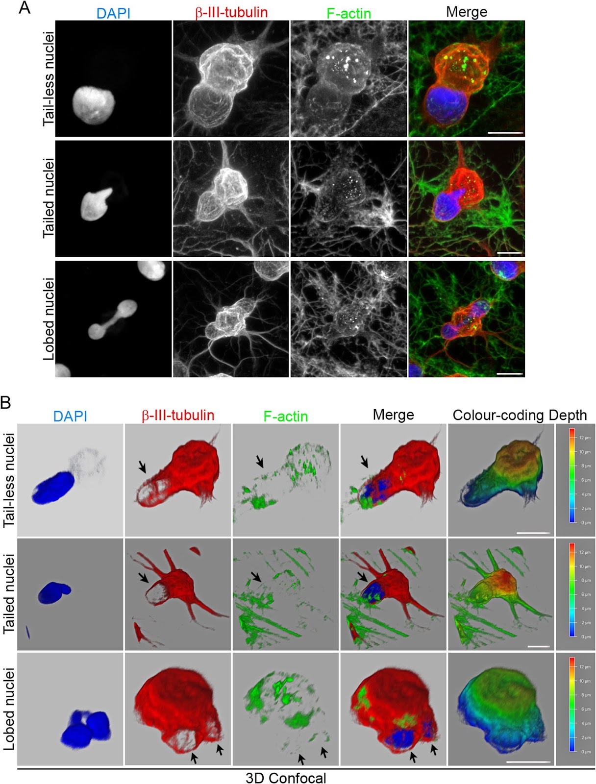
Background The neuronal transdifferentiation of adult bone marrow cells (BMCs) is still considered an artifact based on an alternative explanation of experimental results supporting this phenomenon obtained over decades. However, recent studies have shown that following neural induction, BMCs enter an intermediate cellular state before adopting neural-like morphologies by active neurite extension and that binucleated BMCs can be formed independent of any cell fusion events. These findings provide evidence to reject the idea that BMC neural transdifferentiation is merely an experimental artifact. Therefore, understanding the intermediate states that cells pass through during transdifferentiation is crucial given their potential application in regenerative medicine and disease modelling. Methods In this study, we examined the functional significance of the variety of morphologies and positioning that cell nuclei of human bone marrow-derived mesenchymal stem cells (hBM-MSCs) can adopt during neural-like differentiation using live-cell nuclear fluorescence labelling, time-lapse microscopy, and confocal microscopy analysis. Results Here, we showed that after neural induction, hBM-MSCs enter an intermediate cellular state in which the nuclei are able to move within the cells, switching shapes and positioning and even generating cellular protrusions as they attempt to contact the cells around them. These findings suggest that changes in nuclear positioning occur because human cell nuclei somehow sense their environment. In addition, we showed the process of direct interactions between cell nuclei, which opens the possibility of a new level of intercellular interaction. Conclusions The present study advances the understanding of the intermediate stage through which hBM-MSCs pass during neural transdifferentiation, which may be crucial to understanding the mechanisms of these cell conversion processes and eventually harness them for use in regenerative medicine. Importantly, our study provides for the first time evidence that the nuclei of hBM-MSC-derived intermediate cells somehow sense their environment, generating cellular protrusions to contact other cells. In summary, human mesenchymal stromal cells could not only help to increase our understanding of the mechanisms underlying cellular plasticity but also facilitate the exact significance of nuclear positioning in cellular function and in tissue physiology.

Read Full Abstract10.1186/s13287-024-03638-y
PLOD2, a key factor for MRL MSC metabolism and chondroprotective propertiesGraphical AbstractVerified
Stem Cell Research & Therapy

PLOD2, a key factor for MRL MSC metabolism and chondroprotective properties

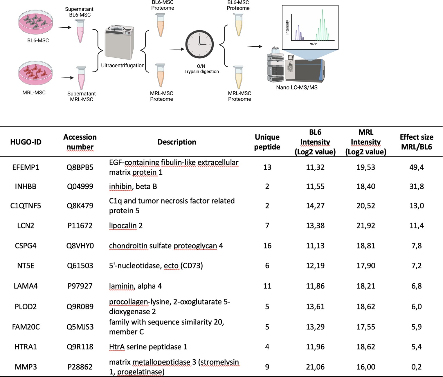
Background Initially discovered for its ability to regenerate ear holes, the Murphy Roth Large (MRL) mouse has been the subject of multiple research studies aimed at evaluating its ability to regenerate other body tissues and at deciphering the mechanisms underlying it. These enhanced abilities to regenerate, retained during adulthood, protect the MRL mouse from degenerative diseases such as osteoarthritis (OA). Here, we hypothesized that mesenchymal stromal/stem cells (MSC) derived from the regenerative MRL mouse could be involved in their regenerative potential through the release of pro-regenerative mediators. Method To address this hypothesis, we compared the secretome of MRL and BL6 MSC and identified several candidate molecules expressed at significantly higher levels by MRL MSC than by BL6 MSC. We selected one candidate, Plod2, and performed functional in vitro assays to evaluate its role on MRL MSC properties including metabolic profile, migration, and chondroprotective effects. To assess its contribution to MRL protection against OA, we used an experimental model for osteoarthritis induced by collagenase (CiOA). Results Among the candidate molecules highly expressed by MRL MSC, we focused our attention on procollagen-lysine,2-oxoglutarate 5-dioxygenase 2 (PLOD2). Plod2 silencing induced a decrease in the glycolytic function of MRL MSC, resulting in the alteration of their migratory and chondroprotective abilities in vitro. In vivo, we showed that Plod2 silencing in MRL MSC significantly impaired their capacity to protect mouse from developing OA. Conclusion Our results demonstrate that the chondroprotective and therapeutic properties of MRL MSC in the CiOA experimental model are in part mediated by PLOD2.

Read Full Abstract10.1186/s13287-024-03650-2
Retraction Note: Secretome of endothelial progenitor cells from stroke patients promotes endothelial barrier tightness and protects against hypoxia-induced vascular leakageGraphical AbstractVerified
Stem Cell Research & Therapy

Retraction Note: Secretome of endothelial progenitor cells from stroke patients promotes endothelial barrier tightness and protects against hypoxia-induced vascular leakage

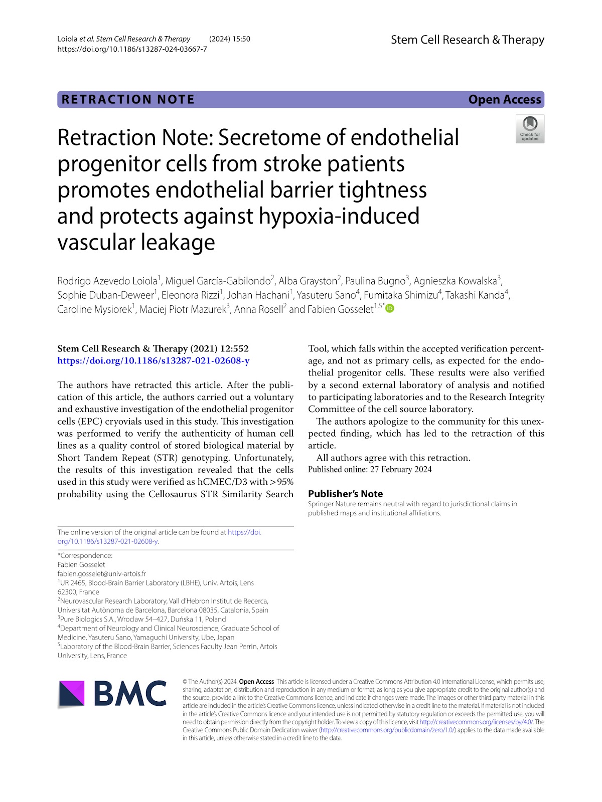
The authors have retracted this article. After the publication of this article, the authors carried out a voluntary and exhaustive investigation of the endothelial progenitor cells (EPC) cryovials used in this study. This investigation was performed to verify the authenticity of human cell lines as a quality control of stored biological material by Short Tandem Repeat (STR) genotyping. Unfortunately, the results of this investigation revealed that the cells used in this study were verified as hCMEC/D3 with >95% probability using the Cellosaurus STR Similarity Search Tool, which falls within the accepted verification percentage, and not as primary cells, as expected for the endothelial progenitor cells. These results were also verified by a second external laboratory of analysis and notified to participating laboratories and to the Research Integrity Committee of the cell source laboratory. The authors apologize to the community for this unexpected finding, which has led to the retraction of this article. All authors agree with this retraction.

Read Full Abstract10.1186/s13287-024-03667-7
Connexin 25 maintains self-renewal and functions of airway basal cells for airway regenerationGraphical AbstractVerified
Stem Cell Research & Therapy

Connexin 25 maintains self-renewal and functions of airway basal cells for airway regeneration

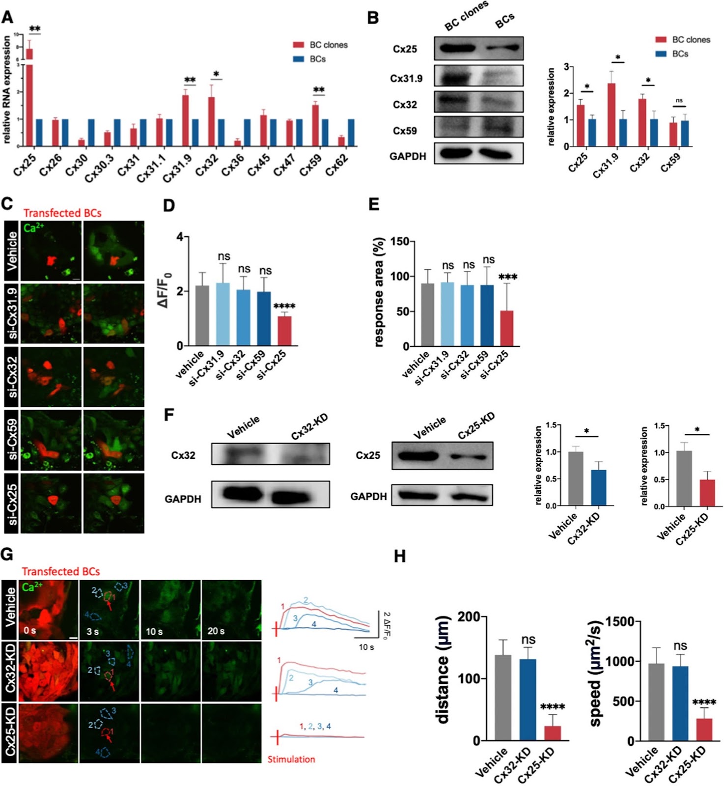
Background The formation of stem cell clones enables close contact of stem cells inside. The gap junctions in such clone spheres establish a microenvironment that allows frequent intercellular communication to maintain self-renewal and functions of stem cells. Nevertheless, the essential gap junction protein for molecular signaling in clones is poorly known. Methods Primary human airway basal cells (hBCs) were isolated from brushing samples through bronchoscopy and then cultured. A tightly focused femtosecond laser was used to excite the local Ca2+ in an individual cell to initiate an internal Ca2+ wave in a clone to screen gap junction proteins. Immunoflourescence staining and clonogenicity assay were used to evaluate self-renewal and functions. RNA and protein levels were assessed by PCR and Western blot. Air–liquid interface assay was conducted to evaluate the differentiation potential. A Naphthalene injury mouse model was used to assess the regeneration potential. Results Herein, we identify Connexin 25 (Cx25) dominates intercellular Ca2+ communications in clones of hBCs in vitro to maintain the self-renewal and pluripotency of them. The self-renewal and in vitro differentiation functions and in vivo regeneration potential of hBCs in an airway damage model are both regulated by Cx25. The abnormal expression of Cx25 is validated in several diseases including IPF, Covid-19 and bronchiectasis. Conclusion Cx25 is essential for hBC clones in maintaining self-renewal and functions of hBCs via gap junctions.

Read Full Abstract10.1186/s13287-024-03908-9
Exosome-mediated repair of spinal cord injury: a promising therapeutic strategyGraphical AbstractVerified
Stem Cell Research & Therapy

Exosome-mediated repair of spinal cord injury: a promising therapeutic strategy

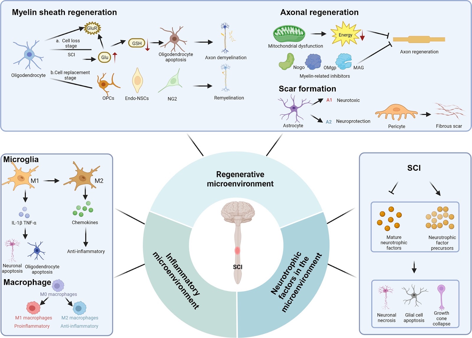
Spinal cord injury (SCI) is a catastrophic injury to the central nervous system (CNS) that can lead to sensory and motor dysfunction, which seriously affects patients’ quality of life and imposes a major economic burden on society. The pathological process of SCI is divided into primary and secondary injury, and secondary injury is a cascade of amplified responses triggered by the primary injury. Due to the complexity of the pathological mechanisms of SCI, there is no clear and effective treatment strategy in clinical practice. Exosomes, which are extracellular vesicles of endoplasmic origin with a diameter of 30–150 nm, play a critical role in intercellular communication and have become an ideal vehicle for drug delivery. A growing body of evidence suggests that exosomes have great potential for repairing SCI. In this review, we introduce exosome preparation, functions, and administration routes. In addition, we summarize the effect and mechanism by which various exosomes repair SCI and review the efficacy of exosomes in combination with other strategies to repair SCI. Finally, the challenges and prospects of the use of exosomes to repair SCI are described.

Read Full Abstract10.1186/s13287-023-03614-y
Long term outcomes of intracarotid arterial transfusion of circulatory-derived autologous CD34+ cells for acute ischemic stroke patients—A randomized, open-label, controlled phase II clinical trialGraphical AbstractVerified
Stem Cell Research & Therapy

Long term outcomes of intracarotid arterial transfusion of circulatory-derived autologous CD34+ cells for acute ischemic stroke patients—A randomized, open-label, controlled phase II clinical trial

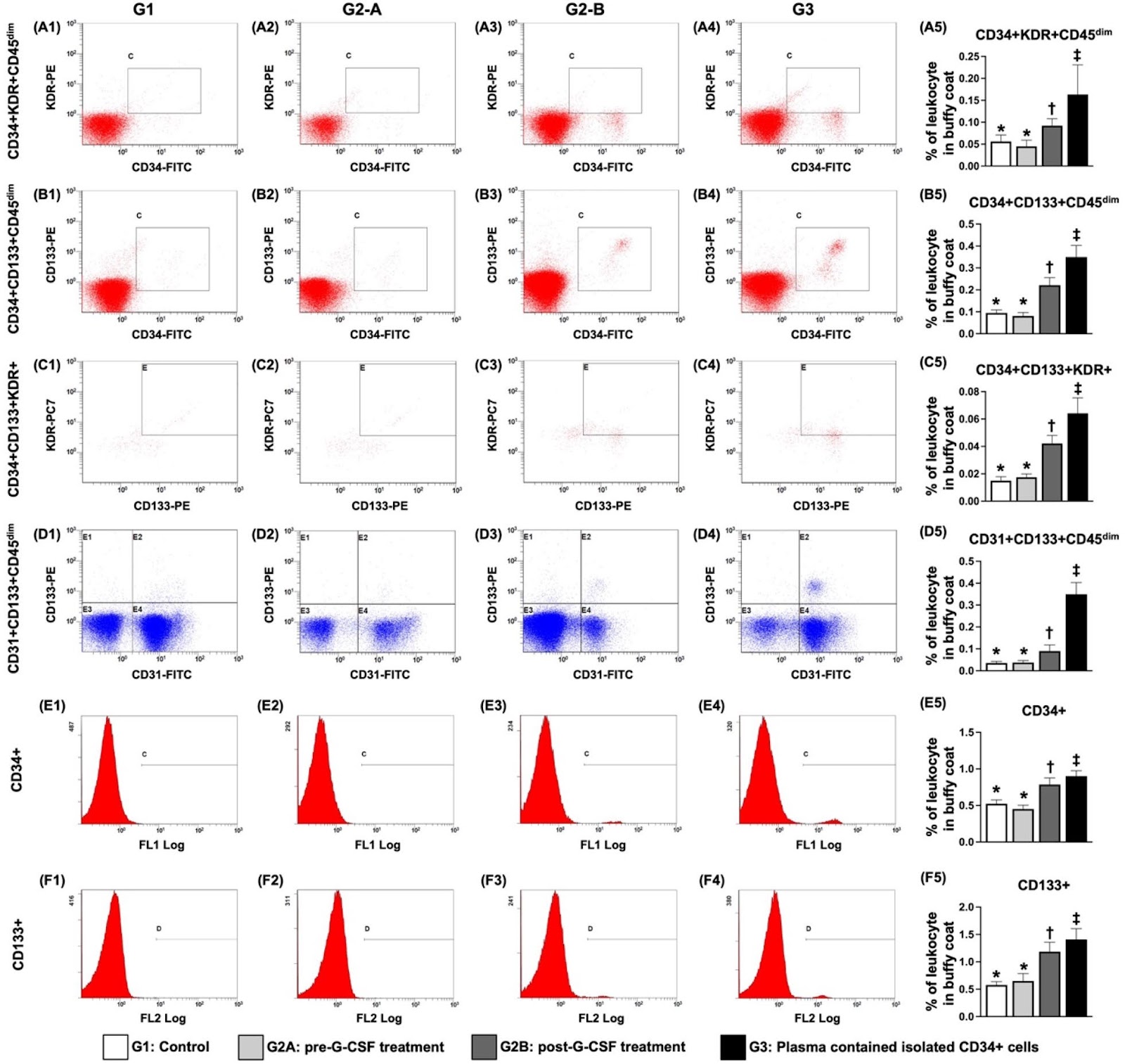
Background This phase II randomized controlled trial tested whether the intracarotid arterial administration (ICAA) of autologous CD34+ cells to patients within 14±7 days after acute ischemic stroke (IS) could be safe and further improve short- and long-term outcomes. Methods Between January 2018 and March 2022, 28 consecutive patients were equally randomly allocated to the cell-treated group (CD34+ cells/3.0Ɨ107/patient) or the control group (receiving optimal medical therapy). CD34+ cells were transfused into the ipsilateral brain infarct zone of cell-treated patients via the ICAA in the catheterization room. Results The results demonstrated 100% safety and success rates for the procedure, and no long-term tumorigenesis was observed in cell-treated patients. In cell-treated patients, the angiogenesis capacity of circulating endothelial progenitor cells (EPCs)/Matrigel was significantly greater after treatment than before treatment with granulocyte colony-stimulating factor (all p<0.001). Blood samples from the right internal jugular vein of the cell-treated patients presented significantly greater levels of the stromal cell-derived factor 1α/EPC at 5, 10 and 30 min compared with 0 min (all p<0.005). The National Institute of Health Stroke Scale scores were similar upon presentation, but a greater response was observed by Days 30 and 90 in the cell-treated group than in the control group. Tc-99 m brain perfusion was significantly greater at 180 days in the cell-treated group than in the control group (p=0.046). The combined long-term end points (defined as death/recurrent stroke/or severe disability) were notably lower in the control group compared with the cell-treated group (14.3% vs. 50.0%, p=0.103). Conclusion Intracarotid transfusion of autologous CD34+ cells is safe and might improve long-term outcomes in patients with acute IS. Trial registration ISRCTN, ISRCTN15677760. Registered 23 April 2018- Retrospectively registered, https://doi.org/10.1186/ISRCTN15677760

Read Full Abstract10.1186/s13287-024-04021-7
Retinoic acid modulation guides human-induced pluripotent stem cell differentiation towards left or right ventricle-like cardiomyocytesGraphical AbstractVerified
Stem Cell Research & Therapy

Retinoic acid modulation guides human-induced pluripotent stem cell differentiation towards left or right ventricle-like cardiomyocytes

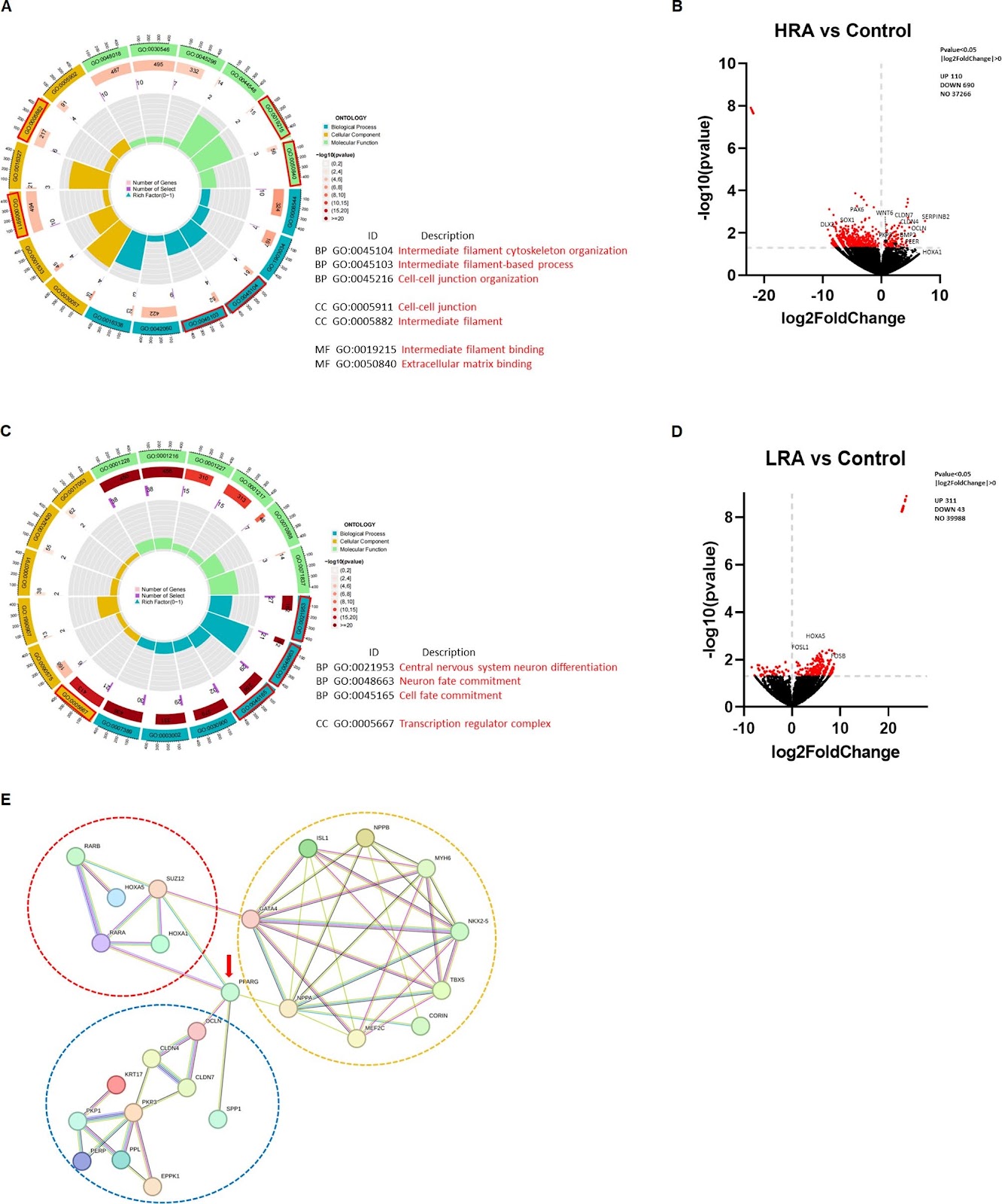
Background Cardiomyocytes (CMs) derived from human induced pluripotent stem cells (hiPSCs) by traditional methods are a mix of atrial and ventricular CMs and many other non-cardiomyocyte cells. Retinoic acid (RA) plays an important role in regulation of the spatiotemporal development of the embryonic heart. Methods CMs were derived from hiPSC (hi-PCS-CM) using different concentrations of RA (Control without RA, LRA with 0.05μM and HRA with 0.1 μM) between day 3-6 of the differentiation process. Engineered heart tissues (EHTs) were generated by assembling hiPSC-CM at high cell density in a low collagen hydrogel. Results In the HRA group, hiPSC-CMs exhibited highest expression of contractile proteins MYH6, MYH7 and cTnT. The expression of TBX5, NKX2.5 and CORIN, which are marker genes for left ventricular CMs, was also the highest in the HRA group. In terms of EHT, the HRA group displayed the highest contraction force, the lowest beating frequency, and the highest sensitivity to hypoxia and isoprenaline, which means it was functionally more similar to the left ventricle. RNAsequencing revealed that the heightened contractility of EHT within the HRA group can be attributed to the promotion of augmented extracellular matrix strength by RA. Conclusion By interfering with the differentiation process of hiPSC with a specific concentration of RA at a specific time, we were able to successfully induce CMs and EHTs with a phenotype similar to that of the left ventricle or right ventricle.

Read Full Abstract10.1186/s13287-024-03741-0
NOD1 deficiency ameliorates the progression of diabetic retinopathy by modulating bone marrow–retina crosstalkGraphical AbstractVerified
Stem Cell Research & Therapy

NOD1 deficiency ameliorates the progression of diabetic retinopathy by modulating bone marrow–retina crosstalk

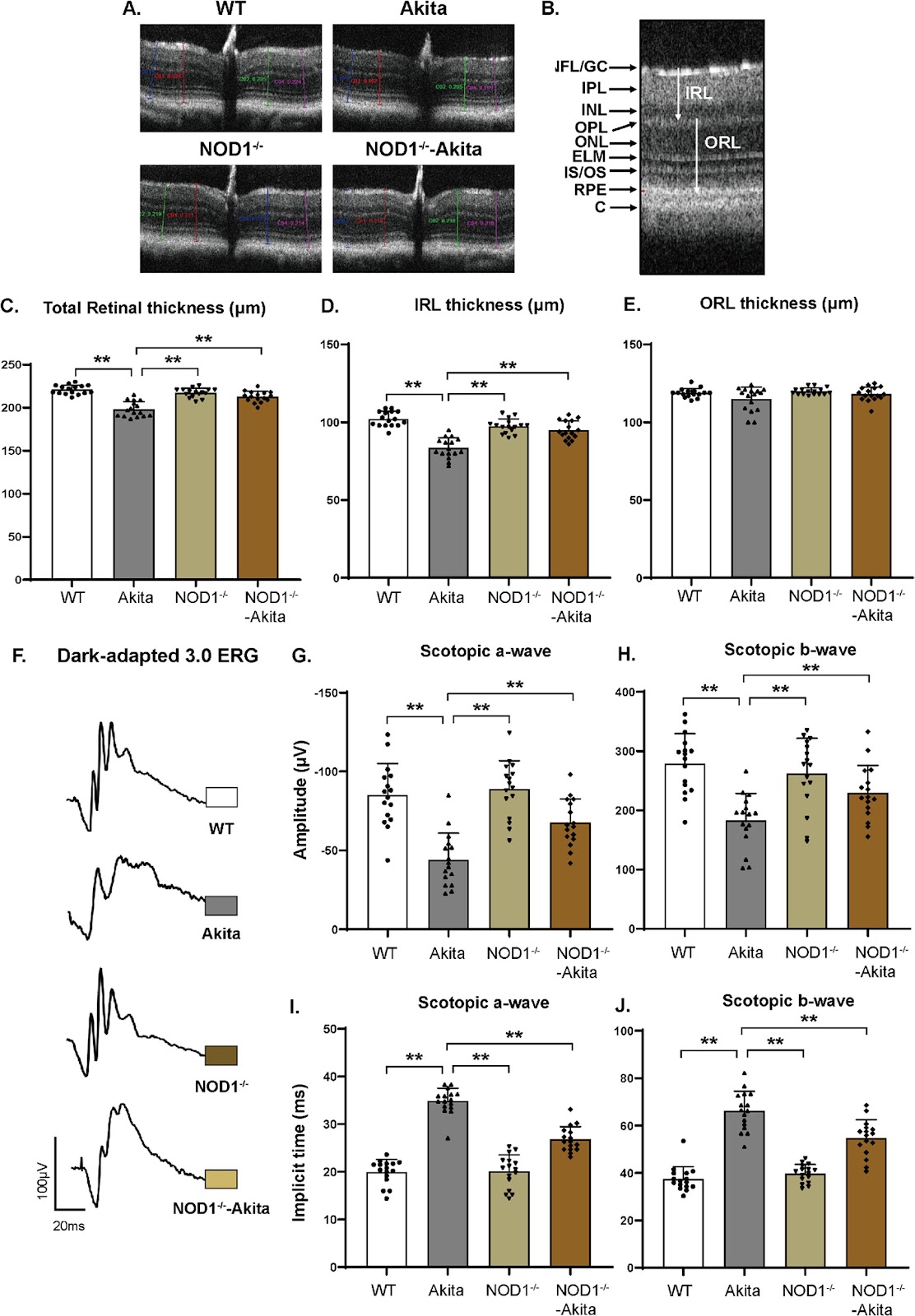
Background Nucleotide-binding oligomerization domain-containing protein 1 (NOD1) plays a pivotal role in inducing metabolic inflammation in diabetes. Additionally, the NOD1 ligand disrupts the equilibrium of bone marrow-derived hematopoietic stem/progenitor cells, a process that has immense significance in the development of diabetic retinopathy (DR). We hypothesized that NOD1 depletion impedes the advancement of DR by resolving bone marrow dysfunction. Methods We generated NOD1āˆ’/āˆ’-Akita double-mutant mice and chimeric mice with hematopoietic-specific NOD1 depletion to study the role of NOD1 in the bone marrow–retina axis. Results Elevated circulating NOD1 activators were observed in Akita mice after 6 months of diabetes. NOD1 depletion partially restored diabetes-induced structural changes and retinal electrical responses in NOD1āˆ’/āˆ’-Akita mice. Loss of NOD1 significantly ameliorated the progression of diabetic retinal vascular degeneration, as determined by acellular capillary quantification. The preventive effect of NOD1 depletion on DR is linked to bone marrow phenotype alterations, including a restored HSC pool and a shift in hematopoiesis toward myelopoiesis. We also generated chimeric mice with hematopoietic-specific NOD1 ablation, and the results further indicated that NOD1 had a protective effect against DR. Mechanistically, loss of hematopoietic NOD1 resulted in reduced bone marrow-derived macrophage infiltration and decreased CXCL1 and CXCL2 secretion within the retina, subsequently leading to diminished neutrophil chemoattraction and NETosis. Conclusions The results of our study unveil, for the first time, the critical role of NOD1 as a trigger for a hematopoietic imbalance toward myelopoiesis and local retinal inflammation, culminating in DR progression. Targeting NOD1 in bone marrow may be a potential strategy for the prevention and treatment of DR.

Read Full Abstract10.1186/s13287-024-03654-y
Evaluation of secretomes derived from human dermal and adipose tissue mesenchymal stem/stromal cells for skin wound healing: not as effective as cellsGraphical AbstractVerified
Stem Cell Research & Therapy

Evaluation of secretomes derived from human dermal and adipose tissue mesenchymal stem/stromal cells for skin wound healing: not as effective as cells

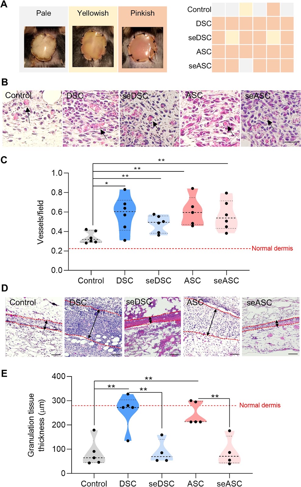
Background Although the paracrine effects of mesenchymal stem/stromal cells (MSCs) have been recognized as crucial mediators of their regenerative effects on tissue repair, the potential of MSC secretomes as effective substitutes for cellular therapies remains underexplored. Methods In this study, we compared MSCs from the human dermis (DSCs) and adipose tissue (ASCs) with their secretomes regarding their efficacy for skin wound healing using a translationally relevant murine model. Results Proteomic analysis revealed that while there was a substantial overlap in protein composition between DSC and ASC secretomes, specific proteins associated with wound healing and angiogenesis were differentially expressed. Despite a similar angiogenic potential in vivo, DSC and ASC secretomes were found to be less effective than cells in accelerating wound closure and promoting tissue remodeling. Conclusions Overall, secretome-treated groups showed intermediary results between cells- and control-treated (empty scaffold) groups. These findings highlight that although secretomes possess therapeutic potential, their efficacy might be limited compared to cellular therapies. This study contributes to the growing understanding of MSC secretomes, emphasizes the need for further protocol optimization, and offers insights into their potential applications in regenerative medicine.

Read Full Abstract10.1186/s13287-023-03630-y
Trehalose extricates impaired mitochondrial and autophagy dysregulation in patient iPSC-derived macular corneal dystrophy disease modelGraphical AbstractVerified
Stem Cell Research & Therapy

Trehalose extricates impaired mitochondrial and autophagy dysregulation in patient iPSC-derived macular corneal dystrophy disease model

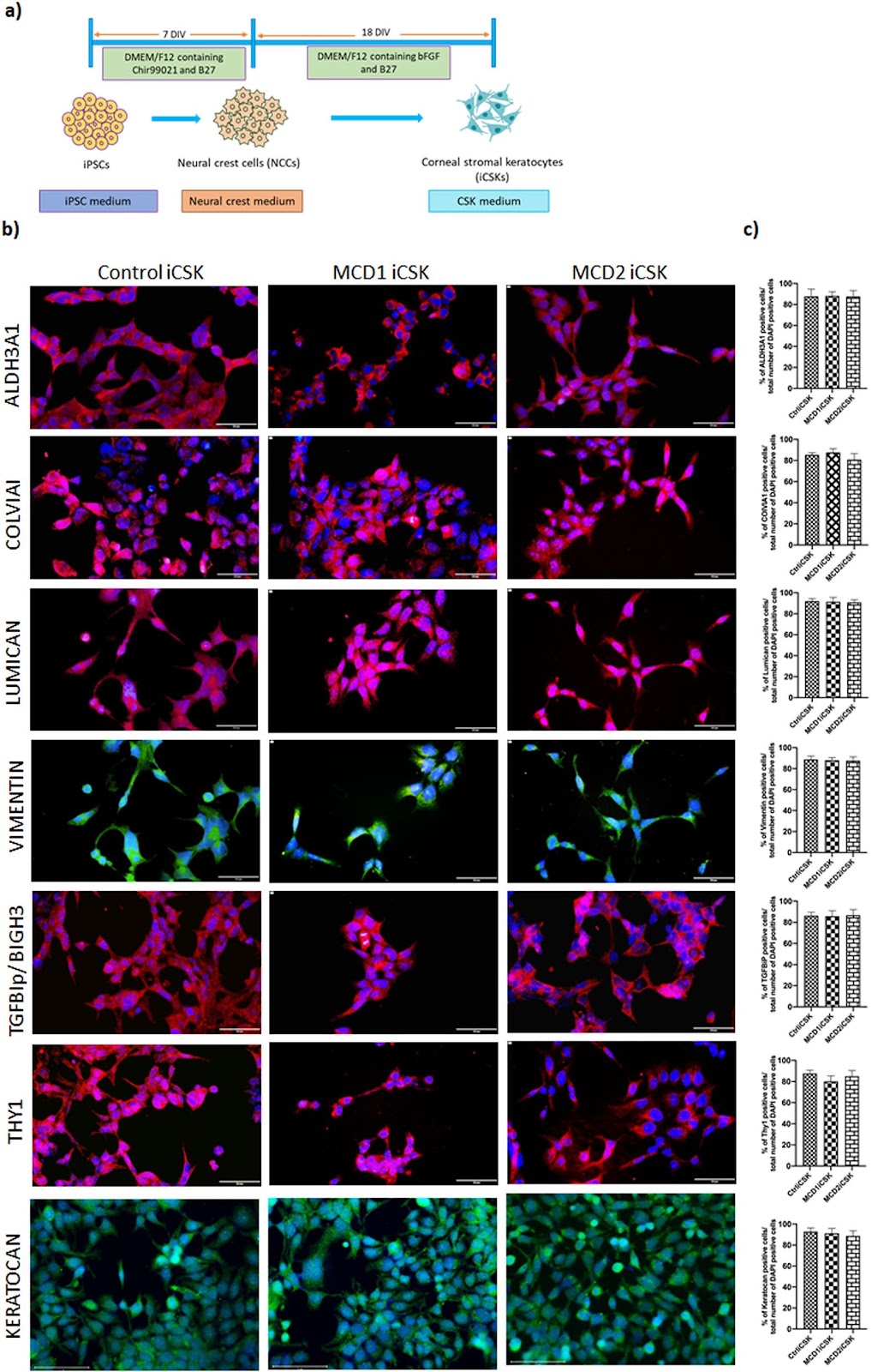
Background Patient-derived induced pluripotent stem cell (iPSCs) represents a powerful tool for elucidating the underlying disease mechanisms. Macular corneal dystrophy (MCD) is an intractable and progressive bilateral corneal disease affecting the corneal stroma due to mutation/s in carbohydrate sulfotransferase 6 (CHST6) gene. The underlying molecular mechanisms leading to MCD are unclear due to a lack of human contextual model and limited access to affected corneal stromal keratocytes (CSKs) from MCD patients. This has restricted the current treatment option for MCD to restorative corneal transplantation thereby lending itself to the use of iPSCs. Methods induced pluripotent stem cells (iPSCs) were generated from two MCD patients and a healthy participant by senai virus based reprogramming of the peripheral mononuclear blood cells (PBMCs). The iPSCs were characterized based on the expression of pluripotent markers and formation of embryoid bodies possessing tri-lineage potential. Directed differentiation of the iPSCs to corneal stromal keratocytes (CSKs) was done via intermediate induction of neural crest cells. The iCSKs were characterized by immunocytochemistry and qPCR. Proteostat staining of the iCSKs was done to validate the disease phenotype invitro. Expression of autophagy markers in the iCSKs and JC staining were visualized by immunochemistry and live-cell imaging in trehalose treated iCSKs. Results We show that the MCD iPSC-derived CSKs (MCDiCSKs) exhibits impaired autophagy assessed by the profiles of autophagy-associated proteins (LAMP1, LC3II/I, p62 and Beclin-1) and mitochondrial membrane potential. Significantly higher protein aggregates in MCDiCSKs was seen compared with the control, which could be rescued upon autophagy modulation. Hence, we treated MCD-iCSKs with trehalose (autophagy inducer) and showed that it protects MCD-iCSKs from mitochondrial dysfunction and maintains autophagic degradation. Conclusion Our study highlights the possible pathological mechanisms involved in MCD. We found trehalose ameliorate the impaired mitochondrial and autophagy dysregulation in patient iPSC-derived macular corneal dystrophy disease model, which could be a potential alternative for MCD management.

Read Full Abstract10.1186/s13287-024-04016-4
Improvement of multilineage hematopoiesis in hematopoietic stem cell-transferred c-kit mutant NOG-EXL humanized miceGraphical AbstractVerified
Stem Cell Research & Therapy

Improvement of multilineage hematopoiesis in hematopoietic stem cell-transferred c-kit mutant NOG-EXL humanized mice

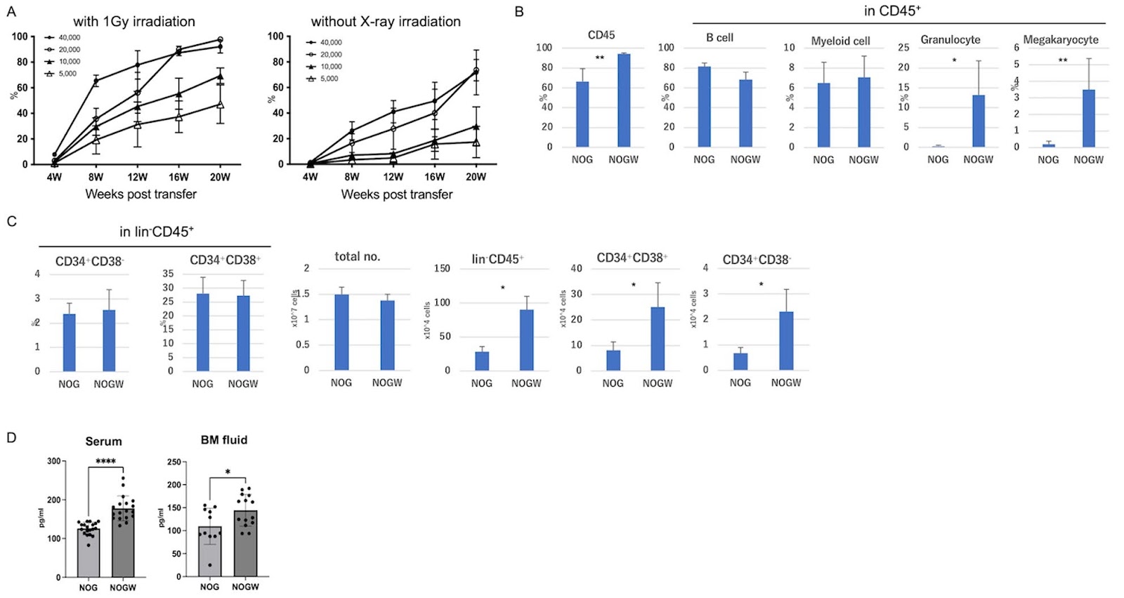
Human hematopoietic stem cell (HSC)-transferred humanized mice are valuable models for exploring human hematology and immunology. However, sufficient recapitulation of human hematopoiesis in mice requires large quantities of enriched human CD34+ HSCs and total-body irradiation for adequate engraftment. Recently, we generated a NOG mouse strain with a point mutation in the c-kit tyrosine kinase domain (W41 mutant; NOGW mice). In this study, we examined the ability of NOGW mice to reconstitute human hematopoietic cells. Irradiated NOGW mice exhibited high engraftment levels of human CD45+ cells in the peripheral blood, even when only 5,000–10,000 CD34+ HSCs were transferred. Efficient engraftment of human CD45+ cells was also observed in non-irradiated NOGW mice transferred with 20,000–40,000 HSCs. The bone marrow (BM) of NOGW mice exhibited significantly more engrafted human HSCs or progenitor cells (CD34+CD38āˆ’ or CD34+CD38+ cells) than the BM of NOG mice. Furthermore, we generated a human cytokine (interleukin-3 and granulocyte-macrophage colony-stimulating factor) transgenic NOG-W41 (NOGW-EXL) mouse to achieve multilineage reconstitution with sufficient engraftment of human hematopoietic cells. Non-irradiated NOGW-EXL mice showed significantly higher engraftment levels of human CD45+ and myeloid lineage cells, particularly granulocytes and platelets/megakaryocytes, than non-irradiated NOGW or irradiated NOG-EXL mice after human CD34+ cell transplantation. Serial BM transplantation experiments revealed that NOGW mice exhibited the highest potential for long-term HSC compared with other strains. Consequently, c-kit mutant NOGW-EXL humanized mice represent an advanced model for HSC-transferred humanized mice and hold promise for widespread applications owing to their high versatility.

Read Full Abstract10.1186/s13287-024-03799-w
A chitosan/acellular matrix-based neural graft carrying mesenchymal stem cells to promote peripheral nerve repairGraphical AbstractVerified
Stem Cell Research & Therapy

A chitosan/acellular matrix-based neural graft carrying mesenchymal stem cells to promote peripheral nerve repair

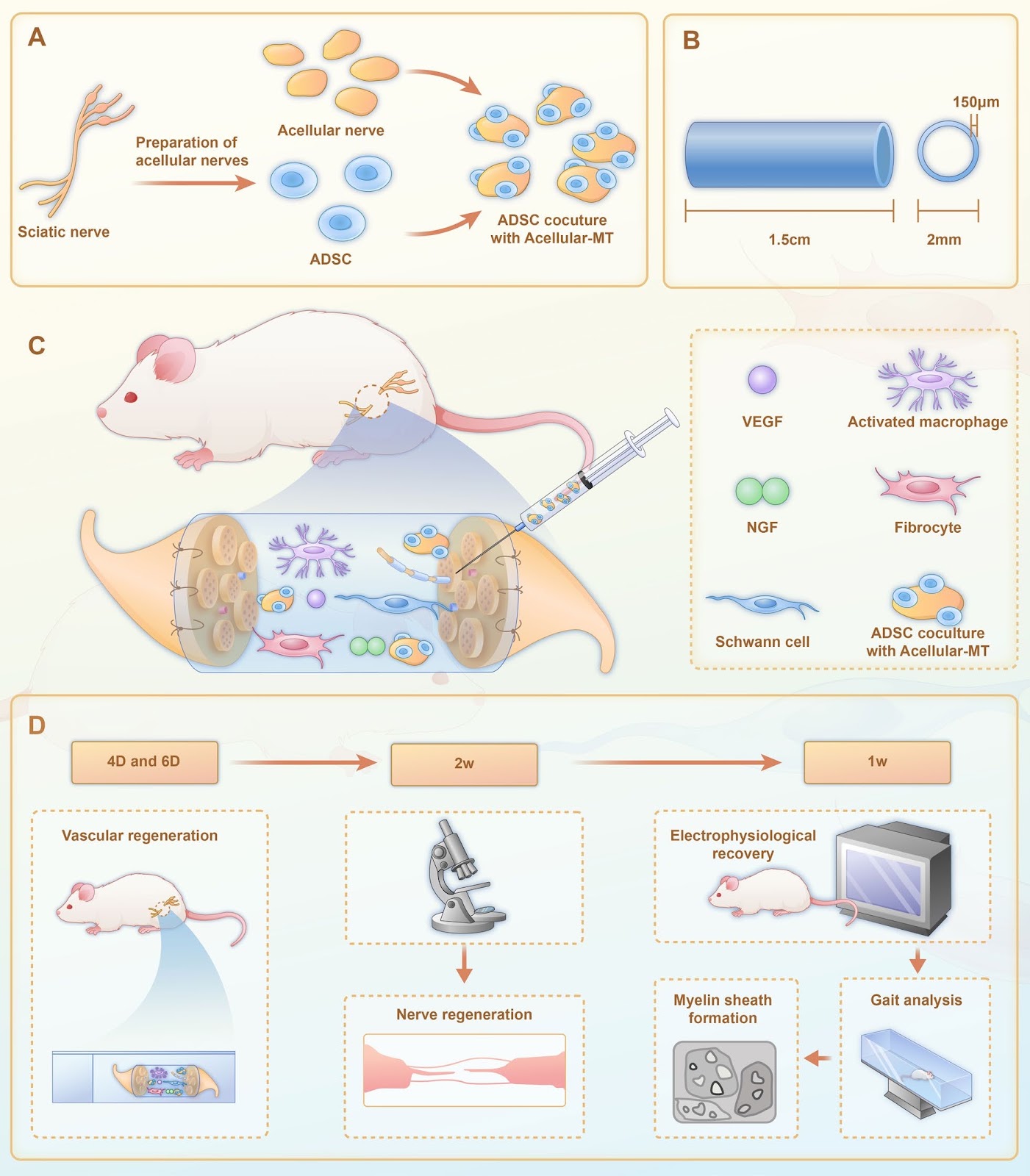
Background Treatment of peripheral nerve defects is a major concern in regenerative medicine. This study therefore aimed to explore the efficacy of a neural graft constructed using adipose mesenchymal stem cells (ADSC), acellular microtissues (MTs), and chitosan in the treatment of peripheral nerve defects. Methods Stem cell therapy with acellular MTs provided a suitable microenvironment for axonal regeneration, and compensated for the lack of repair cells in the neural ducts of male 8-week-old Sprague Dawley rats. Results In vitro, acellular MTs retained the intrinsic extracellular matrix and improved the narrow microstructure of acellular nerves, thereby enhancing cell functionality. In vivo neuroelectrophysiological studies, gait analysis, and sciatic nerve histology demonstrated the regenerative effects of active acellular MT. The Chitosan + Acellular-MT + ADSC group exhibited superior myelin sheath quality and improved neurological and motor function recovery. Conclusions Active acellular-MTs precellularized with ADSC hold promise as a safe and effective clinical treatment method for peripheral nerve defects.

Read Full Abstract10.1186/s13287-024-04093-5
Exploring non-viral methods for the delivery of CRISPR-Cas ribonucleoprotein to hematopoietic stem cellsGraphical AbstractVerified
Stem Cell Research & Therapy

Exploring non-viral methods for the delivery of CRISPR-Cas ribonucleoprotein to hematopoietic stem cells

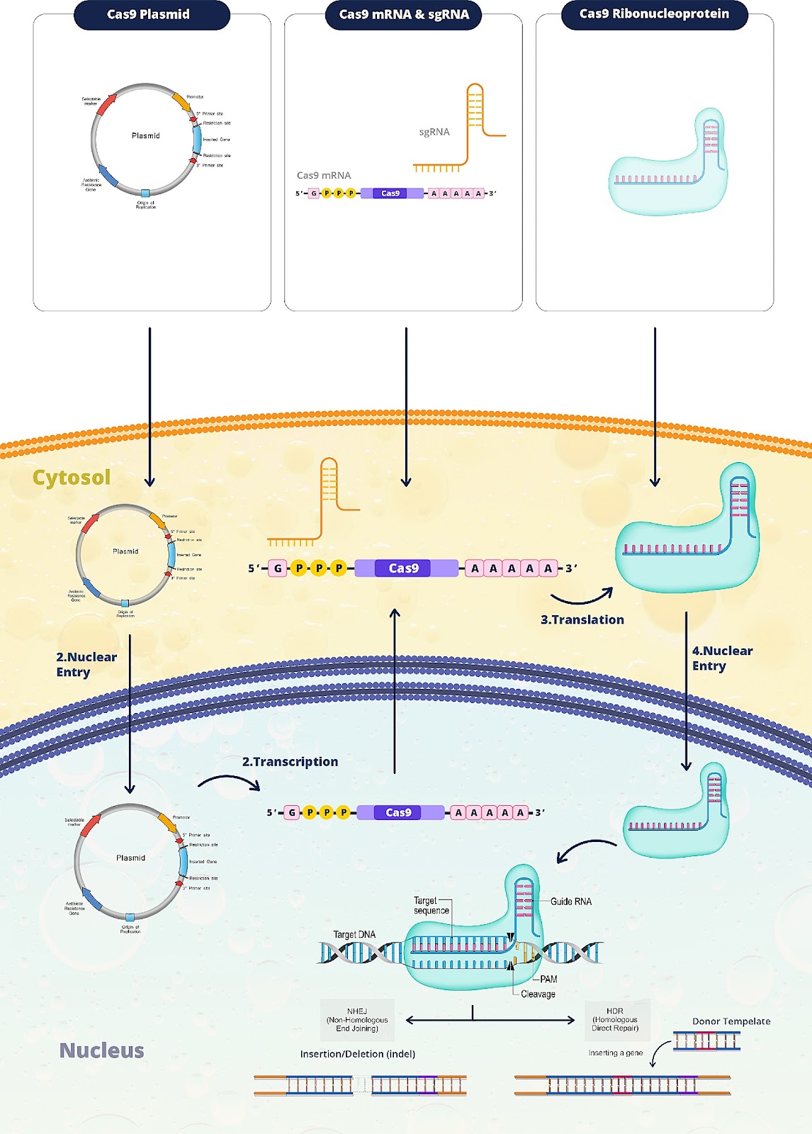
Gene manipulation of hematopoietic stem cells (HSCs) using the CRISPR/Cas system as a potent genome editing tool holds immense promise for addressing hematologic disorders. An essential hurdle in advancing this treatment lies in effectively delivering CRISPR/Cas to HSCs. While various delivery formats exist, Ribonucleoprotein complex (RNP) emerges as a particularly efficient option. RNP complexes offer enhanced gene editing capabilities, devoid of viral vectors, with rapid activity and minimized off-target effects. Nevertheless, novel delivery methods such as microfluidic-based techniques, filtroporation, nanoparticles, and cell-penetrating peptides are continually evolving. This study aims to provide a comprehensive review of these methods and the recent research on delivery approaches of RNP complexes to HSCs.

Read Full Abstract10.1186/s13287-024-03848-4
Regenerative therapies for femoral head necrosis in the past two decades: a systematic review and network meta-analysisGraphical AbstractVerified
Stem Cell Research & Therapy

Regenerative therapies for femoral head necrosis in the past two decades: a systematic review and network meta-analysis

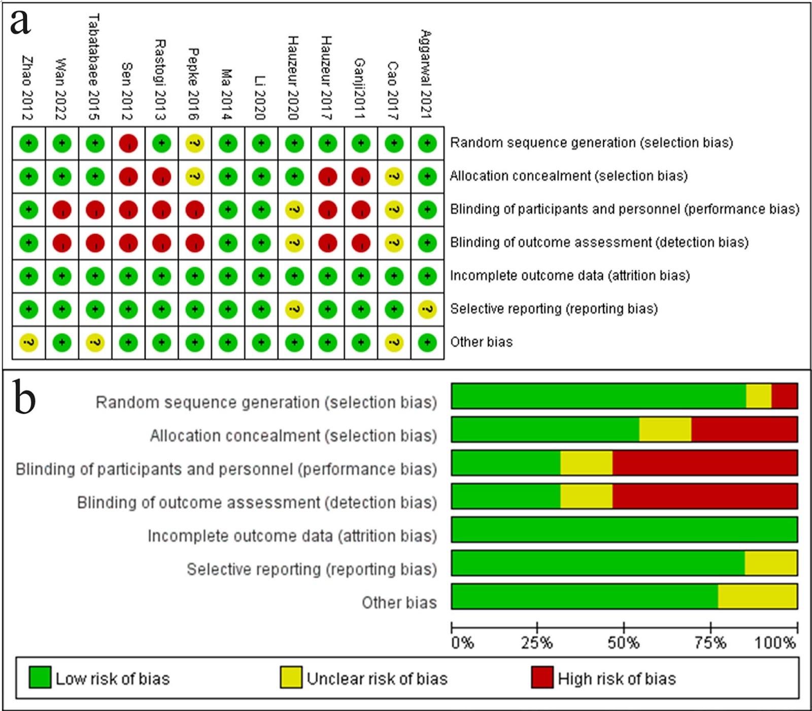
Background Regenerative techniques combined with core decompression (CD) are commonly used to treat osteonecrosis of the femoral head (ONFH). However, no consensus exists on regeneration therapy combined with CD that performs optimally. Therefore, we evaluated six regenerative therapies combined with CD treatment using a Bayesian network meta-analysis (NMA). Methods We searched PubMed, Embase, Cochrane Library, and Web of Science databases. Six common regeneration techniques were categorized into the following groups with CD as the control group: (1) autologous bone graft (ABG), (2) autologous bone graft combined with bone marrow aspirate concentrate (ABG+BMAC), (3) bone marrow aspirate concentrate (BMAC), (4) free vascular autologous bone graft (FVBG), (5) expanded mesenchymal stem cells (MSCs), and (6) platelet-rich plasma (PRP). The conversion rate to total hip arthroplasty (THA) and progression rate to femoral head necrosis were compared among the six treatments. Result A total of 17 literature were included in this study. In the NMA, two of the six treatment strategies demonstrated higher response in preventing the progression of ONFH than CD: MSCs (odds ratio [OR]: 0.098, 95% confidence interval [CI]: 0.0087–0.87) and BMAC (OR: 0.27, 95% CI: 0.073–0.73). Additionally, two of the six treatment strategies were effective techniques in preventing the conversion of ONFH to THA: MSCs (OR: 0.062, 95% CI: 0.0038–0.40) and BMAC (OR: 0.32, 95% CI: 0.1–0.074). No significant difference was found among FVBG, PRP, ABG+BMAC, ABG, and CD in preventing ONFH progression and conversion to THA (P>0.05). Conclusions Our NMA found that MSCs and BMAC were effective in preventing ONFH progression and conversion to THA among the six regenerative therapies. According to the surface under the cumulative ranking value, MSCs ranked first, followed by BMAC. Additionally, based on our NMA results, MSCs and BMAC following CD may be necessary to prevent ONFH progression and conversion to THA. Therefore, these findings provide evidence for the use of regenerative therapy for ONFH.

Read Full Abstract10.1186/s13287-024-03635-1
Transplanted human photoreceptors transfer cytoplasmic material but not to the recipient mouse retinaGraphical AbstractVerified
Stem Cell Research & Therapy

Transplanted human photoreceptors transfer cytoplasmic material but not to the recipient mouse retina

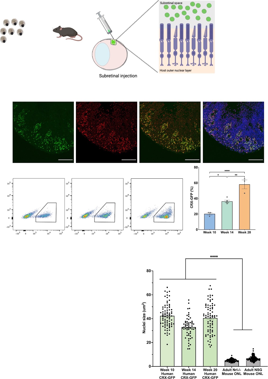
Background The discovery of material transfer between transplanted and host mouse photoreceptors has expanded the possibilities for utilizing transplanted photoreceptors as potential vehicles for delivering therapeutic cargo. However, previous research has not directly explored the capacity for human photoreceptors to engage in material transfer, as human photoreceptor transplantation has primarily been investigated in rodent models of late-stage retinal disease, which lack host photoreceptors. Methods In this study, we transplanted human stem-cell derived photoreceptors purified from human retinal organoids at different ontological ages (weeks 10, 14, or 20) into mouse models with intact photoreceptors and assessed transfer of human proteins and organelles to mouse photoreceptors. Results Unexpectedly, regardless of donor age or mouse recipient background, human photoreceptors did not transfer material in the mouse retina, though a rare subset of donor cells (<5%) integrated into the mouse photoreceptor cell layer. To investigate the possibility that a species barrier impeded transfer, we used a flow cytometric assay to examine material transfer in vitro. Interestingly, dissociated human photoreceptors transferred fluorescent protein with each other in vitro, yet no transfer was detected in co-cultures of human and mouse photoreceptors, suggesting that material transfer is species specific. Conclusions While xenograft models are not a tractable system to study material transfer of human photoreceptors, these findings demonstrate that human retinal organoid-derived photoreceptors are competent donors for material transfer and thus may be useful to treat retinal degenerative disease.

Read Full Abstract10.1186/s13287-024-03679-3
Effectiveness of extracellular vesicles derived from hiPSCs in repairing hyperoxia-induced injury in a fetal murine lung explant modelGraphical AbstractVerified
Stem Cell Research & Therapy

Effectiveness of extracellular vesicles derived from hiPSCs in repairing hyperoxia-induced injury in a fetal murine lung explant model

Background Despite advances in neonatal care, the incidence of Bronchopulmonary Dysplasia (BPD) remains high among preterm infants. Human induced pluripotent stem cells (hiPSCs) have shown promise in repairing injury in animal BPD models. Evidence suggests they exert their effects via paracrine mechanisms. We aim herein to assess the effectiveness of extracellular vesicles (EVs) derived from hiPSCs and their alveolar progenies (diPSCs) in attenuating hyperoxic injury in a preterm lung explant model. Methods Murine lung lobes were harvested on embryonic day 17.5 and maintained in air–liquid interface. Following exposure to 95% O2 for 24 h, media was supplemented with 5 Ɨ 10^6 particles/mL of EVs isolated from hiPSCs or diPSCs by size-exclusion chromatography. On day 3, explants were assessed using Hematoxylin–Eosin staining with mean linear intercept (MLI) measurements, immunohistochemistry, VEGFa and antioxidant gene expression. Statistical analysis was conducted using one-way ANOVA and Multiple Comparison Test. EV proteomic profiling was performed, and annotations focused on alveolarization and angiogenesis signaling pathways, as well as anti-inflammatory, anti-oxidant, and regenerative pathways. Results Exposure of fetal lung explants to hyperoxia induced airspace enlargement, increased MLI, upregulation of anti-oxidants Prdx5 and Nfe2l2 with decreased VEGFa expression. Treatment with hiPSC-EVs improved parenchymal histologic changes. No overt changes in vasculature structure were observed on immunohistochemistry in our in vitro model. However, VEGFa and anti-oxidant genes were upregulated with diPSC-EVs, suggesting a pro-angiogenic and cytoprotective potential. EV proteomic analysis provided new insights in regard to potential pathways influencing lung regeneration. Conclusion This proof-of-concept in vitro study reveals a potential role for hiPSC- and diPSC-EVs in attenuating lung changes associated with prematurity and oxygen exposure. Our findings pave the way for a novel cell free approach to prevent and/or treat BPD, and ultimately reduce the global burden of the disease.

Read Full Abstract10.1186/s13287-024-03687-3
Exosomes derived from hypoxic mesenchymal stem cells restore ovarian function by enhancing angiogenesisGraphical AbstractVerified
Stem Cell Research & Therapy

Exosomes derived from hypoxic mesenchymal stem cells restore ovarian function by enhancing angiogenesis

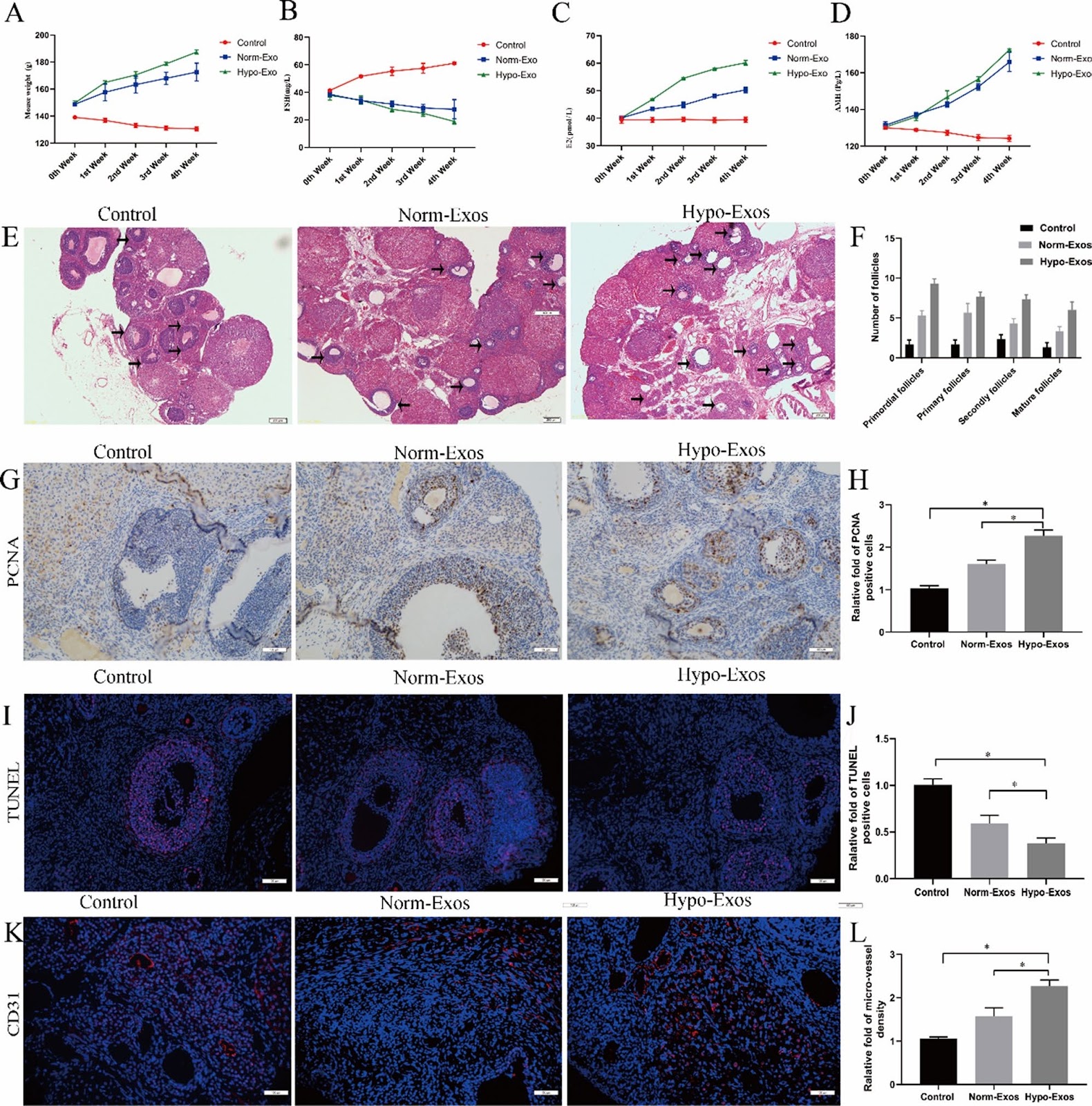
Background hucMSC-exosomes can be engineered to strengthen their therapeutic potential, and the present study aimed to explore whether hypoxic preconditioning can enhance the angiogenic potential of hucMSC-exosomes in an experimental model of POF. Methods Primary hucMSCs and ROMECs were isolated from fresh tissue samples and assessed through a series of experiments. Exosomes were isolated from hucMSCs under normoxic or hypoxic conditions (norm-Exos and hypo-Exos, respectively) and then characterized using classic experimental methods. Based on a series of angiogenesis-related assays, we found that hypo-Exos significantly promoted ROMEC proliferation, migration, and tube formation and increased angiogenesis-promoting molecules in vitro. Histology, immunohistochemistry, and immunofluorescence experiments in a rat model of POF demonstrated that hypoxia pretreatment strengthens the therapeutic angiogenic effect of hucMSC-exosomes in vivo. Subsequently, high-throughput miRNA sequencing, qRT-PCR analysis, and western blotting were employed to identify the potential molecular mechanism. Results We found that hypo-Exos enhance endothelial function and angiogenesis via the transfer of miR-205-5p in vitro and in vivo. Finally, based on the results of bioinformatics analysis, dual luciferase reporter assays, and gain- and loss-of-function studies, we found evidence indicating that exosomal miR-205-5p enhances angiogenesis by targeting the PTEN/PI3K/AKT/mTOR signalling pathway. These results indicated for the first time that exosomes derived from hypoxia-conditioned hucMSCs strongly enhance angiogenesis via the transfer of miR-205-5p by targeting the PTEN/PI3K/AKT/mTOR signalling pathway. Conclusions Our findings provide a theoretical basis and demonstrate the potential application of a novel cell-free approach with stem cell-derived products in the treatment of POF.

Read Full Abstract10.1186/s13287-024-04111-6
Improvement of androgenic alopecia by extracellular vesicles secreted from hyaluronic acid-stimulated induced mesenchymal stem cellsGraphical AbstractVerified
Stem Cell Research & Therapy

Improvement of androgenic alopecia by extracellular vesicles secreted from hyaluronic acid-stimulated induced mesenchymal stem cells

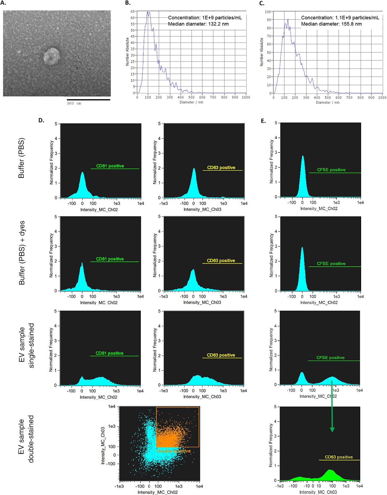
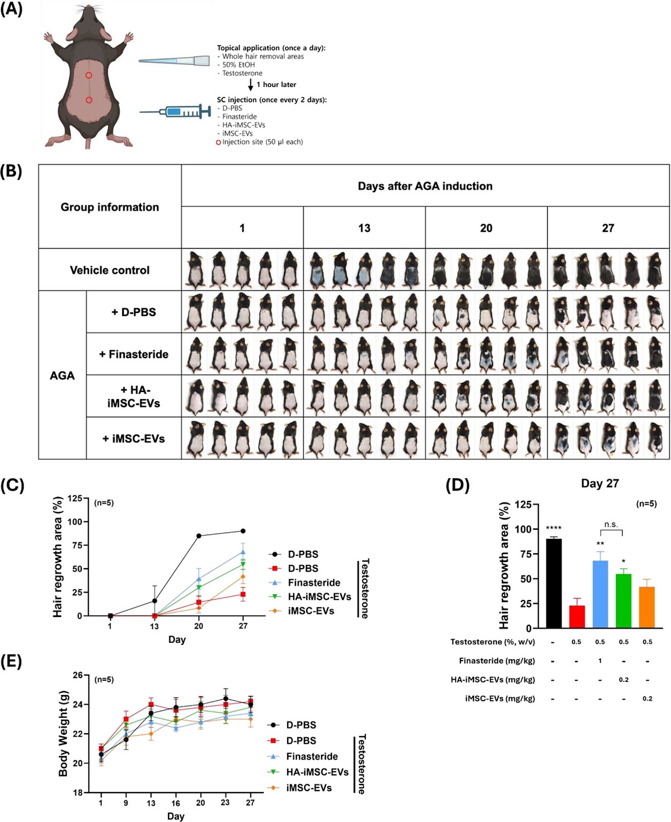
Background: Androgenetic alopecia (AGA) is a common form of hair loss. Androgens, such as testosterone and dihydrotestosterone, are the main causes of AGA. Extracellular vesicles (EVs) derived from mesenchymal stem cells (MSCs) can reduce AGA. However, preparing therapeutic doses of MSCs for clinical use is challenging. Induced pluripotent stem cell-derived MSCs (iMSCs) are homogenous and easily expandable, enabling scalable production of EVs. Hyaluronic acid (HA) can exert various functions including free radical scavenging, immune regulation, and cell migration. Herein, we examined whether hyaluronic acid (HA) stimulation of iMSCs could produce EVs with enhanced therapeutic outcomes for AGA. Methods: EVs were collected from iMSCs primed with HA (HA–iMSC–EVs) or without HA (iMSC–EVs). The characteristics of EVs were examined using dynamic light scattering, cryo-transmission electron microscopy, immunoblotting, flow cytometry, and proteomic analysis. In vitro, we compared the potential of EVs in stimulating the survival of hair follicle dermal papilla cells undergoing testosterone-mediated AGA. Additionally, the expression of androgen receptor (AR) and relevant growth factors as well as key proteins of Wnt/β-catenin signaling pathway (β-catenin and phosphorylated GSK3β) was analyzed. Subsequently, AGA was induced in male C57/BL6 mice by testosterone administration, followed by repeated injections of iMSC–EVs, HA–iMSC–EVs, finasteride, or vehicle. Several parameters including hair growth, anagen phase ratio, reactivation of Wnt/β-catenin pathway, and AR expression was examined using qPCR, immunoblotting, and immunofluorescence analysis. Results: Both types of EVs showed typical characteristics for EVs, such as size distribution, markers, and surface protein expression. In hair follicle dermal papilla cells, the mRNA levels of AR, TGF-β, and IL-6 increased by testosterone was blocked by HA–iMSC–EVs, which also contributed to the augmented expression of trophic genes related to hair regrowth. However, no notable changes were observed in the iMSC–EVs. Re-activation of Wnt/β-catenin was observed in HA–iMSC–EVs but not in iMSC–EVs, as shown by β-catenin stabilization and an increase in phosphorylated GSK3β. Restoration of hair growth was more significant in HA–iMSC–EVs than in iMSC–EVs.

Read Full Abstract10.1186/s13287-024-03906-x
A protein-RNA complex orchestrated by EMB1006, EMB1270, EMB976, and CFM2 facilitates clpP1 intron 2 splicing in Arabidopsis chloroplastsGraphical AbstractVerified
Acta Biochimica et Biophysica Sinica

A protein-RNA complex orchestrated by EMB1006, EMB1270, EMB976, and CFM2 facilitates clpP1 intron 2 splicing in Arabidopsis chloroplasts

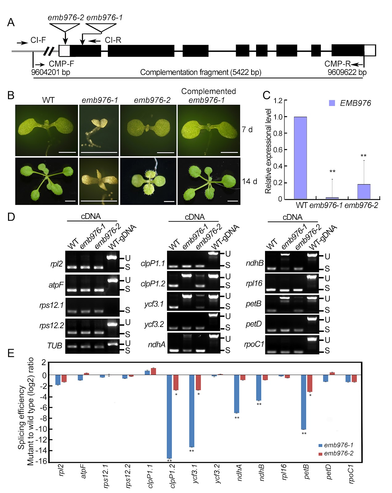
In Arabidopsis, the PPR proteins EMB1006, EMB1270, and EMB976 are all essential for the splicing of plastid clpP1 intron 2 (clpP1.2), although each also targets other distinct RNAs. The precise mechanism underlying their co-ordinated action in clpP1.2 splicing remains unclear. In this study, RNA electrophoretic mobility shift assays, guided by PPR code prediction, confirm that EMB1006 specifically binds to a sequence near the 3′ end of clpP1 exon 2. Additionally, immunoprecipitation coupled with mass spectrometry reveals that EMB1006 forms a complex with EMB1270, EMB976, and CFM2. Direct interactions between EMB1006 and EMB1270 or CFM2 are further supported by yeast two-hybrid (Y2H) and semi-in vivo pull-down assays. However, no direct interactions between EMB976 and EMB1006, CFM2 or EMB1270 are detected by Y2H. Based on these findings and previous evidence that EMB1270 binds to clpP1 intron 2 and interacts with CFM2, we propose a model in which EMB1006 and EMB1270 bind to distinct sites on clpP1 pre-mRNA. Together with CFM2 and possible indirect association with EMB976, they assemble into a protein-RNA complex that facilitates the splicing of clpP1.2 in chloroplasts.

Read Full Abstract10.3724/abbs.2026076
Tanshinones from Salvia miltiorrhiza alleviate ulcerative colitis via reprogramming the gut microbiota-metabolite axisGraphical AbstractVerified
Acta Biochimica et Biophysica Sinica

Tanshinones from Salvia miltiorrhiza alleviate ulcerative colitis via reprogramming the gut microbiota-metabolite axis

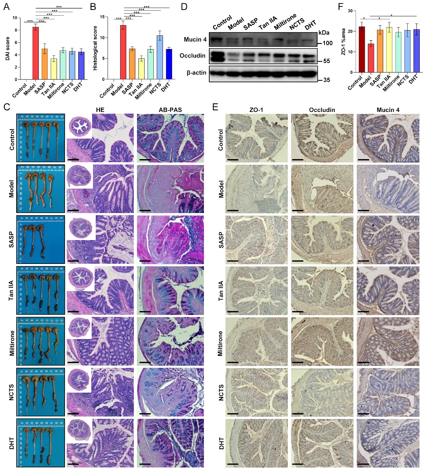
The anti-inflammatory properties of the traditional herb Salvia miltiorrhiza Bunge are well-established, yet its precise mechanism of action in ulcerative colitis (UC) remains unclear. Herein, we evaluate the therapeutic potential of four major tanshinones–tanshinone IIA (Tan IIA), miltirone, neocryptotanshinone, and dihydrotanshinone I–in a murine dextran sulfate sodium (DSS)-induced colitis model. Our results show that tanshinones effectively alleviate disease severity, suppress systemic and local inflammation, and restore intestinal barrier integrity. Integrated multi-omics analysis reveals that the therapeutic efficacy originates from a comprehensive reprogramming of the gut microbiota-metabolite axis. Specifically, tanshinones reverse colitis-associated dysbiosis and rectify metabolic disturbances in linoleic acid metabolism, bile acid biosynthesis, and amino acid utilization. Correlation network analysis identifies key functional modules linking beneficial microbes (e.g., Akkermansia) to anti-inflammatory lipid mediators and associating pathobionts (e.g., Desulfovibrio) with disrupted bile acid metabolism. Notably, supplementation with Akkermansia muciniphila synergizes with Tan IIA to amplify barrier restoration and metabolic normalization. Our findings establish that tanshinones ameliorate UC through microbiota-driven metabolic reprogramming, wherein the restructured microbial community actively shapes a therapeutic metabolic output. This work elucidates a metabolite-mediated mechanism of action and positions tanshinones as promising microbiome-targeting therapeutics for inflammatory bowel disease.

Read Full Abstract10.3724/abbs.2026054
Effects of Staphylococcus aureus on stem cells and potential targeted treatment of inflammatory disordersGraphical AbstractVerified
Stem Cell Research & Therapy

Effects of Staphylococcus aureus on stem cells and potential targeted treatment of inflammatory disorders

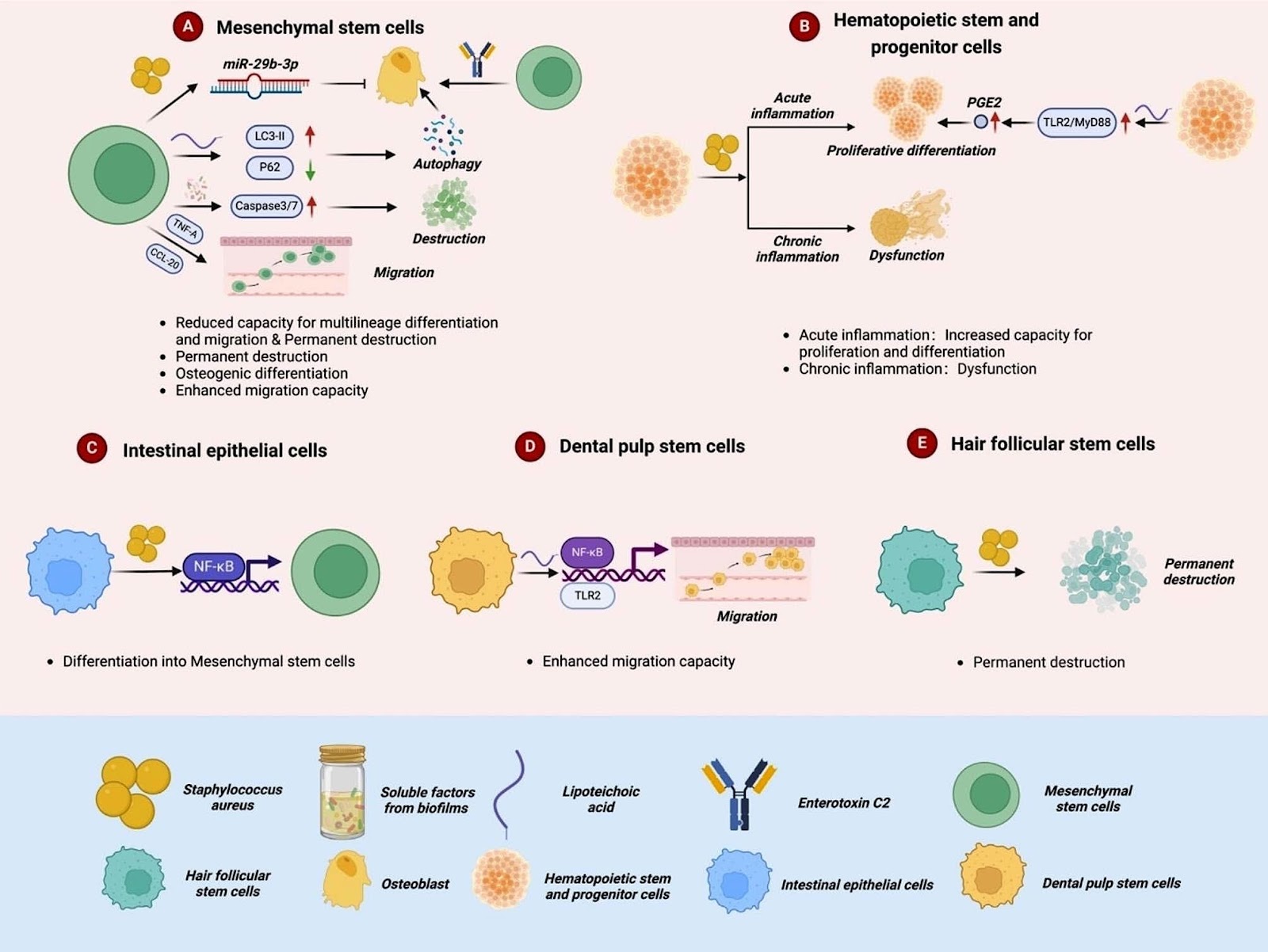
Due to the advanced studies on stem cells in developmental biology, the roles of stem cells in the body and their phenotypes in related diseases have not been covered clearly. Meanwhile, with the intensive research on the mechanisms of stem cells in regulating various diseases, stem cell therapy is increasingly being attention because of its effectiveness and safety. As one of the most widely used stem cell in stem cell therapies, hematopoietic stem cell transplantation shows huge advantage in treatment of leukemia and other blood-malignant diseases. Besides, due to the effect of anti-inflammatory and immunomodulatory, mesenchymal stem cells could be a potential therapeutic strategy for variety infectious diseases. In this review, we summarized the effects of Staphylococcus aureus (S. aureus) and its components on different types of adult stem cells and their downstream signaling pathways. Also, we reviewed the roles of different kinds of stem cells in various disease models caused by S. aureus, providing new insights for applying stem cell therapy to treat infectious diseases.

Read Full Abstract10.1186/s13287-024-03781-6
Reprogramming of 3D genome structure underlying HSPC development in zebrafishGraphical AbstractVerified
Stem Cell Research & Therapy

Reprogramming of 3D genome structure underlying HSPC development in zebrafish

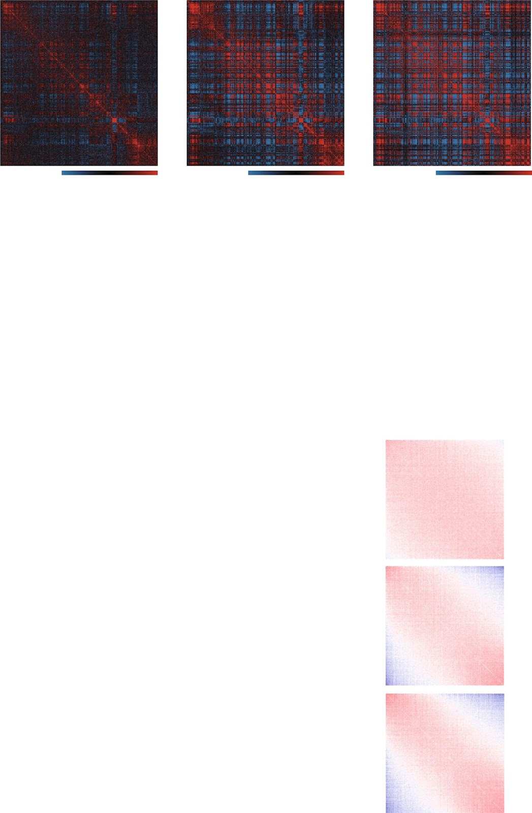
Background Development of hematopoietic stem and progenitor cells (HSPC) is a multi-staged complex process that conserved between zebrafish and mammals. Understanding the mechanism underlying HSPC development is a holy grail of hematopoietic biology, which is helpful for HSPC clinical application. Chromatin conformation plays important roles in transcriptional regulation and cell fate decision; however, its dynamic and role in HSPC development is poorly investigated. Methods We performed chromatin structure and multi-omics dissection across different stages of HSPC developmental trajectory in zebrafish for the first time, including Hi-C, RNA-seq, ATAC-seq, H3K4me3 and H3K27ac ChIP-seq. Results The chromatin organization of zebrafish HSPC resemble mammalian cells with similar hierarchical structure. We revealed the multi-scale reorganization of chromatin structure and its influence on transcriptional regulation and transition of cell fate during HSPC development. Nascent HSPC is featured by loose conformation with obscure structure at all layers. Notably, PU.1 was identified as a potential factor mediating formation of promoter-involved loops and regulating gene expression of HSPC. Conclusions Our results provided a global view of chromatin structure dynamics associated with development of zebrafish HSPC and discovered key transcription factors involved in HSPC chromatin interactions, which will provide new insights into the epigenetic regulatory mechanisms underlying vertebrate HSPC fate decision.

Read Full Abstract10.1186/s13287-024-03798-x