🧬 SinoBioData Academic Portal
📚 Peer-Reviewed Translated Literature

All Biomedical & Clinical Articles (Page 57)

Browse complete peer-reviewed translations from top Chinese biomedical, oncology, and genomics journals. Read verified previews and download full authentic clinical reports.

Published Research Papers

Showing 24 of 1542 peer-reviewed translated articles (Page 57 of 65)

Angelicin attenuates sepsis-associated acute liver injury via p38 MAPK inhibition and NF-κB-mediated Nrf2/Keap1 activation to suppress inflammation and oxidative stressGraphical AbstractVerified
Acta Biochimica et Biophysica Sinica

Angelicin attenuates sepsis-associated acute liver injury via p38 MAPK inhibition and NF-κB-mediated Nrf2/Keap1 activation to suppress inflammation and oxidative stress

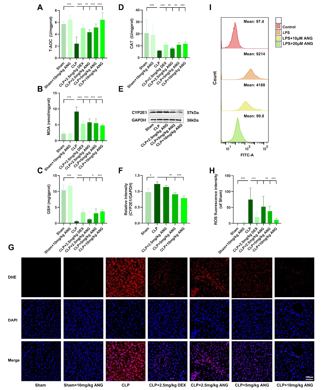
Sepsis-associated acute liver injury (SALI) is a frequent and clinically severe complication of sepsis, in which inflammatory responses and oxidative stress are involved. Angelicin (ANG), one of the main active components in the traditional Chinese medicine Psoralea corylifolia Linn., has anti-inflammatory and antioxidant bioactivities. In this study, the protective effect of ANG on SALI and its specific mechanism are investigated by establishing a mouse model of caecal ligation and puncture (CLP)-induced SALI and an in vitro sepsis model in LPS-stimulated AML12 cells. These results show that ANG can alleviate liver injury and improve liver function in SALI mice. ANG decreases the mRNA expression levels of the pro-inflammatory factors Il-1β, Il-6, and Tnf-α and increases the mRNA expression level of the anti-inflammatory factor Il-10, which suggests its anti-inflammatory effects. The results of the biochemical kit assay and DHE staining show that ANG can decrease the levels of MDA and ROS and increase the level of GSH and the activities of CAT and SOD, which suggests that ANG has antioxidant effects. Mechanistically, ANG exerts anti-inflammatory effects by inhibiting the NF-κB and p38 MAPK pathways and exerting antioxidant effects by activating the Nrf2/Keap1 pathway. Additionally, cell transfection experiments indicate that activation of the Nrf2/Keap1 pathway by ANG may depend on the inhibition of the NF-κB pathway. In conclusion, ANG attenuates SALI by inhibiting the NF-κB and p38 MAPK pathways, thereby activating the Nrf2/Keap1 pathway and making it a promising therapeutic intervention for SALI.

Read Full Abstract10.3724/abbs.2025139
The host gene CSTF2 regulates HBV replication via HBV PRE-induced nuclear exportGraphical AbstractVerified
Acta Biochimica et Biophysica Sinica

The host gene CSTF2 regulates HBV replication via HBV PRE-induced nuclear export

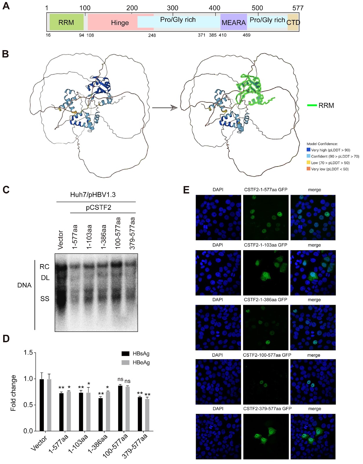
The persistent global burden of hepatitis B virus (HBV) infection has prompted ongoing investigations into host determinants of viral control. In this study, we investigate the regulatory influence of the host gene cleavage stimulation factor subunit 2 (CSTF2) on HBV replication dynamics. We demonstrate differential CSTF2 expression across the spectrum of HBV infection phases, with upregulated expression noted during the immune-reactive and inactive carrier states compared with the immune-tolerant phase. Notably, dose-responsive attenuation of HBV DNA, as well as surface and core protein levels, is observed subsequent to CSTF2 overexpression, whereas HBV RNA levels remain unaffected. Upon HBV transfection, a notable alteration in CSTF2 subcellular localization is discerned, suggesting active relocalization to the cytoplasm, potentially mediated through interaction with the HBV posttranscriptional regulatory element (PRE). This interaction appears to impede the nuclear export of HBV RNA. Additionally, distinct antiviral efficacies are attributed to the functional domains of the CSTF2 protein, indicating a multifaceted host defense mechanism. These insights increase the understanding of host-virus interplay and identify CSTF2 as a candidate for antiviral therapeutic strategies.

Read Full Abstract10.3724/abbs.2024216
MiR-133b-3p attenuates angiotensin II-induced cardiac hypertrophy through the inhibition of apoptosis by targeting CDIP1Graphical AbstractVerified
Acta Biochimica et Biophysica Sinica

MiR-133b-3p attenuates angiotensin II-induced cardiac hypertrophy through the inhibition of apoptosis by targeting CDIP1

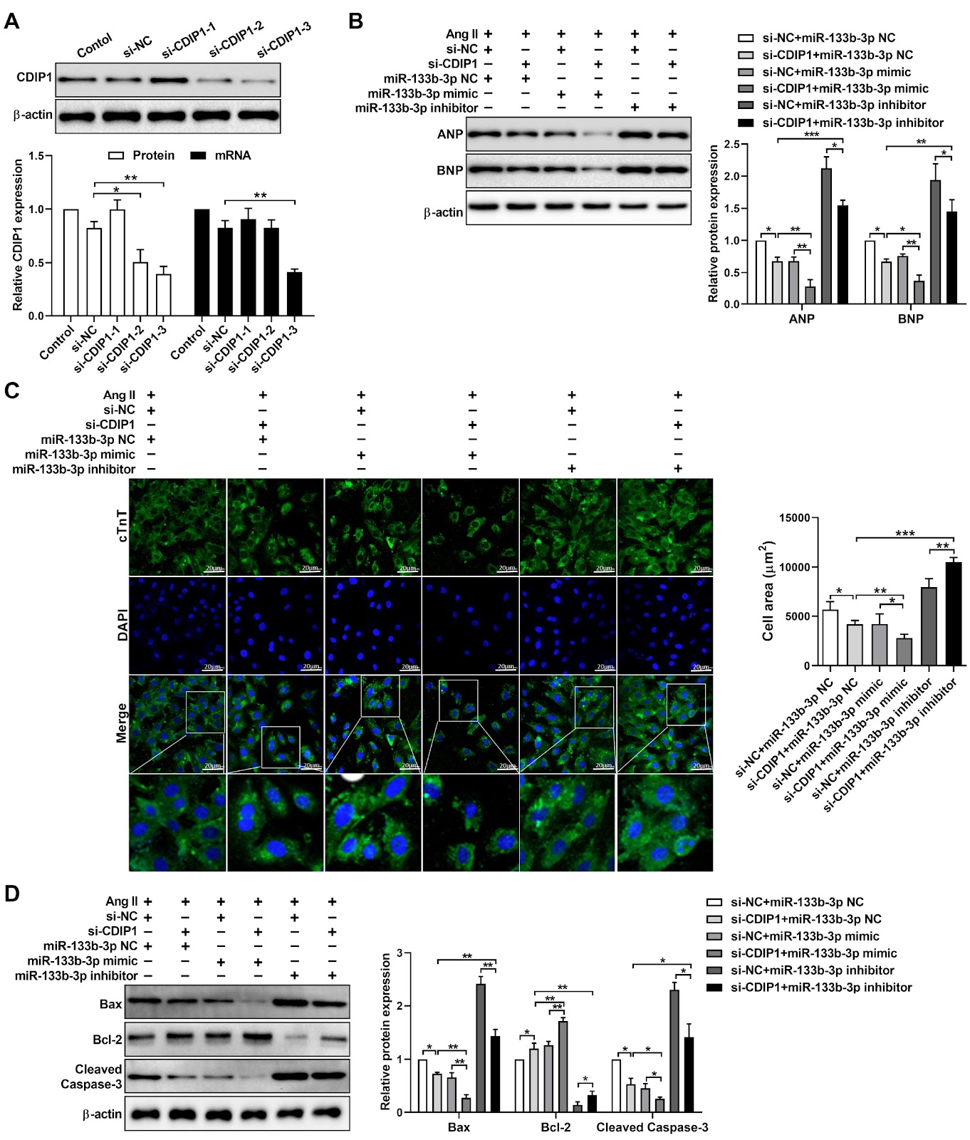
MicroRNAs (miRNAs) have emerged as essential regulators that play important roles in the development of multiple systems. Recent studies have identified significant roles for miRNAs in the progression of cardiac hypertrophy. This study aims to investigate the effects of miR-133b-3p on angiotensin II (Ang II)-induced cardiac hypertrophy and apoptosis, as well as explore its underlying mechanisms. Our experimental results reveal that miR-133b-3p expression is significantly decreased in both animal and cell models of cardiac hypertrophy induced by Ang II. Overexpression of miR-133b-3p reverses the hypertrophic manifestations and apoptosis induced by Ang II. Through bioinformatics analysis and dual-luciferase reporter assays, CDIP1 (cell death inducing p53 target 1) is identified as a direct target of miR-133b-3p, and the overexpression of miR-133b-3p reduces CDIP1 expression. Additionally, CDIP1 silencing suppresses cardiomyocyte hypertrophy and apoptosis induced by Ang II. In summary, these results suggest that miR-133b-3p may serve as a potential diagnostic marker for cardiac hypertrophy and that the upregulation of miR-133b-3p inhibits cardiac hypertrophy by targeting CDIP1.

Read Full Abstract10.3724/abbs.2024181
Structural insights into H2A-H2B and H2A.Z-H2B sliding on histone chaperone NAP1Graphical AbstractVerified
Acta Biochimica et Biophysica Sinica

Structural insights into H2A-H2B and H2A.Z-H2B sliding on histone chaperone NAP1

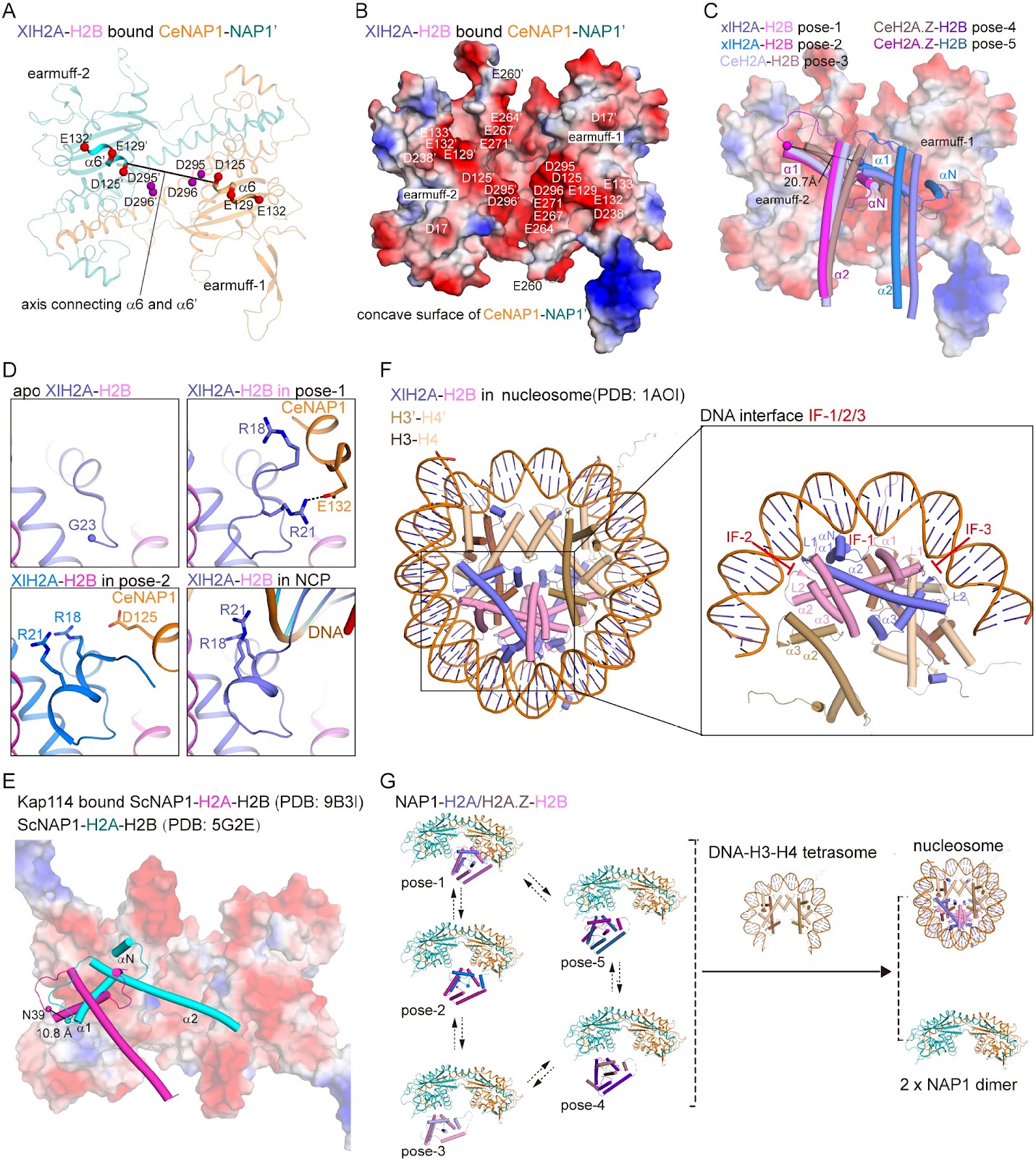
The evolutionarily conserved nucleosome assembly protein 1 (NAP1) functions as a histone chaperone for H2A-H2B, regulating nucleosome assembly and maintaining chromatin integrity. However, the dynamic and variable nature of the interactions between acidic NAP1 and basic H2A-H2B has obscured the molecular basis of its chaperoning activity. Here, we report the crystal structures of Caenorhabditis elegans NAP1 (CeNAP1) in complex with Xenopus laevis H2A-H2B (XlH2A-H2B) and with C. elegans H2A.Z-H2B (CeH2A.Z-H2B) at 3.35 Å and 2.8 Å, respectively. In our structures, H2A/H2A.Z-H2B binds to the acidic concave surface of CeNAP1 in three distinct poses, with two in the CeNAP1-XlH2A-H2B complex and one in the CeNAP1-CeH2A.Z-H2B complex. These poses are different from the two poses observed in the previously reported CeNAP1-CeH2A/H2A.Z-H2B structures. The predominant interaction involves engagement of the acidic CeNAP1 α6-carboxy-terminal (C-terminal) tail by the basic H2A/H2A.Z αN–α1 region, stabilized by salt bridges and electrostatic interactions. A comparative analysis of all five known poses reveals that H2A/H2A.Z-H2B can shift approximately 20.7 Å along the α6-C-terminal tail-C′-terminal tail-α6′ axis. These findings demonstrate a sliding binding mode of H2A/H2A.Z-H2B on NAP1, providing new mechanistic insights into nucleosome assembly activity of histone chaperones.

Read Full Abstract10.3724/abbs.2025241
Astatine-211 and actinium-225: two promising nuclides in targeted alpha therapyGraphical AbstractVerified
Acta Biochimica et Biophysica Sinica

Astatine-211 and actinium-225: two promising nuclides in targeted alpha therapy

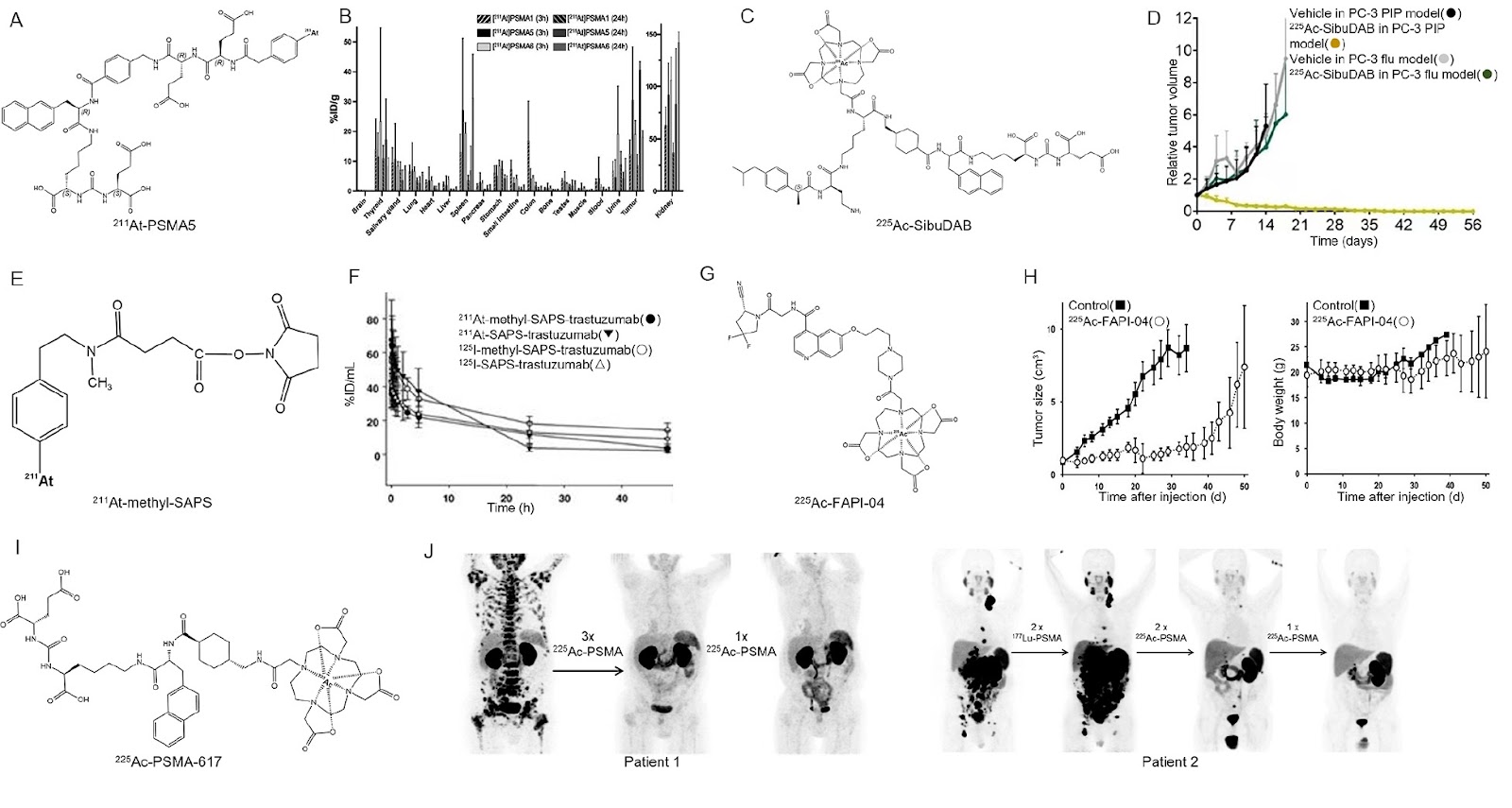
Nuclear medicine therapy offers a promising approach for tumor treatment, as the energy emitted during radionuclide decay causes irreparable damage to tumor cells. Notably, α-decay exhibits an even more significant destructive potential. By conjugating α-nuclides with antibodies or small-molecule inhibitors, targeted alpha therapy (TAT) can enhance tumor destruction while minimizing toxic side effects, making TAT an increasingly attractive antineoplastic strategy. Astatine-211 (211At) and actinium-225 (225Ac) have emerged as highly effective agents in TAT due to their exceptional physicochemical properties and biological effects. In this review, we highlight the applications of 211At-/225Ac-radiopharmaceuticals, particularly in specific tumor targets, such as prostate-specific membrane antigen (PSMA) in prostate cancers, cluster of differentiation (CD) in hematological malignancies, human epidermal growth factor receptor-2 (HER2) in ovarian cancers, and somatostatin receptor (SSTR) in neuroendocrine tumors. We synthesize the progress from preclinical and clinical trials to provide insights into the promising potential of 211At-/225Ac-radiopharmaceuticals for future treatments.

Read Full Abstract10.3724/abbs.2024206
Lysyl oxidase exacerbates rheumatoid arthritis through promoting angiogenesis and the proliferation of fibroblast-like synoviocytesGraphical AbstractVerified
Acta Biochimica et Biophysica Sinica

Lysyl oxidase exacerbates rheumatoid arthritis through promoting angiogenesis and the proliferation of fibroblast-like synoviocytes

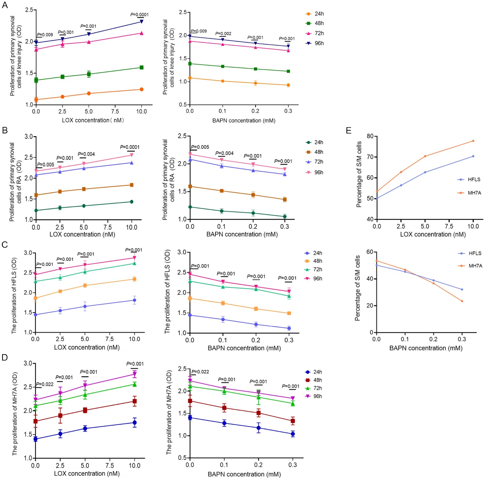
Rheumatoid arthritis (RA) is an autoimmune disorder characterized by synovial hyperplasia and pannus formation, which serves as its primary pathological feature and may ultimately result in joint deformities. Lysyl oxidase (LOX) is involved in the formation and remodeling of the extracellular matrix, but its role in RA is not yet clear. This study aims to investigate the mechanism of lysyl oxidase (LOX) in synovial hyperplasia and pannus formation associated with rheumatoid arthritis (RA). Synovial, serum, and synovial fluid samples are collected from RA, osteoarthritis (OA), and knee injury patients and subsequently analyzed via HE staining, immunohistochemistry, and ELISA. Compared with those of the OA and injury groups, the RA synovium presents increased thickness, disorganized cell layers, increased microvascular density (MVD), and elevated LOX expression. Moreover, LOX levels are positively correlated with the MVD. Both synovial fluid and fibroblast-like synoviocytes (FLSs) derived from RA patients present significantly elevated concentrations of LOX. In vitro experiments reveal that LOX dose-dependently promotes the proliferation of FLSs derived from both RA patients and healthy individuals (MH7A/HFLS) by accelerating S/M-phase cell cycle progression while simultaneously stimulating angiogenesis in human umbilical vein endothelial cells (HUVECs). In contrast, the LOX inhibitor BAPN suppresses these effects. Mechanistic analysis further reveals that LOX increases the phosphorylation of the PI3K-AKT signaling pathway, an effect that is reversible by BAPN. In conclusion, LOX may induce abnormal fibroblast proliferation and endothelial neovascularization via activation of the PI3K/AKT pathway, thus aggravating synovial hyperplasia and pathological membrane formation in RA. These findings provide a theoretical foundation for the development of targeted LOX treatments for RA.

Read Full Abstract10.3724/abbs.2025162
IL15RA-STAT3-GPX4/ACSL3 signaling leads to ferroptosis resistance in pancreatic cancerGraphical AbstractVerified
Acta Biochimica et Biophysica Sinica

IL15RA-STAT3-GPX4/ACSL3 signaling leads to ferroptosis resistance in pancreatic cancer

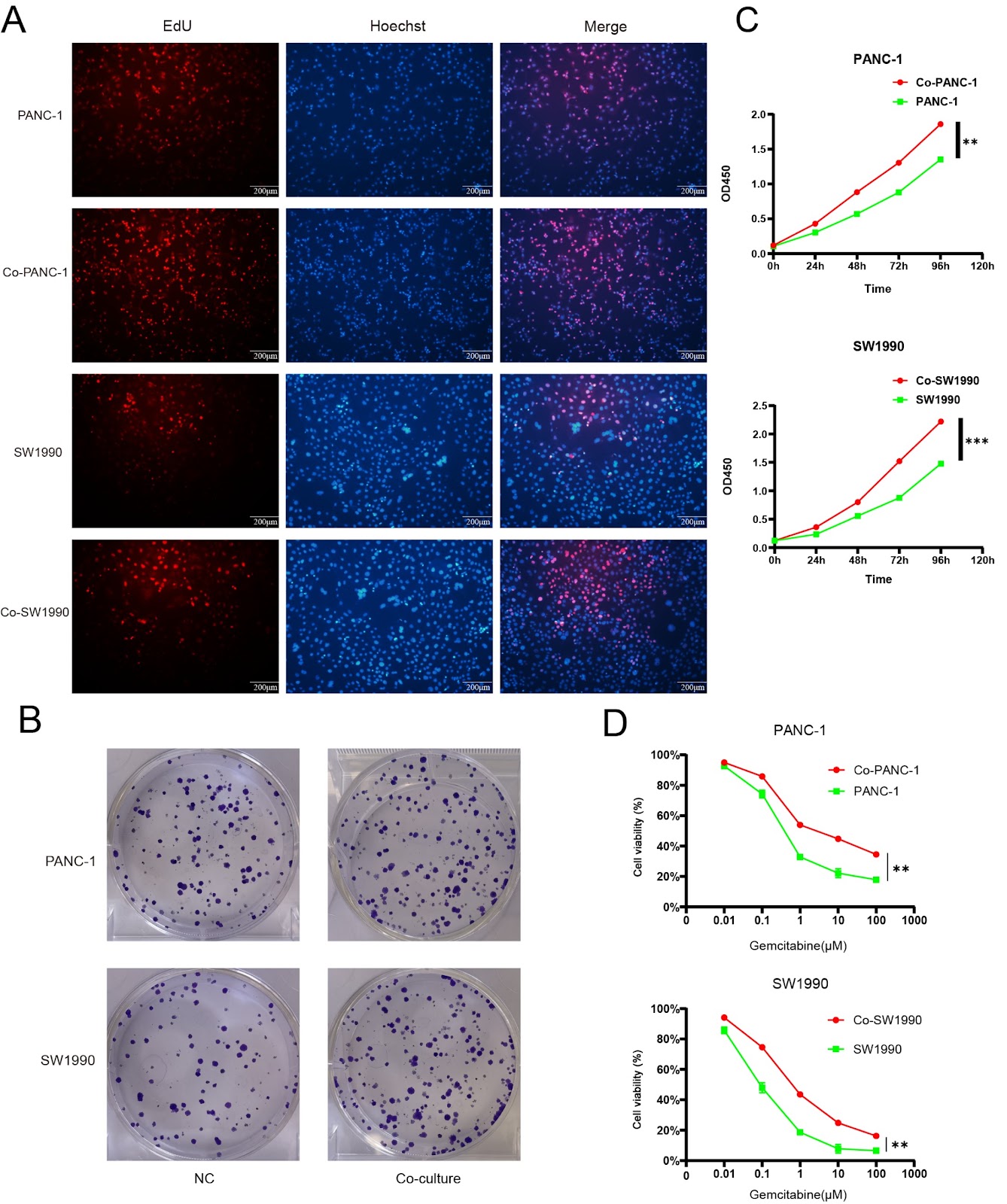
Pancreatic ductal adenocarcinoma (PDAC) is a highly malignant disease with a poor prognosis, and the lack of effective treatment methods accounts for its high mortality. Pancreatic stellate cells (PSCs) in the tumor microenvironment play an important role in the development of PDAC. Previous studies have reported that patients with PDAC are more vulnerable to ferroptosis inducers. To investigate the relationship between PSCs and pancreatic cancer cells, a coculture system is used to further reveal the influence of PSCs on ferroptosis resistance in PDAC using many in vitro and in vivo experiments. Our results show that PSCs promote ferroptosis resistance in pancreatic cancer cells. We further demonstrate that IL15 secretion by PSCs activates the IL15RA-STAT3-GPX4/ACSL3 axis. The simultaneous upregulation of GPX4 and ACSL3 prevents lipid peroxidation and ultimately protects pancreatic cancer cells from ferroptosis both in vitro and in vivo. This study demonstrates that PSCs protect pancreatic cancer cells in a paracrine manner and may indicate a novel strategy for the treatment of PDAC.

Read Full Abstract10.3724/abbs.2024153
ANT1 suppression inhibits the progression of colorectal cancer by suppressing PINK1/Parkin-mediated mitophagyGraphical AbstractVerified
Acta Biochimica et Biophysica Sinica

ANT1 suppression inhibits the progression of colorectal cancer by suppressing PINK1/Parkin-mediated mitophagy

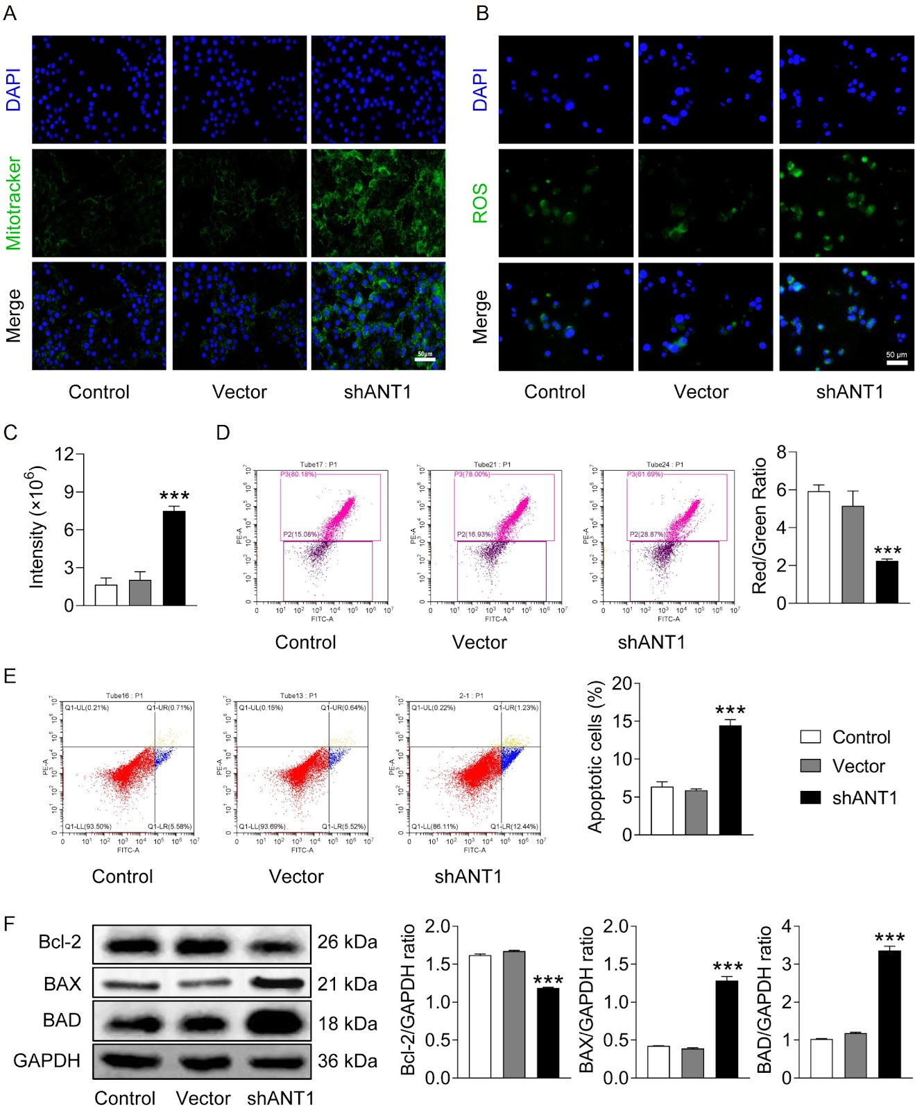
Mitochondrial dysfunction is closely related to tumor development. Adenine nucleotide translocator 1 (ANT1), which promotes ADP/ATP translocation across the inner mitochondrial membrane, is an important protein involved in mitochondrial function and plays a role in a variety of diseases, including cancers. However, its role in colorectal cancer (CRC) progression remains poorly understood. This study aims to explore the potential role of ANT1 in CRC and its relationship with mitophagy. Through immunohistochemical analysis, we find that ANT1 expression is significantly higher in the tumor tissues of CRC patients than in adjacent normal tissues and that its overexpression is associated with poor prognosis. Further experiments demonstrate that ANT1 knockdown significantly inhibits CRC cell proliferation, migration, and invasion and leads to mitochondrial dysfunction, increased ROS production, and apoptosis by suppressing mitophagy. Mechanistically, ANT1 knockdown downregulates the PINK1/Parkin pathway, thereby inhibiting mitophagy activity. Notably, PINK1 overexpression partially rescues the cellular dysfunction induced by ANT1 knockdown, suggesting a potential role for PINK1 in reversing the suppression of mitophagy. In vivo xenograft models also show that ANT1 knockdown markedly inhibits tumor growth. In conclusion, ANT1 may play a critical role in CRC progression by regulating mitophagy, providing a basis for its potential as a therapeutic target.

Read Full Abstract10.3724/abbs.2025154
Phillyrin prevents sepsis-induced acute lung injury through inhibiting the NLRP3/caspase-1/GSDMD-dependent pyroptosis signaling pathwayGraphical AbstractVerified
Acta Biochimica et Biophysica Sinica

Phillyrin prevents sepsis-induced acute lung injury through inhibiting the NLRP3/caspase-1/GSDMD-dependent pyroptosis signaling pathway

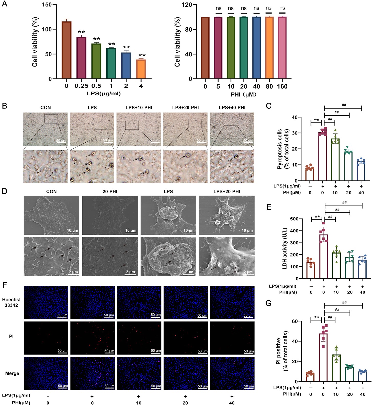
Acute lung injury (ALI) is a severe pulmonary disorder of sepsis with high clinical incidence and mortality. Nucleotide-binding oligomerization domain-like receptor family pyrin domain containing 3 (NLRP3)-cysteinyl aspartate specific proteinase 1-gasdermin D (GSDMD)-dependent pyroptosis of alveolar epithelial cells (AECs) has emerged as a crucial contributor to ALI during sepsis. Phillyrin (PHI), a natural lignan isolated from the traditional Chinese herbal medicine Forsythia suspensa, has been shown to have anti-inflammatory, antioxidant and antiviral properties. However, little is known about the protective role and potential mechanism of PHI in sepsis-induced ALI, and it is uncertain whether the protective effect of PHI in sepsis-induced ALI is connected to pyroptosis. This study aims to examine the preventive effects of PHI on sepsis-induced ALI via the inhibition of NLRP3/caspase-1/GSDMD-mediated pyroptosis in AECs. Our findings demonstrate that preadministration of PHI successfully reduces sepsis-induced pulmonary edema, systemic/pulmonary inflammation, and pulmonary histological damage in lung tissues, bronchoalveolar lavage fluid, and the serum of septic mice. Intriguingly, PHI preadministration suppresses sepsis-induced protein expressions of pyroptosis-specific markers, especially their active forms. In vitro assays show that PHI pretreatment also protects type II AECs (MLE-12) from lipopolysaccharide-induced pyroptosis by preventing the activation of the pyroptosis signaling pathway. The results from molecular docking and surface plasmon resonance reveal that PHI has a significant affinity for direct binding to the GSDMD protein, suggesting that GSDMD is a potential pharmacological target for PHI. In conclusion, PHI can prevent sepsis-triggered ALI by effectively suppressing the activation of the canonical pyroptosis signaling pathway and pyroptosis of AECs.

Read Full Abstract10.3724/abbs.2024161
Characterization of the association and sequestration of RNA-binding proteins by single-stranded DNA chimeraGraphical AbstractVerified
Acta Biochimica et Biophysica Sinica

Characterization of the association and sequestration of RNA-binding proteins by single-stranded DNA chimera

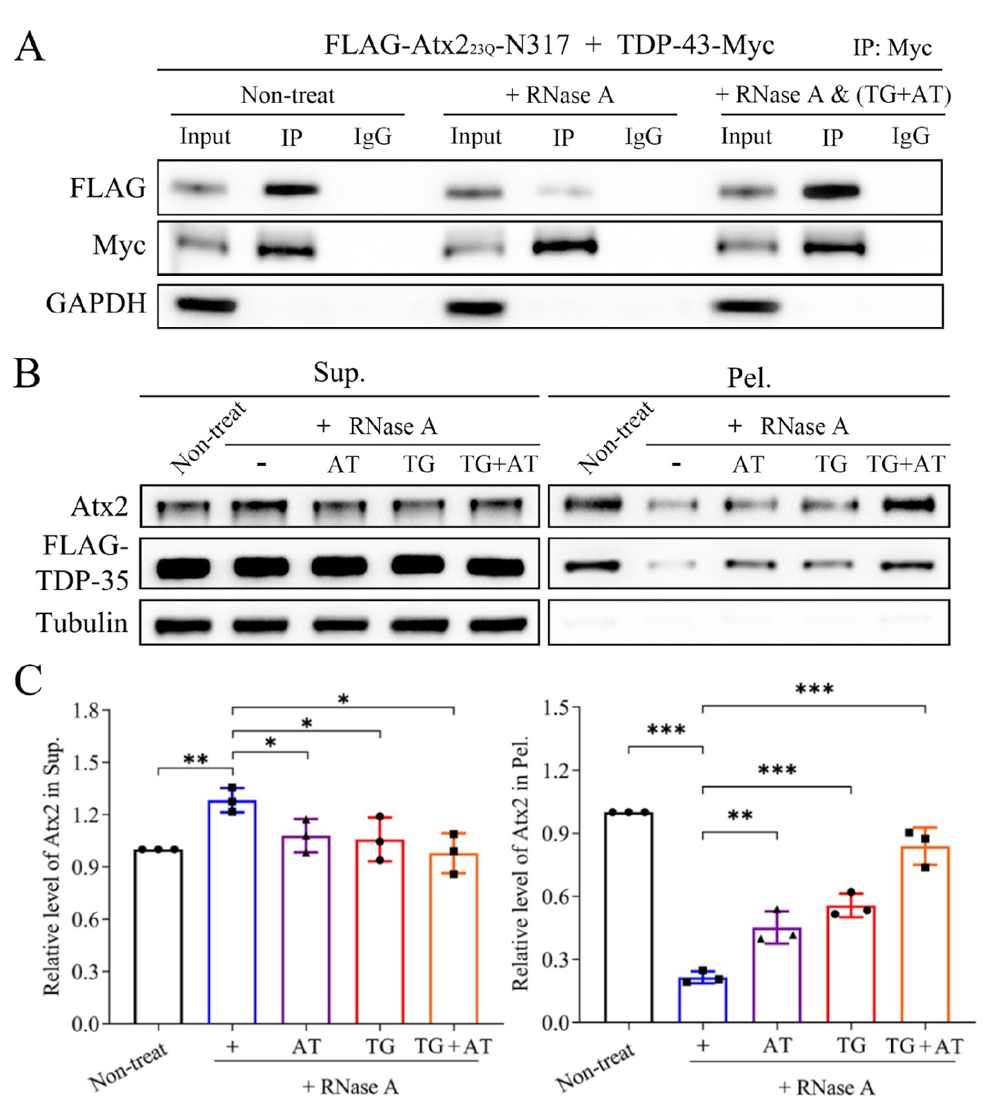
The biomolecular assemblies (condensates or aggregates) formed by mutant proteins are a pathological hallmark of neurodegenerative diseases. Some RNA-binding proteins (RBPs) are typically prone to aggregation that is closely associated with disease pathologies. These RBPs include numerous well-recognized pathogenic proteins, such as TAR DNA binding protein of 43 kDa (TDP-43), fused in sarcoma (FUS), ataxin-2 (Atx2), and poly(A)-binding protein nuclear 1 (PABPN1). Recent studies have revealed that liquid-liquid phase separation (LLPS), as a mechanism, underlies the highly dynamic and reversible granule formation of RBPs, and highlighted that multivalent RNA molecules play crucial roles in this process. These granules are necessary for diverse physiological functions, such as RNA splicing, trafficking, and even RNA storage, during stress. However, the aberrant phase transition of these mutant RBPs usually results in the formation of solid-like aggregates or inclusions within both the cytoplasm and nucleus. More importantly, aggregates formed by RBPs can sequester specific proteins, RNAs or other interacting partners, consequently contributing to RBP-related pathologies. For example, wild-type PABPN1 forms dynamic nuclear speckles with the assistance of poly(A) RNAs, whereas Ala expansion of PABPN1 results in the formation of aggregates, which are involved in the disease progression of oculopharyngeal muscular dystrophy (OPMD). Although the biological importance of various RBP granules is realized in either the cytoplasm or nucleus, how RNA regulates the formation of granules and the transition to aberrant RBP aggregates remains largely unknown. The interaction of a protein with other biomolecules (proteins, nucleic acids, etc.) is the prerequisite for the protein executing its normal biological function in cells. Identifying protein-protein and protein-RNA interactions is fundamental for the biochemical investigation of an individual protein and for attempts to understand the functional role of the protein. To date, many methods for studying protein-protein interactions have been developed on the basis of various principles, but it is still difficult to clarify whether the interactions between proteins, especially the RBPs involved, are direct or indirect, since RBPs generally bind to diverse RNAs closely and are incorporated into macromolecular ribonucleoprotein (RNP) complexes. We have taken several pairs of RBPs as examples, including TDP-35 (C-terminal 35-kDa fragment of TDP-43) with TDP-43 or TIA1, PABPN1 with a 25-kDa component of the mammalian cleavage factor I complex (CFIm25) and Atx2 with DEAD-box RNA helicase 6 (DDX6), and applied modified co-immunoprecipitation (Co-IP) and supernatant/pellet (S/P) fractionation experiments to characterize the association and sequestration of RBPs by using single-stranded DNA (ssDNA) chimera under ribonuclease (RNase) treatment. We designed several pieces of ssDNA oligonucleotides to mimic particular RNAs in cells that may mediate the association and sequestration of RBPs. The association of RBP proteins generally requires binding with multivalent RNA chains, since the bound RNAs tend to incorporate into a large protein-RNA complex with the help of RNA molecules. In Co-IP assay, especially for RBPs, RNase is often utilized to digest RNA in cell lysates to characterize whether the association of different RBPs is direct or indirect. It is important for us to demonstrate the active role of particular RNAs in the association or interaction of RBPs. Therefore, we designed and synthesized ssDNA chimeras to mimic the corresponding RNA that specifically bind to both RBPs simultaneously. In this case, ssDNA is used for rescuing the association of RBPs under the condition of RNase treatment, since the ssDNA oligonucleotide is resistant to nuclease activity. To design ssDNA chimeras for the RBPs of interest, first, the RNA sequences that bind to the two RPBs should be defined. The ssDNA should contain at least two portions (motifs) that specifically bind to each RBP, and each ssDNA portion may include 2–3 repeats of the binding sequence, so that the ssDNA can be recognized and bound efficiently by each RBP. Notably, the T base in ssDNA may sometimes be replaced with the U base (dU) for some more specific-binding RBPs, such as PABPN1. In the case of TDP-43 with Atx2, the binding specificities of the RNA sequences for TDP-43 and Atx2 are UG-rich and AUUUUU (AU5), respectively; then, the TG repeat portion is designed to bind to TDP-43, and the AT5 repeat is to bind to Atx2. Thus, an integrated method of co-IP and S/P fractionation was applied to characterize the association and sequestration of RBPs by combining ribonuclease (RNase) and ssDNA treatments.
Read Full Abstract10.3724/abbs.2024157
NLRP3 inflammasome-mediated disruption of mitochondrial homeostasis in alveolar macrophages contributes to ozone-induced acute lung inflammatory injuryGraphical AbstractVerified
Acta Biochimica et Biophysica Sinica

NLRP3 inflammasome-mediated disruption of mitochondrial homeostasis in alveolar macrophages contributes to ozone-induced acute lung inflammatory injury

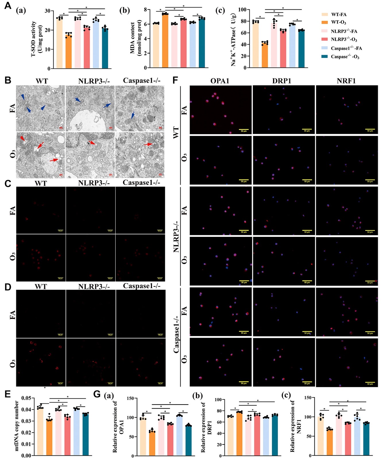
Ozone (O3), a prevalent atmospheric pollutant, can induce lung injury. However, the molecular mechanisms of O3-induced acute lung inflammatory injury remain unclear. In this study, we investigate the abnormal changes in and molecular mechanism of mitochondrial homeostasis in alveolar macrophages (AMs) in O3-induced acute lung inflammatory injury mice. Mitochondria and mitochondrial reactive oxygen species (mtROS) are labeled with Mito-Tracker® Deep Red and MitoSOX Red, respectively. Mitochondrial DNA (mtDNA) in AMs from the bronchoalveolar lavage fluid (BALF) is detected via real-time PCR, and the expressions of mitochondrial fusion/fission-related and biogenesis-related proteins in AMs are determined via immunofluorescence staining. Our data show that in O3-induced acute lung inflammatory injury mice, the number of AMs and the protein expression of the NLRP3 inflammasome complex in the lung tissue are increased. In AMs from O3-exposed mice, the number of mitochondria, mtROS, and fission-related protein DRP1 are increased, but the levels of Na+-K+-ATPase, fusion-related protein OPA1, biogenesis-related protein NRF1 and mtDNA are significantly decreased. Compared with that in O3-exposed WT mice, lung inflammation is attenuated, especially the indicators of mitochondrial homeostatic imbalance in AMs, which are alleviated in NLRP3‒/‒ and Caspase-1‒/‒ mice after O3 exposure. These findings indicate that the NLRP3 inflammasome-mediated imbalance in mitochondrial homeostasis in AMs contributes to O3-induced acute lung inflammatory injury. This study may provide a new target for the prevention of lung inflammation induced by O3.

Read Full Abstract10.3724/abbs.2024171
CircMALAT1 promotes the proliferation and metastasis of intrahepatic cholangiocarcinoma via the miR-512-5p/VCAM1 axisGraphical AbstractVerified
Acta Biochimica et Biophysica Sinica

CircMALAT1 promotes the proliferation and metastasis of intrahepatic cholangiocarcinoma via the miR-512-5p/VCAM1 axis

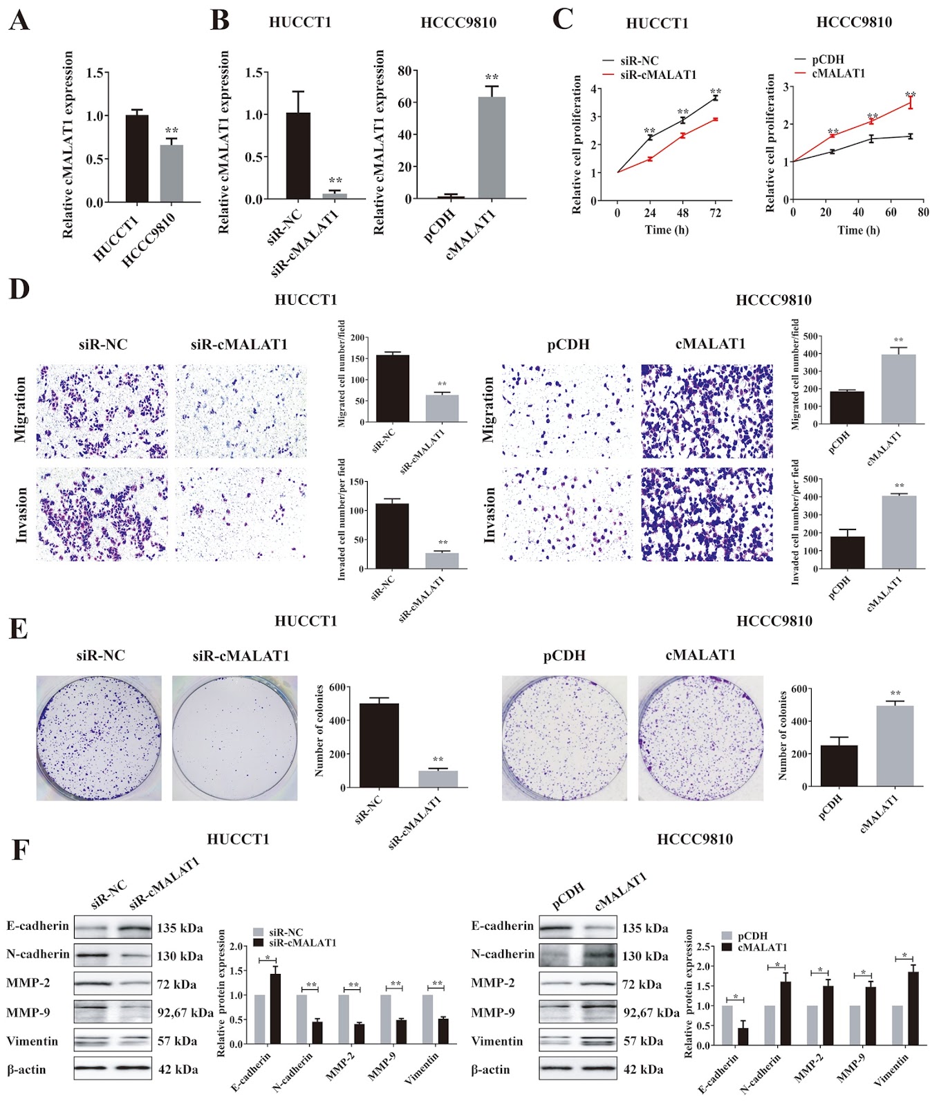
Circular RNAs play a pivotal role in the progression of various cancers. In our previous study, we observed high expression of the circRNA MALAT1 (cMALAT1) in intrahepatic cholangiocarcinoma (ICC) cells co-incubated with activated hepatic stellate cells. This study is designed to explore the roles of cMALAT1 and the underlying mechanisms in ICC. We find that cMALAT1 significantly facilitates the progression of ICC both in vitro and in vivo. The binding between cMALAT1 and miR-512-5p is subsequently confirmed through RNA pull-down experiments. As anticipated, the application of miR-512-5p mimics noticeably reverses the cMALAT1 overexpression-induced malignant phenotypes of ICC cells. Furthermore, VCAM1 is identified as a downstream gene of the cMALAT1/miR-512-5p axis. Importantly, silencing of VCAM1 not only effectively suppresses the malignant phenotypes of ICC cells but also significantly impairs the functions of cMALAT1. Our study reveals that cMALAT1 promotes the progression of ICC by competitively binding to VCAM1 mRNA with miR-512-5p, leading to the upregulation of VCAM1 expression and the activation of the PI3K/AKT signaling pathway.

Read Full Abstract10.3724/abbs.2024185
Nicotinamide mononucleotide ameliorates ionizing radiation-induced spermatogenic dysfunction in mice by modulating the glycolytic pathwayGraphical AbstractVerified
Acta Biochimica et Biophysica Sinica

Nicotinamide mononucleotide ameliorates ionizing radiation-induced spermatogenic dysfunction in mice by modulating the glycolytic pathway

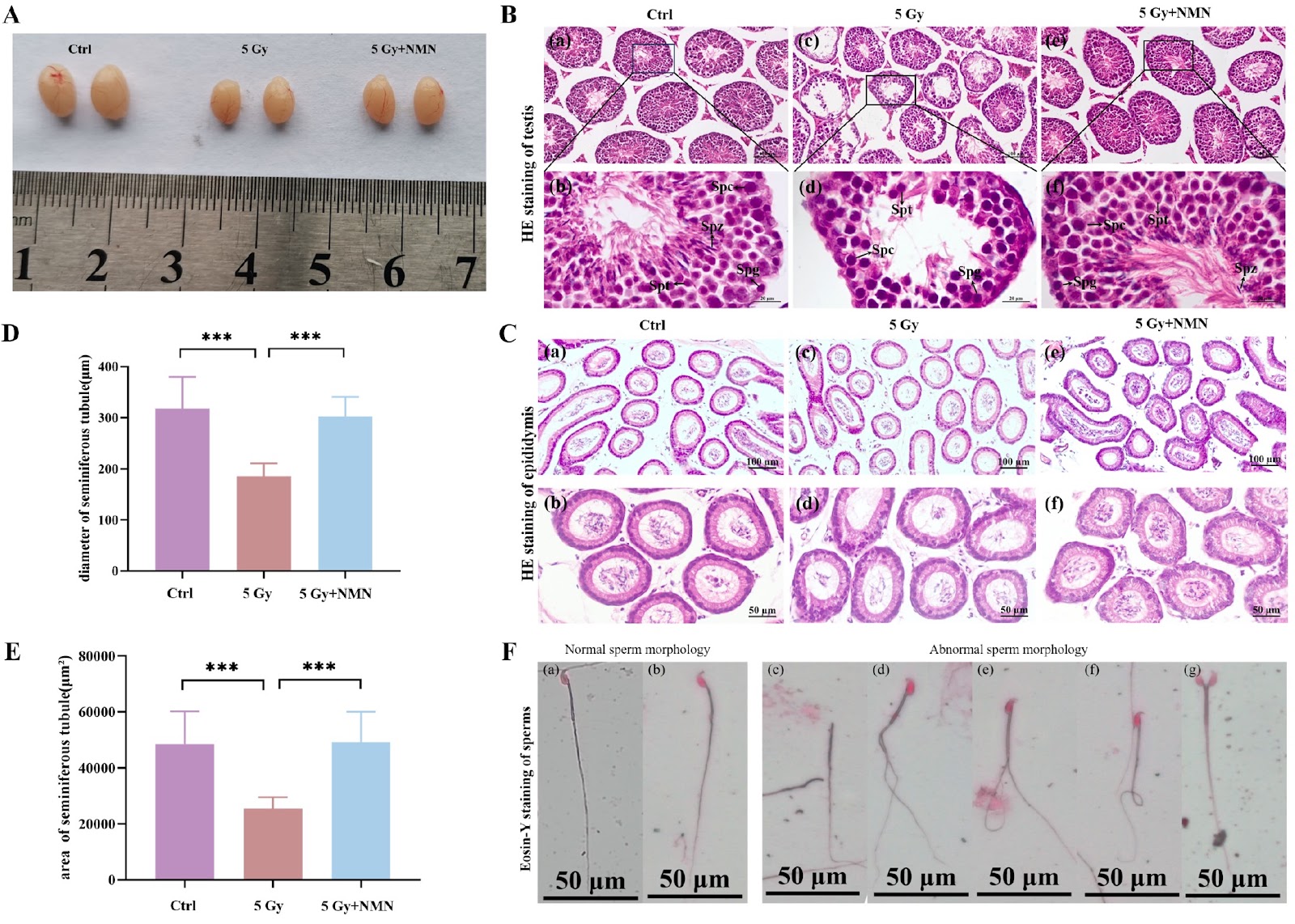
Radiotherapy, a common cancer treatment, leads to infertility in male cancer survivors, particularly young and middle-aged patients. Nicotinamide mononucleotide (NMN), a precursor of nicotinamide adenine dinucleotide (NAD+), plays crucial roles in energy metabolism, DNA repair, and gene expression. The purpose of this study is to investigate the protective effects and underlying mechanisms of NMN against ionizing radiation (IR)-induced testicular injury and spermatogenic dysfunction in an adult male mouse model. To assess the effects of NMN, single whole-body γ-ray irradiation is used to induce testicular injury and spermatogenic dysfunction in adult male mice. NMN is orally administered at 500 mg/kg before and after IR exposure. The structural and cellular damage to the testes caused by 5 Gy γ-ray irradiation, as well as the protective effect of NMN on testicular spermatogenic dysfunction, are evaluated. The serum hormone testosterone, LH, and FSH levels, as well as testicular NAD+, lactate, and pyruvate levels, are detected. Furthermore, the expressions of the apoptosis-related genes Bcl-2, Bax, and Caspase-3 and the rate-limiting enzymes HK2, PKM2, and LDHA, which are potentially associated with the mechanism of injury, are examined. The results demonstrate that 5 Gy γ-ray irradiation exposure causes a decrease in the serum testosterone, LH, and FSH levels in adult male mice, as well as in the testicular NAD+, lactate, and pyruvate levels, and causes damage to the testicular structure and cells. Morphometric analysis reveal a decrease in the testis mass, seminiferous tubule diameter, and height of the germinal epithelium. The sperm quantity, motility, and testicular volume are reduced in the 5 Gy group but are restored by NMN supplementation. NMN intervention downregulates the expressions of proapoptotic genes (Bax and Caspase-3) and upregulates the expression of an antiapoptotic gene (Bcl-2). Sertoli cells marker genes (WT-1, GATA-4, SOX9, and vimentin) and glycolysis rate-limiting enzyme-encoding genes (HK2, PKM2, and LDHA) are significantly upregulated. In summary, NMN has a positive regulatory effect on testicular spermatogenic dysfunction in male mice induced by ionizing radiation. This positive effect is likely achieved by promoting the proliferation of spermatogenic cells and activating glycolytic pathways. These findings suggest that NMN supplementation may be a potential protective strategy to prevent reproductive damage to male subjects from ionizing radiation.

Read Full Abstract10.3724/abbs.2024167
Transcription factor occupancy limits DNA methylation and determines ICAM1 expression in breast cancerGraphical AbstractVerified
Acta Biochimica et Biophysica Sinica

Transcription factor occupancy limits DNA methylation and determines ICAM1 expression in breast cancer

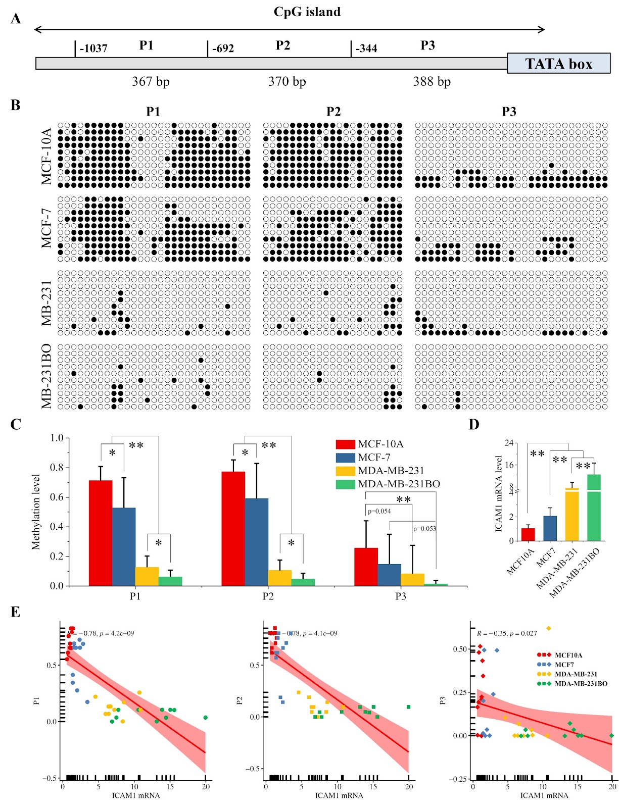
The interaction between TF binding and DNA methylation is increasingly recognized as a key player in the regulation of gene expression. However, the role of this interaction in regulating ICAM1 expression in breast cancer has not been elucidated. CpG methylation in the ICAM1 promoter is negatively correlated with ICAM1 expression, and ICAM1 expression is significantly positively correlated with DNMT and TET3 expression in breast cancer. TF binding attenuates ICAM1 promoter CpG methylation and promotes ICAM1 transcription. DNA methylation regulation enhances ICAM1 expression in breast cancer by promoting the transcription of transcription factors. In terms of mechanisms, RELA and STATs recruit TET3 to prevent DNMT-mediated DNA methylation, thereby maintaining CpG island hypomethylation in the ICAM1 promoter. Therefore, TF occupancy limits DNA methylation and affects ICAM1 expression in breast cancer.

Read Full Abstract10.3724/abbs.2024237
Multiple allostery in the regulation of PDGFR beta kinase activitiesGraphical AbstractVerified
Acta Biochimica et Biophysica Sinica

Multiple allostery in the regulation of PDGFR beta kinase activities

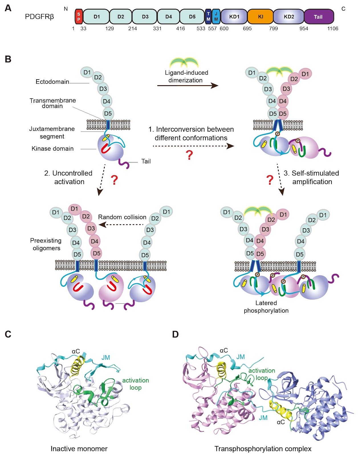
Platelet-derived growth factor receptor beta (PDGFRβ), a type III receptor tyrosine kinase (RTK) with a featured kinase insert, regulates important cellular functions. Dysregulation of PDGFRβ is associated with cardiovascular and fibrosis diseases. Thus, its kinase activity needs to be precisely regulated under physiological conditions. Early studies demonstrated that its kinase is autoinhibited by its juxtamembrane segment and activated by transphosphorylation. However, additional mechanisms are required for the comprehensive regulation of the receptor kinase. Herein, we provide evidence that dimerization of activated kinases, autoinhibition by the kinase insert, and dimerization of inactive kinase, all contribute to the regulation of the receptor kinase. Moreover, we find such multiple allosteric regulation is also conserved in other type III RTKs, including colony stimulating factor 1 receptor (CSF1R). Impaired allosteric regulation of CSF1R is associated with malfunctions of microglia and demyelination of neurons in hereditary diffuse leukoencephalopathy with spheroids (HDLS).

Read Full Abstract10.3724/abbs.2024205
DDX11 interacts with PARP1 to facilitate PARylation, thereby promoting gallbladder cancer progression and conferring gemcitabine resistanceGraphical AbstractVerified
Acta Biochimica et Biophysica Sinica

DDX11 interacts with PARP1 to facilitate PARylation, thereby promoting gallbladder cancer progression and conferring gemcitabine resistance

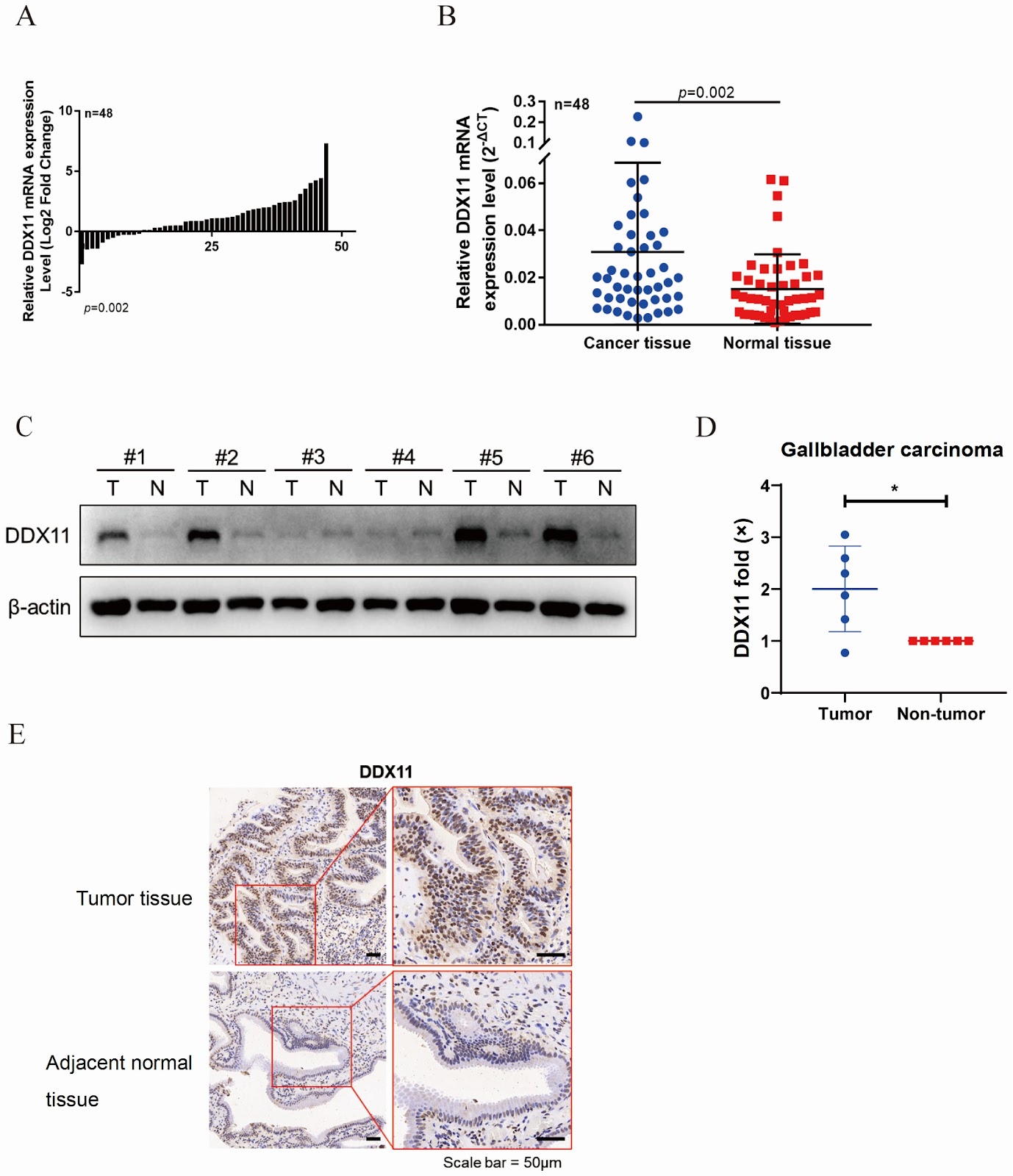
Gemcitabine resistance poses a significant challenge in gallbladder cancer (GBC) treatment, necessitating exploration of its molecular mechanisms. This study focuses on DDX11, which is highly expressed in gemcitabine-resistant GBC cells, suggesting a potential role in DNA damage repair. We establish gemcitabine-resistant GBC cell lines and observe significantly higher DDX11 expression in these cells than in parental cells. Clinical tissue analysis through qRT-PCR, western blot analysis, and immunohistochemistry confirms elevated DDX11 levels in tumors compared with adjacent normal tissues. Functional assays demonstrate that DDX11 knockdown inhibits cell proliferation, colony formation, and tumor growth, while restoring gemcitabine sensitivity. Mechanistically, proteomic analysis and co-immunoprecipitation reveal that the interaction of DDX11 with PARP1 leads to increased poly(ADP-ribosyl)ation (PARylation), which promotes DNA repair and drug resistance. Notably, combining gemcitabine with the PARP inhibitor olaparib has synergistic anti-tumor effects on resistant cells. These findings indicate that DDX11 contributes to GBC progression and chemoresistance by regulating PARP1-mediated PARylation and that targeting this pathway with PARP inhibitors may overcome gemcitabine resistance. This study provides new insights into GBC drug resistance mechanisms and suggests that combining conventional chemotherapy with PARP inhibition is a potential therapeutic strategy for resistant patients. The DDX11-PARP1-PARylation axis represents a promising target for improving GBC treatment outcomes, particularly in gemcitabine-resistant patients.

Read Full Abstract10.3724/abbs.2025155
Eupalinolide B exerts cytotoxic effects against KRAS-mutant NSCLC through Nrf2/HO-1-regulated ferroptosisGraphical AbstractVerified
Acta Biochimica et Biophysica Sinica

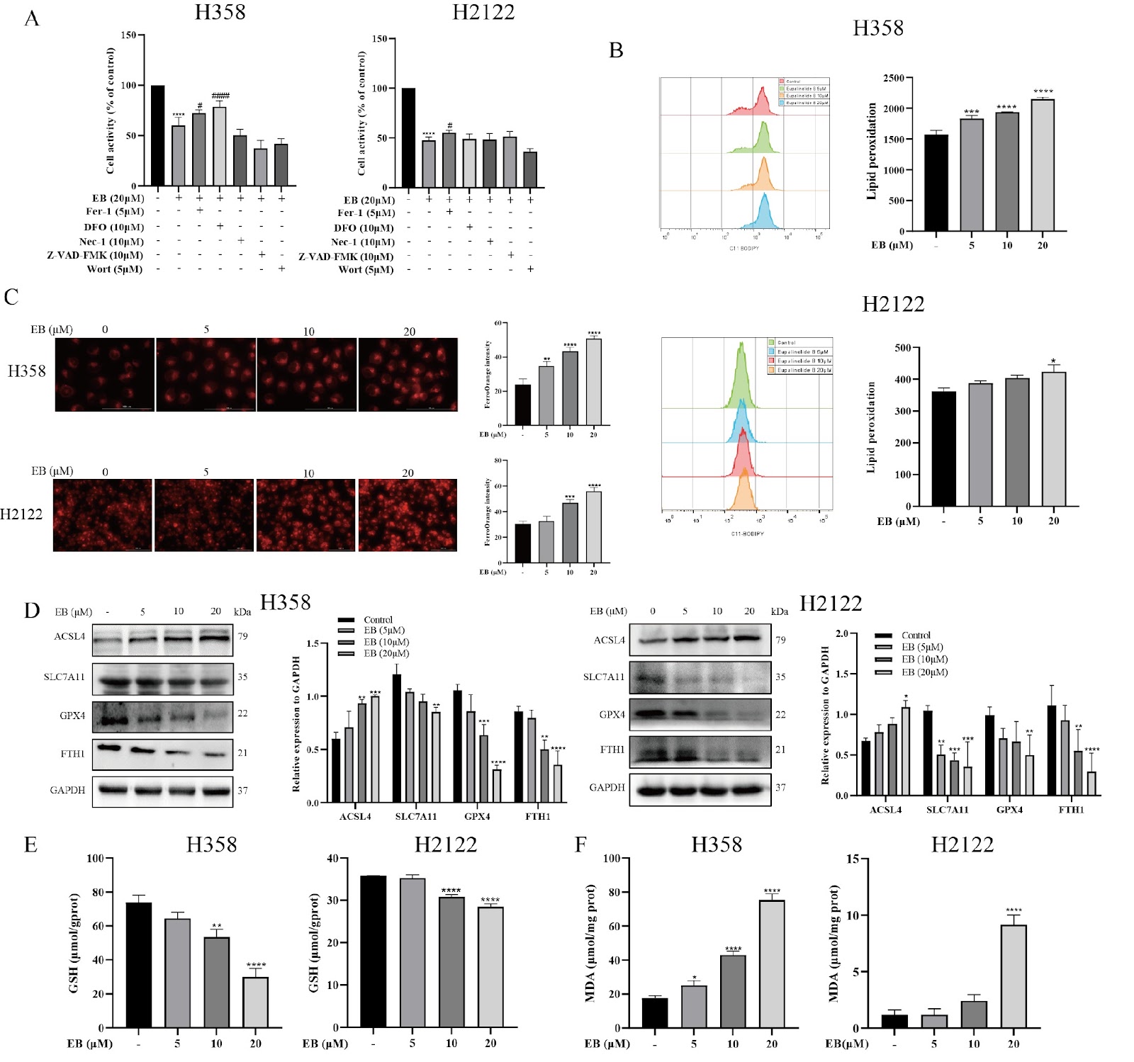
Eupalinolide B exerts cytotoxic effects against KRAS-mutant NSCLC through Nrf2/HO-1-regulated ferroptosis

Ferroptosis, an iron-dependent form of regulated cell death, is characterized by excessive reactive oxygen species (ROS) accumulation and lipid peroxidation of polyunsaturated fatty acids (PUFAs) in cellular membranes. Non-small cell lung cancer (NSCLC) harboring KRAS mutations often exhibits therapeutic resistance but may display high susceptibility to ferroptosis. Eupalinolide B (EB), a natural compound with documented anti-cancer activity, has not been thoroughly explored for its ferroptosis-inducing potential in KRAS-mutant NSCLC. In this study, we demonstrate that EB treatment significantly elevates ROS levels, intracellular iron accumulation, and lipid peroxidation in KRAS-mutant NSCLC cells, resulting in ferroptotic cell death. Molecular docking and cellular thermal shift assays reveal that EB directly binds to and activates heme oxygenase-1 (HO-1), a critical component of the Kelch-like ECH-associated protein 1 (Keap1)-Nrf2/HO-1 oxidative stress response pathway. Genetic or pharmacological inhibition of HO-1 attenuates EB-induced ferroptosis, underscoring the pivotal role of HO-1-mediated oxidative stress in this process. Furthermore, in vivo studies using KRAS-mutant H358 xenograft models confirm the potent anti-tumor effects of EB. Collectively, our findings establish that EB triggers ferroptosis in KRAS-mutant NSCLC by activating the Keap1-Nrf2/HO-1 pathway, suggesting a promising therapeutic strategy for this challenging malignancy.

Read Full Abstract10.3724/abbs.2025211
Mitochondrial dysfunction in adipocyte differentiation: implications for obesity and metabolic syndrome interventionGraphical AbstractVerified
Acta Biochimica et Biophysica Sinica

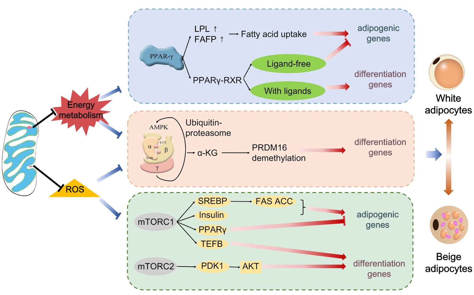
Mitochondrial dysfunction in adipocyte differentiation: implications for obesity and metabolic syndrome intervention

Mitochondrial dysfunction critically disrupts adipocyte remodeling by impairing the thermogenic browning process essential for combating obesity through the upregulation of uncoupling protein 1 (UCP1) and mitochondrial biogenesis. Deficiencies in mitochondrial metabolism, dynamics (including fusion/fission), and autophagy suppress adipocyte plasticity, directly inhibiting UCP1 expression and destabilizing the PPAR-γ/PGC-1α and adenosine 5′-monophosphate (AMP)-activated protein kinase (AMPK)/mammalian target of rapamycin (mTOR) signaling pathways. These disruptions reduce energy expenditure, exacerbate insulin resistance, and promote metabolic syndrome. Moreover, mitochondrial inactivation intersects with neurodegenerative disorders via oxidative stress induced by β-amyloid and α-synuclein aggregation, amplifying systemic metabolic dysregulation. Structural mitochondrial anomalies further impede lipid utilization and adipose tissue adaptation, but unresolved crosstalk between mtDNA and nuclear DNA complicates therapeutic targeting. Future research must prioritize spatiotemporal mapping of mitochondrial dynamics in adipocyte differentiation via single-cell omics to identify key regulatory nodes. Addressing these mechanisms could unlock precision therapies, such as gene editing, to restore mitochondrial function, enhance adipocyte browning, and mitigate obesity, related pathologies alongside neurodegenerative and age-associated diseases.

Read Full Abstract10.3724/abbs.2025153
Decreased CCL5 expression in endometrial stromal cells induces deficient CCR5+CD4+ T cells in endometriosisGraphical AbstractVerified
Acta Biochimica et Biophysica Sinica

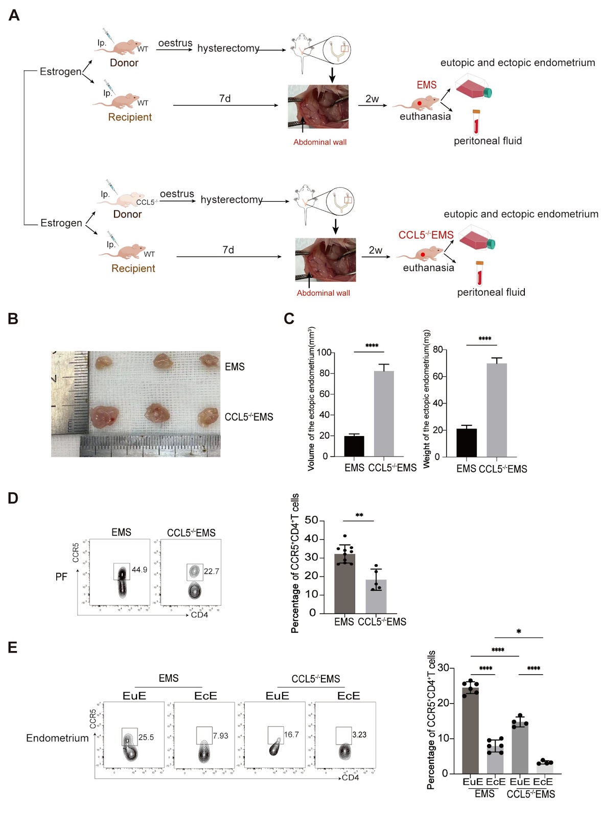
Decreased CCL5 expression in endometrial stromal cells induces deficient CCR5+CD4+ T cells in endometriosis

Endometriosis (EMS) is a benign gynecological disease characterized by the growth of endometrial tissue outside the uterine cavity. Evidence shows that the survival of patients with ectopic endometrial implants is associated with a dysregulated immune microenvironment. CD4+ T cells can regulate EMS through diverse cytokines, the inflammatory response, and angiogenesis. CCR5+CD4+ T cells exhibit increased cellular immunogenicity and play a role in infectious diseases, host defense, and cancer progression. However, the specific mechanisms of CCR5+CD4+ T cells in EMS remain unknown. In the present study, flow cytometry and RNA-seq are utilized to assess the proportions and features of CCR5+CD4+ T cells in EMS patients, RT-PCR and ELISA are used to assess the production of CCL5 by ectopic endometrial stromal cells (ecESCs). Two EMS models are established through C57B6 wild-type and CCL5‒/‒ mice and utilized to explore the in vivo effects of CCR5+CD4+ T cells on ectopic lesions. Compared with CCR5‒CD4+ T cells, CCR5+CD4+ T cells display a more activated and cytotoxic phenotype. Diminished CCR5+CD4+ T cells and their impaired ability to produce IFN-γ are observed in the ectopic lesions of EMS patients and in murine EMS models. Impaired production of CCL5 has been detected in human ecESCs. Moreover, endometria stripped from CCL5‒/‒ mice are more likely to generate ectopic lesions in the peritoneum of recipient mice. These findings demonstrate that the attenuated recruitment of CCR5+CD4+ T cells in ectopic lesions caused by decreased production of CCL5 in ecESCs may facilitate the progression of EMS.

Read Full Abstract10.3724/abbs.2024178
Continuous carbon source supply is essential for high rifamycin productivity of Amycolatopsis mediterranei in nitrate-stimulated fermentation revealed by a metabolomic studyGraphical AbstractVerified
Acta Biochimica et Biophysica Sinica

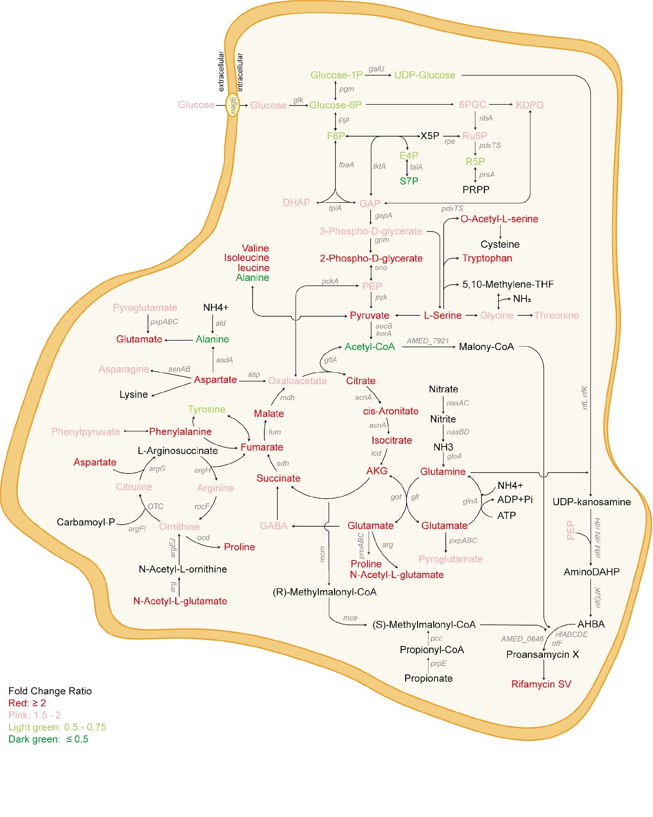
Continuous carbon source supply is essential for high rifamycin productivity of Amycolatopsis mediterranei in nitrate-stimulated fermentation revealed by a metabolomic study

Amycolatopsis mediterranei U32 is an industrial strain capable of producing therapeutically useful rifamycin SV. In early days of fermentation studies, nitrate was found to increase the yield of rifamycin along with globally, affecting both carbon and nitrogen metabolism in favor of antibiotic biosynthesis; thus, the nitrate-stimulating effect (NSE) hypothesis was proposed. Although GlnR is likely the master regulator of the pleotropic effect of NSE, the global metabolism affected by NSE has never been systematically examined. In this study, we use mass spectrometry-based metabolomics to quantitatively monitor the metabolomic responses of A. mediterranei U32 to nitrate supplementation. The concentrations of many metabolites involved in central carbon metabolism, including glucose 6-phosphate, glucose 1-phosphate, UDP-glucose, and acetyl-coenzyme A, decrease significantly after the addition of 80 mM potassium nitrate to the medium. We find that the rifamycin SV production yield could be increased by the addition of glucose during the logarithmic growth phase. Moreover, at multiple time points during glucose supplementation in the mid- and late-exponential phases, the yield of rifamycin SV further increases, reaching 354.3%. Quantitative real-time PCR assays of the key genes corresponding to the synthesis of the rifamycin SV precursor combined with data from metabolomics analysis confirm that carbon source deficiency is compensated for after glucose supplementation and that the expression of genes involved in the pathway of 3-amino-5-hydroxybenzoic acid synthesis by UDP-glucose and glutamine is significantly increased. This preliminary exploration of dynamic metabolomic profiles has the potential to increase our understanding of the NSE.

Read Full Abstract10.3724/abbs.2024245
Noncanonical functions of microRNAs in the nucleusGraphical AbstractVerified
Acta Biochimica et Biophysica Sinica

Noncanonical functions of microRNAs in the nucleus

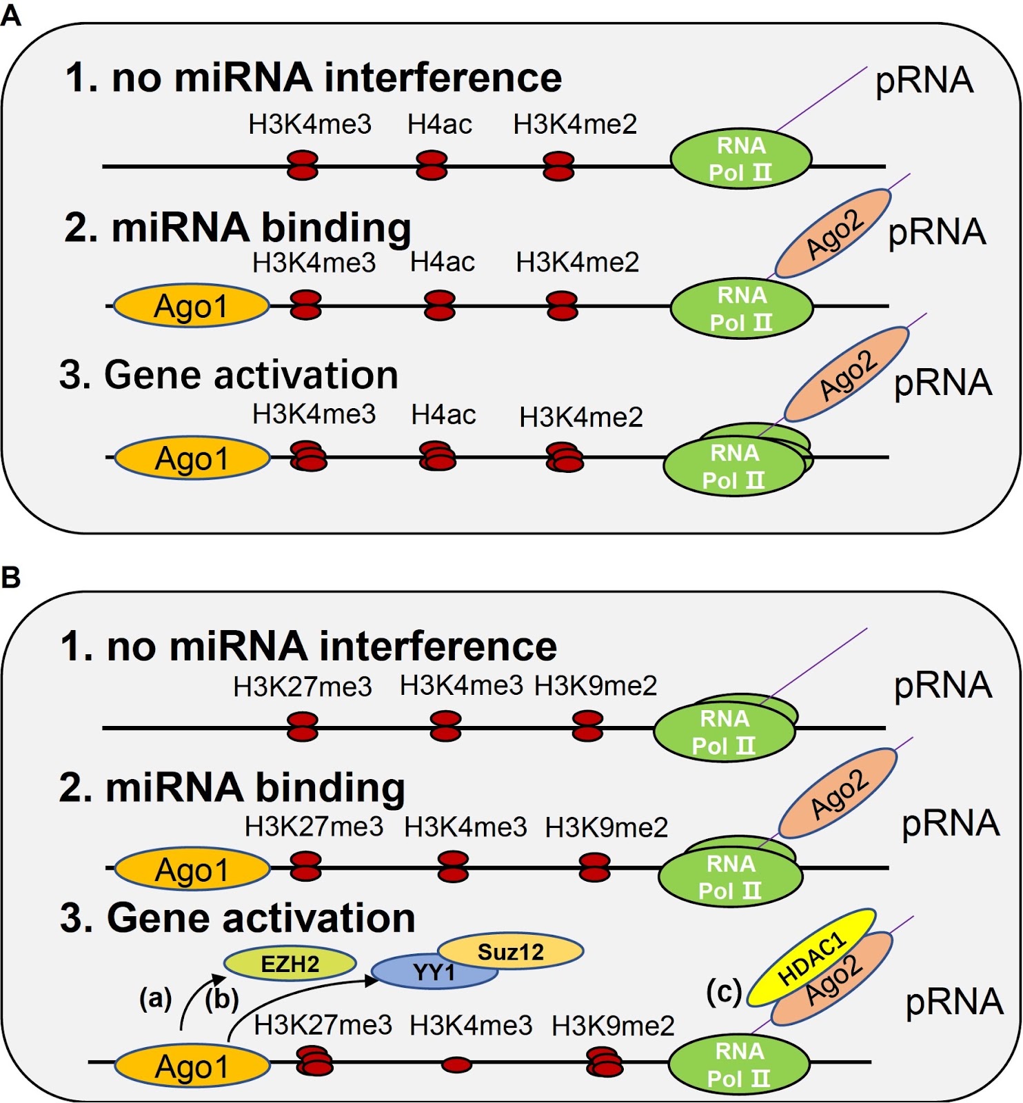
MicroRNAs (miRNAs) are small noncoding RNAs (ncRNAs) that play their roles in the regulation of physiological and pathological processes. Originally, it was assumed that miRNAs only modulate gene expression post-transcriptionally in the cytoplasm by inducing target mRNA degradation. However, with further research, evidence shows that mature miRNAs also exist in the cell nucleus, where they can impact gene transcription and ncRNA maturation in several ways. This review provides an overview of novel models of nuclear miRNA functions. Some of the models remain to be verified by experimental evidence, and more details of the miRNA regulation network remain to be discovered in the future.

Read Full Abstract10.3724/abbs.2023268
PROS1/AXL signaling protects mice from lethal influenza infection by inducing M2 macrophage polarizationGraphical AbstractVerified
Acta Biochimica et Biophysica Sinica

PROS1/AXL signaling protects mice from lethal influenza infection by inducing M2 macrophage polarization

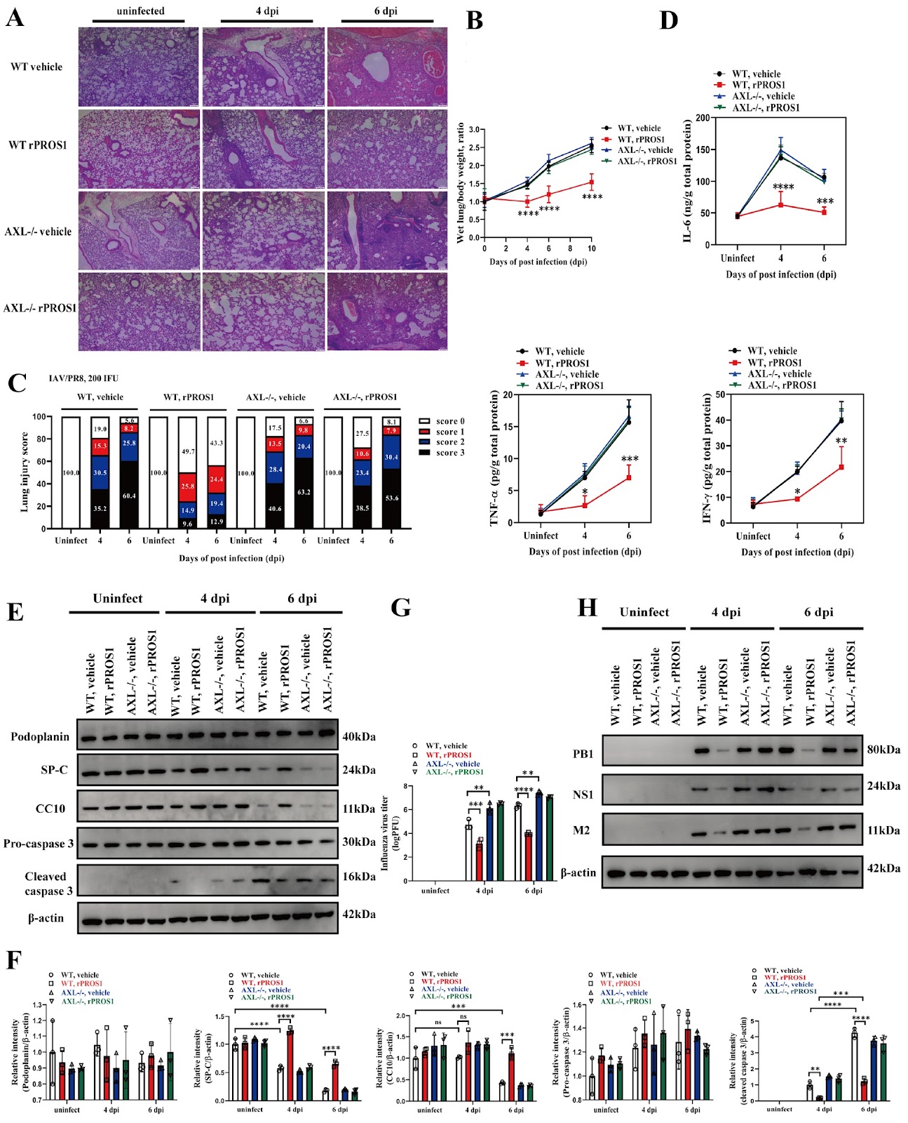
AXL, a member of the TAM (Tyro3, AXL, and Mertk) subfamily of RTKs, is abundantly expressed in lung tissue and has been implicated in viral infections and lung injury. PROS1, one of the ligands known to activate AXL, functions as an immunomodulator in many diseases. However, the role of PROS1/AXL signaling in influenza A virus (IAV) infection and infection-induced lung injury is largely unknown. In this study, we find that the exogenous administration of PROS1 mitigates lung injury and protects mice from lethal infection by IAVs through the activation of AXL. PROS1 induces the phosphorylation of AXL, which in turn recruits Gab1 and p85, a regulatory subunit of PI3K, to form a complex that activates Gab1 and its downstream PI3K/AKT/mTOR in alveolar macrophages. Gab1 knockdown in vivo, or LY294002 (a PI3K inhibitor), abolishes the PROS1/AXL-induced protective activity against lethal influenza infection in mice. We also show that PROS1/AXL signaling induces M2 polarization of alveolar macrophages through Gab1 activation both in vitro and in vivo. Gab1 knockdown inhibits M2 macrophage accumulation in IAV-infected lungs and attenuates the protective effect of PROS1. These results indicate that PROS1/AXL signaling can activate Gab1 in macrophages and induce macrophage polarization to an anti-inflammatory M2 phenotype, thereby eliciting protective activity against lethal infection with IAVs. These data also highlight the PROS1/AXL signal as a novel therapeutic target for IAV infection.

Read Full Abstract10.3724/abbs.2025169
The SRC-TOPK positive feedback loop promotes RB1 phosphorylation and drives the development of lung squamous cell carcinomaGraphical AbstractVerified
Acta Biochimica et Biophysica Sinica

The SRC-TOPK positive feedback loop promotes RB1 phosphorylation and drives the development of lung squamous cell carcinoma

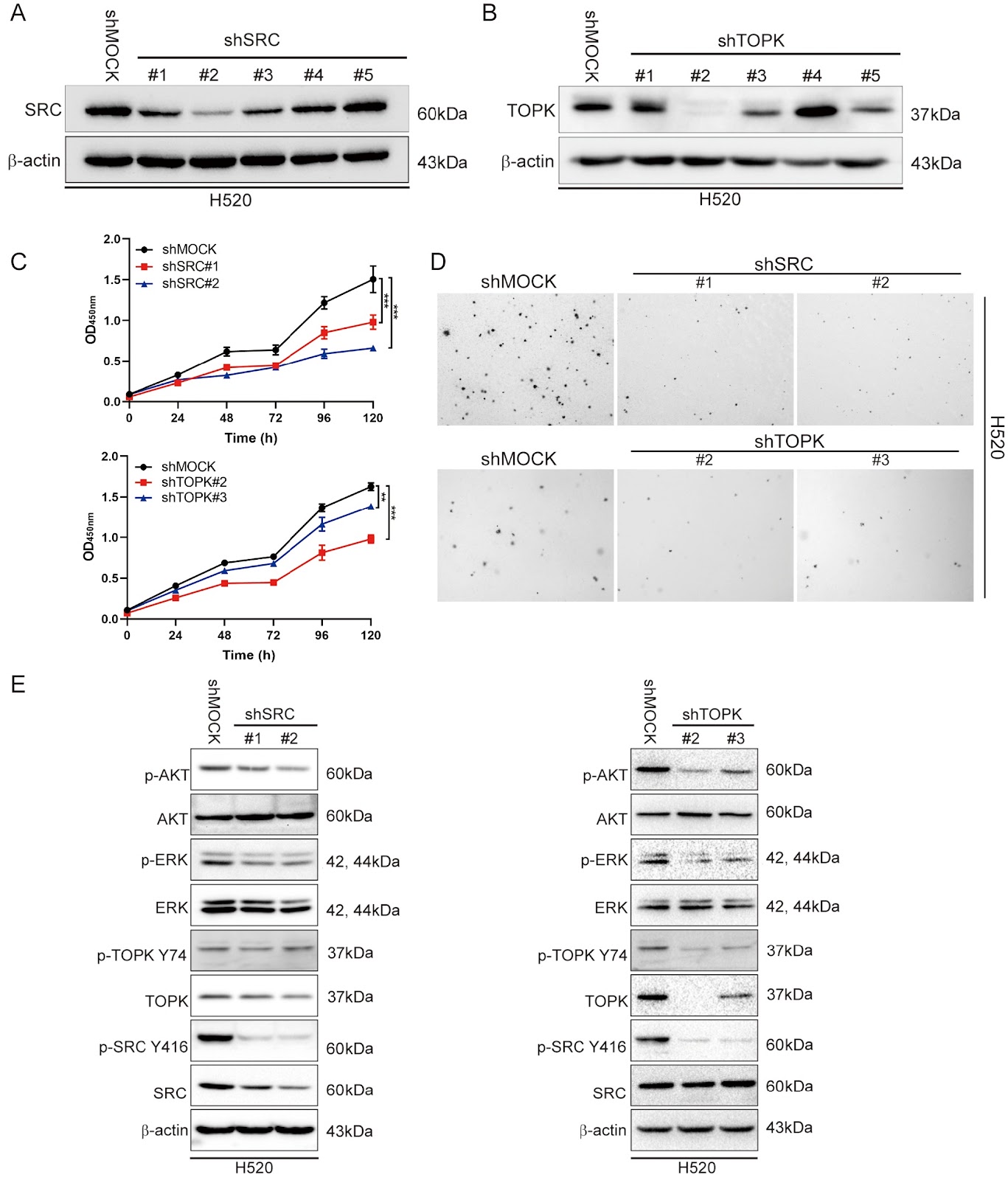
Lung squamous cell carcinoma (LUSC) is a common subtype of non-small cell lung cancer, with limited treatment options and poor patient prognosis. Currently, common driver mutations in lung adenocarcinoma rarely occur in LUSC; the mutated genes found in LUSCs lack corresponding targeted drugs. Therefore, it is necessary to discover new therapeutic targets for LUSC and provide patients with more treatment options. By analyzing different databases and tissue microarray immunohistochemistry staining, we firstly find that the expression of SRC/TOPK is elevated and positively correlated in LUSC and that patients with high SRC/TOPK expression have shorter survival time. Changing the expression levels of SRC/TOPK in LUSC cells can affect cell growth and colony formation, as there is a positive feedback loop between SRC and TOPK that regulates the transcription factor RB1, thereby altering the expressions of key factors in some growth-related signaling pathways. These inhibitors can synergistically promote apoptosis and have been validated in vivo. Therefore, the positive feedback loop between SRC and TOPK promotes tumorigenicity by inhibiting RB1 function, and has the potential to become a precise therapeutic target for LUSC, providing new possibilities for targeted therapy.

Read Full Abstract10.3724/abbs.2025149
CD47 blockade enhances cisplatin sensitivity by inhibiting DNA repair gene expressionGraphical AbstractVerified
Acta Biochimica et Biophysica Sinica

CD47 blockade enhances cisplatin sensitivity by inhibiting DNA repair gene expression

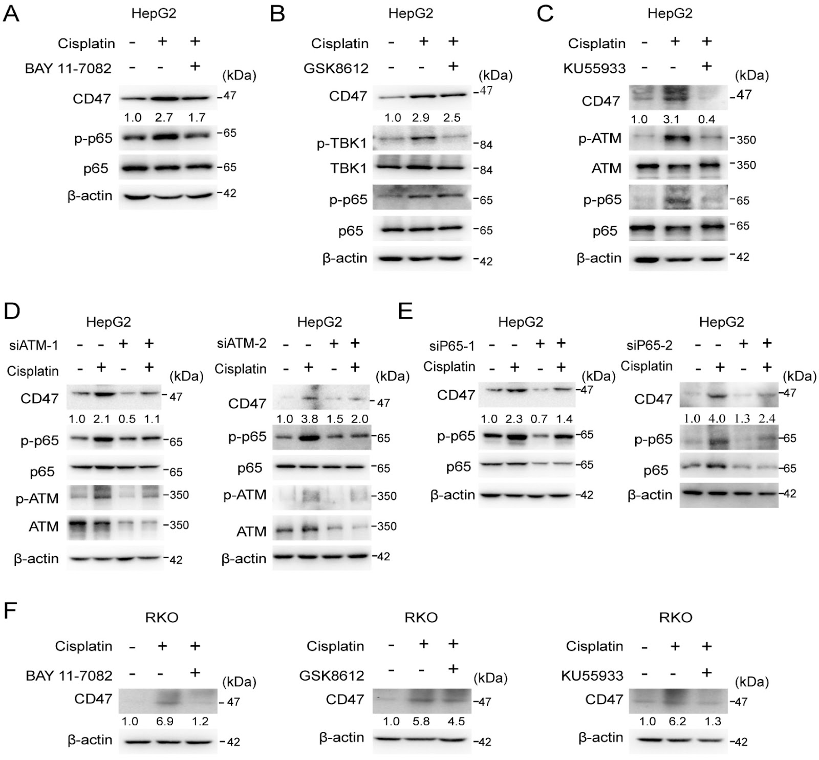
CD47, a cell surface transmembrane glycoprotein, is an innate immune checkpoint that suppresses phagocytic clearance. Emerging evidence suggests that CD47 has noncanonical functions. However, its involvement in chemotherapy resistance is not well understood. Our study reveals that cisplatin treatment upregulates CD47 expression across multiple cancer cell lines. Cisplatin induces the expression of CD47 through the ATM/NF-κB signaling pathway. Genetic ablation of CD47 dramatically sensitizes cancer cells to cisplatin. Mechanistically, CD47 depletion potentiates cisplatin-induced DNA damage, as demonstrated by elevated γH2AX formation and ATM phosphorylation. Knockdown of CD47 inhibits the expression of DNA repair genes ERCC1, FANCA, and BRCA2 through the ATM/NF-κB pathway. Remarkably, CD47 blockade with neutralizing antibodies recapitulates these effects, synergistically potentiating cisplatin’s DNA-damaging capacity while suppressing DNA repair capacity. CD47 blockade also potentiates cisplatin’s tumor inhibitory effect in vivo. These findings establish a novel mechanism whereby CD47 promotes cisplatin resistance through transcriptional regulation of DNA repair pathway, providing rationale for combining CD47-targeted therapies with conventional chemotherapy. This dual approach could simultaneously overcome immune evasion while enhancing treatment efficacy.

Read Full Abstract10.3724/abbs.2025147