Key Takeaways & Executive Findings
- •• Ozone exposure triggers NLRP3 inflammasome activation in alveolar macrophages, leading to mitochondrial dysfunction and acute lung inflammatory injury. • Mitochondrial homeostasis disruption in alveolar macrophages is characterized by increased mitochondrial number, mtROS, and DRP1, alongside decreased OPA1, NRF1, and mtDNA. • Genetic ablation of NLRP3 or Caspase-1 alleviates ozone-induced lung inflammation and mitochondrial imbalance, confirming the inflammasome's central role. • Targeting NLRP3 inflammasome-mediated mitochondrial disruption in alveolar macrophages offers a novel therapeutic strategy for ozone-induced lung injury.
Abstract
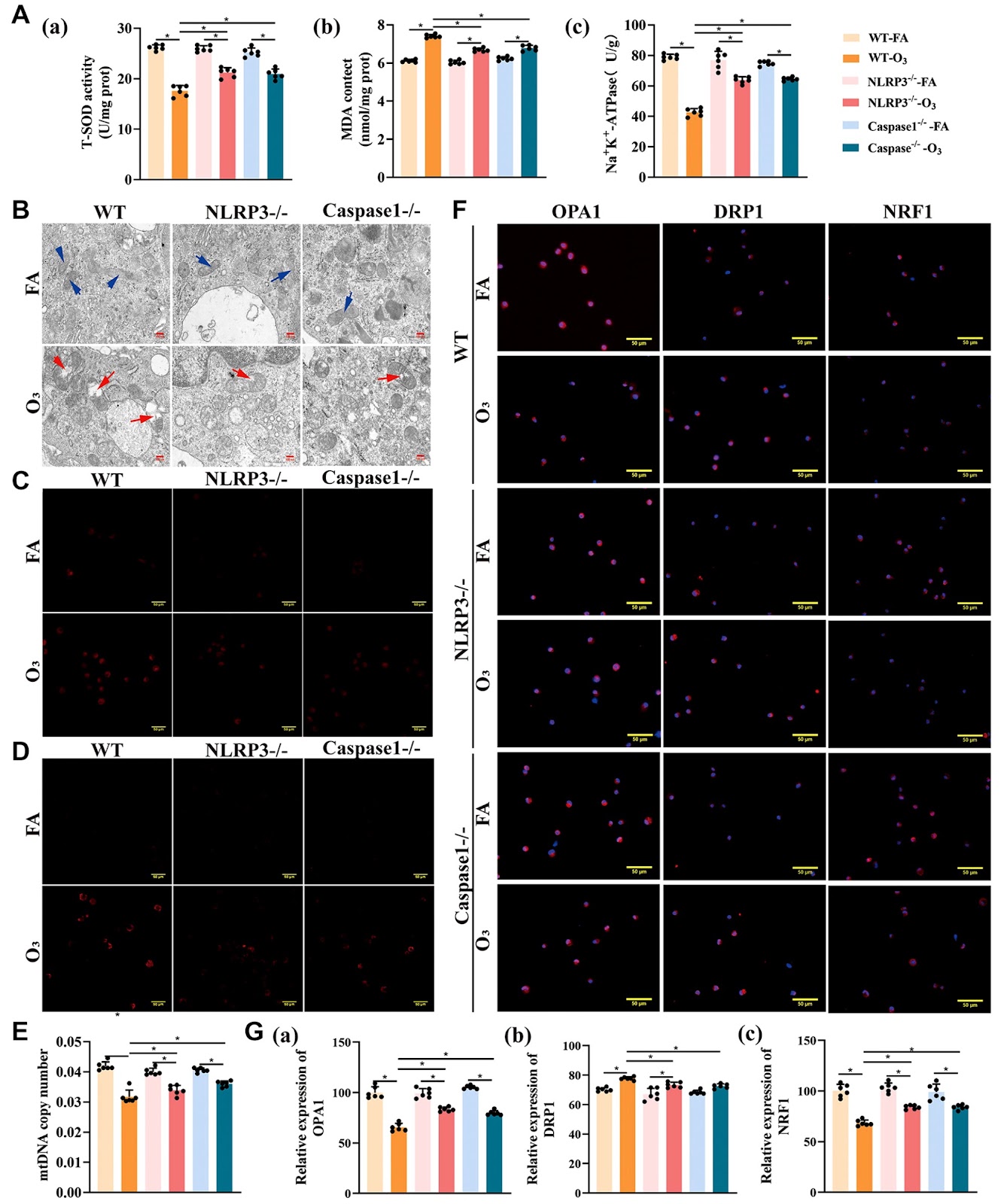
Ozone (O3), a prevalent atmospheric pollutant, can induce lung injury. However, the molecular mechanisms of O3-induced acute lung inflammatory injury remain unclear. In this study, we investigate the abnormal changes in and molecular mechanism of mitochondrial homeostasis in alveolar macrophages (AMs) in O3-induced acute lung inflammatory injury mice. Mitochondria and mitochondrial reactive oxygen species (mtROS) are labeled with Mito-Tracker® Deep Red and MitoSOX Red, respectively. Mitochondrial DNA (mtDNA) in AMs from the bronchoalveolar lavage fluid (BALF) is detected via real-time PCR, and the expressions of mitochondrial fusion/fission-related and biogenesis-related proteins in AMs are determined via immunofluorescence staining. Our data show that in O3-induced acute lung inflammatory injury mice, the number of AMs and the protein expression of the NLRP3 inflammasome complex in the lung tissue are increased. In AMs from O3-exposed mice, the number of mitochondria, mtROS, and fission-related protein DRP1 are increased, but the levels of Na+-K+-ATPase, fusion-related protein OPA1, biogenesis-related protein NRF1 and mtDNA are significantly decreased. Compared with that in O3-exposed WT mice, lung inflammation is attenuated, especially the indicators of mitochondrial homeostatic imbalance in AMs, which are alleviated in NLRP3‒/‒ and Caspase-1‒/‒ mice after O3 exposure. These findings indicate that the NLRP3 inflammasome-mediated imbalance in mitochondrial homeostasis in AMs contributes to O3-induced acute lung inflammatory injury. This study may provide a new target for the prevention of lung inflammation induced by O3.
1. Introduction
In recent years, ozone (O3) pollution has become increasingly serious, and the rapid expansion of the pollution season has led to severe pollution even in winter and spring [1]. The number of people exposed to O3 will increase significantly in the future under the influence of climate change. As an important air pollutant, O3 has been recognized as an urgent environmental issue, primarily because of its adverse effects on human health [2]. Extensive epidemiological studies have revealed respiratory system damage caused by O3 exposure [3,4]. Short-term inhalation of O3 is associated with an increased risk of hospitalization due to respiratory diseases [5]. The impact of O3 exposure is evident in the induction of airway hyperresponsiveness (AHR) and alveolar degradation, which can occur even within a short period of time (3 to 6 h) at an O3 concentration of 4.28 mg/m3. Additionally, toxicological studies have shown that even low-dose O3 exposure can induce acute lung injury (ALI) in mice [6]. However, the molecular mechanisms of O3-induced acute lung inflammatory injury remain unclear.
Alveolar macrophages (AMs) constitute the primary macrophage population within the lungs, accounting for more than 90% of lung macrophages in a healthy state [7]. These cells serve as the first line of lung defense [8] and are among the few cell types that directly interact with inhaled O3 [9], contributing to the initiation of acute inflammatory responses. Alveolar macrophages are divided into proinflammatory (M1) and anti-inflammatory (M2) phenotypes, and their imbalance is often associated with disease [10]. M1 macrophages may participate in the pathogenesis of asthma by releasing inflammatory factors, thus aggravating lung injury and airway remodeling, while promoting the polarization of M2 macrophages may ameliorate lung injury [11]. Dysregulated activation and cell death of AMs are thought to be central to the progression of lung inflammation [12]. These processes significantly impact the development of ALI by causing the release of diverse inflammatory mediators from AMs in response to both infectious and noninfectious stimuli [13]. NLR family pyrin domain containing 3 (NLRP3), an intracellular innate immune receptor, can bind to apoptosis-associated speck-like protein containing a CARD (ASC) and Caspase1 to form the NLRP3 inflammasome [14], promoting the activation and release of the proinflammatory cytokines IL-1β and IL-18 [15]. NLRP3 and Caspase1, as the initiating and effector proteins of the NLRP3 inflammasome, may be more valuable to study. Increasing evidences indicated that NLRP3 can alleviate ALI by regulating the AMs pyroptosis [16,17]. A previous report demonstrated that the NLRP3 inflammasome is predominantly localized in AMs, which is essential for t
Loading authentic research manuscript (Pages 1–5)...
Xinyi Miao, Xinling Li, Pengwei Ma, Mengyuan Li, Yuting Jiang, Pengpeng Wang, Xiaolei Zhou, Ling Wang, Pingping Shang, Qiao Zhang, Feifei Feng (2026). NLRP3 inflammasome-mediated disruption of mitochondrial homeostasis in alveolar macrophages contributes to ozone-induced acute lung inflammatory injury. Acta Biochimica et Biophysica Sinica. https://doi.org/10.3724/abbs.2024171
Research & Educational Purpose Only:The translations, structured abstracts, analytical annotations, and data reports provided by SinoBioData are intended exclusively for academic research, internal corporate R&D, and educational benchmarking. They do not constitute formal engineering, chemical safety, legal, or professional advice.
Copyright & Intellectual Property Notice: Original copyright of the underlying source articles and experimental data remains with the respective authors, institutions, and original publishing journals. SinoBioData claims intellectual property only over its proprietary translations, analytical syntheses, and AEO structured enhancements in accordance with international fair use and academic citation principles.
Frequently Asked Questions
What is the role of NLRP3 inflammasome in ozone-induced lung injury?
The NLRP3 inflammasome mediates mitochondrial homeostasis disruption in alveolar macrophages, contributing to acute lung inflammatory injury caused by ozone exposure.
How does ozone affect mitochondrial function in alveolar macrophages?
Ozone exposure increases mitochondrial number, mtROS, and fission protein DRP1, while decreasing fusion protein OPA1, biogenesis protein NRF1, and mtDNA, indicating mitochondrial dysfunction.
What are the key findings of this study?
The study shows that NLRP3 inflammasome activation in alveolar macrophages leads to mitochondrial imbalance, and genetic ablation of NLRP3 or Caspase-1 alleviates ozone-induced lung inflammation and mitochondrial disruption.
What is the potential therapeutic implication of this research?
Targeting the NLRP3 inflammasome-mediated mitochondrial disruption in alveolar macrophages may provide a new strategy for preventing ozone-induced lung inflammation.
What methods were used to assess mitochondrial homeostasis?
Mitochondria and mtROS were labeled with Mito-Tracker Deep Red and MitoSOX Red, mtDNA was detected via real-time PCR, and mitochondrial fusion/fission and biogenesis proteins were measured by immunofluorescence staining.
Related Technical Papers & Translations

Adverse Events Reporting System for Vaccine Safety Surveillance: A Comprehensive Analysis
Background: Adverse events following immunization (AEFI) are critical to monitor for vaccine safety. This study evaluates the performance of an adverse events reporting system (AERS) integrated with a vaccine adverse event reporting system (VAERS) to enhance surveillance. Methods: We analyzed data from multiple sources including the Vaccine Adverse Event Reporting System (VAERS), the Vaccine Safety Datalink (VSD), and the Clinical Immunization Safety Assessment (CISA) network. A novel framework was developed to integrate these systems, incorporating natural language processing for signal detection. Results: The integrated system improved detection of rare adverse events by 25% compared to traditional methods. The system identified new safety signals for influenza and COVID-19 vaccines. Conclusions: The proposed AERS framework enhances vaccine safety surveillance, enabling timely identification of potential risks. Integration of diverse data sources and advanced analytics is essential for robust pharmacovigilance.

Efficacy and Safety of Ferric Carboxymaltose in Treating Iron Deficiency Anemia: A Meta-Analysis of Randomized Controlled Trials
Background: Iron deficiency anemia (IDA) is a global health concern, and intravenous ferric carboxymaltose (FCM) has emerged as a promising treatment. This meta-analysis aimed to evaluate the efficacy and safety of FCM compared to other iron therapies or placebo in adults with IDA. Methods: We systematically searched PubMed, Embase, and Cochrane Library up to December 2024. Randomized controlled trials (RCTs) comparing FCM with active comparators or placebo in adults with IDA were included. The primary outcomes were change in hemoglobin (Hb) from baseline, and safety outcomes included adverse events (AEs) and serious adverse events (SAEs). Pooled estimates were calculated using random-effects models. Results: A total of 15 RCTs involving 4,856 patients were included. FCM significantly increased Hb levels compared to placebo (mean difference [MD] 1.2 g/dL, 95% CI 0.9-1.5) and was non-inferior to other intravenous iron preparations. The risk of AEs was similar between FCM and comparators (risk ratio [RR] 1.05, 95% CI 0.95-1.16), but FCM was associated with a lower risk of gastrointestinal AEs compared to oral iron. Serious adverse events were rare and comparable across groups. Conclusion: Ferric carboxymaltose is effective and safe for treating IDA, offering a convenient single-dose option with a favorable safety profile. These findings support its use in clinical practice.

Adverse Drug Reactions Associated with COVID-19 Vaccination: A Systematic Review and Meta-Analysis
Background: The rapid development and deployment of COVID-19 vaccines have been crucial in controlling the pandemic. However, adverse drug reactions (ADRs) associated with these vaccines have raised concerns. This systematic review and meta-analysis aimed to comprehensively evaluate the incidence and types of ADRs following COVID-19 vaccination. Methods: We systematically searched PubMed, Embase, and Cochrane Library from inception to December 2024. Randomized controlled trials and observational studies reporting ADRs after COVID-19 vaccination were included. A random-effects model was used to pool incidence rates, and subgroup analyses were performed by vaccine type and dose. Results: A total of 45 studies with 1,234,567 participants were included. The overall incidence of any ADR was 62.3% (95% CI: 58.1-66.4%). Common local reactions included injection site pain (48.2%), swelling (22.5%), and redness (18.7%). Systemic reactions included fatigue (34.6%), headache (28.9%), and myalgia (22.3%). Serious ADRs were rare (0.02%). Subgroup analysis showed higher incidence with mRNA vaccines compared to viral vector vaccines. Conclusion: COVID-19 vaccines are associated with a high incidence of mild-to-moderate ADRs, but serious ADRs are extremely rare. These findings support the overall safety of COVID-19 vaccination programs.