Acta Biochimica et Biophysica Sinica
Validation of six commercially available angiotensin II type 1 receptor antibodies
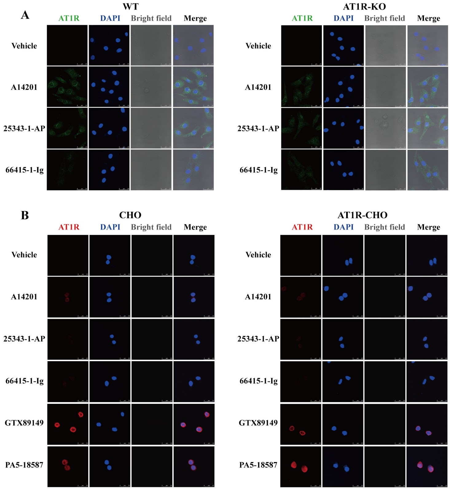
The renin-angiotensin system (RAS) is a crucial regulatory mechanism for cardiovascular function. The angiotensin II (Ang II) type 1 receptor (AT1R) is the principal receptor responsible for mediating RAS function. AT1R belongs to the G protein-coupled receptor (GPCR) family and is present in multiple tissues, including vascular smooth muscle, endothelium, heart, brain, kidney, adrenal gland, and adipose tissue. Physiologically, AT1R mediates second messenger signaling through classical G proteins. Ang II binding to AT1R predominantly activates Gq/11, leading to the activation of phospholipase C (PLC), which results in the production of inositol 1,4,5-trisphosphate (IP3) and diacylglycerol (DG). Then, increased Ca2+ is released from the sarcoplasmic reticulum to mediate the processes of vasoconstriction, enhance cardiac contractility, regulate water‒salt balance, etc. Under pathological conditions, AT1R aberrantly activates G proteins, including mitogen-activated protein kinases (MAPKs: ERK1/2, JNK, and p38MAPK), receptor tyrosine kinases (PDGF, EGFR, and insulin receptor), non-receptor tyrosine kinases [Src, JAK/STAT, and focal adhesion kinase (FAK)], and NADPH oxidase, to influence downstream pathways. This activation exacerbates inflammatory responses, fibrosis, and pathological cardiovascular remodeling. AT1Rs within the nervous system can also induce excessive activity in the sympathetic nervous system, which increases myocardial strain and facilitates the progression of heart failure. Owing to the importance of AT1R in a variety of diseases, greater demands have been placed on the accuracy of AT1R detection. The structural complexity and low immunogenicity of GPCRs pose considerable challenges in the development of specific antibodies. Many commercially available antibodies for GPCRs, such as those against muscarinic and adrenergic receptors, lack specificity. Current studies on AT1R often use these commercial antibodies, but many fail to demonstrate specificity when AT1R-knockdown or AT1R-overexpressing tissues and cells are tested. This study aims to specifically validate six newly available commercial AT1R antibodies (Supplementary Table S1). Using AT1R global knockout SD rats, cardiomyocyte conditional AT1R knockout C57BL/6N mice, AT1R-overexpressing CHO stable-transformed cell lines and AT1R-overexpressing HEK293 cells, we assessed AT1R expression and localization through receptor-ligand binding assays, RT-PCR, western blot analysis, and immunocytochemistry. Materials and methods are available in Supplementary Materials and Methods. To verify the specificity of the antibody, we generated AT1R-global knockout SD rats (AT1R-KO) using CRISPR-Cas9 technology. Agarose gel electrophoresis revealed bands at approximately 470 bp for AT1R-KO rats and 531 bp for wild-type (WT) rats, confirming successful AT1R knockout at the gene level (Figure 1A). RT-PCR analysis of vascular tissue RNA revealed the absence of AT1R in AT1R-KO rats (Figure 1B). Ligand-receptor binding assays revealed significantly less 125I-Ang II binding to vascular tissue proteins in AT1R-KO rats than in WT rats (Figure 1C). Additionally, primary cardiomyocytes extracted from 0–3-day-old WT and AT1R-KO neonatal rats presented a significant increase in beating rate upon Ang II stimulation in WT rats, whereas no response was observed in AT1R-KO rats (Figure 1D). These results confirmed successful AT1R global knockout in AT1R-KO rats. AT1R-KO rats were thus utilized to verify the specificity of AT1R antibodies (A14201, 25343-1-AP, and 66415-1-Ig). Western blot analysis was conducted on protein extracts from the heart, vascular, liver, and kidney tissues of WT and AT1R-KO rats. Under room temperature denaturation conditions, the A14201 antibody detected AT1R bands at the expected molecular weight (42 kDa) in all tissues from WT rats, the 25343-1-AP antibody detected AT1R in heart and kidney tissues, and the 66415-1-Ig antibody detected AT1R only in the heart tissues. Compared with WT control rats, the A14201 antibody revealed a reduction in AT1R protein expression in AT1R-KO rats.
 Graphical AbstractVerified