Acta Biochimica et Biophysica Sinica
The combination of fatigue with the serum GCSF improves the performance of serological screening for frailty
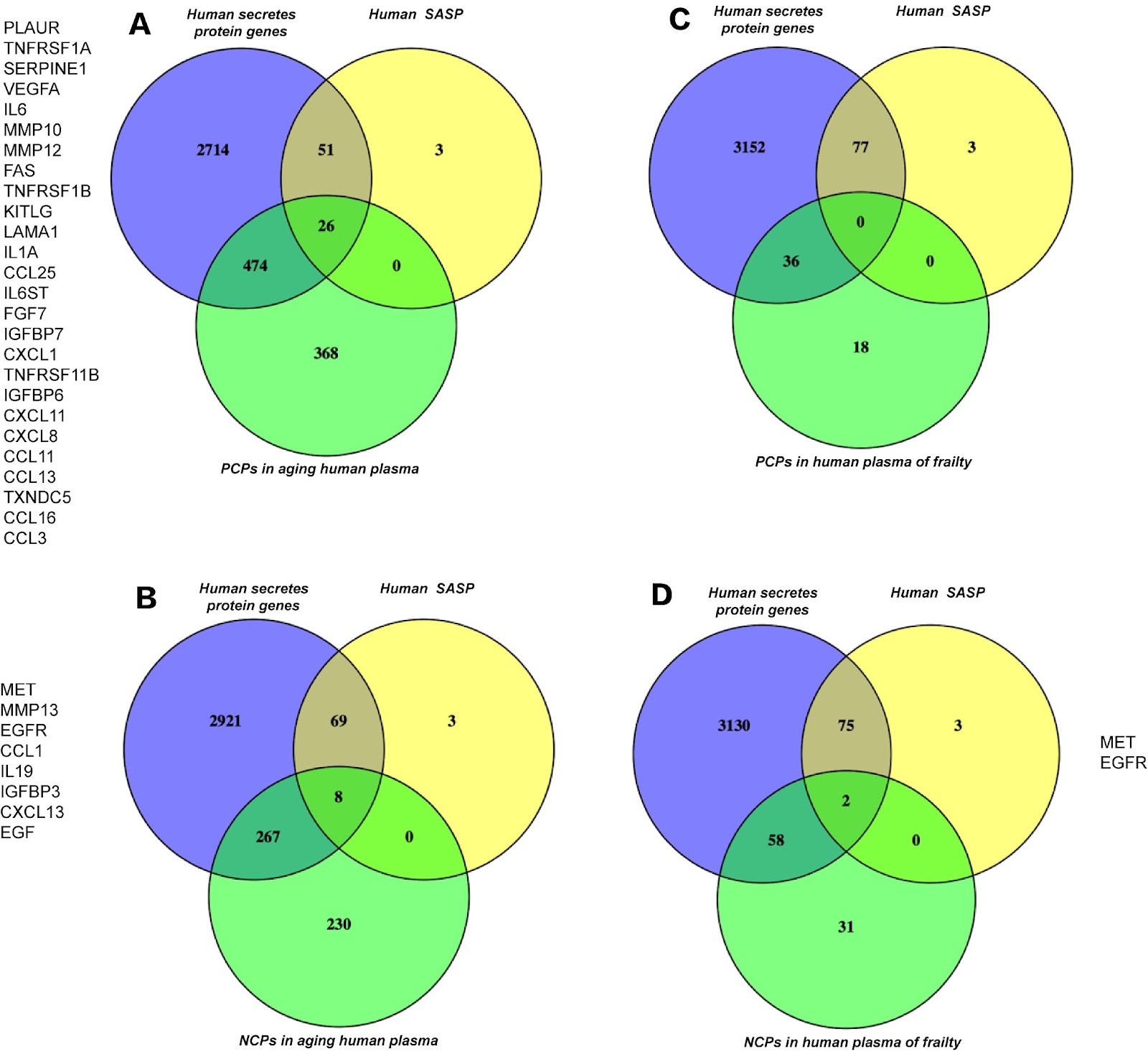
Frailty is a common geriatric disease characterized by accelerated aging and the loss of biological reserves across multiple organs. Approximately 10% of people aged 65 years and older and 25%–50% of people older than 85 years are in a frail state. The increasing institutionalization, hospitalization, and mortality caused by frailty incur massive medical costs and impose a heavy health service burden. For elderly individuals with chronic and/or infectious diseases, such as COVID-19, concomitant frailty can lead to extremely high mortality rates. Moreover, except exercise and nutritional intervention, no effective medicine for treating frailty is available. However, frailty can be prevented, and prefrailty can be reversed. Therefore, effectively screening frailty in elderly individuals is a public health priority. Two main methods for assessing frailty exist: the Fried phenotype and the Rockwood frailty index. The Fried phenotype uses five items, namely, fatigue, weakness, slowness, low physical activity, and weight loss, whereas the Rockwood frailty index is based on the accumulation of age-related deficits. However, these two diagnostic tools are subjective, challenging to use and time-consuming, and are therefore unsuitable for simple, rapid, and extensive screening of frailty in clinical practice. Here, we propose a new strategy to address the above issue. We first harnessed common professional databases to perform inflammatory niche analysis for plasma proteomics from normal aging and frailty patients. We subsequently performed frailty screening and blood sample collection. A total of 852 elderly people were included in the study from January 2018 to August 2018. The assessments included demographic information collection, frailty evaluation, and physical and body composition tests. Blood samples for the determination of inflammatory cytokines were taken from 67 participants. We utilized ELISA to detect the expressions of inflammatory cytokines and chemokines. All blood samples were collected and centrifuged at 4°C and 2000 g for 20 min. The serum was aliquoted and stored properly for ELISA. Inflammatory cytokines in human sera were quantified using corresponding human ELISA kits according to the manufacturer’s protocols. The following markers were measured: IL1A, IL2, IL6, IL8, IL10, IL17, TNFα, IFNγ, GCSF, MCP2, CXCL1, CX3CL1, MMP7, and SOD1. All the statistical analyses were performed with Prism v.6.0. P < 0.05 was considered statistically significant. The receiver operating characteristic (ROC) curve was used to evaluate the performance of all the screening tools. Our inflammatory niche analysis revealed intriguing results when five datasets containing a large number of proteins related to normal aging and inflammation were utilized. The Venn diagrams in Figure 1 show 77 human senescence-associated secretory phenotype genes. Among these genes, 26 are positively correlated with normal aging, whereas 8 are negatively correlated with normal aging. However, neither are positively correlated with frailty, and only two genes are negatively correlated with frailty. Therefore, frailty is obviously distinct from normal aging. Given that most of these differential proteins are inflammatory factors, frailty and normal aging may involve different inflammatory niches. These results suggest that inflammatory factors may be candidates for frailty screening. Our ELISA detection of 15 frailty-related inflammatory factors in the serum of frailty patients screened from 852 volunteers also provided valuable results. The prevalence of frailty was 7.16% (61/852), and that of prefrailty was 41.90% (357/852). The volunteers were 65.22% female and 34.78% male, with a mean age of 70.18 ± 0.73 years. As shown in Table 1, the frail and prefrail groups were significantly older than the robust group (P < 0.001). We observed statistically significant differences in RASM, gait speed, CCI, SARC-F, ADL and MNA scores (P < 0.05), whereas no significant differences were detected with respect to sex, WHR, grip strength or drug count among the three groups. A total of 67 serum samples (nonfrail, n = 20; prefrail, n = 28; and frail, n = 19) were subjected to inflammatory factor screening. No significant difference in the expression of most inflammatory factors was detected.
 Graphical AbstractVerified