🧬 SinoBioData Academic Portal
šŸ“š Peer-Reviewed Translated Literature

All Biomedical & Clinical Articles (Page 55)

Browse complete peer-reviewed translations from top Chinese biomedical, oncology, and genomics journals. Read verified previews and download full authentic clinical reports.

Published Research Papers

Showing 24 of 1542 peer-reviewed translated articles (Page 55 of 65)

Combined treatment with cetuximab and STA9090 has synergistic anticancer effects on human non-small cell lung cancerGraphical AbstractVerified
Acta Biochimica et Biophysica Sinica

Combined treatment with cetuximab and STA9090 has synergistic anticancer effects on human non-small cell lung cancer

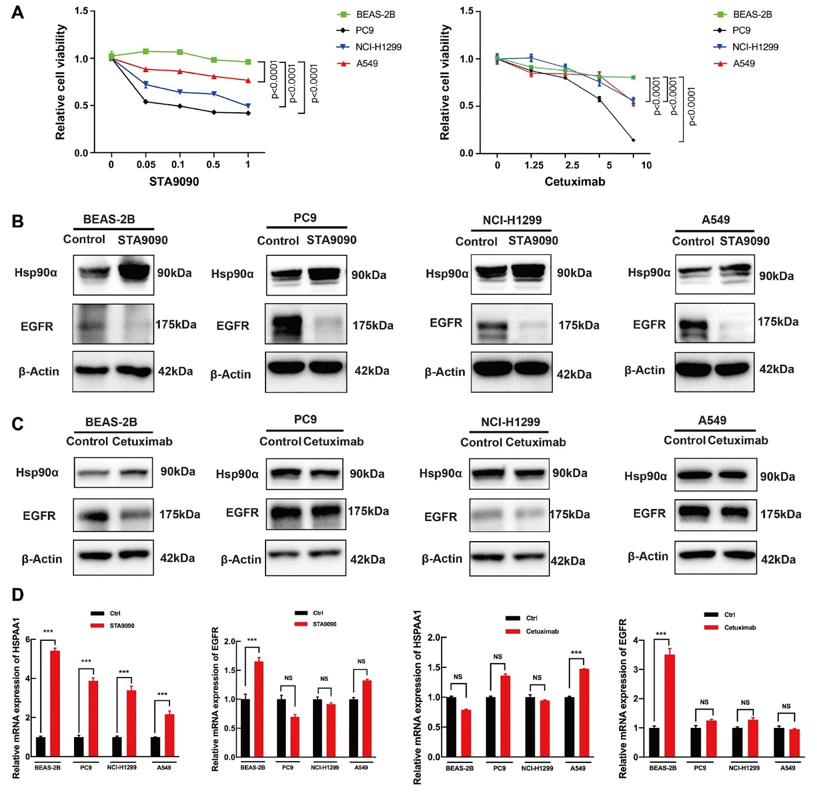
Cetuximab (CET), a human murine chimeric IgG monoclonal antibody and an inhibitor of epidermal growth factor receptor (EGFR), has been shown to be effective in treating various types of cancer. However, its use is hindered by limitations such as resistance development, variability in patient response, side effects, and challenges in biomarker identification. Therefore, CET is often combined with other targeted therapies or chemotherapies to enhance its effectiveness. In this study, we investigate the anticancer effects and underlying mechanisms of the combination of CET, an EGFR inhibitor, and STA9090, an inhibitor of heat shock protein 90 (Hsp90), in both in vitro and in vivo models of non-small cell lung cancer (NSCLC). The results demonstrate significantly stronger effects on NSCLC cells in response to combination therapy than to treatment with either agent alone, indicating that the combination of CET and STA9090 has potential synergistic effects. Additionally, the combination therapy inhibits tumor growth in a xenograft nude mouse model more effectively than treatment with either agent alone, suggesting improved efficacy when used together. Furthermore, the synergistic effects of the combination therapy are likely due to inactivation of the receptor tyrosine kinase (RTK) pathway, which is overly activated in cancer and contributes to tumor growth, angiogenesis, and metastasis. Consequently, our findings suggest that STA9090 has potent direct antitumor activity and synergizes with CET against NSCLC tumors. It is highly likely that these synergistic effects are mediated through RTK pathway inactivation caused by the combination. Therefore, our findings strongly and consistently support the potential synergistic effect of STA9090, an RTK inhibitor, in combination with EGFR-targeting agents.

Read Full Abstract10.3724/abbs.2024069
Macrophage NLRP3-dependent IL-1β production contributes to aortic fibrosis in heart failure with preserved ejection fractionGraphical AbstractVerified
Acta Biochimica et Biophysica Sinica

Macrophage NLRP3-dependent IL-1β production contributes to aortic fibrosis in heart failure with preserved ejection fraction

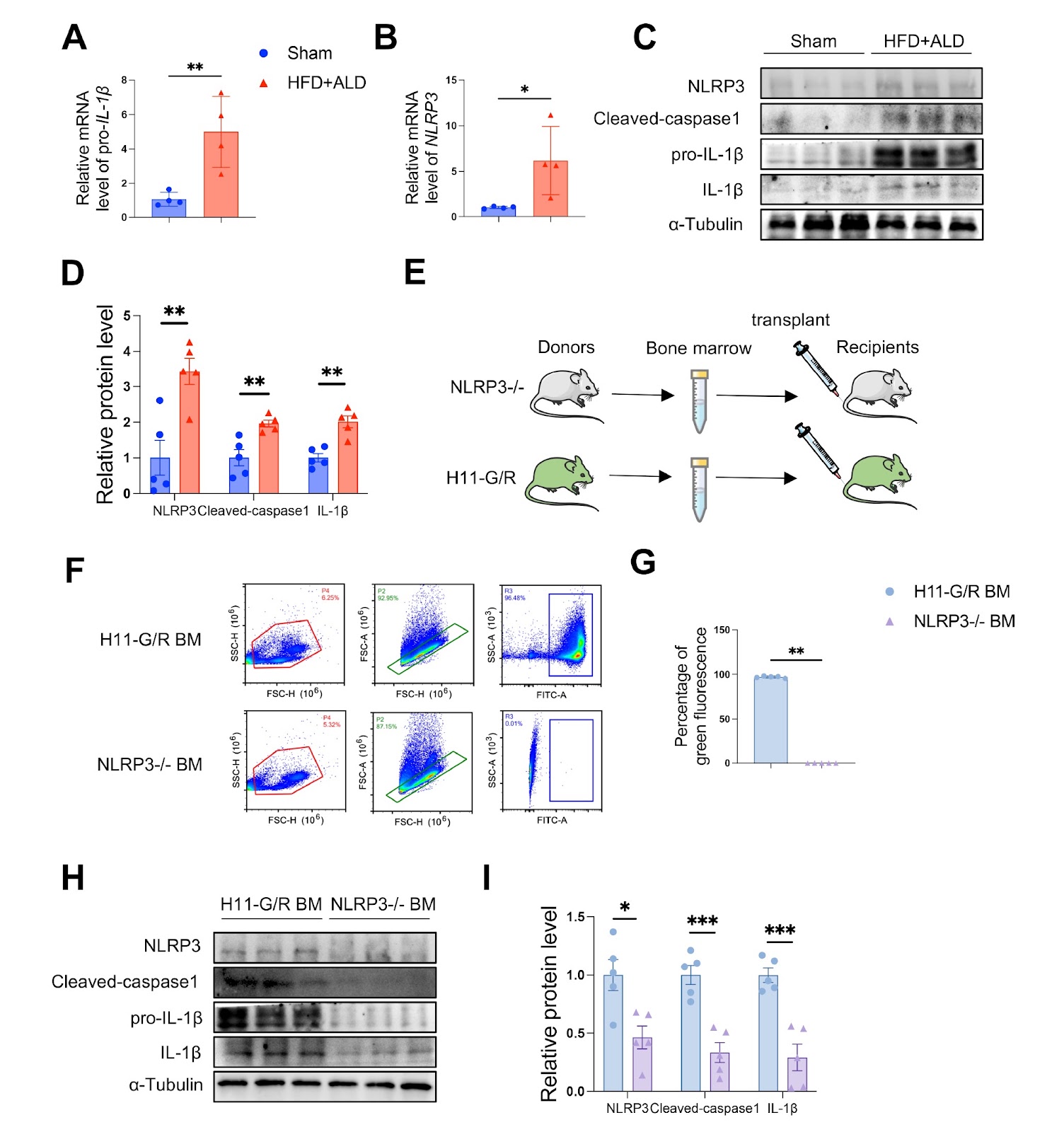
Fibrosis is the main pathological feature of aortic stiffness, which is a common extracardiac comorbidity of heart failure with preserved ejection fraction (HFpEF) and a contributor to left ventricular (LV) diastolic dysfunction. Systemic low-grade inflammation plays a crucial role in the pathogenesis of HFpEF and the development of vascular fibrosis. In this study, we investigate the inflammatory mechanism of aortic fibrosis in HFpEF using a novel mouse model. LV diastolic dysfunction with preserved ejection fraction and aortic fibrosis induced by a high-fat diet (HFD) combined with subcutaneous aldosterone infusion are utilized. The constructed model exhibits augmented macrophage recruitment and NLR family pyrin domain containing 3 (NLRP3)-dependent interleukin (IL)-1β production in fibrotic aortas. In addition, a bone marrow transplant is employed to induce macrophage-specific NLRP3 deficiency in the HFpEF mouse model. These mice show almost completely suppressed cleaved-caspase-1 and mature IL-1β protein expression in the aortas, indicating that macrophage NLRP3 inflammasome activation enhances the IL-1β overproduction in fibrotic aortas. Furthermore, we show that macrophage NLRP3 inflammasome inhibition improves aortic fibrosis and LV diastolic dysfunction. In conclusion, this study demonstrates the pivotal effect of macrophage NLRP3-dependent IL-1β production on aortic fibrosis and cardiac function in HFpEF, suggesting a potential target for HFpEF therapy.

Read Full Abstract10.3724/abbs.2024238
Epigallocatechin-3-gallate inhibits osteogenic differentiation of vascular smooth muscle cells through the transcription factor JunBGraphical AbstractVerified
Acta Biochimica et Biophysica Sinica

Epigallocatechin-3-gallate inhibits osteogenic differentiation of vascular smooth muscle cells through the transcription factor JunB

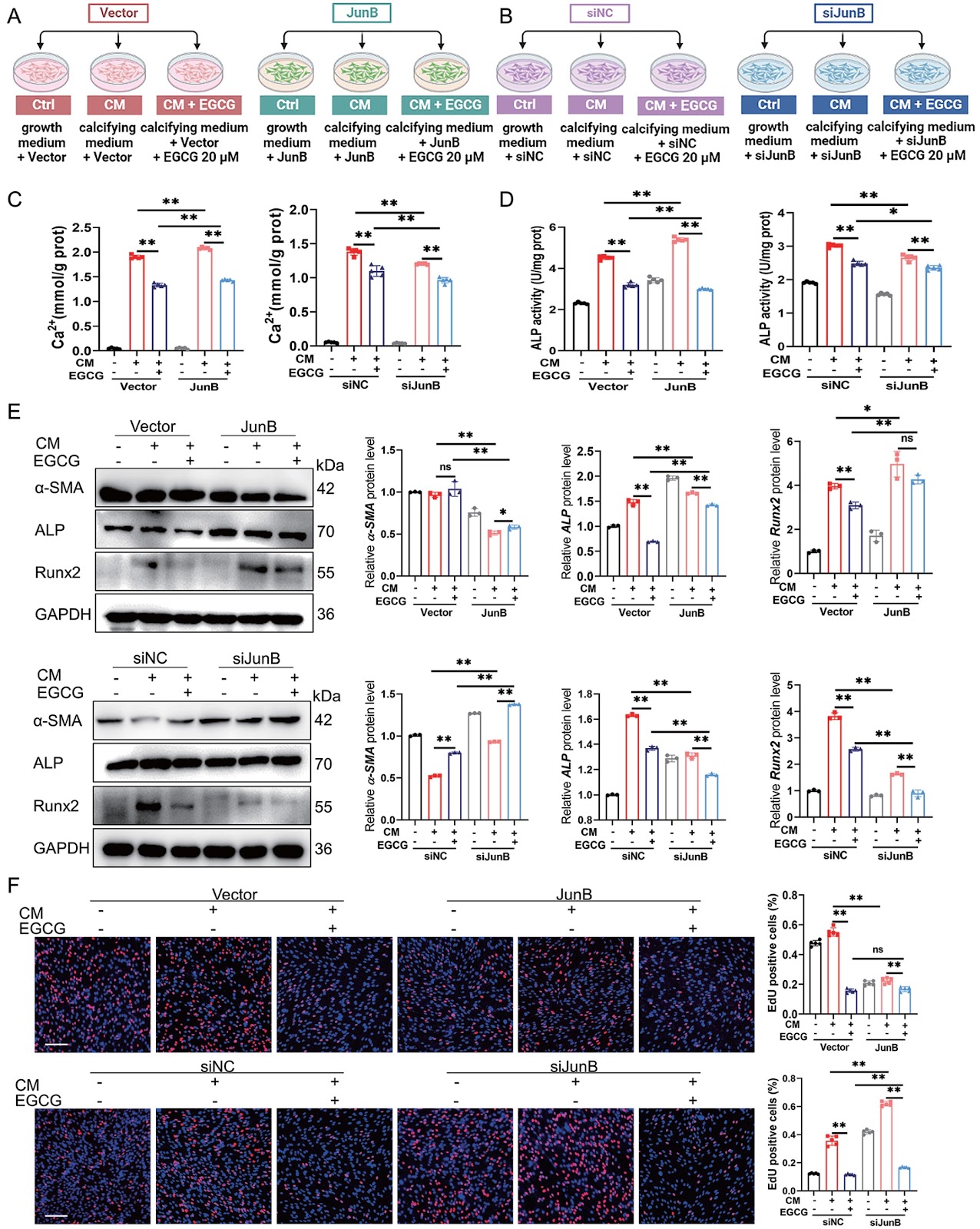
Medial arterial calcification (MAC) accompanying chronic kidney disease (CKD) leads to increased vessel wall stiffness, myocardial ischemia, heart failure, and increased cardiovascular morbidity and mortality. Unfortunately, there are currently no drugs available to treat MAC. The natural polyphenol epigallocatechin-3-gallate (EGCG) has been demonstrated to protect against cardiovascular disease; however, whether EGCG supplementation inhibits MAC in CKD remains unclear. In this study, we utilize a CKD-associated MAC model to investigate the effects of EGCG on vascular calcification and elucidate the underlying mechanisms involved. Our findings demonstrate that EGCG treatment significantly reduces calcium phosphate deposition and osteogenic differentiation of VSMCs in vivo and in vitro in a dose-dependent manner. In addition, through RNA sequencing (RNA-seq) analysis, we show a significant activation of the transcription factor JunB both in CKD mouse arteries and in osteoblast-like VSMCs. Notably, EGCG effectively suppresses CKD-associated MAC by inhibiting the activity of JunB. In addition, overexpression of JunB can abolish while knockdown of JunB can enhance the inhibitory effect of EGCG on the osteogenic differentiation of VSMCs. Furthermore, EGCG supplementation inhibits MAC in CKD via modulation of the JunB-dependent Ras/Raf/MEK/ERK signaling pathway. In conclusion, our study highlights the potential therapeutic value of EGCG for managing CKD-associated MAC, as it mitigates this pathological process through targeted inactivation of JunB.

Read Full Abstract10.3724/abbs.2024060
EZH2 inhibition induces senescence via ERK1/2 signaling pathway in multiple myelomaGraphical AbstractVerified
Acta Biochimica et Biophysica Sinica

EZH2 inhibition induces senescence via ERK1/2 signaling pathway in multiple myeloma

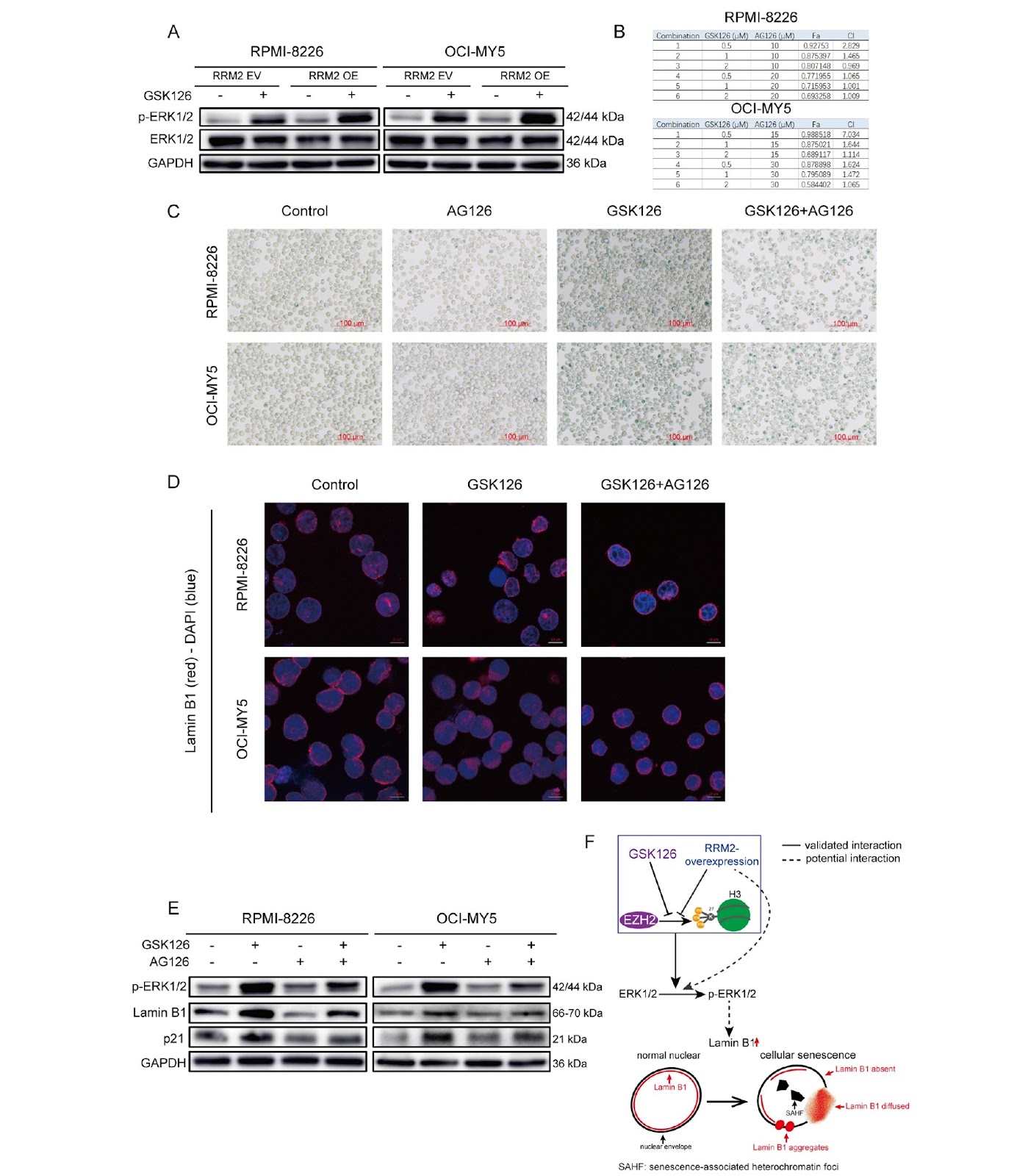
Epigenetic modifications play an important role in cellular senescence, and enhancer of zeste homolog 2 (EZH2) is a key methyltransferase involved in epigenetic remodeling in multiple myeloma (MM) cells. We have previously demonstrated that GSK126, a specific EZH2 inhibitor, exhibits anti-MM therapeutic efficacy and safety in vivo and in vitro; however, its specific mechanism remains unclear. This study shows that GSK126 induces cellular senescence in MM, which is characterized by the accumulation of senescence-associated heterochromatin foci (SAHF) and p21, and increased senescence-associated β galactosidase activity. Furthermore, EZH2 is inhibited in ribonucleotide reductase regulatory subunit M2 (RRM2)-overexpressing OCI-MY5 and RPMI-8226 cells. RRM2 overexpression inhibits the methyltransferase function of EZH2 and promotes its degradation through the ubiquitin-proteasome pathway, thereby inducing cellular senescence. In this senescence model, Lamin B1, a key component of the nuclear envelope and a marker of senescence, does not decrease but instead undergoes aberrant accumulation. Meanwhile, phosphorylation of extracellular signal-regulated protein kinase (ERK1/2) is significantly increased. The inhibition of ERK1/2 phosphorylation in turn partially restores Lamin B1 level and alleviates senescence. These findings suggest that EZH2 inhibition increases Lamin B1 level and induces senescence by promoting ERK1/2 phosphorylation. These data indicate that EZH2 plays an important role in MM cellular senescence and provide insights into the relationships among Lamin B1, p-ERK1/2, and cellular senescence.

Read Full Abstract10.3724/abbs.2024077
Antitumor potential of polyamines in cancerGraphical AbstractVerified
Acta Biochimica et Biophysica Sinica

Antitumor potential of polyamines in cancer

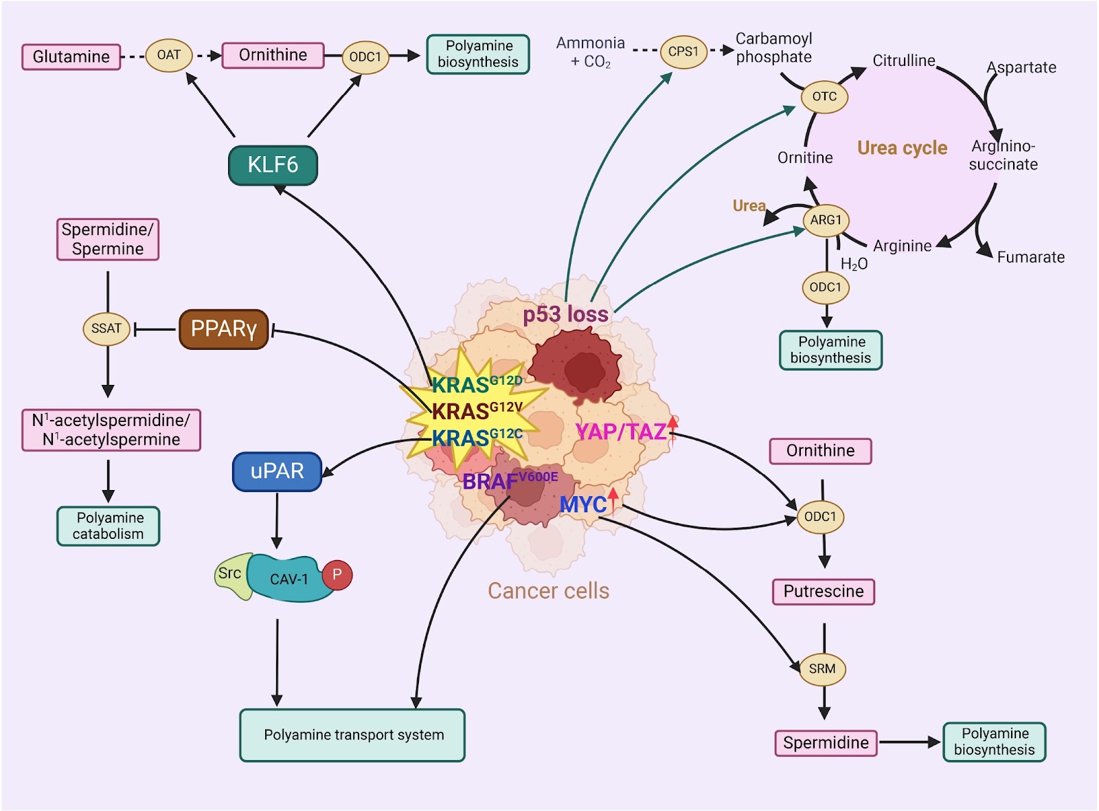
The dysregulation of polyamines in tumors has made polyamine metabolism an appealing target for cancer therapy. Gene mutations drive the reprogramming of polyamine metabolism in tumors, presenting promising opportunities for clinical treatment. The proposed strategies involve inhibiting polyamine biosynthesis while also targeting the polyamine transport system as antitumor approaches. A growing number of drugs aimed at polyamine biosynthesis and transport systems are undergoing clinical trials. Polyamine metabolism plays a role in regulating cancer signaling pathways, suggesting potential combination therapies for cancer treatment. Furthermore, supplemental polyamine substances have demonstrated antitumor activity, indicating that combining polyamines with downstream targets or immunotherapy could offer significant clinical benefits. These discoveries open new avenues for leveraging polyamine metabolism in anticancer therapy.

Read Full Abstract10.3724/abbs.2025030
Function and mechanism of action of the TRPV1 channel in the development of triple-negative breast cancerGraphical AbstractVerified
Acta Biochimica et Biophysica Sinica

Function and mechanism of action of the TRPV1 channel in the development of triple-negative breast cancer

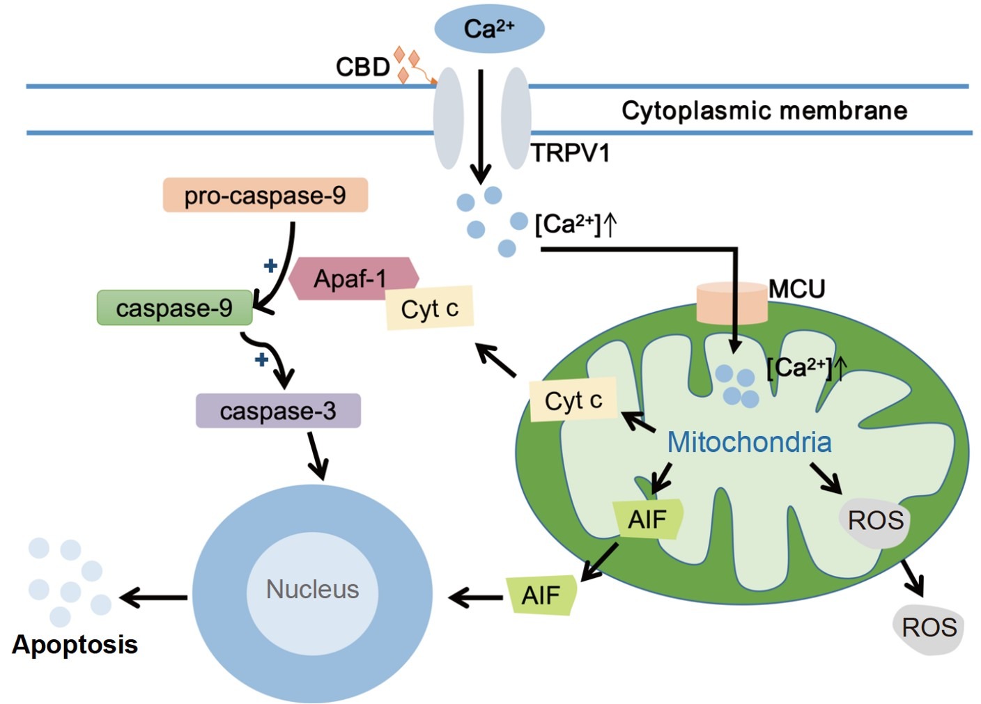
Transient receptor potential channel subfamily vanilloid 1 (TRPV1) is a member of the transient receptor potential family of nonselective cationic transmembrane channel proteins that are involved in the regulation of calcium homeostasis. It is expressed in various tumor types and has been implicated in the regulation of cancer growth, metastasis, apoptosis, and cancer-related pain. TRPV1 is highly expressed in triple-negative breast cancer (TNBC), and both its agonists and antagonists may exert anti-cancer effects. In this review, we provide an overview of the effect of TRPV1 on TNBC development and its influence on immunotherapy in an attempt to facilitate the development of future treatment strategies.

Read Full Abstract10.3724/abbs.2024068
Skeletal muscle-derived musclin attenuates glycolysis, oxidative stress, and pulmonary hypertension through the NPR3/AKT/mTORC1 pathwayGraphical AbstractVerified
Acta Biochimica et Biophysica Sinica

Skeletal muscle-derived musclin attenuates glycolysis, oxidative stress, and pulmonary hypertension through the NPR3/AKT/mTORC1 pathway

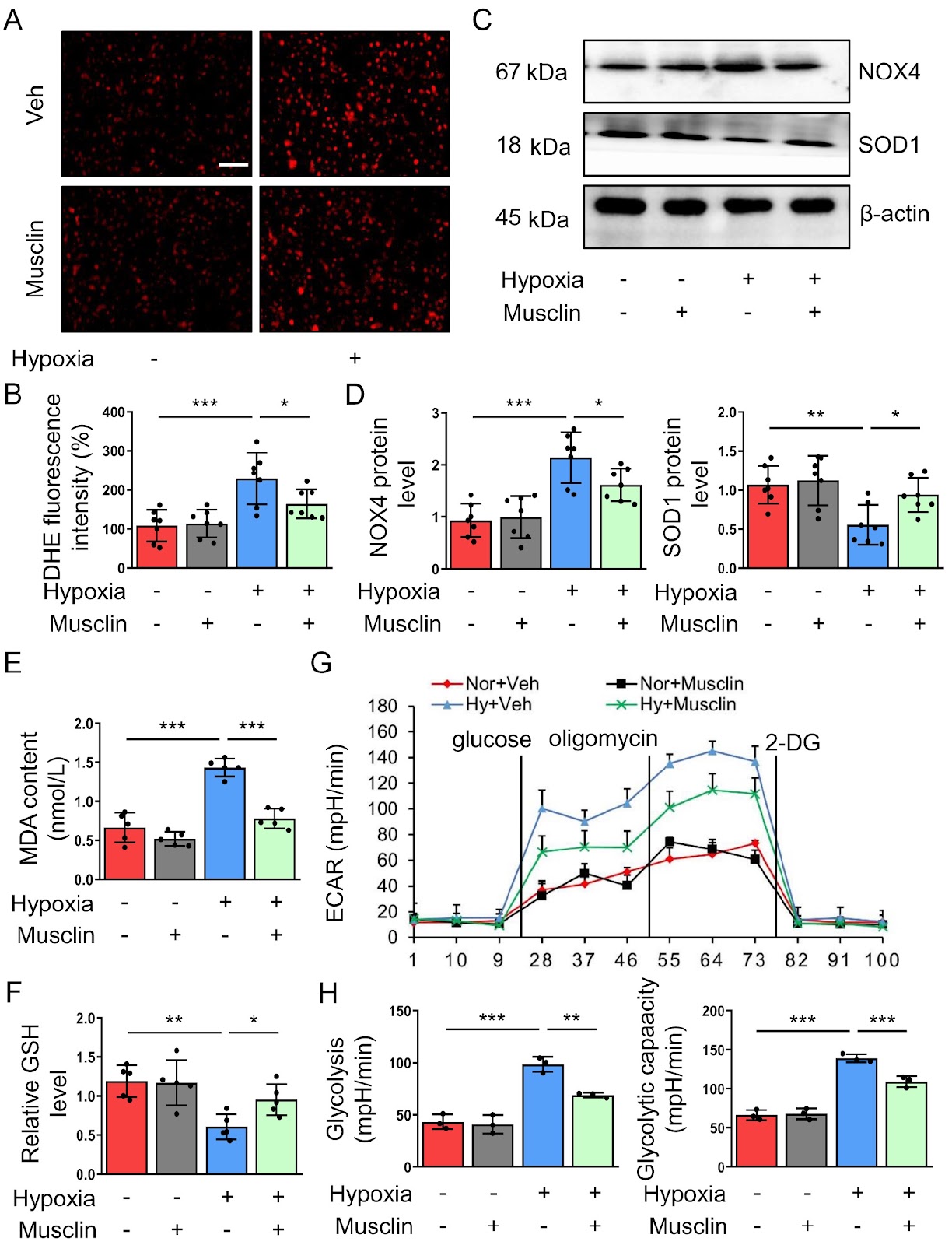
Exercise ameliorates pulmonary hypertension (PH) progression. However, the underlying mechanisms are largely unclear. Musclin is an exercise-responsive myokine that exerts protective effects on cardiovascular diseases. The current study aims to explore the role of musclin in the development of PH. A monocrotaline (MCT)-induced mouse PH model is established. Adeno-associated virus serotype 6 (AAV6)-mediated gene transfer is used to induce musclin overexpression in skeletal muscle. Ultrasound and morphological analyses are utilized to assess the severity of PH. Cell viability assay, Ki-67 immunofluorescence staining, wound healing assay, and transwell assay are used to evaluate the proliferation and migration of pulmonary arterial smooth muscle cells (PASMCs). We find that the musclin levels in both plasma and skeletal muscle are decreased in MCT-treated mice. The external expression of musclin in skeletal muscle ameliorates pulmonary arterial remodeling and right ventricular dysfunction. In vitro, musclin treatment suppresses hypoxia-induced glycolysis, oxidative stress, proliferation, and migration. Further experiments reveal that musclin inhibits mechanistic target of rapamycin complex 1 (mTORC1) activity in hypoxia-stimulated PASMCs and pulmonary arteries of MCT-treated mice. Reactivating mTORC1 abolishes the protective role of musclin against PH. Additionally, musclin enhances its interaction with natriuretic peptide receptor 3 (NPR3) in PASMCs. Silencing of NPR3 reverses the inhibitory effects of musclin on AKT phosphorylation, mTORC1 activity, glycolysis, oxidative stress, proliferation, and migration in hypoxia-challenged PASMCs. In conclusion, our study highlights the inhibitory role of musclin in the proliferation and migration of PASMCs and PH progression, thereby providing a novel potent therapeutic strategy for treating PH and partly clarifying the mechanism of exercise-mediated protection against PH.

Read Full Abstract10.3724/abbs.2024214
Magnolol promotes the autophagy of esophageal carcinoma cells by upregulating HACE1 gene expressionGraphical AbstractVerified
Acta Biochimica et Biophysica Sinica

Magnolol promotes the autophagy of esophageal carcinoma cells by upregulating HACE1 gene expression

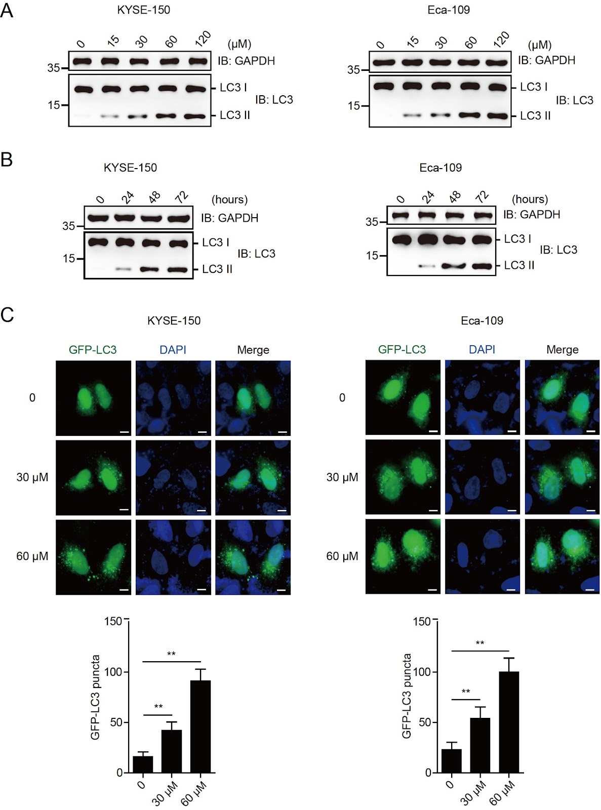
Esophagus cancer (EC) is one of the most aggressive malignant digestive system tumors and has a high clinical incidence worldwide. Magnolol, a natural compound, has anticancer effects on many cancers, including esophageal carcinoma, but the underlying mechanism has not been fully elucidated. Here, we first find that magnolol inhibits the proliferation of esophageal carcinoma cells and enhances their autophagy activity in a dose- and time-dependent manner. This study demonstrates that magnolol increases the protein levels of LC3 II, accompanied by increased HACE1 protein levels in both esophageal carcinoma cells and xenograft tumors. HACE1-knockout (KO) cell lines are generated, and the ablation of HACE1 eliminates the anti-proliferative and autophagy-inducing effects of magnolol on esophageal carcinoma cells. Additionally, our results show that magnolol primarily promotes HACE1 expression at the transcriptional level. Therefore, this study shows that magnolol primarily exerts its antitumor effect by activating HACE1-OPTN axis-mediated autophagy. It can be considered a promising therapeutic drug for esophageal carcinoma.

Read Full Abstract10.3724/abbs.2024044
Inhibition of USP22 by miR-200b-5p represses gastric cancer cell proliferation and migration by targeting the NF-ĪŗB signaling pathwayGraphical AbstractVerified
Acta Biochimica et Biophysica Sinica

Inhibition of USP22 by miR-200b-5p represses gastric cancer cell proliferation and migration by targeting the NF-ĪŗB signaling pathway

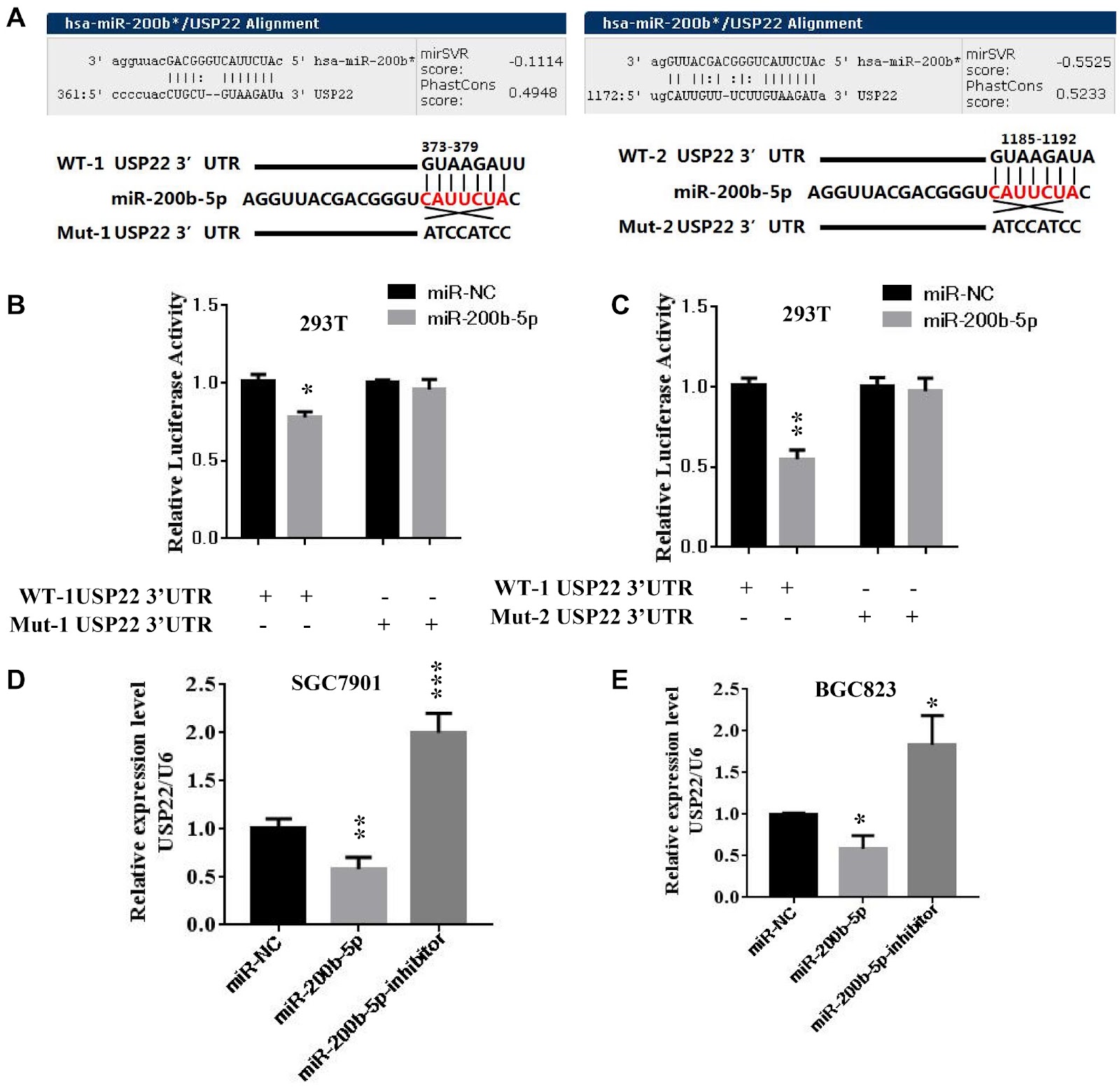
Gastric cancer (GC) is an aggressive tumor type with an intricate pathogenesis and limited therapeutic options. Ubiquitin-specific protease 22 (USP22) is a protein implicated in cell proliferation, metastasis, and tumorigenesis. However, the regulatory mechanisms governing USP22 in GC are still not fully understood. In this study, we perform bioinformatics analysis to identify conserved miRNA recognition sites for miR-200b-5p within the 3′UTR of USP22. Validation via luciferase reporter assay confirms the transcriptional regulation of USP22 by miR-200b-5p. Overexpression of miR-200b-5p markedly inhibits the proliferation and migration of GC cells in vitro and suppresses tumor growth in vivo. Conversely, ectopic expression of USP22 reversed this effect by modulating the NF-ĪŗB signaling pathway. Additionally, qPCR analysis reveals an inverse correlation between the miR-200b-5p level and USP22 expression in GC. Collectively, our findings indicate that miR-200b-5p-mediated inhibition of USP22 attenuates cell proliferation by targeting the NF-ĪŗB signaling pathway in GC, suggesting that miR-200b-5p and USP22 could serve as potential diagnostic or therapeutic targets for gastric cancer and other related human diseases.

Read Full Abstract10.3724/abbs.2024231
The E2F1-KIF14 axis drives focal adhesion formation and promotes colorectal cancer metastasisGraphical AbstractVerified
Acta Biochimica et Biophysica Sinica

The E2F1-KIF14 axis drives focal adhesion formation and promotes colorectal cancer metastasis

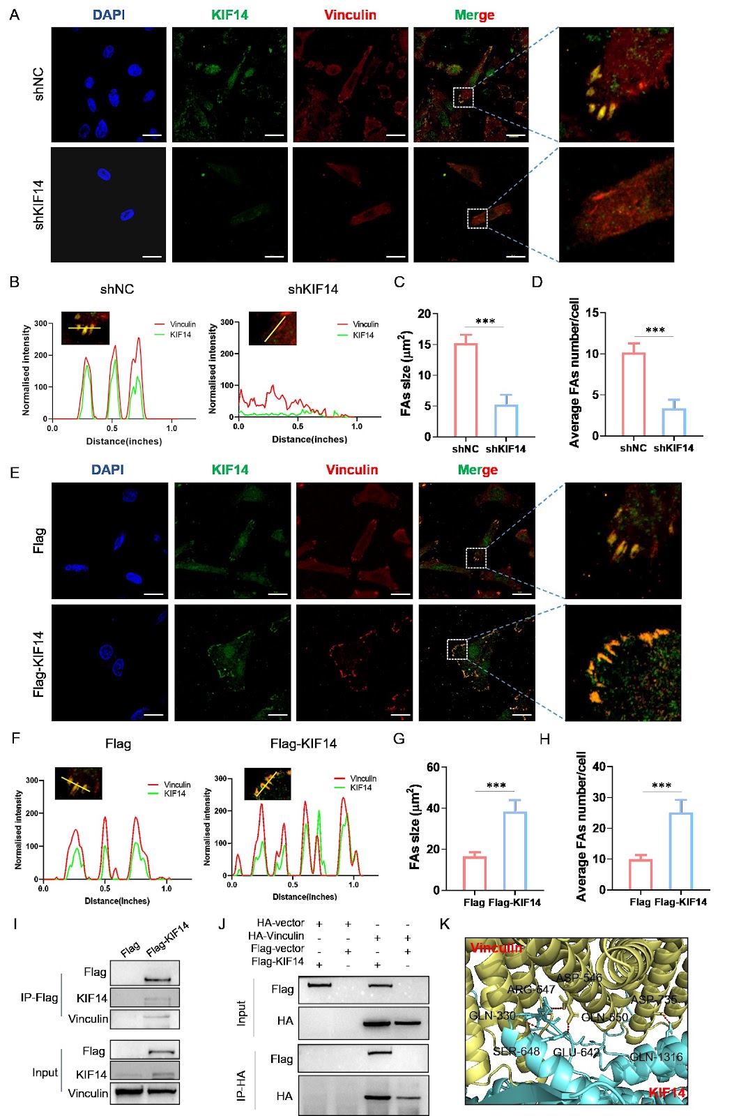
Kinesin family member 14 (KIF14) has been implicated in the progression of multiple cancer types, yet its role in colorectal cancer (CRC) metastasis remains undefined. Here, we assesse KIF14 expression in CRC specimens and explore its clinical and functional significance. KIF14 upregulation is frequently observed in CRC tissues and is correlated with advanced tumor stage and reduced overall survival. Functional assays reveal that KIF14 depletion in CRC cells inhibits migration, invasion, and in vivo metastatic colonization, whereas KIF14 overexpression induces the opposite effects. Transcriptomic and pathway enrichment analyses reveal that KIF14 functions as a critical regulator of focal adhesion and cell-matrix adhesion signaling. This finding is further supported by experimental evidence showing that KIF14 overexpression promotes focal adhesion assembly, whereas KIF14 knockdown disruptes this process. Mechanistically, we demonstrate that KIF14 binds directly to the focal adhesion protein vinculin and mediates its delivery to the leading edge of migrating cells. Moreover, bioinformatics prediction and chromatin immunoprecipitation confirm that E2F1 directly binds the KIF14 promoter to drive its transcription. Rescue experiments reveal that ectopic KIF14 expression restores the prometastatic phenotypes suppressed by E2F1 silencing, indicating that the effects of E2F1 are mediated by the E2F1-KIF14 axis. Collectively, our findings reveal a novel E2F1-KIF14-vinculin signaling axis that drives CRC metastasis by modulating focal adhesion dynamics, highlighting KIF14 as a potential therapeutic target.

Read Full Abstract10.3724/abbs.2025158
Cx58 is associated with the metastasis of non-small cell lung cancer via MEF2B/Cx58 axisGraphical AbstractVerified
Acta Biochimica et Biophysica Sinica

Cx58 is associated with the metastasis of non-small cell lung cancer via MEF2B/Cx58 axis

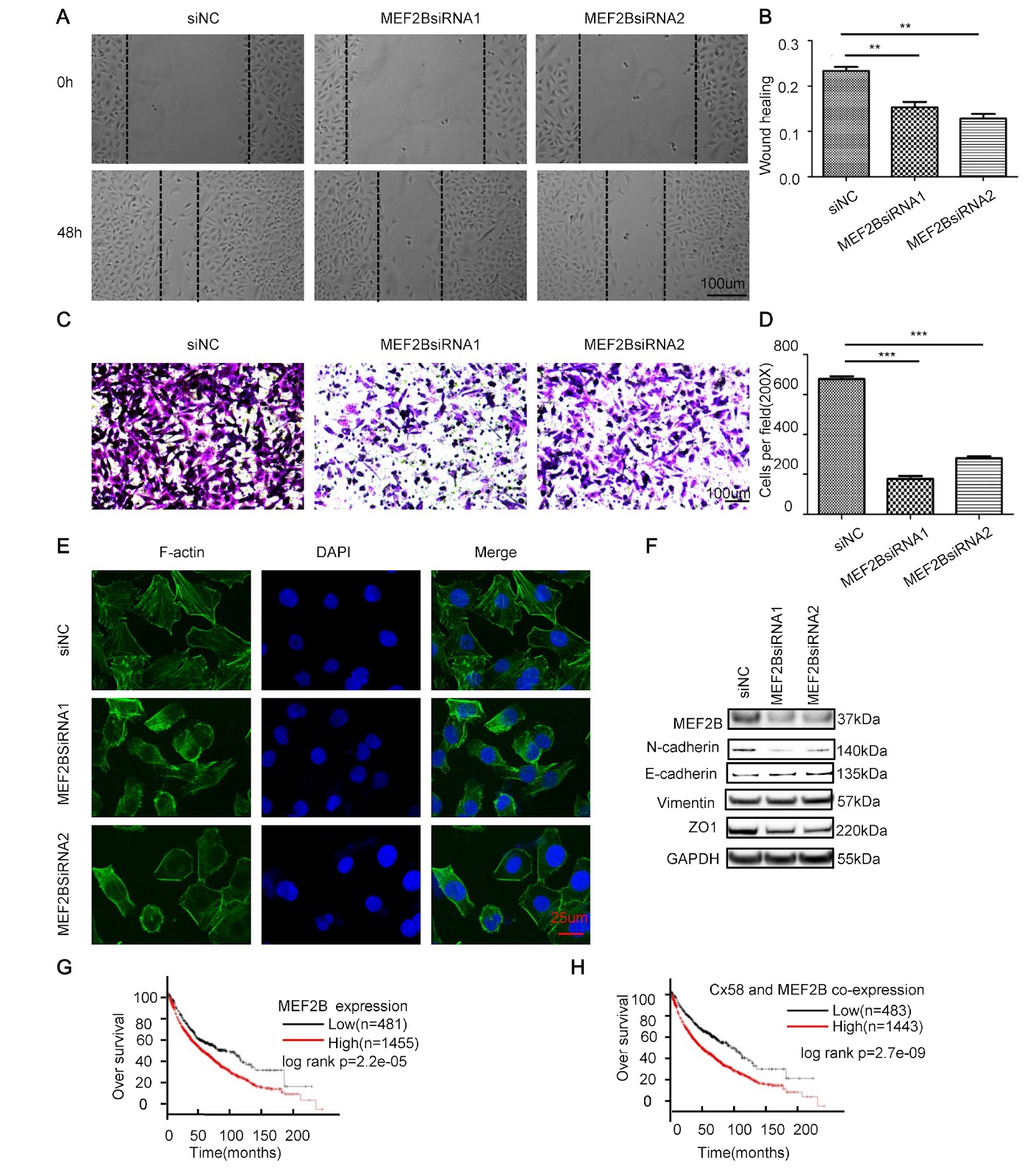
Connexins (Cxs), also known as gap junction proteins, are structurally related transmembrane proteins and have been implicated in carcinogenesis. Although some evidence suggests that these proteins are tumor suppressors due to their reduced expression in cancers, recent research indicates their complicated roles in tumor progression during different stages, including metastasis. Here, we show that Cx58, which is upregulated in non-small cell lung cancer (NSCLC), is modulated by myocyte-enhancer binding factor 2B (MEF2B). Either Cx58 or MEF2B knockdown attenuates the migration and invasion of NSCLC cells by inducing cytoskeleton rearrangement. Additionally, the prometastatic role of Cx58 in NSCLC is demonstrated in vivo. In conclusion, our findings suggest that Cx58 is transcriptionally activated by MEF2B and is involved in the metastasis of NSCLC by regulating cytoskeleton organization. Targeting the MEF2B/Cx58 axis may be exploited as a modality for improving NSCLC therapy.

Read Full Abstract10.3724/abbs.2025049
A novel mutation in SMARCB1 associated with adult Coffin-Siris syndrome and meningiomaGraphical AbstractVerified
Acta Biochimica et Biophysica Sinica

A novel mutation in SMARCB1 associated with adult Coffin-Siris syndrome and meningioma

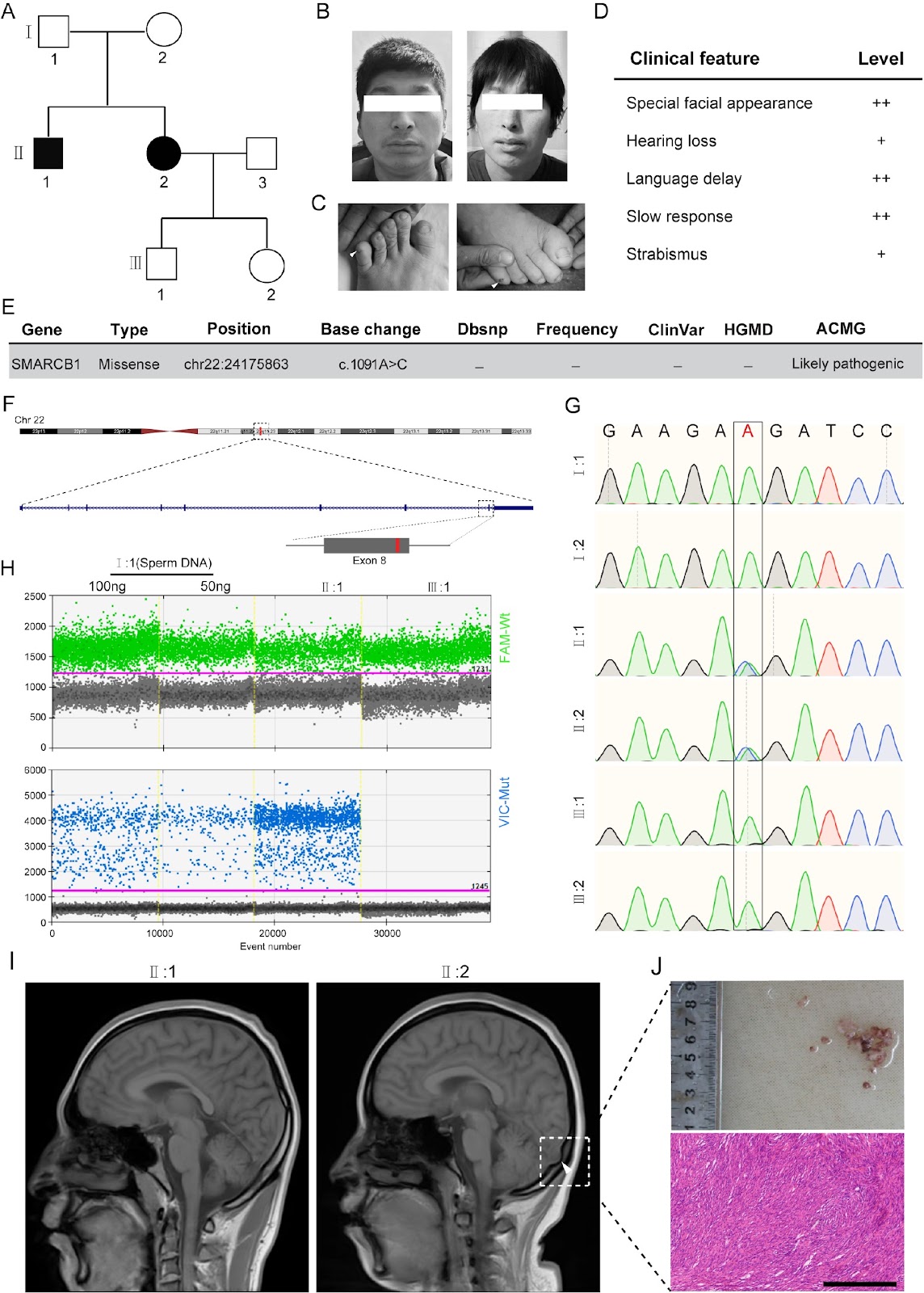
SMARCB1 encodes a core subunit of the SWI/SNF chromatin remodeling complex, which plays a crucial role in the regulation of gene expression. Germline mutations in the SMARCB1 gene have been linked to early childhood Coffin-Siris syndrome type 3 (CSS3), a rare congenital malformation syndrome characterized by severe developmental delay and intellectual disability. In this study, we report a family of two adult CSS3 patients with a novel missense SMARCB1 mutation (c.1091A>C, p.Lys364Thr) identified through whole-exome sequencing (WES). Both patients exhibit selective difficulties in verbal learning and experience language delays. Additionally, the development of meningioma is confirmed in one of the patients. Mechanistic studies suggest that this missense mutation may abnormally activate the MAPK signaling pathway, which is implicated in the pathogenesis of tumor progression and neurodevelopmental disorders. This is the first reported case of a germline mutation in the SMARCB1 gene associated with both CSS3 and meningioma, thereby expanding the phenotypic spectrum of SMARCB1-related disorders.

Read Full Abstract10.3724/abbs.2024204
PIPKI-PIP2 promotes cell migration by recruiting Smurf1 to the membrane and increasing its activityGraphical AbstractVerified
Acta Biochimica et Biophysica Sinica

PIPKI-PIP2 promotes cell migration by recruiting Smurf1 to the membrane and increasing its activity

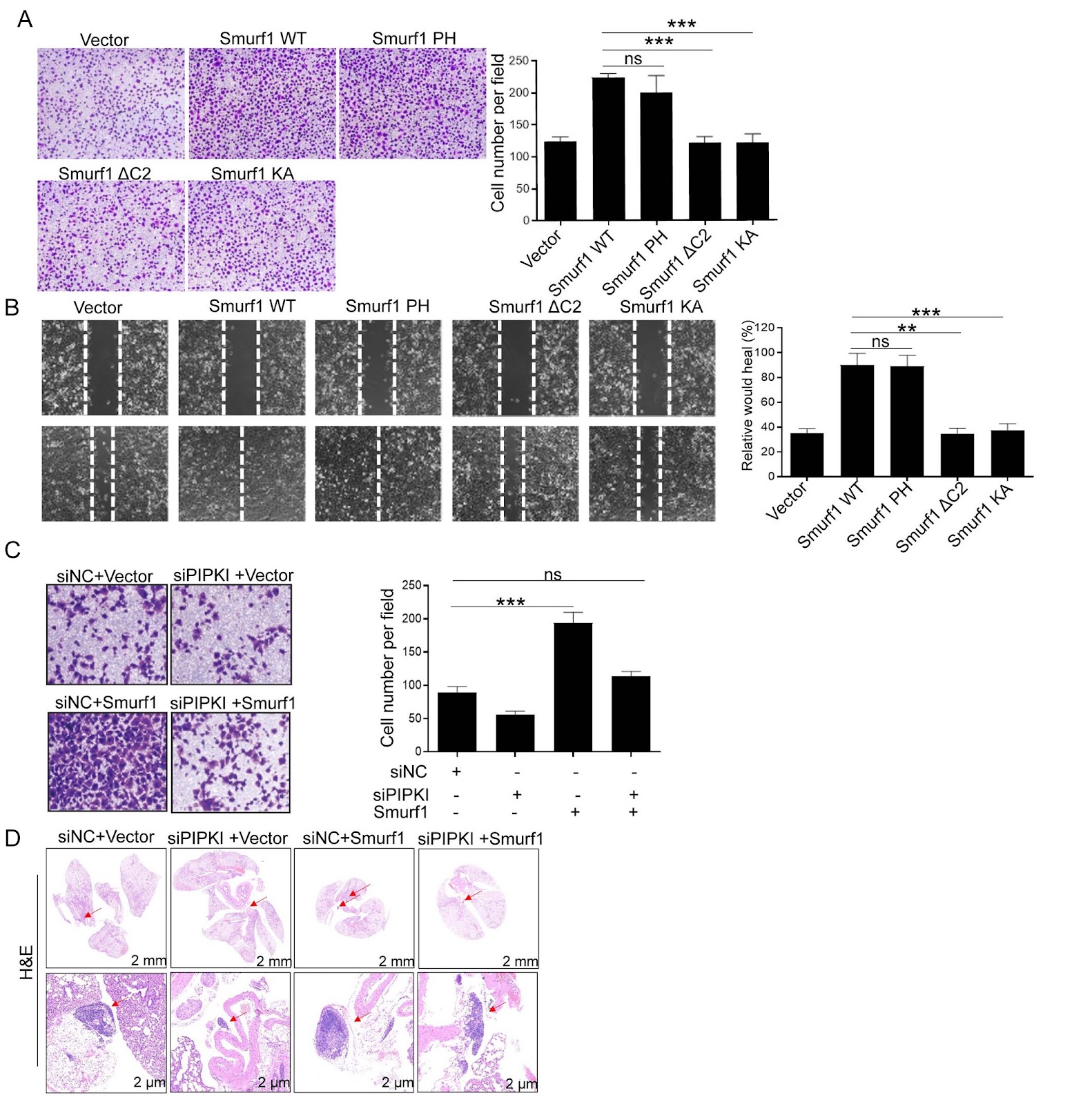
Smurf1 is a member of the Nedd4 family of E3 ubiquitin ligases. Numerous lines of evidence indicate that the membrane localization of Smurf1 is essential for its activity. However, the underlying mechanisms that regulate the membrane localization of Smurf1 remain unclear. Type I phosphatidylinositol phosphate kinase (PIPKI) is a phosphatidylinositol kinase that generates phosphatidylinositol 4,5-bisphosphate (PIP2), which is located in the plasma membrane and regulates cellular processes, including ion channel activity and cell migration. In this study, we show that PIP2 and PIPKI regulate the membrane translocation of Smurf1. Importantly, the recruitment of Smurf1 to the cell membrane through the association of its C2 domain with PIPKI-produced PIP2 is essential for Smurf1-mediated E3 ligase activity and cell migration. Therefore, we identify a PIPKI-PIP2-Smurf1 signaling axis that regulates cell migration.

Read Full Abstract10.3724/abbs.2025217
Psychological stress induces dysfunction in the lacrimal gland through the sympathetic nervous system and the hypothalamic-pituitary-adrenal axisGraphical AbstractVerified
Acta Biochimica et Biophysica Sinica

Psychological stress induces dysfunction in the lacrimal gland through the sympathetic nervous system and the hypothalamic-pituitary-adrenal axis

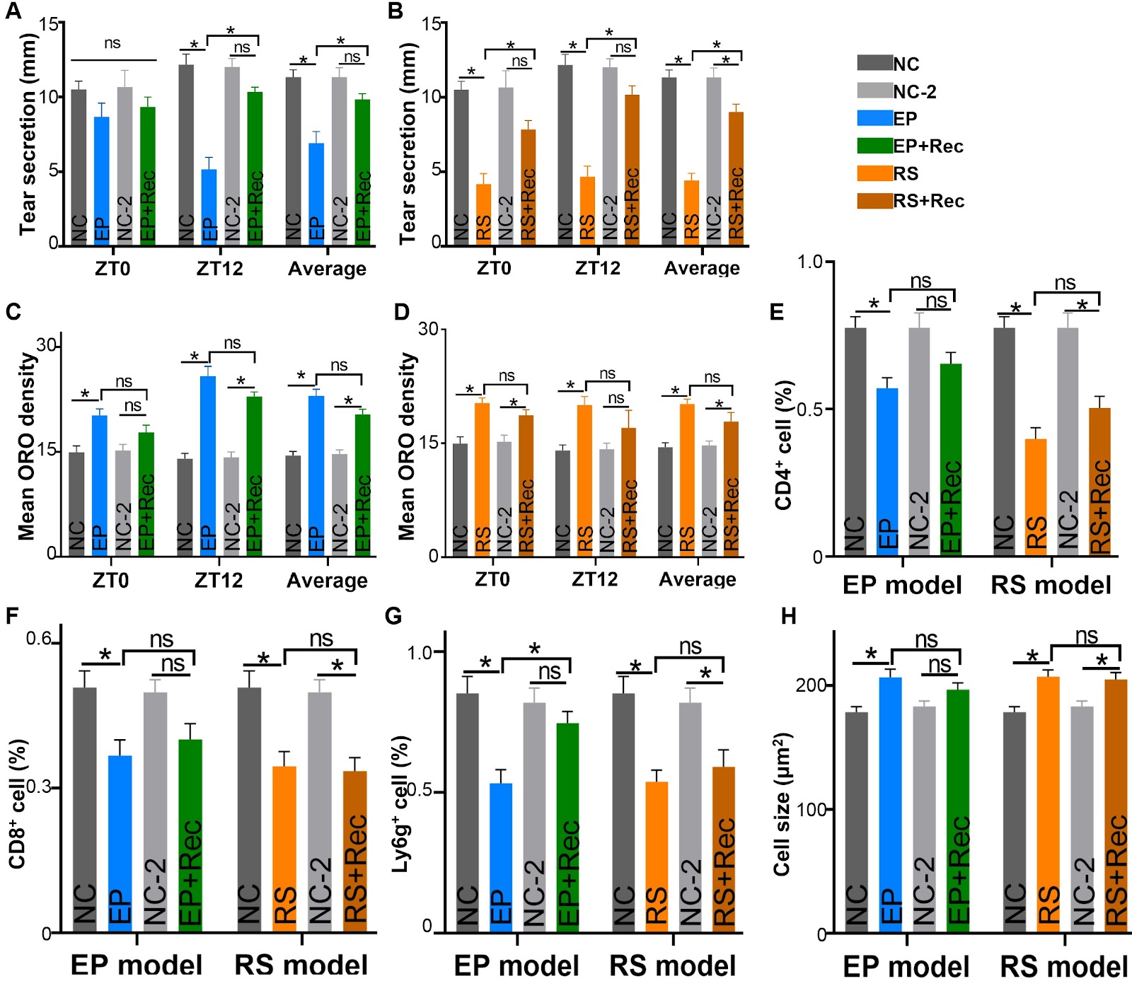
Chronic psychosocial stress is increasingly recognized as a key risk factor for dry eye disease, potentially because of its disruption of the circadian transcriptome and lacrimal gland function, which impacts eye health. In this study, we test this hypothesis by using two mouse models (high platform and restraint experiments) of psychological stress and report that both models uniquely alter the circadian transcriptome and signaling pathways of the lacrimal gland. Psychosocial stress significantly affects the normal rhythmic oscillations of extraorbital lacrimal gland (ELG) immune cell trafficking, secretion response, and lipid deposition. Both models significantly reduce the volume of stimulated lacrimal secretions as well as the recruitment of immune cells to the lacrimal gland. Importantly, treatment with beta-adrenergic receptor blockers or glucocorticoid synthesis inhibitors significantly improves these secretory functions and histopathological changes. Collectively, these findings demonstrate the detrimental effects of chronic psychosocial stress on lacrimal gland circadian transcriptome homeostasis and suggest potential clinical applications for patients with both psychological stress and dry eye disease.

Read Full Abstract10.3724/abbs.2025195
PDK1 promotes epithelial ovarian cancer progression by upregulating BGNGraphical AbstractVerified
Acta Biochimica et Biophysica Sinica

PDK1 promotes epithelial ovarian cancer progression by upregulating BGN

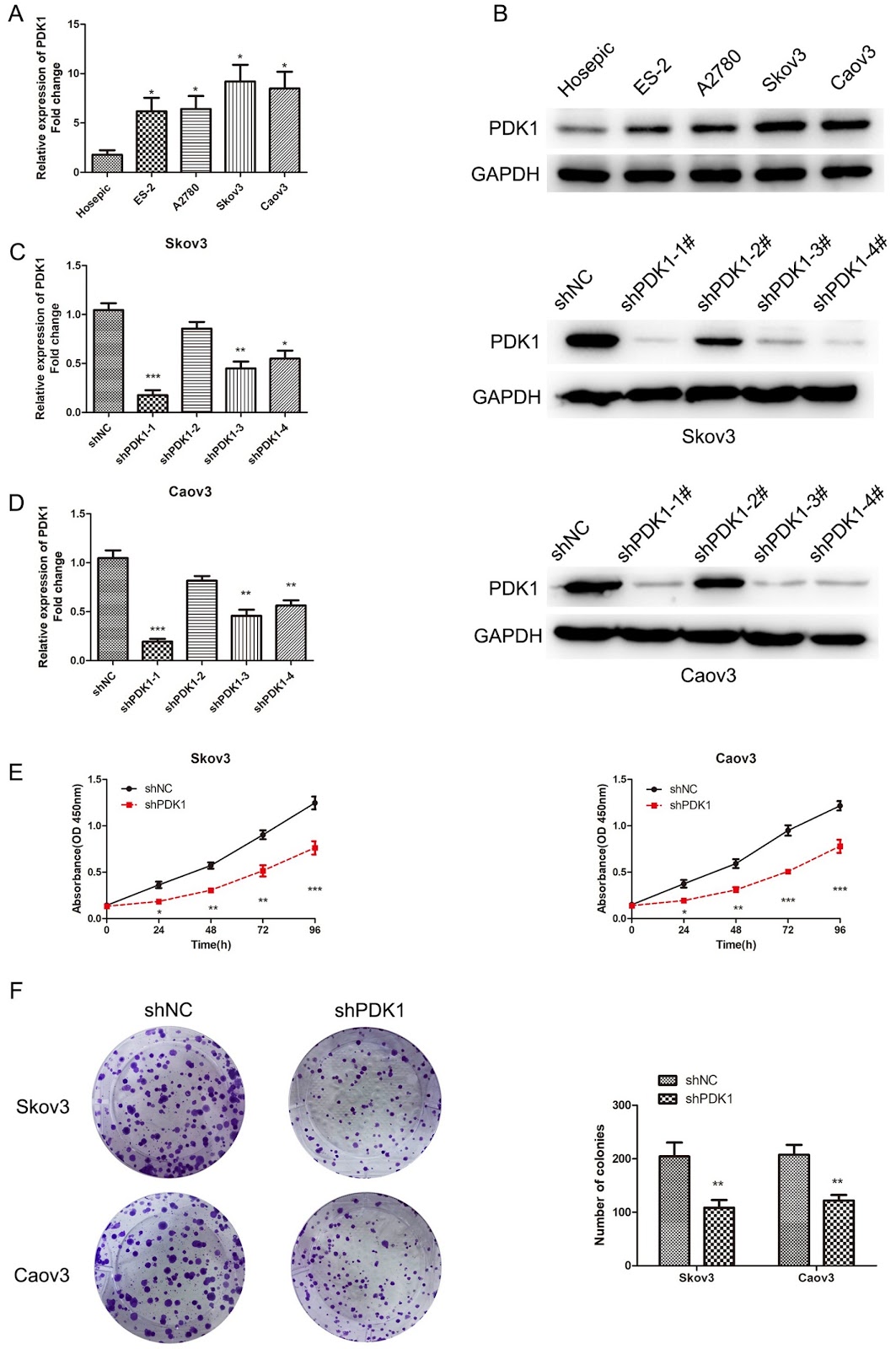
Pyruvate dehydrogenase kinase 1 (PDK1) is a new therapeutic target that is dysregulated in multiple tumors. This study aims to explore the potential role and regulatory mechanism of PDK1 in epithelial ovarian cancer (EOC). We detect PDK1 expression in EOC tissues and cells using qRT-PCR and western blot analysis, and the effects of PDK1 on EOC cell malignant behaviors are explored. RNA sequencing analyses are performed to explore the differentially expressed genes in PDK1-silenced EOC cells. Furthermore, tumor-bearing mouse models are established to assess the impacts of PDK1 and BGN on EOC tumor growth and metastasis in vivo. The results show that PDK1 is upregulated in EOC tissues and cell lines. Biglycan (BGN) is downregulated in PDK1-silenced EOC cells, and its expression is positively correlated with PDK1 levels in EOC tissues. PDK1 depletion inhibits EOC cell proliferation, migration and invasion. Mechanistically, PDK1 and BGN are colocalized in the cytoplasm of EOC cells and interact with each other. PDK1 positively regulates BGN expression by enhancing BGN mRNA stability. BGN overexpression partially reverses the anti-tumor effects of PDK1 depletion on EOC cell malignant behaviors. PDK1 has also been revealed to upregulate BGN to activate the NF-ĪŗB oncogenic pathway in EOC cells. Additionally, PDK1 accelerates tumor growth and metastasis by modulating BGN expression. In conclusion, PDK1 functions as an oncogene, facilitating EOC progression by upregulating BGN and activating the NF-ĪŗB pathway. These findings may provide valuable biomarkers for the diagnosis and treatment of EOC.

Read Full Abstract10.3724/abbs.2024186
Unraveling the metabolic potential of biocontrol fungi through omics data: a key to enhancing large-scale application strategiesGraphical AbstractVerified
Acta Biochimica et Biophysica Sinica

Unraveling the metabolic potential of biocontrol fungi through omics data: a key to enhancing large-scale application strategies

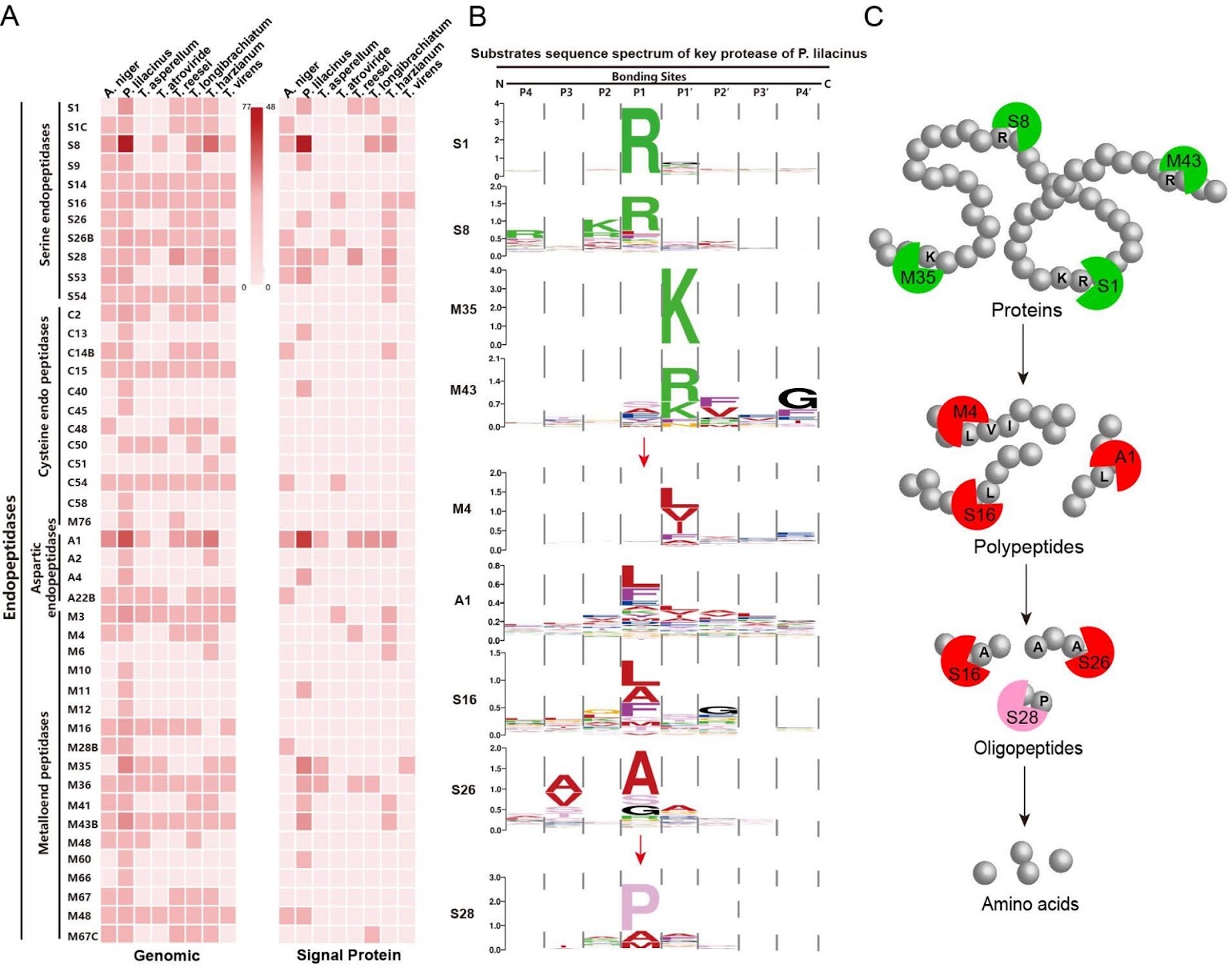
Biological control of pests and pathogens has attracted much attention due to its green, safe and effective characteristics. However, it faces the dilemma of insignificant effects in large-scale applications. Therefore, an in-depth exploration of the metabolic potential of biocontrol fungi based on big omics data is crucial for a comprehensive and systematic understanding of the specific modes of action operated by various biocontrol fungi. This article analyzes the preferences for extracellular carbon and nitrogen source degradation, secondary metabolites (nonribosomal peptides, polyketide synthases) and their product characteristics and the conversion relationship between extracellular primary metabolism and intracellular secondary metabolism for eight different filamentous fungi with characteristics appropriate for the biological control of bacterial pathogens and phytopathogenic nematodes. Further clarification is provided that Paecilomyces lilacinus, encoding a large number of hydrolase enzymes capable of degrading pathogen protection barrier, can be directly applied in the field as a predatory biocontrol fungus, whereas Trichoderma, as an antibiosis-active biocontrol control fungus, can form dominant strains on preferred substrates and produce a large number of secondary metabolites to achieve antibacterial effects. By clarifying the levels of biological control achievable by different biocontrol fungi, we provide a theoretical foundation for their application to cropping habitats.

Read Full Abstract10.3724/abbs.2024056
Ivermectin inhibits the growth of ESCC by activating the ATF4-mediated endoplasmic reticulum stress-autophagy pathwayGraphical AbstractVerified
Acta Biochimica et Biophysica Sinica

Ivermectin inhibits the growth of ESCC by activating the ATF4-mediated endoplasmic reticulum stress-autophagy pathway

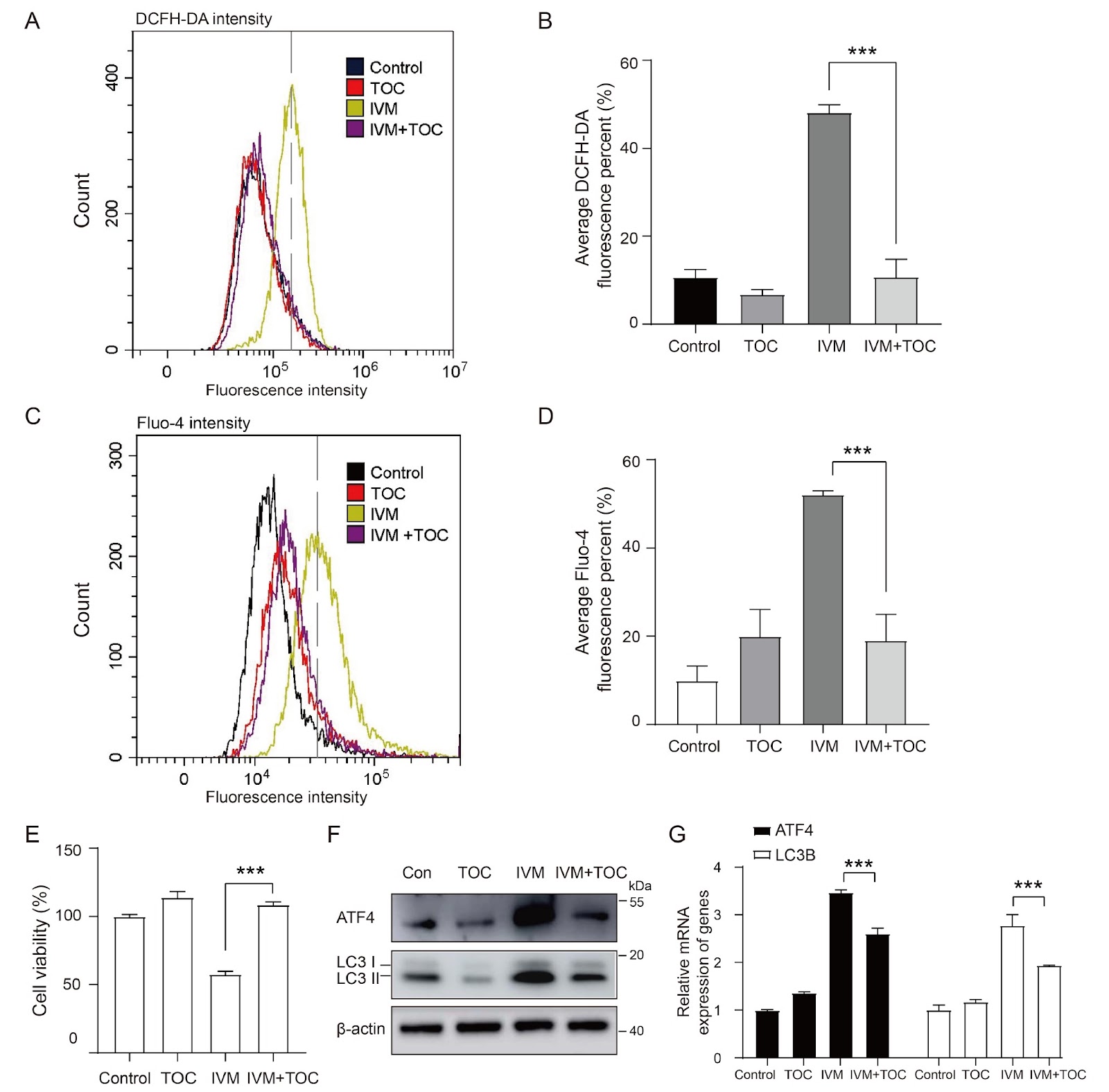
Esophageal squamous cell carcinoma (ESCC) is one of the most common forms of malignancy worldwide. However, there is currently a lack of effective chemotherapeutic drugs for ESCC. Ivermectin is a broad-spectrum antiparasitic drug with notable antitumor activity. However, the cellular and molecular mechanisms by which ivermectin inhibits cancer growth remain unclear. In this study, we elucidate the role of ivermectin in ESCC suppression by activating the endoplasmic reticulum (ER) stress and autophagy pathways. In transcriptome analyses, we find that activating transcription factor 4 (ATF4) and DNA damage inducible transcript 3 (DDIT3) are involved in the activation of ER stress by ivermectin. Moreover, ivermectin treatment suppresses the growth of ESCC xenograft tumors in nude mice. Taken together, our results establish the antitumor molecular role of ivermectin in targeting the ER stress-autophagy pathway and suggest that ivermectin is a potential drug candidate for the treatment of ESCC.

Read Full Abstract10.3724/abbs.2024210
Artemisinin alleviates arsenic-induced myocardial injury in rats by modulating oxidative stress and inflammatory responsesGraphical AbstractVerified
Acta Biochimica et Biophysica Sinica

Artemisinin alleviates arsenic-induced myocardial injury in rats by modulating oxidative stress and inflammatory responses

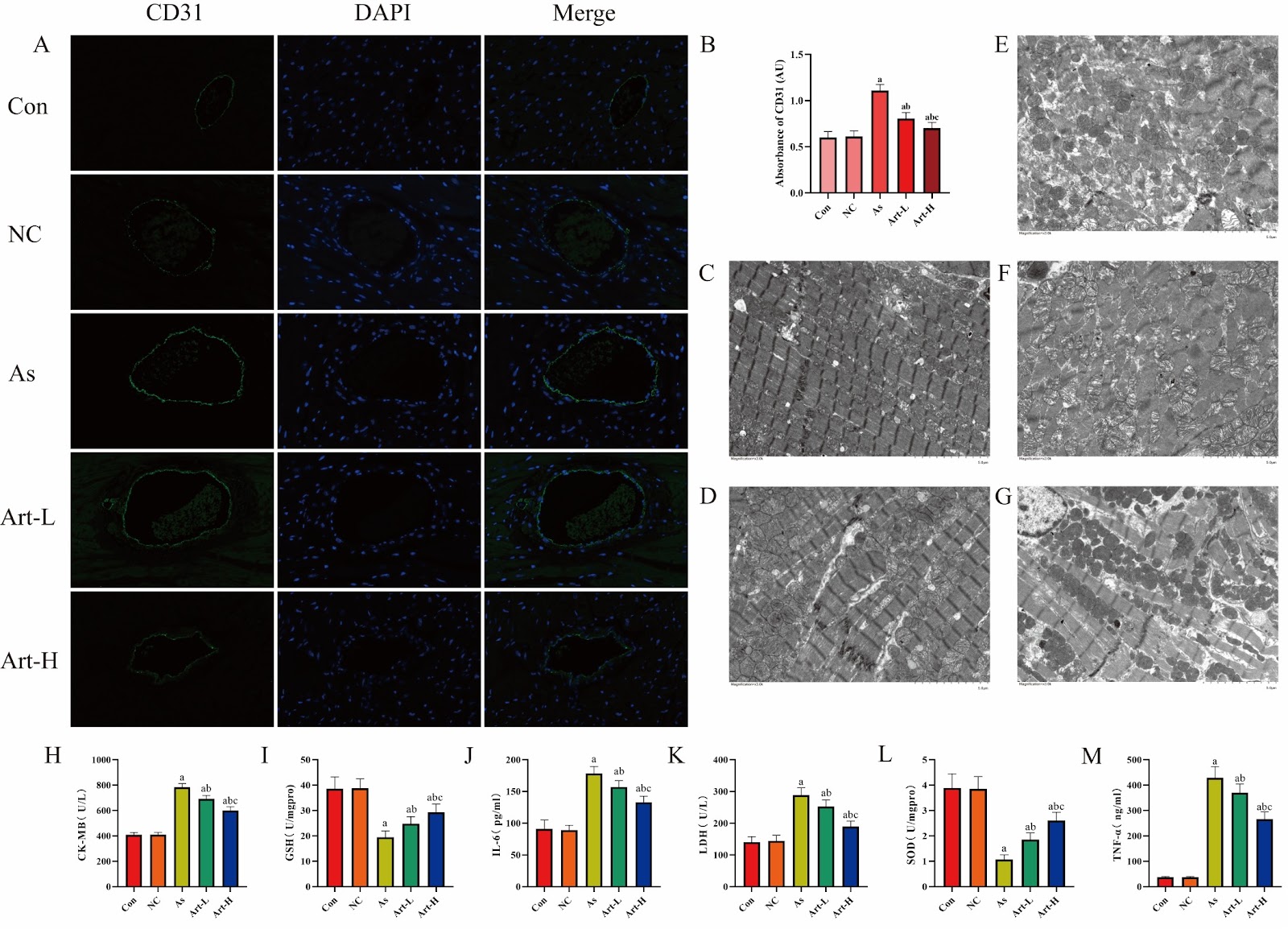
Arsenic is widely present in nature, and its compounds are extensively used in industrial, agricultural, and medical fields. Arsenic trioxide (As2O3) is specifically used as a therapeutic agent for acute promyelocytic leukemia because it induces cancer cell differentiation and apoptosis, significantly reduces the cancer cell count and has unique medical value. However, owing to its high toxicity and carcinogenicity, long-term use can induce cardiovascular diseases such as arrhythmia and myocardial contractile dysfunction. However, research on the treatment of arsenic-induced cardiotoxicity remains relatively scarce. Notably, artemisinin has anti-inflammatory and antioxidative effects on various heart diseases, effectively inhibiting reactive oxygen species (ROS) production, preventing myocardial damage and apoptosis caused by arsenic poisoning, and improving cardiac contractile and diastolic functions, thus enhancing cardiac function. This study aims to discuss the impact of artemisinin on the myocardium of arsenic-poisoned rats. Forty 12-week-old male SD rats were randomly divided into five groups: control, arsenic poisoning, drug control, low-dose artemisinin, and high-dose artemisinin. As2O3 was intraperitoneally injected at 5 mg/kg/day for 10 days in the arsenic poisoning, low-dose, and high-dose groups, whereas the control and drug control groups received equal volumes of physiological saline. Artemisinin was subsequently injected at corresponding doses for three weeks. Myocardial contrast echocardiography (MCE) was used to assess myocardial blood perfusion. Blood and myocardial tissue samples were collected for biochemical and histological analyses. Results showed that arsenic poisoning significantly decreased myocardial blood perfusion (AUC and WISƗPI) and increased CD31 expression, indicating microvascular damage and inflammation. Artemisinin intervention, especially at high dose, restored perfusion and reduced CD31 expression, suggesting a protective effect. Electron microscopy confirmed that artemisinin alleviated arsenic-induced myocardial structural damage. These findings suggest that artemisinin alleviates arsenic-induced myocardial injury by modulating oxidative stress and inflammatory responses.

Read Full Abstract10.3724/abbs.2024225
Anthocyanins and flavonoids derived from Clitoria ternatea L. flower inhibit bladder cancer growth via suppressing fatty acid synthesis mediated by SREBP1 pathwayGraphical AbstractVerified
Acta Biochimica et Biophysica Sinica

Anthocyanins and flavonoids derived from Clitoria ternatea L. flower inhibit bladder cancer growth via suppressing fatty acid synthesis mediated by SREBP1 pathway

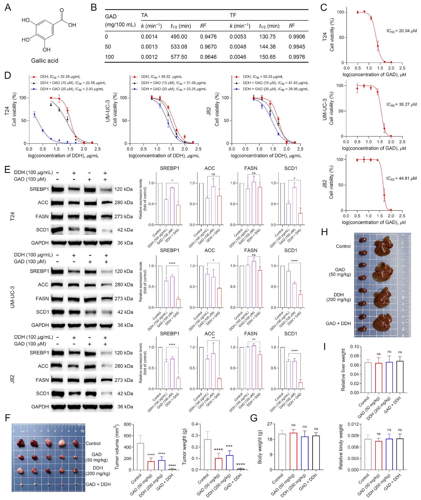
Clitoria ternatea L. flowers are used as traditional herbal medicines and are known for their advanced pharmacological activities. Flavonoids and anthocyanins reportedly contribute to the therapeutic properties of C. ternatea flowers; however, their potential anti-bladder cancer effects and molecular mechanisms remain unknown. In this study, flavonoid- and anthocyanin-rich samples from C. ternatea flowers (DDH) are prepared via macroporous resin-based extraction coupled with an efficient and reliable two-dimensional UPLC-DAD-MS/MS method. In vitro and in vivo studies reveal that DDH can inhibit bladder cancer cell growth and enhance the anti-bladder cancer activity of cisplatin. RNA-seq combined with KEGG analysis reveals that fatty acid synthesis is closely related to the anti-bladder cancer effect of DDH. Furthermore, DDH dose-dependently reduces cellular fatty acid levels in bladder cancer cells, and the addition of fatty acids significantly mitigates DDH-induced cell growth inhibition. Subsequent findings reveal that DDH downregulates sterol regulatory element-binding protein 1 (SREBP1), a key transcriptional regulator of de novo fatty acid synthesis in cancer cells, and its downstream targets (FASN, SCD1, and ACC). Additionally, this study demonstrates that gallic acid not only enhances the stability of DDH but also synergistically potentiates its anti-bladder cancer activity. Our study suggests that targeting the SREBP1 pathway is an effective strategy in bladder cancer therapy, and the ability of DDH to induce cell death by inhibiting the SREBP1 pathway and its good tolerance in mice make it a promising strategy for preventing and treating bladder cancer.

Read Full Abstract10.3724/abbs.2024192
Undaria pinnatifida extract attenuates combined allergic rhinitis and asthma syndrome by the modulation of epithelial cell dysfunction and oxidative stressGraphical AbstractVerified
Acta Biochimica et Biophysica Sinica

Undaria pinnatifida extract attenuates combined allergic rhinitis and asthma syndrome by the modulation of epithelial cell dysfunction and oxidative stress

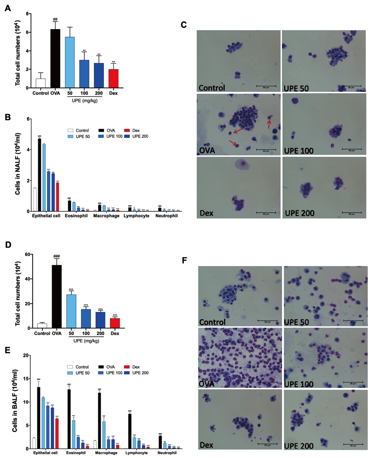
Undaria pinnatifida (U. pinnatifida) has long been a part of the human diet and medicine. Although U. pinnatifida has been reported to have immunomodulatory, anti-inflammatory, anti-diabetic and antibacterial activities, its specific effect on patients with combined allergic rhinitis and asthma syndrome (CARAS) has not been clarified. In this study, the anti-allergic and anti-inflammatory effects of U. pinnatifida extract (UPE) are investigated in a mouse model of ovalbumin (OVA)-induced CARAS. The oral administration of UPE inhibits allergic responses by reducing OVA-specific immunoglobulin levels. As a result, the symptoms of early reactions are also improved. UPE inhibits the accumulation of inflammatory cells and attenuates the expression of Th2 cytokines in both nasal and bronchoalveolar lavage fluid. Furthermore, UPE treatment inhibits the NF-ĪŗB/MAPK signaling pathway in lung homogenates. Additionally, UPE prevents shedding of the nasal mucosal epithelium, protects the integrity of the epithelium, enhances the expression of E-cadherin at the junction of epithelial cells, and inhibits the degradation of ZO-1 and occludin in the airway epithelium. In addition, UPE ameliorates dysfunction of the nasal epithelial barrier by enhancing antioxidant properties and downregulating the expression of the inflammatory factor IL-33. These results suggest that UPE may treat CARAS by modulating epithelial cell dysfunction and oxidative stress.

Read Full Abstract10.3724/abbs.2024190
Immune signatures of megakaryocytes in persistent inflammation-immunosuppression and catabolism syndromeGraphical AbstractVerified
Acta Biochimica et Biophysica Sinica

Immune signatures of megakaryocytes in persistent inflammation-immunosuppression and catabolism syndrome

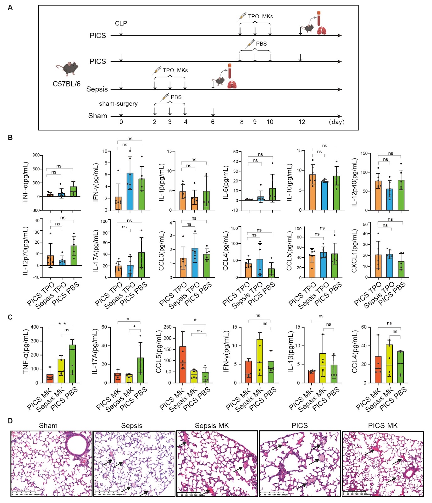
Persistent inflammation-immunosuppression and catabolism syndrome (PICS) is a severe condition that may follow sepsis and is characterized by ongoing inflammation and immune suppression, diminishing quality of life and potentially causing death. The role of megakaryocytes (MKs) in PICS, despite their association with thrombopoiesis, is not well understood. In this study, we use single-cell RNA sequencing to profile MKs in peripheral blood mononuclear cell samples obtained from 11 patients, including six with PICS, five with sepsis, and five healthy controls, to determine the diversity and molecular signatures of the MKs. Five subgroups of MKs are identified (MK1–MK5), and their proportions vary across the groups. MK1 and MK2 are predominant in PICS. Gene Ontology analysis shows that genes related to antigen processing and presentation and IL-17 signaling are enriched in MK1, whereas genes associated with platelet degranulation and neutrophil activation are enriched in MK2. Moreover, the expression level of CCL5 is markedly increased in MKs. Ligand-receptor analysis reveals dynamic interactions among MKs and T cells, B cells, natural killer cells, monocytes, and macrophages, suggesting a broad role of MKs in immune homeostasis. In PICS model mice, MKs regulate systemic inflammation by reducing the levels of the proinflammatory cytokines TNF-α and IL-17A and promoting lung tissue repair. Our findings establish MKs as essential components of the immune system in PICS and provide new insights into their potential as therapeutic targets for post-sepsis immune dysfunction.

Read Full Abstract10.3724/abbs.2025087
SIRPα modulates the podocyte cytoskeleton through influencing the phosphorylation of FAK at tyrosine residue 597Graphical AbstractVerified
Acta Biochimica et Biophysica Sinica

SIRPα modulates the podocyte cytoskeleton through influencing the phosphorylation of FAK at tyrosine residue 597

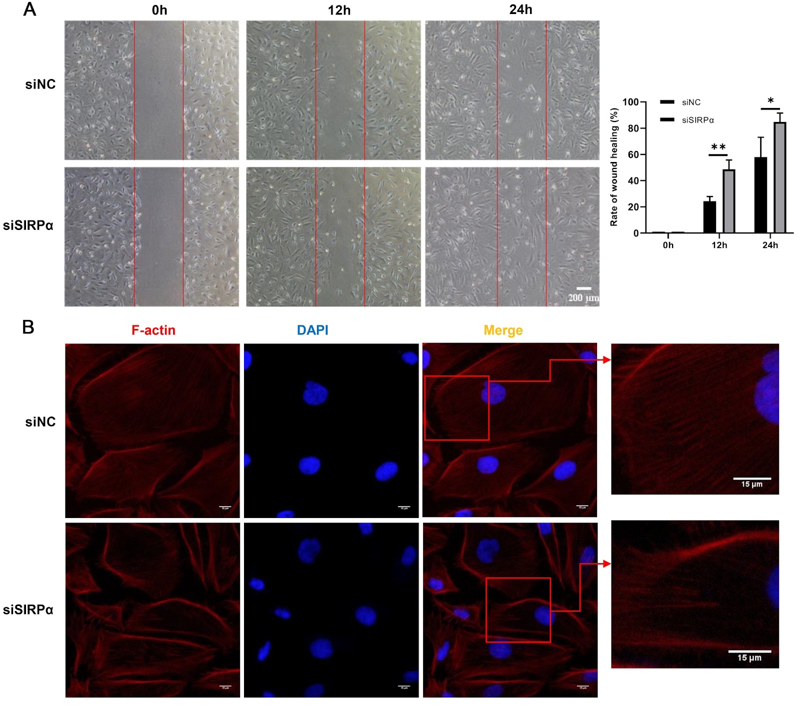
Signal regulatory protein α (SIRPα) is recognized as a significant transmembrane protein within the glomeruli that is specifically localized in podocytes, where it plays a role in modulating downstream signaling pathways through phosphorylation. Upon tyrosine phosphorylation of the immunoreceptor tyrosine-based inhibitory motif (ITIM) within SIRPα, protein tyrosine phosphatases are recruited to facilitate the dephosphorylation of downstream signals. Nevertheless, the specific downstream signaling pathways affected by this mechanism have yet to be elucidated. In this study, phosphoproteomic analysis is conducted on podocytes with SIRPα deficiency to identify proteins whose phosphorylation is regulated by SIRPα and the associated signaling pathways in human podocytes. The results reveal significant alterations in biological processes related to cytoskeleton arrangement and cytoskeleton protein binding. Specifically, an increase in FAK tyrosine phosphorylation at Y576 is identified as a potentially crucial signal of the influence of SIRPα on the podocyte cytoskeleton. Our study suggests that SIRPα may facilitate podocyte cytoskeleton rearrangement and migration through the Src/FAK/p38 MAPK signaling pathway. For the first time, we discover increased level of SIRPα, which is strongly linked to urinary protein, in the urine of patients with nephrotic syndrome (NS). Additionally, an increase in urinary FAK level is observed in NS patients, which is positively correlated with both urinary protein level and urinary SIRPα level. These findings suggest that SIRPα and FAK may serve as promising biomarkers for podocytopathies.

Read Full Abstract10.3724/abbs.2024198
CRISPR-based shuttle cloning of 1397 human genes into UAS vectorsGraphical AbstractVerified
Acta Biochimica et Biophysica Sinica

CRISPR-based shuttle cloning of 1397 human genes into UAS vectors

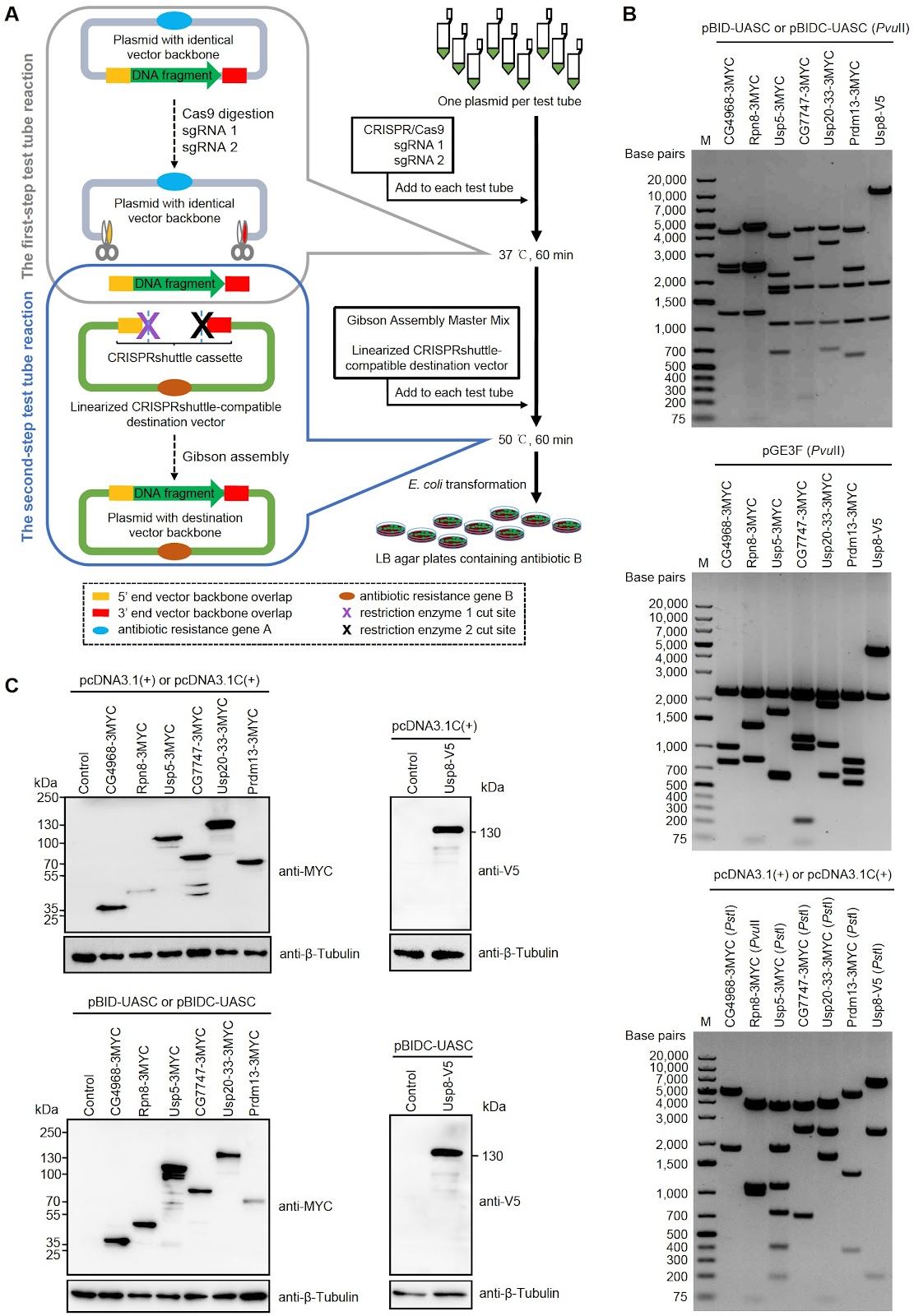
Functional genomics is a powerful tool for elucidating the function of all genes in an organism and primarily relies on construction and manipulation of genome-wide DNA libraries such as cDNAs, ORFs, gene promoters and inverted repeats for RNAi. Typically, a DNA library must be transferred to a destination vector in a high-throughput manner for functional genomics studies. However, the construction of genome-wide DNA libraries, such as cDNA/ORF overexpression libraries, has been challenging due to limitations in high-throughput DNA cloning methods. This severely restricts the use of functional genomics. The prevailing high-throughput cloning methods for constructing and manipulating genome-wide DNA libraries primarily include Gateway, In-Fusion, Creator, and Univector cloning systems, all of which are based on site-specific recombination. Among these, the Gateway cloning system is the most extensively employed high-throughput cloning method. All the aforementioned high-throughput cloning methods predominantly rely on PCR amplification of the DNA fragments of interest. This step requires individualized manipulations for each DNA fragment, including primer design and synthesis, gel purification, and DNA sequencing, which are laborious and time-consuming. Additionally, PCR amplification is particularly problematic for long DNA fragments. Consequently, the PCR amplification of DNA fragments of interest is not only costly but also a rate-limiting step in high-throughput cloning. For example, although cDNA and ORF resources for human, mouse and Drosophila have been publicly available for nearly two decades, the construction of a genome-wide GAL4/UAS (upstream activating sequence)-based UAS-cDNA/ORF plasmid library from these resources has been severely impeded by the PCR amplification of cDNAs and ORFs [1]. We previously developed a high-throughput cloning method, CRISPRmass, for constructing a genome-wide UAS-cDNA/ORF plasmid library from publicly available cDNA/ORF resources [2]. However, CRISPRmass is applicable solely to the insertion of an identical DNA fragment (e.g., a UAS module) into the identical backbones of different plasmids [2]. It does not allow for the transfer of DNA fragments (e.g., cDNAs or ORFs) between vectors, thereby limiting its use in DNA cloning. By introducing the concept of a CRISPRshuttle cassette, we developed a novel high-throughput DNA cloning method termed CRISPR-based shuttle cloning (CRISPRshuttle cloning). This method allows for the transfer of numerous DNA fragments of interest from original plasmids with identical backbones to a different vector background through two-step test tube reactions prior to bacterial transformation, thereby eliminating the need for PCR amplification of the DNA fragments (Figure 1A). In the first-step test tube reaction, different DNA fragments of interest are excised from their original plasmids by digesting the plasmid backbones with Cas9/sgRNA 1 and Cas9/sgRNA 2. Cas9/sgRNA 1 targets the backbone sequence adjacent to the 5′ end of the DNA fragments, while Cas9/sgRNA 2 targets the backbone sequence adjacent to the 3′ end. The released DNA fragments do not need to be purified, and the reaction products can be directly used in the second-step test tube reaction. In the second-step test tube reaction, the released DNA fragments are transferred to the CRISPRshuttle cassette of a CRISPRshuttle-compatible destination vector via Gibson assembly, yielding the desired plasmids. A CRISPRshuttle cassette consists of approximately 20‒40 bp of backbone sequence flanking both the 5′ and 3′ ends of the DNA fragments, and one or two unique restriction enzyme recognition sites between these sequences. These recognition sites are used for linearizing the CRISPRshuttle-compatible destination vector and must be unique within the vector.
Read Full Abstract10.3724/abbs.2025050
Proline/serine-rich coiled-coil protein 1 alleviates pyroptosis in murine bone marrow-derived macrophagesGraphical AbstractVerified
Acta Biochimica et Biophysica Sinica

Proline/serine-rich coiled-coil protein 1 alleviates pyroptosis in murine bone marrow-derived macrophages

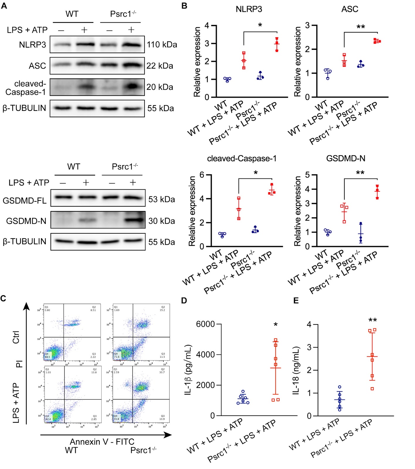
Pyroptosis is a regulated inflammatory cell death process that plays an essential role in various diseases. This study investigates the role of proline/serine-rich coiled-coil protein 1 (PSRC1) in pyroptosis and inflammation in macrophages. This study reports that PSRC1 expression is decreased in pyroptotic macrophages and that knockout of PSRC1 exacerbates pyroptosis and inflammation. PSRC1 overexpression alleviates pyroptosis and inflammation in macrophages. RNA-seq analysis reveals that PSRC1 regulates the expression of genes involved in the extracellular matrix (ECM). Specifically, PSRC1 downregulates the expression of periostin (POSTN), an ECM component. Knockdown of POSTN suppresses macrophage pyroptosis mediated by low expression of PSRC1. These findings suggest that PSRC1 can alleviate pyroptosis and inflammation in bone marrow-derived macrophages (BMDMs) by regulating the ECM and negatively regulating POSTN. This study provides insights into the role of PSRC1 in macrophage pyroptosis and identifies a potential target for the treatment of inflammatory diseases. Further research is needed to confirm these findings in vivo and in various disease models.

Read Full Abstract10.3724/abbs.2025012